Orthodontic Extrusion: Periodontal Considerations and Applications
Orthodontic Extrusion: Periodontal Considerations and Applications
Orthodontic Extrusion: Periodontal Considerations and Applications

Create successful ePaper yourself
Turn your PDF publications into a flip-book with our unique Google optimized e-Paper software.
In everyday practice, the general practitioner sometimes<br />
sees cases of subgingival trauma or carious lesions.<br />
Possible therapeutic options include extraction <strong>and</strong><br />
prosthetic restoration (by a bridge) or surgical lengthening<br />
of the crown, as well as orthodontic extrusion. The last of<br />
these options provides undeniable benefits for the patient.<br />
<strong>Orthodontic</strong> <strong>Extrusion</strong><br />
Movement of a tooth by extrusion involves applying<br />
traction forces in all regions of the periodontal ligament to<br />
stimulate marginal apposition of crestal bone. Because the<br />
gingival tissue is attached to the root by connective tissue,<br />
the gingiva follows the vertical movement of the root<br />
during the extrusion process. Similarly, the alveolus is<br />
attached to the root by the periodontal ligament <strong>and</strong> is in<br />
turn pulled along by the movement of the root.<br />
Rapid <strong>Extrusion</strong><br />
In the normal course of events, bone <strong>and</strong> gingival movements<br />
are produced under low-intensity extrusive forces.<br />
When stronger traction forces are exerted, as in rapid extrusion,<br />
coronal migration of the tissues supporting the tooth<br />
is less pronounced because the rapid movement exceeds<br />
their capacity for physiologic adaptation. 1 As well, rapid<br />
Journal of the Canadian Dental Association<br />
C L I N I C A L P R A C T I C E<br />
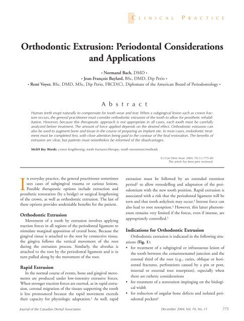
<strong>Orthodontic</strong> <strong>Extrusion</strong>: <strong>Periodontal</strong> <strong>Considerations</strong><br />
<strong>and</strong> <strong>Applications</strong><br />
• Norm<strong>and</strong> Bach, DMD •<br />
• Jean-François Baylard, BSc, DMD, Dip Perio •<br />
• René Voyer, BSc, DMD, MSc, Dip Perio, FRCD(C), Diplomate of the American Board of Periodontology •<br />
A b s t r a c t<br />
Human teeth erupt naturally to compensate for tooth wear <strong>and</strong> tear. When a subgingival lesion such as crown fracture<br />
occurs, the general practitioner must consider orthodontic extrusion of the tooth to allow for prosthetic rehabilitation.<br />
However, because this therapeutic approach is not appropriate in all cases, each tooth must be carefully<br />
analyzed before treatment. The amount of force applied depends on the desired effect. <strong>Orthodontic</strong> extrusion can<br />
also be used to augment bone <strong>and</strong> tissue in the course of preparing an implant site. In most cases, endodontic treatment<br />
must be completed first, with close attention being paid to the contour of the final restoration. The benefits of<br />
extrusion are clear, but patients must nonetheless be informed of the disadvantages.<br />
MeSH Key Words: crown lengthening; tooth fractures/therapy; tooth movement/methods<br />
© J Can Dent Assoc 2004; 70(11):775–80<br />
This article has been peer reviewed.<br />
extrusion must be followed by an extended retention<br />
period 2 to allow remodelling <strong>and</strong> adaptation of the periodontium<br />
with the new tooth position. Rapid extrusion is<br />
associated with a risk that the periodontal ligament will be<br />
torn <strong>and</strong> that tooth ankylosis may occur. 3 Intense force can<br />
also lead to root resorption. 4 However, this latter phenomenon<br />
remains very limited if the forces, even if intense, are<br />
appropriately controlled. 5<br />
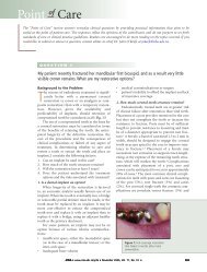
Indications for <strong>Orthodontic</strong> <strong>Extrusion</strong><br />
<strong>Orthodontic</strong> extrusion is indicated in the following situations<br />
(Fig. 1):<br />
• for treatment of a subgingival or infraosseous lesion of<br />
the tooth between the cementoenamel junction <strong>and</strong> the<br />
coronal third of the root (e.g., caries, oblique or horizontal<br />
fractures, perforations caused by a pin or post,<br />
internal or external root resorption), especially when<br />
there are esthetic considerations<br />
• for treatment of a restoration impinging on the biological<br />
width<br />
• for reduction of angular bone defects <strong>and</strong> isolated periodontal<br />
pockets 6<br />
December 2004, Vol. 70, No. 11 775
Bach, Baylard, Voyer<br />
a b c d e<br />
Figure 1: Examples of indications for orthodontic extrusion: a) subgingival or infraosseous<br />
dental lesion, such as a fracture; b) restoration impinging on the biological width; c) reduction<br />
of localized angular bone defects; d) preimplant extraction; e) trauma or impacted teeth.<br />
• for preimplant extraction to maintain or re-establish the<br />
integrity of an alveolar ridge (see “<strong>Extrusion</strong> for Implant<br />
Purposes”)<br />
• for orthodontic extraction where surgical extraction is<br />
contraindicated (e.g., in patients receiving chemotherapy<br />
or radiotherapy) 7<br />
• for treatment of trauma 8,9 or impacted teeth 10 (canines).<br />
Contraindications to <strong>Orthodontic</strong> <strong>Extrusion</strong><br />
<strong>Extrusion</strong> is contraindicated in patients with the following<br />
conditions:<br />
• ankylosis or hypercementosis 11 (the extra load would<br />
cause intrusion of the anchor teeth)<br />
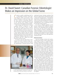
• vertical root fracture<br />
• root proximity <strong>and</strong> premature closure of embrasures<br />
(Fig. 2).<br />
Additional contraindications come into play when<br />
extrusion is used for prosthetic purposes:<br />
• short roots, which do not allow for adequate support of<br />
the restoration 12 (that is, when the crown–root ratio is<br />
less than 1:1)<br />
• insufficient prosthetic space<br />
• exposure of the furcation.<br />
These criteria are not absolute <strong>and</strong> do not apply if the<br />
purpose of orthodontic extrusion is to increase the quantity<br />
of bone in a ridge before placing a dental implant.<br />
Advantages<br />
<strong>Orthodontic</strong> extrusion is a conservative procedure that<br />
allows retention of a tooth without the disadvantages of a<br />
fixed bridge (e.g., the mutilation of adjacent dental tissue<br />
that typically occurs during bridge fabrication). As well,<br />
extrusion does not involve loss of bone or periodontal<br />
support, as commonly occurs during extraction. Simple<br />
surgical crown lengthening involves additional resection of<br />
bone of the teeth adjacent to the tooth that is to be lengthened,<br />
<strong>and</strong> such osteotomy can sometimes be avoided by use<br />
of orthodontic extrusion. Finally, this simple technique<br />
requires a relatively easy movement of the tooth.<br />
776 December 2004, Vol. 70, No. 11<br />
Figure 2: Root proximity, a major contraindication<br />
for orthodontic extrusion of a molar.<br />
Disadvantages<br />
Wearing an orthodontic device, as is required for orthodontic<br />
extrusion, may cause esthetic problems <strong>and</strong> may<br />
adversely affect oral hygiene. As well, the duration of treatment<br />
(4 to 6 weeks of extrusion <strong>and</strong> 4 weeks to 6 months<br />
of retention for implant cases in which tissue <strong>and</strong> bone<br />
remodelling are the objectives 6 ) may discourage some<br />
patients. Indeed, some authors recommend 4 weeks of<br />
retention for every millimetre of extrusion. 4 At the end of<br />
the procedure, conservative periodontal surgery may be<br />
necessary to correct any discrepancy that has developed<br />
between adjacent periodontal levels. 13<br />
Forces Exerted<br />
Forces of 15 g for the fine root of a lower incisor <strong>and</strong> 60 g<br />
for a molar are sufficient for slow extrusion. Some authors<br />
recommend that the maximum force for a slow movement<br />
should not exceed 30 g, 4,14 whereas rapid extrusions are<br />
accomplished with forces higher than 50 g. 15<br />
After a latency period of a few days to a few weeks, including<br />
a period of hyalinization, slow extrusion occurs at a rate of<br />
approximately 1 mm or less per week. 4 The force used will<br />
vary depending on the physiologic response of the patient <strong>and</strong><br />
other factors such as root surface morphology. The extent of<br />
the force exerted can only be approximated, since it is difficult<br />
to quantify the force applied. The forces must be adjusted on<br />
the basis of the clinically verified speed of extrusion.<br />
It is imperative that constant force be maintained between<br />
the extrusion <strong>and</strong> hyalinization phases; otherwise, the desired<br />
orthodontic movement will not take place. <strong>Periodontal</strong> ligament<br />
tension is needed for bone remodelling <strong>and</strong> movement<br />
of the periodontal attachment. 8 Finally, the force must be<br />
applied along the tooth axis to prevent any undesirable tilting.<br />
<strong>Periodontal</strong> Effects<br />
<strong>Orthodontic</strong> extrusion forces coronal migration of the<br />
root <strong>and</strong> increases the bone ridge as well as the quantity of<br />
attached gingiva, in particular when weak to moderate<br />
forces are applied. 16,17 The amount of attached gingiva is<br />
Journal of the Canadian Dental Association
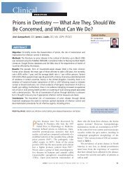
Figure 3: Development of a b<strong>and</strong> of immature<br />
nonkeratinized tissue (“red patch”).<br />
Figure 5: Restoration phase. Care is needed<br />
to prevent overcontouring of the crowns.<br />
increased through eversion of the sulcular epithelium,<br />
appearing first as immature nonkeratinized tissue (known as<br />
“red patch”) (Fig. 3) <strong>and</strong> then as keratinized tissue; the<br />
process of keratinization requires 28 to 42 days. 4 After coronal<br />
movement of the periodontal attachment has occurred,<br />
minor surgical correction may be necessary. To avoid or<br />
minimize this correction, some authors recommend weekly<br />
fibrotomy (incision of the supracrestal gingival fibres). 18,19<br />
Others recommend a single fibrotomy procedure when the<br />
movement is completed, 5,17 before bone <strong>and</strong> gingiva remodelling<br />
occurs. However, according to several clinicians, fibrotomy<br />
has proved unpredictable, <strong>and</strong> gingiva <strong>and</strong>/or bone<br />
remodelling may still be required after the stabilization<br />
period. 1,20 In a study carried out on dogs, repeated fibrotomy<br />
failed to prevent coronal migration of the gingival<br />
attachment. 21 In-depth studies on human subjects to<br />
demonstrate the usefulness of this procedure <strong>and</strong> to define<br />
the appropriate frequency have yet to be carried out. 5<br />
<strong>Extrusion</strong> for Implant Purposes<br />
<strong>Orthodontic</strong> extrusion, which preserves or regenerates<br />
the volume of bone in the ridge, makes the placement of<br />
dental implants more favourable. Conservation of the ridge<br />
allows placement of the dental implant within the thickness<br />
of the bone in a suitable axis. It also optimizes the potential<br />
for a guided bone regeneration technique. 7 Finally, the<br />
newly keratinized tissue improves the esthetic appearance at<br />
the site. 22 More specifically, extrusion is appropriate for<br />
Journal of the Canadian Dental Association<br />
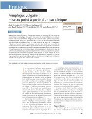
Figure 4: Steps in orthodontic extrusion for the purpose of preimplant extraction.<br />
<strong>Orthodontic</strong> <strong>Extrusion</strong><br />
teeth with average bone loss <strong>and</strong> when esthetics is a determining<br />
factor (Fig. 4), because it facilitates esthetic corrections<br />
<strong>and</strong> ensures stabilization of the implant within<br />
adequate bone mass. 23 The eruptive phase, lasting between<br />
4 <strong>and</strong> 6 weeks, is followed by 6 to 8 weeks of stabilization,<br />
during which tissues are remodelled before extraction of the<br />
“condemned” tooth <strong>and</strong> placement of the dental implant. 7<br />
However, some authors recommend up to 6 months of<br />
retention in preimplant cases to maximize remodelling of<br />
the ridge. 6 Indeed, prolonged stabilization allows more time<br />
for tissue remodelling, which, in turn, promotes more voluminous<br />
bone remodelling <strong>and</strong> decreases the risk of relapse<br />
before placement of the dental implant. 6<br />
<strong>Extrusion</strong> <strong>and</strong> Endodontics<br />
In some cases, the tooth to be extruded must be treated<br />
endodontically to prevent sensitivity <strong>and</strong> exposure of the pulp<br />
during the occlusal reduction required during the extrusion. 4<br />
A canal that cannot be adequately treated (because of subgingival<br />
fracture <strong>and</strong> lack of an adequate operative field) can be<br />
filled with calcium hydroxide before extrusion <strong>and</strong> subsequent<br />
treatment. 14 However, when the tooth must be<br />
extracted <strong>and</strong> the purpose of extrusion is to obtain an optimal<br />
ridge (e.g., in cases of preimplant extraction), pulpectomy<br />
may be sufficient. 15 Moreover, if the tooth is to be saved <strong>and</strong><br />
its pulp kept intact, slow orthodontic extrusion, over a period<br />
of 3 to 6 months, is the preferred method of reducing the risk<br />
of pulpal necrosis; rapid extrusion could be traumatic to the<br />
pulpal tissue. 24 A histologic study demonstrated odontoblastic<br />
degeneration after 1 week of activation <strong>and</strong> pulpal fibrosis<br />
after 4 weeks in a tooth subject to an extrusion force of 50 g. 25<br />
The authors assumed that the pulpal reaction would differ<br />
depending on the diameter of the apical foramen. Pulp<br />
prolapse would be due to ischemia secondary to rapid movement.<br />
25 During rapid extrusion, a pseudo-apical lesion (an<br />
apical radiolucency) appears, which must be differentiated<br />
from a true lesion of endodontic origin. However, a tooth that<br />
has undergone incomplete root canal treatment, although<br />
asymptomatic, could eventually develop a true apical lesion<br />
because of inflammatory mediators involved in the root apex<br />
during orthodontic movement. 26<br />
December 2004, Vol. 70, No. 11 777
Bach, Baylard, Voyer<br />
Figure 6a: System of orthodontic brackets<br />
attached by a nickel–titanium wire<br />
Figure 7a: <strong>Extrusion</strong> accomplished by treatment<br />
with a traditional orthodontic bracket.<br />
<strong>Extrusion</strong> <strong>and</strong> Prosthodontics<br />
The mesiodistal diameter of the root, which is naturally<br />
“strangled” at the cementoenamel junction of single-rooted<br />
teeth, is reduced with progression of the extrusion (especially<br />
in the case of conical roots), which involves expansion<br />
of interproximal gingival embrasures. The contour shape of<br />
the crowns must not be exaggerated to compensate for this<br />
reduction in diameter (Fig. 5). Similarly, embrasures<br />
should not be filled to prevent an overcontour, which could<br />
adversely affect the marginal periodontium. 27<br />
Techniques<br />
Several extrusion methods are available, depending on<br />
the clinical conditions encountered. A variety of mechanical<br />
strategies can be used to control the forces applied.<br />
One technique involves placing orthodontic brackets on<br />
the buccal aspect of the teeth adjacent to the tooth that is to<br />
undergo extrusion in a passive position that will not cause<br />
any orthodontic movement of the anchor teeth. The bracket<br />
on the target tooth is positioned more apically than the<br />
brackets on the adjacent teeth; the difference in distance<br />
represents the desired extrusion. A 0.016-in. nickel–<br />
titanium arch wire is attached to the brackets (Figs. 6a, 6b<br />
<strong>and</strong> 6c). If greater movement is desired, a second, more rigid<br />
wire (0.016 in. x 0.022 in.), attached only to the brackets of<br />
the adjacent teeth, is used to stabilize everything (Figs. 7a,<br />
7b <strong>and</strong> 7c). Following extrusion, a more rigid 0.018-in.<br />
stainless steel arch wire is inserted <strong>and</strong> set by means of a<br />
778 December 2004, Vol. 70, No. 11<br />
Figure 6b: <strong>Extrusion</strong> is accomplished by the<br />
orthodontic brackets over a 1-month period.<br />
Figure 7b: The stabilization wire is attached<br />
to the brackets adjacent to the tooth that is<br />
to be extruded.<br />
Figure 6c: Coronal migration of the gingiva<br />
in the buccal aspect of extruded tooth 21.<br />
(Treatment by Dr. René Voyer.)<br />
Figure 7c: Active extrusion is carried out<br />
over a 1-month period. (Treatment by<br />
Dr. Martin Vallois.)<br />
metal ligature for a minimum retention period of 12<br />
weeks. 11 If the dental tissues are inadequate for cementing a<br />
bracket, a composite reconstruction of the crown can be<br />
done or another consolidation strategy can be used.<br />
It is possible to avoid positioning the bracket apically by<br />
shaping a stainless steel wire (0.018 in. diameter) into a<br />
horizontal loop (Fig. 8). This activated extrusion system<br />
will produce movement of 1 mm per month. A wire in the<br />
form of a spiral (a spring) can also be used to provide the<br />
necessary traction force (Fig. 9).<br />
Another strategy consists of inserting a rigid wire into<br />
the restorations of the anchor teeth. A metal wire, 0.7 mm<br />
in diameter, hooked at one end, is cemented into the canal<br />
of the tooth that is to undergo extrusion. An elastic<br />
connects the hook to the rigid anchor wire to activate the<br />
mechanism (Fig. 10). The elastic is changed every 2 weeks.<br />
This method can be difficult to use on posterior teeth<br />
because occlusion can interfere with the mechanism.<br />
If the anchor teeth have not been restored, a rectangular<br />
stainless steel arch wire (0.018 or 0.019 in. x 0.025 in.) can be<br />
folded <strong>and</strong> affixed with composite to the buccal aspect of each<br />
tooth (Figs. 11a <strong>and</strong> 11b). Forces must be applied according<br />
to the position of the long axis of the tooth that is to undergo<br />
extrusion to prevent buccal or lingual tipping. A temporary<br />
crown cemented on a final post can be used as a traction<br />
attachment point; this approach maintains the esthetic<br />
appearance. 3 If necessary, the proximal contours of the tooth<br />
undergoing extrusion can be carefully reduced to prevent the<br />
Journal of the Canadian Dental Association
Figure 8: A system of orthodontic brackets<br />
attached by a horizontal loop wire.<br />
Figure 11a: <strong>Orthodontic</strong> wire cemented by<br />
composite to the buccal aspect of the<br />
anchor teeth. An elastic activates the extrusion<br />
in the vertical axis only.<br />
Figure 13: Removable device (Hawley<br />
retainer with spring) <strong>and</strong> bracket on the<br />
tooth that is to be extruded.<br />
tooth from interfering with the movement.<br />
An extrusion device can also be prepared from a b<strong>and</strong><br />
<strong>and</strong> a soldered spring (Fig. 12); however, this method is<br />
more labour-intensive.<br />
A removable Hawley device <strong>and</strong> an anchoring tip<br />
cemented to the buccal aspect is a good mechanical alternative<br />
(Fig. 13). This method is useful when the adjacent<br />
teeth are mobile or offer inadequate anchorage because of<br />
trauma or when mild force is required.<br />
The number of teeth required for anchoring depends on<br />
the type of tooth to be extruded, root number <strong>and</strong> conformation,<br />
<strong>and</strong> the quantity of periodontal attachment.<br />
Follow-up evaluations should be done every 2 weeks to<br />
Journal of the Canadian Dental Association<br />
Figure 9: <strong>Extrusion</strong> of a central incisor<br />
affected by traumatic impaction is accomplished<br />
with an orthodontic bracket system<br />
activated by a spring.<br />
Figure 11b: The extrusion is accomplished<br />
by means of an orthodontic wire activated<br />
by an elastic. The temporary restoration in<br />
acrylic resin is cemented to the wire to<br />
improve the esthetic appearance.<br />
(Treatment by Dr. René Voyer.)<br />
<strong>Orthodontic</strong> <strong>Extrusion</strong><br />
Figure 10: <strong>Orthodontic</strong> wire embedded in<br />
the restorations adjacent to the tooth that is<br />
to be extruded. Movement is effected by an<br />
elastic that is changed regularly.<br />
Figure 12: A spring welded to a b<strong>and</strong><br />
(molar anchor) can be used to activate<br />
extrusion of the first premolar.<br />
ensure adequate oral hygiene <strong>and</strong> to correct any changes in<br />
occlusion as movement occurs. As well, it is important to<br />
ensure that movement of the tooth being treated actually<br />
occurs, because tooth ankylosis will cause the anchoring<br />
mechanism to be intruded.<br />
Evaluation <strong>and</strong> Preparation of Cases<br />
Before any therapeutic decision can be made, a detailed<br />
dental history, including that of any potential dental trauma,<br />
must be obtained. The evaluation must also take into account<br />
oral hygiene; bacterial plaque control must be exceptional<br />
before starting orthodontic extrusion treatment, so as to<br />
reduce the risk of dental demineralization <strong>and</strong> periodontal<br />
inflammation, which would adversely affect marginal bone<br />
gain or induce hyperplasia of the soft tissues.<br />
Before starting treatment, the dentist must evaluate the<br />
following:<br />
• periodontal status<br />
• quality <strong>and</strong> quantity of attached gingiva<br />
• depth of periodontal (or gingival) pockets for the<br />
targeted teeth<br />
• esthetic appearance of the site<br />
• gingival clearance when smiling<br />
• gingival contour line<br />
• occlusion<br />
• overjet <strong>and</strong> overbite<br />
December 2004, Vol. 70, No. 11 779
Bach, Baylard, Voyer<br />
• interference with movement (occlusal excursion)<br />
• postextrusion prosthetic space<br />
• general condition of the dentition.<br />
It is imperative to maintain an appropriate crown–root<br />
ratio (at least 1:1 after extrusion) <strong>and</strong> to ensure adequate<br />
width of the pulpal canal (a wide pulpal canal may indicate<br />
root fracture) so as to provide a favourable prognosis for the<br />
restored tooth. <strong>Periodontal</strong> probing, radiographic analysis<br />
<strong>and</strong> examination of the fragment of the fractured tooth (if<br />
available) can help in determining the extent of the fracture<br />
or carious lesion or in detecting a vertical root fracture.<br />
The informed consent form must describe, among other<br />
items, the risks of ankylosis, root resorption, relapse, movement<br />
of adjacent teeth <strong>and</strong> failure of treatment, any of<br />
which could lead to extraction of the tooth <strong>and</strong> another<br />
treatment option, such as a dental implant or other prosthetic<br />
replacement. As well, the consent form should point<br />
out the advantages <strong>and</strong> disadvantages of each alternative<br />
solution, specifying the duration of therapy, the approximate<br />
number of visits required <strong>and</strong> the costs.<br />
Conclusions<br />
In spite of the relative difficulties, orthodontic extrusion<br />
remains an accessible technique for general practitioners<br />
<strong>and</strong> a beneficial technique for the patient who wishes to<br />
keep a tooth, if only to keep the bone ridge volume intact<br />
<strong>and</strong> thereby to maximize the benefits of dental implants. C<br />
Acknowledgements: The authors wish to thank Dr. Sylvain<br />
Arsenault, director, department of stomatology, Notre-Dame<br />
Hospital, Montreal, Quebec; Dr. Fannie Brousseau, orthodontist,<br />
Rosemère, Quebec; Dr. Sylvain Gagnon, orthodontist, Montreal,<br />
Quebec; Dr. Martin Vallois, orthodontic resident, University of<br />
Montreal; <strong>and</strong> Danielle Mongrain, graphic artist, Notre-Dame<br />
Hospital, Montreal (Quebec).<br />
Dr. Bach is clinical instructor at the University of<br />
Montreal, Montreal, Quebec. He also maintains a<br />
private practice in Montreal.<br />
Dr. Baylard is lecturer <strong>and</strong> clinical instructor in the<br />
faculty of dentistry, University of Montreal, Montreal,<br />
Quebec. He also maintains a private practice in<br />
Montreal.<br />
Dr. Voyer is assistant professor <strong>and</strong> director, division of<br />
clinical periodontology, faculty of dentistry, University<br />
of Montreal, Montreal, Quebec. He also maintains a<br />
private practice in Montreal.<br />
Corresponence to: Dr. Norm<strong>and</strong> Bach, 12660 Odette Oligny,<br />
Montreal QC H4J 2R4. E-mail : normbach@hotmail.com.<br />
The authors have no declared financial interests.<br />
780 December 2004, Vol. 70, No. 11<br />
References<br />
1. Sabri R. L’allongement coronaire par l’égression orthodontique.<br />
Principes et techniques. J Parodontol 1989; 8(2):197–204.<br />
2. Antrim DD. Vertical extrusion of endodontically treated teeth. US<br />
Navy Med 1981; 72:23–8.<br />
3. Oesterle LJ, Wood LW. Raising the root. A look at orthodontic extrusion.<br />
J Am Dent Assoc 1991; 122(7):193–8.<br />
4. Minsk L. <strong>Orthodontic</strong> tooth extrusion as an adjunct to periodontal<br />
therapy. Compend Contin Educ Dent 2000; 21(9):768–70, 772, 774.<br />
5. Malmgren O, Malmgren B, Frykholm A. Rapid orthodontic extrusion<br />
of crown root <strong>and</strong> cervical root fractured teeth. Endod Dent Traumatol<br />
1991; 7(2):49–54.<br />
6. Mantzikos T, Shamus I. Case report: forced eruption <strong>and</strong> implant site<br />
development. Angle Orthod 1998; 68(2):179–86.<br />
7. Buskin R, Castellon P, Hochstedler JL. <strong>Orthodontic</strong> extrusion <strong>and</strong><br />
orthodontic extraction in preprosthetic treatment using implant therapy.<br />
Pract Periodontics Aesthet Dent 2000; 12(2):213–9.<br />
8. Alves LD, Donnelly JC, Lugo A, Carter DR. Reeruption <strong>and</strong> extrusion<br />
of a traumatically intruded immature permanent incisor: case report. J<br />
Endod 1997; 23(4):246–8.<br />
9. Jacobs SG. The treatment of traumatized permanent anterior teeth :<br />
case report & literature review. Part I — management of intruded<br />
incisors. Aust Orthod J 1995; 13(4):213–8.<br />
10. Quirynen M, Op Heij DG, Adriansens A, Opdebeeck HM, van<br />
Steenberghe D. <strong>Periodontal</strong> health of orthodontically extruded impacted<br />
teeth. A split-mouth, long-term clinical evaluation. J Periodontol 2000;<br />
71(11):1708–14.<br />
11. Nappen DL, Kohlan DJ. <strong>Orthodontic</strong> extrusion of premolar teeth: an<br />
improved technique. J Prosthet Dent 1989; 61(5):549–54.<br />
12. Shillingburg HT. Fundamentals of fixed prosthodontics. 3rd ed.<br />
Carol Stream (IL): Quintessence Publishing; 1997.<br />
13. Heithersay GS, Moule AJ. Anterior subgingival fractures : a review of<br />
treatment alternatives. Aust Dent J 1982; 27(6):368–76.<br />
14. Reitan K. Clinical <strong>and</strong> histological observations on tooth movement<br />
during <strong>and</strong> after orthodontic treatment. Am J Orthod 1967;<br />
53(10):721–45.<br />
15. Bondemark L, Kurol J, Hallonsten AL, Andreason JO. Attractive<br />
magnets for orthodontic extrusion of crown-root fractured teeth. Am J<br />
Orthod Dentofacial Orthop 1997; 112(2):187–93.<br />
16. Rosenberg ES, Cho SC, Garber DA. Crown lengthening revisited.<br />
Compend Contin Educ Dent 1999; 20(6):527–32, 534, 536-8.<br />
17. Ainama J, Talari A. The increase with age of the width of attached<br />
gingiva. J <strong>Periodontal</strong> Res 1976; 11(4):182–8.<br />
18. Palomo F, Kopezyk RA. Rationale <strong>and</strong> methods for crown lengthening.<br />
J Am Dent Assoc 1978; 96(2):257–60.<br />
19. Lythgoe JR, Torabinejad M, Simon JH. <strong>Extrusion</strong> techniques for the<br />
general dentist. Gen Dent 1980; 28(1):42–3, 46–9.<br />
20. Lovdahl PE. <strong>Periodontal</strong> management <strong>and</strong> root extrusion of traumatized<br />
teeth. Dent Clin North Am 1995; 39(1):169–79.<br />
21. Berglundh T, Marinello CP, Lindhe J, Thil<strong>and</strong>er B, Liljenber B.<br />
<strong>Periodontal</strong> tissue reactions to orthodontic extrusion. An experimental<br />
study in the dog. J Clin Periodontol 1991; 18(5):330–6.<br />
22. Mantzikos T, Shamus I. Forced eruption <strong>and</strong> implant site development:<br />
soft tissue response. Am J Orthod Dentofacial Orthop 1997;<br />
112(6):596–606.<br />
23. Salama H, Salama M. The role of orthodontic extrusion remodeling<br />
in the enhancement of soft <strong>and</strong> hard tissue profiles prior to implant placement:<br />
a systematic approach to the management of extraction site defects.<br />
Int J Periodontics Restorative Dent 1993; 13(4):312–33.<br />
24. Kahnberg KE. Surgical extrusion of root-fractured teeth — a followup<br />
study of two surgical methods. Endod Den Traumatol 1988; 4(2):<br />
85–9.<br />
25. Mostafa YA, Isk<strong>and</strong>er KG, el-Mangoury NH. Iatrogenic pulpal reactions<br />
to orthodontic extrusion. Am J Orthod Dentofacial Orthop 1991;<br />
99(1):30–4.<br />
26. Blase D, Bercy P. Une technique esthétique d’allongement de la<br />
couronne clinique. L’égression orthodontique rapide. Rev Belge Med Dent<br />
1993; 48(3):9–28.<br />
27. Cronin RJ, Wardle WL. Prosthodontic management of vertical root<br />
extrusion. J Prosthet Dent 1981; 46(5):498–504.<br />
Journal of the Canadian Dental Association