X-AutofIt - Accelrys
X-AutofIt - Accelrys
X-AutofIt - Accelrys

Create successful ePaper yourself
Turn your PDF publications into a flip-book with our unique Google optimized e-Paper software.
DATASHEET<br />
X-<strong>AutofIt</strong><br />
X-<strong>AutofIt</strong> is a powerful X-ray crystallography application that enables you to trace and<br />
build protein model coordinates into electron density maps phased by single isomorphous<br />
replacement (SIr), multiple isomorphous replacement (MIr), or multi-wavelength anomalous<br />
dispersion (MAD). with X-<strong>AutofIt</strong>, what previously took weeks or months can now be<br />
accomplished in a single day. The new X-AUTOFIT module combines functionality previously<br />
available in X-AUTOFIT and X-POWERFIT. Functionality includes three fully automated protocols<br />
for tracing, and for sequence assignment, and model building.<br />
X-AUTOFIT is a unique module that automatically<br />
determines the Cα trace of a protein from MAD,<br />
MIR, or SIR electron density maps. New methods<br />
in this tool can speed up electron density fitting<br />
up to 500-fold over conventional methods. Fully<br />
automated protocols in X-AUTOFIT then allow you<br />
to proceed to sequence assignment and fitting of<br />
an all-atom model all with a few clicks of the mouse.<br />
Tracing an initial map is a time consuming and<br />
laborious process that can take many months of<br />
work, especially for large maps. The tracing process<br />
is also significantly slower for maps generated from<br />
data with low resolution or poor phases. X-AUTOFIT<br />
has been fully automated and optimized so that<br />
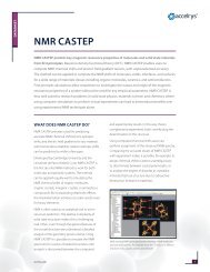
Cα trace and final structure of truB, a tRNA pseudo-uridine synthase.<br />
Data courtesy of Robert Fletterick at UCSF.<br />
accelrys.com<br />
it traces high resolution maps extremely fast (in<br />
less than a second). Furthermore, for data in the<br />
2-4 Å resolution X-AUTOFIT range, the automated<br />
protocols not only tease out features in the maps<br />
using a unique approach, but they do so in less<br />
than a minute. Once a Cα trace is obtained,<br />
sequence assignment, generation of an all-atom<br />
model, and real-space refinement of the final model<br />
can all be done in a few clicks with fully automated<br />
protocols. X-AUTOFIT represents a complete<br />
solution for tracing of de novo electron density<br />
maps, both in the low- and high-resolution ranges.<br />
About the SoftwAre<br />
When using the X-AUTOFIT module, your electron<br />
density map is first skeletonized. X-AUTOFIT allows<br />
you to edit the skeleton (bones) so that it only<br />
covers one molecule in the map. You can then<br />
calculate a map mask from the bones, and use this<br />
as a boundary for subsequent calculations.<br />
You then have two options for automatically<br />
determining the Cα trace from the skeletonized<br />
density. The first option is geared towards<br />
highresolution maps, generally between 2.2<br />
and 1.5 Å. This is a pathway analysis method in<br />
which the program tries to identify a unique<br />
1
path through the skeleton in a single pass. The second method 1<br />
is tailored for lower resolution maps (between 4.0 and 2.2 Å)<br />
and is based on the calculation and placement of vectors that<br />
represent the principle components of the secondary structural<br />
elements2 . These vectors are automatically converted into a Cα<br />
trace. X-AUTOFIT then takes these Cα traces and automatically<br />
extends them through the rest of the map. The end result is a<br />
close-to-complete trace of the structure. Semiautomated tools<br />
within X-AUTOFIT can then be used to extend the Cα trace into a<br />
complete structure. X-AUTOFIT also permits real space refinement<br />
of the Cα atoms with respect to the map.<br />
An additional option is available in X-AUTOFIT that automatically<br />
performs the low-resolution tracing protocol nine times with<br />
different starting parameters (bones sigma values). A ‘consensus<br />
trace’ is generated at the end of this process. This option saves<br />
time by automating a process that would otherwise have to be<br />
repeated manually, and it also ensures that the final trace is more<br />
likely to identify features in your low-resolution map that would<br />
otherwise have gone undetected in a single trace.<br />
Once a Cα trace is obtained, the sequence can be assigned to<br />
the trace with a single click (for high resolution maps). For low<br />
resolution maps, you are often able to identify characteristics of<br />
the amino acid side chain (e.g. large, medium, small, aromatic,<br />
or aliphatic). Using unique fuzzy descriptors, and any specific<br />
assignments, X-AUTOFIT quickly realigns against the protein<br />
sequence.<br />
Fully automated tools also exist for building an all-atom model<br />
of your structure, and to fit this model into the electron density<br />
using real-space refinement techniques. These steps used to take<br />
significant time and effort, and involved many interactive steps.<br />
But now, the entire process has been completely automated. The<br />
X-AUTOFIT application only requires an extended map. Although<br />
space group knowledge of the molecule under construction is<br />
highly recommended, it is not absolutely necessary.<br />
benefItS<br />
The availability of fully-automated tools means that you can<br />
quickly complete the de novo tracing of your structure. For<br />
high-resolution maps, the single pass tracing option is extremely<br />
accelrys.com<br />
DATASHEET: DiScovEry STuDio<br />
reliable and allows you to trace a majority of the Cα atoms in the<br />
structure with one click. For lowresolution maps, the automated<br />
tracing protocols enable you to arrive at a Cα trace within one<br />
minute. Furthermore, X-AUTOFIT can initially provide qualitative<br />
structural information when fitting Cα atoms into electron density.<br />
This can help you speed up the structure determination process<br />
in a number of ways, such as:<br />
• by providing you a good starting point in a map which has<br />
been difficult to trace<br />
• by providing a simple, fully-automated map fitting approach to<br />
accelerate the map fitting process<br />
• by helping you to diagnose whether a map is worth the effort<br />
of model building, or whether you just need better data<br />
Key feAtureS<br />
• Automatically generates a Cα trace from skeletonized electron<br />
density map utilizing built-in intelligence of protein structure<br />
features 3<br />
• Provides three tracing options uniquely suited for either low or<br />
high-resolution maps<br />
• Provides real space refinement of the Cα trace<br />
• Unique ‘knot analysis’ tool identifies incorrect tracing by<br />
checking for internal knots in the Cα trace<br />
• Automatically assigns the sequence to the Cα trace<br />
• For poor quality maps, quickly identifies unique fragments<br />
in the chain by using fuzzy descriptors for amino acids with<br />
forward and reverse automatic sequence alignment<br />
• Builds and refines a full coordinate representation of each<br />
Cα segment in seconds using one of four available methods,<br />
including a unique torsion angle real space refinement<br />
technique and a robust Cα correlation method<br />
2
CoMpleMentAry SoftwAre<br />
• X-BUILD contains functionality previously found in X-LIGAND<br />
that automatically fits ligand molecules to electron density<br />
of protein-ligand complexes. This module also features an<br />
extensive suite of tools that modify and construct the atomic<br />
model during rounds of crystallographic refinement<br />
• X-SOLVATE for rapid searching and placement of water<br />
molecules into an electron density map<br />
• CNX for x-ray and NMR structure determination<br />
• CHARMm for simulation of biological macromolecules<br />
• MODELER for automatic homology model generation<br />
To learn more about Discovery Studio, go to<br />
accelrys.com/discovery-studio<br />
referenCeS:<br />
1. Oldfi eld, T.J., “Automated Tracing of Electron- Density Maps of Proteins,” Acta Cryst., 2003, D59, 483-491<br />
accelrys.com © 2011 <strong>Accelrys</strong> Software Inc. All brands or product names may be trademarks of their respective holders.<br />
DATASHEET: DiScovEry STuDio<br />
2. Oldfi eld, T.J., “Patter-recognition Methods to Identify Secondary Structure within X-ray Crystallographic Electron<br />
Density Maps,” Acta Cryst. 2002, D58, 487-493<br />
3. Oldfi eld, T.J. and Hubbard, R. E., “Analysis of Cα Geometry in Protein Structures,” Structure, Function, and Genetics,<br />
1994, 18, 324-337.<br />
DS-3032-0811<br />
3