Lecture Craniofacial Limoudei Hemsech
Lecture Craniofacial Limoudei Hemsech
Lecture Craniofacial Limoudei Hemsech

You also want an ePaper? Increase the reach of your titles
YUMPU automatically turns print PDFs into web optimized ePapers that Google loves.
1<br />
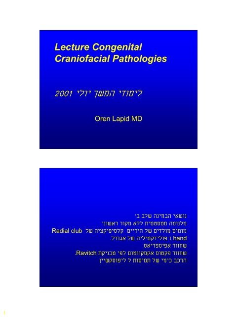
<strong>Lecture</strong> Congenital<br />
<strong>Craniofacial</strong> Pathologies<br />
2001 ילוי ךשמה ידומיל<br />
Radial club<br />
Oren Lapid MD<br />
'ב<br />
בלש הניחבה יאשונ<br />
ינושאר רוקמ אלל תיטטסטמ המונלמ<br />
לש היצקיפיסלק םיידיה לש םידלומ םימומ<br />
. לדוגא לש היליטקדילופ ו hand<br />
. Ravitch<br />
סאידפסיפא רוזחש<br />
תקינכט יפל םוטווקסקא סוטקפ רוזחש<br />
ןיישקסופיל ל תוסימת לש ימיכ בכרה
2<br />
<strong>Craniofacial</strong> surgery<br />
• Plastic surgery of the cephalic extremity<br />
– skull, face, Orbit<br />
– Congenital<br />
– Post traumatic<br />
– Tumors<br />
Craniosynostosis:<br />
• A general term encompassing a variety of<br />
developmental disorders characterized by<br />
inadequate capacity of the cranium to<br />
accommodate the growing brain, which<br />
results in compensatory deformities. (Less<br />
accurate for the minor forms where there is<br />
deformity with out problems of capacity)
3<br />
Simple: involves the cranial sutures,<br />
although there often is an extension into<br />
the base of the skull.<br />
Syndromatic: involves also the face with<br />
possible involvement of the limbs.<br />
Impact<br />
Aesthetic disfigurement<br />
Functional disorders: Increased ICP,<br />
hydrocephalus, visual impairment.
4<br />
Epidemiology<br />
• Detecteble abnormalities due to<br />
craniosynostosis are seen in<br />
1:1700-1900 births.<br />
• Most common types<br />
• Sagital schaphocephaly<br />
• Metopic trignocephaly<br />
• Unilateral coronal Plagiocephaly<br />
1:10,000<br />
• Bilateral coronal Brachicephaly<br />
Pathogenesis mechanisms to<br />
be completed
6<br />
ICP<br />
• Increased ICP is seen in cases of<br />
craniosynostosis more in cases where<br />
several sutures are involved.<br />
• Surgery decreases ICP.<br />
• A small skull doesn’t imply ICP elevation but<br />
ICP elevations usually seen in small skulls.<br />
Not all authors agree.<br />
• Signs of increased ICP<br />
• Papiledema, headaches, vomiting, lethargy,<br />
anorexia, beaten silver appearance on X-ray.
7<br />
Hydrocephalus<br />
• Hydrocephalus<br />
• Rare, seen more in the syndromatic<br />
cases.<br />
• Enlarged ventricles may be seen as part<br />
of the syndrome as in Apert’s. Changes<br />
in ventricle size are more accurate for<br />
diagnosis.<br />
Mental retardation<br />
• It is not clear how many of these children<br />
really have mental retardation. It is not clear if<br />
it is related to the problem or independent<br />
• Some just look retarded - environmental<br />
influences.<br />
• In children with non syndromatic<br />
craniosynostosis there was no increase in IQ<br />
after surgery. Other studies claim there was<br />
an improvmentt.
8<br />
Visual abnormalities<br />
• Optic atrophy and papilledema,<br />
stretching of optic nerve, pressure on<br />
the nerve. Damage from increased ICP.<br />
Morphology
9<br />
Acrocephaly ( also tower skull or<br />
oxycephaly of Virchow)<br />
Cranium in which the anterior part is<br />
higher than the posterior part, slanting<br />
in a front to back direction.
10<br />
Brachycephaly<br />
AP shortening, forehead and suprorbital<br />
bar are retropositioned.<br />
Results from bi-coronal stenosis. Typical<br />
of Apert’s and Crouzon.<br />
Oxycephaly<br />
Extreme upward growth with loss of AP &<br />
Lat diameters. Obtuse or absent<br />
nasofrontal angle. Usually a result of<br />
closure of the coronal suture.
11<br />
Turricephaly<br />
Exaggerated upward growth caused by<br />
frontoparietal suture closure. Seen in<br />
Apert’s<br />
Scaphocephaly<br />
Hull shaped cranium,reduced width with<br />
increased length, seen with premature<br />
sagital suture closure. The most<br />
common type. M:F 4:1
12<br />
Trigonocephaly<br />
Narrow and sharp forehead caused by<br />
premature closure of<br />
metopic suture. May be accompanied by<br />
hypotelorism.<br />
Clover leaf (Kleebattscaädel)<br />
Multiple sutural fusions, constriction ring<br />
in the lamboid-squamosal zone bulging<br />
of frontal and temporal lobes
13<br />
Plagiocephaly<br />
• Cranial asymmetry (various sutures)<br />
Plagiocephaly<br />
• Frontal<br />
– Ipsilateral coronal and frontosphenoidal sutures, flat<br />
forehead, backward displacement of orbit, bulging of<br />
contarlateral parietal area. Mandibular<br />
asymmetry.”harlequin” orbit.<br />
• Occipital<br />
– Flattening Lamboid suture must be distinguished from<br />
positional plagiocephaly.
14<br />
Plagiocephaly<br />
•Positional caused by extrinsic<br />
forces<br />
•Positional plagiocephaly does not<br />
require surgical treatment.<br />
Plagiocephaly
15<br />
Syndromes:<br />
• Different sutures may be involved in<br />
different patients with the same<br />
syndrome.<br />
• The difference may be in other parts of<br />
body.<br />
• Mental retardation found to some extent<br />
in all syndromes.
16<br />
Crouzon (<strong>Craniofacial</strong><br />
dysostosis)<br />
(Acrocephalosyndactyly, type<br />
II)<br />
• 1:25,000<br />
• Autosomal dominant (Most cases sporadic)<br />
Chromosome 10<br />
• Craniosynostosis, midfacial hypoplasia,<br />
exopthalmos.<br />
Crouzon cont<br />
• Various dformations of skull- usually brachy.<br />
• Midfacial retrusion – class III bite and shallow<br />
orbits. May cause damage to eyes. Beaked<br />
nose, High arched palate<br />
• Conductive hearing loss is common.<br />
• Synostosis develops during 1 st year of life<br />
usually complete by 3 rd .<br />
• Increased ICP common.
17<br />
Apert<br />
(Acrocephalosyndactyly,<br />
type I)<br />
• 1:160,000<br />
• Autosomal dominant (Most cases sporadic)<br />
• High cranial vault, flat posteriorly while<br />
bulging in the front. Brachycephalic,<br />
Turribracephalic.<br />
• Syndactyly of all four extremities- symmetrical<br />
middle three digits or more.
18<br />
Apert cont<br />
• Mild telorbitism, midfacial retrusion,<br />
deficient maxilla, abnormal dentition,<br />
Cleft palate 11-30%, low hair line,<br />
hypertrichosis of the eye brows, ptosis<br />
(mainly lateral), well developed<br />
protruding tongue.<br />
• Mental reardation seen in most.
19<br />
Pfeiffer<br />
(Acrocephalosyndactyly,<br />
type V)<br />
• Autosomal dominant (Most cases sporadic)<br />
Chromosome 8<br />
• Brachy-turricephalic, broad thumbs and great<br />
toes, occasional minor syndactyly,maxillary<br />
retrusion, possible high arched palate and<br />
submucous cleft.<br />
• Intelligence normal to mild mental retardation.
20<br />
Sathre-Chotzen<br />
(Acrocephalosyndactyly,<br />
type III)<br />
• Autosomal dominant (Most cases sporadic)<br />
chromosome 7<br />
• Brachycephalic cranium, various degrees of<br />
craniosynostosis, facial asymmetry, shallow<br />
orbits, telecantus, nasal septal deviation, low<br />
hair line minor syndactyly. Maxillary<br />
hypoplasia.<br />
• Intelligence normal to mild mental retardation.
21<br />
Carpenter<br />
• Autosomal recesive (Most cases sporadic)<br />
• Various cranial malformations, flat nasal<br />
canthi, dystrophic orbits, (possible anomalies<br />
of globe), low set ears<br />
• Hand (obligatory, syndactyly, brachydactyly,<br />
polydactyly)<br />
• Obesity, hypogonadism.<br />
• Mental retardation.<br />
Clefts<br />
• Major facial clefts 1.5-4.5/100,000 (CL<br />
CLP 1:750-1000)<br />
• Various degrees of such defects.
22<br />
Classification of Tessier<br />
• The orbit is the point of reference as it<br />
belongs both to cranial and facial clefts.<br />
• Counter clockwise, South to North<br />
• There may be both north and south<br />
components<br />
• Clefts always have hypoplasia (which may be<br />
minimal)<br />
• Pictures clefts 0-14
23<br />
Cleft 0-14 Cleft 1-13 bilateral<br />
Cleft 3 Cleft 4<br />
Cleft 5 bilateral<br />
Cleft 10<br />
Cleft 6 bilateral
24<br />
Clasification of Van der<br />
Mulen<br />
• The facial skeleton develops along an<br />
“S”<br />
• Focal fetal dysplasia,
25<br />
Telorbitism-Hypertelorism<br />
• Distance between<br />
anterior lacrimal<br />
crests<br />
• Men 19.5-<br />
30.7 (28) mm<br />
• Women 18.5-29.5<br />
(25) mm<br />
• 1 0 30-35 mm<br />
• 2 0 35-39 mm<br />
• 3 0 >40 mm<br />
Telorbitism-Hypertelorism<br />
• Up to 40 mm no problem<br />
• Tessier claims it is secondary to clefts or<br />
craniostenosis while Van-der-Mulen attributes<br />
it to arrest during 5 th -8 th weeks of<br />
embryogenesis.<br />
• Repair is undertaken to try and restore<br />
binocular vision and to improve the esthetic<br />
results.
26<br />
Frontoethmoidal<br />
Encephalomeningoceles<br />
• Failure of closure of the neural tube with<br />
herniation of CNS tissues from the<br />
forehead until the sacrum.<br />
• Nasofrontal, Naasoethmoidal and<br />
Nasoorbital<br />
• Rare seen mostly in Far East in<br />
Thailand 1:6000<br />
• Clefts 0,14, 1,13<br />
Oculomotor disturbances in<br />
craniofacial malformation.<br />
• Abnormal vectors of extraocular<br />
muscles or absence of muscles.<br />
• Altered intraocular distance and angle<br />
• Involvement of cranial nerves
27<br />
Laterofacial Microsomias<br />
• Represent groups of clefts 6,7,8<br />
Treacher Collins Complex<br />
(Franceschetti-Zwahlen-<br />
Klein Mandibulofacial<br />
dysostosis)<br />
• 1:25,000-50,000<br />
• Autosomal dominant. Chromosome 5<br />
• Variable penetration
28<br />
Treacher Collins<br />
• Bilateral and symetric<br />
• Convex profile, prominent nasal dorsum<br />
over a retrusive jaw and chin. Typical<br />
eyes, toungue shaped sideburns.<br />
Maldeveloped ears.<br />
• Complete form absence of the malar<br />
bone and the zygomatic arch.
29<br />
Treacher Collins<br />
• EYES<br />
– Eye<br />
• Strabismus, amblyopia<br />
• Lower eyelid<br />
Notch between lateral and<br />
middle third, absent eye lashes<br />
– Upper eyelid Microform coloboma<br />
– Eyebrow Notch<br />
– Lacrimal Absence of lower punctum<br />
– Lateral CantusNo insertion, free, small<br />
palpabrale fissure.<br />
– Orbit Open laterally because of the<br />
absent structures. The<br />
orbital floor is inclined<br />
outwards 45 0 .<br />
• Nose<br />
– Narrow, deviated, hooked, possible cohanal atresia.<br />
• Cheek<br />
– Sclerodermic furrow from the lower eye lid notch to the mandibular angle.<br />
• Oral comissure<br />
– Frequently macromastia<br />
• Alveola Palate<br />
– Narrow dental arch, high palate,<br />
• Maxilla& sinus<br />
– Under developed and small<br />
• Temporalis<br />
– The muscle is usually atrophic the masseter is not absent.<br />
• TMJ<br />
– Frequently hypoplastic<br />
• Mandible<br />
– Short ascending ramus. Lower body is hyoplastic.<br />
• Chin<br />
– Retrusion and increase in height.<br />
• Ear<br />
– Microtia and Cryptotia, Middle ear abnormalities<br />
• Cleft palate<br />
– May be present
30<br />
Nager syndrome (Acrofacial<br />
Dysostosis)<br />
• Similar to Treacher Collins but rarer<br />
• Autosomal recesive<br />
Binder’s Syndrome<br />
(Maxillonasal Dysplasia)<br />
• Short nose with flat bridge short<br />
columella
31<br />
Pierre Robin Sequence<br />
• Retrognathia, glosoptosis, airway obstruction.<br />
Cleft of soft and occasionally hard palate<br />
seen in 50%.<br />
• Etiology- late straightening of the fetus or late<br />
descent of the tongue.<br />
• Children have FTT de to difficulty breathing<br />
and feeding. Vicious cycle<br />
• Treatment- prone position, tongue adhesion.<br />
Hemifacial microsomia<br />
First and second branchiall<br />
arch syndrome<br />
Otomandibular dystosis<br />
<strong>Craniofacial</strong> microsomia<br />
Lateral facial dysplasia<br />
Otomandibular syndrome
33<br />
Hemifacial microsomia<br />
• No genetic background 1:3500-5600 births<br />
• Unilateral microtia, macrostomia, and failure of<br />
formation of the mandibular ramus and condyle.<br />
• Orbit, zygoma, temporal bone, maxilla and nose may<br />
be involved.<br />
• The defect may seem minor at birth but progresses<br />
with growth.<br />
• Soft tissue deficits range from normal to major<br />
deficiency of sub cutaneous tissue and muscle, the<br />
parotid may also be affected.<br />
• CNS Abnormalities: agenesis the corpus callosum,<br />
hydrocephalus, cranial nerve anomalies facial palsy)<br />
• Tongue depressor test.<br />
Pruzansky’s clasification by<br />
grades<br />
I Minimal hypoplasia<br />
II Deformed TMJ, distortion of<br />
condyle, ramusand sigmoid notch.<br />
•III complete absence of the ramus<br />
and glenoid fossa, the mandibular<br />
body ends in the molar area.
34<br />
Goldenhar Syndrome<br />
• Similar to Treacher Collins<br />
• Asymetrical skull, prominent frontal<br />
bossing, low hair line, mandibular<br />
hypoplasia, low set ears, colobomas of<br />
upper eyelids, epibulbar dermoids,<br />
accessory ear lobes, vertebral<br />
anomalies and a change in the clefting<br />
pattern.
35<br />
Romberg’s disease<br />
(Progresive facial<br />
hemiatrophy)<br />
• First or second decade of life. F:M 1.5:1<br />
• Unilateral in 95% of cases<br />
Etiology?<br />
• Infection, scleroderma,cervical<br />
sympathetic loss.<br />
• Progresive hemifacial atrophy of skin,<br />
soft tissue and bone.<br />
• Coup de sabre sign in 50%.
36<br />
Reconstruction<br />
• Reconstruction is under taken at least a<br />
year after the disease is static.<br />
• Reconstruction by soft tissue flaps and<br />
grafts and bone. Fat injection is also an<br />
option.<br />
Surgical repair
37<br />
Pre operative evaluation<br />
• History<br />
– Family, pregnancy..<br />
• Physical<br />
– Observation, palpation of sutures,<br />
fontanels, neurological exam.<br />
• Other abnormalities<br />
– search for other abnormalities<br />
Pre operative evaluation<br />
• Imaging<br />
– X-ray (including C-spine)<br />
– Cephalometris<br />
– CT 3DCT<br />
– MRI<br />
• Imaging has a major importance in the<br />
management of these cases with the<br />
possibility of performing mock surgery
38<br />
Timing of surgery<br />
• The consensus is before one year.<br />
• Earlier<br />
– Better growth, early relief of ICP.<br />
• Later<br />
– Child stands surgery better, nearer to adult<br />
size.<br />
• Facial osteotomies<br />
– are performed at a later age.<br />
Principles of craniofacial<br />
surgery<br />
• Work is on a dynamic structure<br />
Surgery is performed on a growing skull,<br />
growth can alter the results and at the same<br />
time may change the growth patterns.<br />
• Patients positioned according to the operative<br />
plan and area.<br />
• Wide dissection of affected areas, Bicoronal<br />
incisions.<br />
• Combined intra and extracranial approaches.
39<br />
• Extensive use of bone grafts<br />
• Bone grafts, free flaps, Distraction<br />
osteogenesis.<br />
• Strip craniotomies.<br />
• Total craniectomy (abandoned)<br />
• Dead space is replaced by brain growth.<br />
• Elevation of scalp flaps and then periostal<br />
flaps.<br />
• Via burr holes the dura is separated from the<br />
skull.<br />
• Starting at the burr holes the bones are<br />
sawed in-situ and also shaped and modified<br />
outside the patient.<br />
• The older the child the more brittle the bones<br />
– less easy to work.<br />
• Methods of fixation. Wires, plates, sutures.<br />
• Absorbable vs. permanent plates, Plate<br />
migration.
40<br />
Correction of simple sutural<br />
synostosis<br />
• Aim: correct functional and esthetic<br />
deficits<br />
• Strip craniotomies<br />
– Not suficent for an esthablished deformity<br />
but may be used in infancy, Bone growth<br />
and regeneration fill the defects.<br />
Correction of simple sutural<br />
synostosis<br />
• Plagiocephaly<br />
– Frontoorbital remodeling<br />
• Brachycephaly<br />
– Frontoorbital remodeling<br />
– The orbital bar is remodeled and repositioned in<br />
an appropriate location<br />
• Metopic<br />
– Deformed frontal bone Remodeled or<br />
replaced by parietal bone
43<br />
Correction of syndromatic<br />
sutural synostosis<br />
• Advancement of forehead and midface<br />
with monoblock frontal osteotomy.<br />
• Le fort osteotomies.<br />
• Facial bipartition for telorbitism.<br />
• Mid face deformities<br />
• Le Fort III, Le Fort III+I, Monoblock<br />
advancment
44<br />
Le Fort III<br />
Facial<br />
Bipartition
45<br />
Forehead Advancement<br />
Forehead Advancement Floating
46<br />
Forehead Advancement
47<br />
Forehead Advancement for Trigonocephaly
48<br />
Correction of Laterofacial<br />
Microsomias<br />
• Dental- orthodontic treatment can improve<br />
results and direct growth.<br />
• Distraction osteogenesis<br />
• Mandibular osteotomies<br />
• Bone grafts transfers for reconstruction of the<br />
mandibular ramus TMJ<br />
• Onlay bone grafts for contour restoration.<br />
Complications<br />
• Death 1.5-2%<br />
• Hemorhagic complications. There is<br />
significantand continous blood loss in<br />
these operations. Replacment required<br />
• Air emboli.
49<br />
Complications<br />
• Infection of soft tissues and bones more when<br />
there is a communication of bone with the<br />
respiratory system. Related to the length of<br />
surgery. Prophylactic antibiotics are given.<br />
• Infection (meningitis)<br />
• CSF leak<br />
• Ophthalmic complications- blindness.<br />
• Cerebral edema<br />
• Respiratory obstruction<br />
• Sagital sinus thrombosis<br />
• Seizures<br />
Thanks<br />
When will you fix me ?