The new generation in ECG interpretation - Philips
The new generation in ECG interpretation - Philips
The new generation in ECG interpretation - Philips
You also want an ePaper? Increase the reach of your titles
YUMPU automatically turns print PDFs into web optimized ePapers that Google loves.
<strong>The</strong> <strong>new</strong> <strong>generation</strong><br />
<strong>in</strong> <strong>ECG</strong> <strong>in</strong>terpretation<br />
DXL<br />
16<br />
<strong>Philips</strong> DXL <strong>ECG</strong> Algorithm, Release PH100B<br />
DXL<br />
16<br />
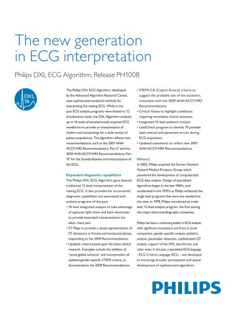
<strong>The</strong> <strong>Philips</strong> DXL <strong>ECG</strong> Algorithm, developed<br />
by the Advanced Algorithm Research Center,<br />
uses sophisticated analytical methods for<br />
<strong>in</strong>terpret<strong>in</strong>g the rest<strong>in</strong>g <strong>ECG</strong>. While <strong>in</strong> the<br />
past <strong>ECG</strong> analysis programs were limited to 12<br />
simultaneous leads, the DXL Algorithm analyzes<br />
up to 16 leads of simultaneously acquired <strong>ECG</strong><br />
waveforms to provide an <strong>in</strong>terpretation of<br />
rhythm and morphology for a wide variety of<br />
patient populations. <strong>The</strong> algorithm reflects <strong>new</strong><br />
recommendations, such as the 2007 AHA/<br />
ACCF/HRS Recommendations, Part II 1 and the<br />
2009 AHA/ACCF/HRS Recommendations, Part<br />
VI 2 for the Standardization and Interpretation of<br />
the <strong>ECG</strong>.<br />
Expanded diagnostic capabilities<br />
<strong>The</strong> <strong>Philips</strong> DXL <strong>ECG</strong> Algorithm goes beyond<br />
traditional 12-lead <strong>in</strong>terpretation of the<br />
rest<strong>in</strong>g <strong>ECG</strong>. It also provides for <strong>in</strong>cremental<br />
diagnostic capabilities not associated with<br />
analysis programs of the past:<br />
• 16-lead <strong>in</strong>tegrated analysis to take advantage<br />
of optional right chest and back electrodes<br />
to provide extended <strong>in</strong>terpretations for<br />
adult chest pa<strong>in</strong><br />
• ST Maps to provide a visual representation of<br />
ST deviations <strong>in</strong> frontal and horizontal planes,<br />
respond<strong>in</strong>g to the 2009 Recommendations<br />
• Updated criteria based upon the latest cl<strong>in</strong>ical<br />
research. Examples <strong>in</strong>clude the addition of<br />
“acute global ischemia” and <strong>in</strong>corporation of<br />
updated gender-specific STEMI criteria, as<br />
documented <strong>in</strong> the 2009 Recommendations<br />
• STEMI-CA (Culprit Artery) criteria to<br />
suggest the probable site of the occlusion,<br />
consistent with the 2009 AHA/ACCF/HRS<br />
Recommendations<br />
• Critical Values to highlight conditions<br />
requir<strong>in</strong>g immediate cl<strong>in</strong>ical attention<br />
• Integrated 15-lead pediatric analysis<br />
• LeadCheck program to identify 19 possible<br />
lead reversal and placement errors dur<strong>in</strong>g<br />
<strong>ECG</strong> acquisition<br />
• Updated statements to reflect <strong>new</strong> 2007<br />
AHA/ACCF/HRS Recommendations<br />
History<br />
In 2002, <strong>Philips</strong> acquired the former Hewlett-<br />
Packard Medical Products Group which<br />
pioneered the development of computerized<br />
<strong>ECG</strong> data analysis. Design of specialized<br />
algorithms began <strong>in</strong> the late 1960’s, and<br />
accelerated <strong>in</strong> the 1970’s as <strong>Philips</strong> enhanced the<br />
s<strong>in</strong>gle lead programs that were the standard at<br />
the time. In 1978, <strong>Philips</strong> <strong>in</strong>troduced its multi-<br />
lead, 12-lead analysis program, the first among<br />
the major electrocardiography companies.<br />
<strong>Philips</strong> has been a cont<strong>in</strong>u<strong>in</strong>g leader <strong>in</strong> <strong>ECG</strong> analysis,<br />
with significant <strong>in</strong>novations and firsts <strong>in</strong> serial<br />
comparison, gender-specific analysis, pediatric<br />
analysis, pacemaker detection, sophisticated QT<br />
analysis, support of the XML data format, and<br />
other areas. In the past, a specialized <strong>ECG</strong> language<br />
- <strong>ECG</strong> Criteria Language (ECL) - was developed<br />
to encourage broader participation and speed<br />
development of sophisticated algorithms.
V1<br />
V3R<br />
V4R<br />
V5R<br />
RA<br />
Today, this experience and expertise is reflected <strong>in</strong> the<br />
<strong>Philips</strong> DXL <strong>ECG</strong> Algorithm with its <strong>in</strong>dustry lead<strong>in</strong>g<br />
<strong>in</strong>tegration of extended leads and expanded analysis<br />
capabilities.<br />
Analysis process<br />
<strong>The</strong> DXL Algorithm has multiple steps to produce<br />
precise and consistent <strong>ECG</strong> measurements that are used<br />
to generate <strong>in</strong>terpretive statements.<br />
Acquisition and quality assessment<br />
• Cont<strong>in</strong>uous signal quality feedback is provided to the<br />
user via color-coded waveforms and messages<br />
• 12 to 16 user-selected leads are captured for 10 seconds<br />
and digitized at 8 KHz per lead<br />
• Unique LeadCheck software identifies 19 possible<br />
swapped leads for correction<br />
Waveform recognition and measurement<br />
• Pacemaker pulses are accurately identified<br />
• Each complex <strong>in</strong> each lead is measured <strong>in</strong>dividually.<br />
Excessive variation <strong>in</strong> a lead can be used to determ<strong>in</strong>e<br />
if that lead is unreliable for global measurements<br />
• Complexes are classified <strong>in</strong>to families based upon rate<br />
and morphology parameters. Paced beats are classified<br />
<strong>in</strong>dependently<br />
• Representative beats are created by averag<strong>in</strong>g aligned<br />
beats from the dom<strong>in</strong>ant family<br />
Patient <strong>ECG</strong> and Patient Data Quality Assessment<br />
RL<br />
LL<br />
V2<br />
V3<br />
V4<br />
V5<br />
V6<br />
LA<br />
Pace Pulse Detection Average Beat Formation<br />
DXL<br />
16<br />
I<br />
II<br />
.<br />
V9<br />
Smart Lead<br />
Quality<br />
Morphology<br />
Rhythm<br />
LeadCheck<br />
Measurements<br />
Culprit Artery<br />
• Rhythms with some paced complexes undergo<br />
morphologic analysis of the non-paced beats<br />
• Individual lead and global measurements of<br />
representative beats are made. Innovative methods<br />
are used for difficult measurements, such as onset of<br />
P and end of T<br />
• Specialized techniques, such as synthesized<br />
vectorcardiograms, are used for transverse plane<br />
measurements (specifically for pediatric <strong>ECG</strong>s)<br />
Application of criteria<br />
• <strong>The</strong> criteria can use any comb<strong>in</strong>ation of lead and<br />
global measurements, enhanc<strong>in</strong>g the flexibility and<br />
power of <strong>in</strong>terpretive capabilities<br />
• Established traditional and modern <strong>in</strong>terpretation<br />
criteria, <strong>in</strong>clud<strong>in</strong>g age- and gender-specific criteria<br />
and criteria based upon latest scientific f<strong>in</strong>d<strong>in</strong>gs and<br />
us<strong>in</strong>g current nomenclature, are applied<br />
• If additional leads beyond the standard 12 are<br />
available, <strong>in</strong>cremental criteria are applied to<br />
enhance specificity and sensitivity<br />
• New measurements and criteria are applied for<br />
STEMI-CA (to suggest Culprit Artery occlusion<br />
sites), ST Maps (to show the spatial orientation<br />
of ST deviations), and Critical Values (to highlight<br />
<strong>ECG</strong>s requir<strong>in</strong>g urgent cl<strong>in</strong>ical attention)<br />
Feedback<br />
to Operator<br />
ST Maps<br />
Critical Values<br />
16-Lead Criteria Interpretive Report Overreader Overread Report<br />
From acquisition to report <strong>generation</strong>, the <strong>Philips</strong> DXL 16-Lead Algorithm uses multiple steps to ensure a high quality <strong>in</strong>terpretation.<br />
2
Waveform Storage<br />
Previous <strong>generation</strong> technologies down-sampled <strong>ECG</strong><br />
waveforms for transmission and long-term data storage<br />
purposes. <strong>The</strong> DXL Algorithm uses lossless data<br />
compression, ensur<strong>in</strong>g that the orig<strong>in</strong>al, raw data is available<br />
for further review and analysis. This <strong>in</strong>creases the value of<br />
collected data for the development of further enhancements<br />
and <strong>in</strong>corporation of <strong>new</strong> scientific f<strong>in</strong>d<strong>in</strong>gs. It also supports<br />
the needs of pharmaceutical cl<strong>in</strong>ical research.<br />
Use of additional right chest and posterior leads<br />
While the limb leads and chest leads comprise the<br />
standard 12-lead <strong>ECG</strong>, it has long been recognized that<br />
additional electrodes can provide <strong>in</strong>formation that is<br />
poorly or not at all seen on the traditional 12-lead <strong>ECG</strong>.<br />
In addition to the standard limb and chest leads,<br />
the DXL Algorithm can <strong>in</strong>corporate any four of six<br />
additional leads from the follow<strong>in</strong>g:<br />
• right chest leads: V3R, V4R, V5R<br />
• posterior leads: V7, V8, V9<br />
Some users may choose to add just one or two leads to<br />
the standard 12, while others may choose to record the<br />
maximum of 16 leads to provide the additional waveform<br />
<strong>in</strong>formation as well to ga<strong>in</strong> the additional sensitivity and<br />
specificity that the DXL Algorithm can provide with the<br />
additional lead <strong>in</strong>formation. Among the areas that can benefit:<br />
Right heart analysis (V3R, V4R, V5R)<br />
Right ventricular <strong>in</strong>farcts may accompany <strong>in</strong>ferior<br />
myocardial <strong>in</strong>farction and have strong prognostic<br />
implications. <strong>The</strong> signs are subtle <strong>in</strong> the right-sided leads<br />
and are generally <strong>in</strong>visible <strong>in</strong> the standard leads.<br />
<strong>The</strong> DXL Algorithm is not limited to a s<strong>in</strong>gle right lead<br />
and can take advantage of <strong>in</strong>formation that may be present<br />
<strong>in</strong> V3R, V4R, and/or V5R. Additional criteria use this<br />
<strong>in</strong>formation, plus criteria based upon the traditional 12<br />
leads, to provide improved specificity and sensitivity. For<br />
example, consistent with the 2009 Recommendations, the<br />
algorithm supports the record<strong>in</strong>g of both V3R and V4R<br />
for patients present<strong>in</strong>g with evidence of acute <strong>in</strong>ferior wall<br />
ischemia or <strong>in</strong>farction. And the DXL Algorithm provides<br />
<strong>in</strong>cremental analysis based upon those additional leads.<br />
Posterior wall analysis (V7, V8, V9)<br />
Posterior myocardial <strong>in</strong>farction has traditionally been<br />
diagnosed from reciprocal changes seen <strong>in</strong> leads V1 and<br />
V2. ST-segment depression may be very subtle <strong>in</strong> these<br />
leads and go unnoticed or misdiagnosed, especially if gender<br />
differences are ignored. ST-segment elevation recorded<br />
<strong>in</strong> the posterior leads can aid <strong>in</strong> the <strong>in</strong>terpretation,<br />
especially <strong>in</strong> the early stages of acute posterior<br />
myocardial <strong>in</strong>farction.<br />
<strong>The</strong> DXL Algorithm can take advantage of <strong>in</strong>formation <strong>in</strong><br />
one or more of leads V7, V8, and V9 to provide greater<br />
specificity and sensitivity <strong>in</strong> detect<strong>in</strong>g posterior <strong>in</strong>farcts.<br />
Pediatrics<br />
Many pediatric <strong>in</strong>stitutions add three leads to the 12 lead<br />
set. <strong>The</strong> most common sites are V3R, V4R, and V7. <strong>The</strong><br />
selected <strong>in</strong>cremental leads are <strong>in</strong>tegrated <strong>in</strong>to the DXL<br />
Algorithm’s pediatric analysis.<br />
ST Maps<br />
<strong>The</strong> 2009 AHA/ACCF/HRS document recommends<br />
the display of the spatial orientation of ST-segment<br />
deviations <strong>in</strong> both frontal and transverse planes. <strong>Philips</strong><br />
cardiographs can display patented ST Maps <strong>in</strong> both<br />
planes to provide for rapid visual assessment of the<br />
degree of ST abnormalities. <strong>The</strong> maps are plotted<br />
<strong>in</strong> “Cabrera sequence” to reflect the anatomical<br />
orientation.<br />
This presentation, previously available only on <strong>Philips</strong><br />
IntelliVue patient monitors, provides dist<strong>in</strong>ct patterns<br />
for different anatomic sites of <strong>in</strong>farcts, global ischemia,<br />
pericarditis, and other conditions that are readily seen<br />
us<strong>in</strong>g such maps.<br />
ST Maps provide for rapid visual assessment of ST abnormalities.<br />
3
STEMI detection<br />
ST-segment elevations <strong>in</strong> <strong>ECG</strong> trac<strong>in</strong>gs provide highly<br />
effective risk stratification <strong>in</strong> early acute coronary<br />
syndromes. For patients with ST-segment elevation<br />
acute myocardial <strong>in</strong>farction (STEMI), the most critical<br />
factor <strong>in</strong> improv<strong>in</strong>g survival rates is the earliest<br />
possible adm<strong>in</strong>istration of reperfusion therapy and<br />
reduction of “door-to-balloon” and “discovery-to-<br />
treatment” times.<br />
<strong>The</strong> DXL Algorithm provides advanced capabilities<br />
for fast, accurate detection of STEMI based on the<br />
<strong>new</strong>est recommendations. Unique to <strong>Philips</strong>, the DXL<br />
Algorithm can provide <strong>in</strong>formation <strong>in</strong> 16 <strong>ECG</strong> leads<br />
selected at the time of acquisition, allow<strong>in</strong>g right<br />
heart (V3R, V4R, and/or V5R) and posterior (V7, V8,<br />
and/or V9) <strong>in</strong>formation to be <strong>in</strong>cluded <strong>in</strong> the data<br />
presentation and subsequent analysis.<br />
Classical STEMI applies only to ST elevation <strong>in</strong> a group<br />
of leads. With obstructions <strong>in</strong> the left ma<strong>in</strong> coronary<br />
artery or multiple obstructions <strong>in</strong> several coronary<br />
arteries, an <strong>ECG</strong> pattern of widespread ST depression<br />
is seen along with ST elevation seen <strong>in</strong> aVR and/or<br />
VI. This pattern, recognized by the DXL Algorithm, is<br />
called global ischemia and has prognostic significance.<br />
Gender-specific <strong>ECG</strong> <strong>in</strong>terpretation<br />
As cardiac medic<strong>in</strong>e cont<strong>in</strong>ues to advance, there has been<br />
cont<strong>in</strong>ued learn<strong>in</strong>g regard<strong>in</strong>g the physiological differences<br />
between men and women. Gender-specific criteria are<br />
not <strong>new</strong> to <strong>Philips</strong>. <strong>The</strong>y have been <strong>in</strong>corporated <strong>in</strong>to the<br />
multi-lead algorithms s<strong>in</strong>ce 1987 and have been enhanced<br />
cont<strong>in</strong>ually based upon the latest research and guidel<strong>in</strong>es.<br />
For example, the DXL Algorithm applies gender, lead and<br />
age limits for improved detection of acute MI. Based upon<br />
the 2009 recommendations, STEMI criteria are subject to<br />
reduced ST thresholds <strong>in</strong> women. <strong>The</strong> algorithm also uses<br />
gender-specific axis deviation and MI criteria, Cornell<br />
gender-specific criteria for detection of left ventricular<br />
hypertrophies, and Rochester and Rautaharju criteria for<br />
the detection of prolonged QT.<br />
Application of these gender-specific criteria results <strong>in</strong> an<br />
<strong>ECG</strong> <strong>in</strong>terpretation that helps cl<strong>in</strong>icians more accurately<br />
assess the cardiac state of patients of either sex.<br />
STEMI-CA for identification of culprit artery<br />
If STEMI criteria are met, the DXL Algorithm’s STEMI-CA<br />
criteria can identify the likely culprit artery or probable<br />
anatomical site caus<strong>in</strong>g the functional ischemia. This may<br />
have prognostic significance as well as help to p<strong>in</strong>po<strong>in</strong>t the<br />
offend<strong>in</strong>g lesion when multiple obstructions are present<br />
and can thus be used to optimize the treatment approach<br />
<strong>in</strong> the cath lab.<br />
Specific anatomic sites can <strong>in</strong>clude the follow<strong>in</strong>g<br />
coronary arteries:<br />
• left anterior descend<strong>in</strong>g artery (LAD)<br />
• right coronary artery (RCA)<br />
• left circumflex artery (LCx)<br />
• left ma<strong>in</strong> or multi-vessel disease (LM/MVD)<br />
Critical Values<br />
Critical Values are highly visible <strong>in</strong>dependent statements<br />
on the <strong>ECG</strong> report that highlight conditions requir<strong>in</strong>g<br />
immediate cl<strong>in</strong>ical attention. Specific statements are<br />
made for:<br />
• acute MI<br />
• global ischemia<br />
• complete heart block<br />
• extreme tachycardia<br />
Use of the Critical Values feature is configurable, and<br />
can be used to support quality <strong>in</strong>itiatives <strong>in</strong> multiple<br />
areas of the medical enterprise. For example, <strong>in</strong> the<br />
emergency department, Critical Values can support<br />
a hospital’s focus on Door-to-Balloon and quality<br />
<strong>in</strong>itiatives.<br />
<strong>The</strong> DXL Algorithm applies gender, age, and lead-specific criteria to<br />
provide for more accurate assessment of the cardiac status across<br />
diverse patient populations.<br />
4
Integrated pediatric analysis<br />
<strong>Philips</strong> <strong>in</strong>troduced the <strong>in</strong>dustry’s first pediatric <strong>ECG</strong><br />
analysis capabilities <strong>in</strong> 1982 and has built upon that<br />
experience to ma<strong>in</strong>ta<strong>in</strong> an advanced, fully <strong>in</strong>tegrated<br />
pediatric <strong>in</strong>terpretation. With the DXL Algorithm, the<br />
<strong>in</strong>terpretation now can use the availability of additional<br />
leads – typically 15 <strong>in</strong> the pediatric application.<br />
<strong>The</strong> pediatric criteria use 12 dist<strong>in</strong>ct age groups –<br />
grouped progressively from hours through days, weeks,<br />
months, and years – to ensure that the most age-<br />
relevant <strong>in</strong>terpretation criteria are applied for analyz<strong>in</strong>g<br />
the available waveform. <strong>The</strong> age is used throughout<br />
the criteria to def<strong>in</strong>e normal limits <strong>in</strong> heart rate, axis<br />
deviation, time <strong>in</strong>tervals, voltage values for <strong>in</strong>terpretation<br />
accuracy <strong>in</strong> tachycardia, bradycardia, prolongation or<br />
shorten<strong>in</strong>g of PR and QT <strong>in</strong>tervals, hypertrophy, early<br />
repolarization, myocardial ischemia and <strong>in</strong>farct, and other<br />
cardiac conditions.<br />
Mild right ventricular hypertrophy and <strong>in</strong>complete right<br />
bundle branch block differentiation is improved with<br />
a unique comb<strong>in</strong>ation of transverse measurements<br />
from a derived vectorcardiogram with more traditional<br />
measurements from the standard 12 leads and the extra<br />
pediatric leads.<br />
Left ventricular hypertrophy criteria<br />
<strong>The</strong> widely accepted Cornell criteria for left ventricular<br />
hypertrophy are <strong>in</strong>cluded with the traditional criteria.<br />
This <strong>in</strong>creases the sensitivity and specificity of detection.<br />
Advanced pacemaker detection<br />
Between 2 and 5% of rout<strong>in</strong>e diagnostic <strong>ECG</strong>s<br />
are acquired from patients with implanted cardiac<br />
pacemakers, devices which have become <strong>in</strong>creas<strong>in</strong>gly<br />
capable, efficient, and complex. To keep pace with<br />
these pacemaker developments, the DXL Algorithm is<br />
designed to handle a variety of atrial, ventricular, and<br />
A-V sequential pac<strong>in</strong>g modes. A sophisticated patented<br />
noise-adaptive pulse detector is run on all acquired leads<br />
with confirmation from multiple leads differentiat<strong>in</strong>g true<br />
pulses from noise.<br />
If the pacemaker is known to be present at the time of<br />
acquisition, the algorithm pulse detector sensitivity can<br />
be <strong>in</strong>creased to improve detection of extremely small<br />
pulses. In addition, analysis of pacemaker pulse locations<br />
with respect to P-waves and QRS complexes permits<br />
classification of a variety of pacemaker rhythms and<br />
identification of non-paced QRS complexes for further<br />
morphologic analysis <strong>in</strong> <strong>ECG</strong>s with paced and non-paced<br />
beats.<br />
Although the detector is quite sensitive, even to low<br />
amplitude pacer spikes typical of modern pacemakers, it<br />
can still differentiate these spikes from the very narrow<br />
and sharp QRS complexes typical of neonatal <strong>ECG</strong>s, a<br />
problem that has plagued previous <strong>generation</strong> algorithms.<br />
QT analysis and cl<strong>in</strong>ical research<br />
Because of the relatively recent appreciation of<br />
the connection between many medications and the<br />
development of Torsade de Po<strong>in</strong>tes, there has been<br />
re<strong>new</strong>ed <strong>in</strong>terest <strong>in</strong> the accurate measurement of the QT<br />
<strong>in</strong>terval and the various QT “corrections.”<br />
<strong>The</strong> DXL Algorithm uses an <strong>in</strong>novative measurement<br />
technique to ensure an accurate and reproducible end of<br />
T measurement, even <strong>in</strong> the presence of large U waves<br />
and biphasic or notched T waves. Because high resolution<br />
<strong>ECG</strong> signals are ma<strong>in</strong>ta<strong>in</strong>ed through analysis, transmission,<br />
and storage, the <strong>in</strong>tegrity of the end of T rema<strong>in</strong>s available<br />
for analysis and subsequent high resolution review typical<br />
<strong>in</strong> cl<strong>in</strong>ical research.<br />
<strong>The</strong> same QT algorithm has been further applied for<br />
use with<strong>in</strong> <strong>Philips</strong> IntelliVue monitor<strong>in</strong>g systems and<br />
ambulatory monitor<strong>in</strong>g products.<br />
Nomenclature<br />
<strong>The</strong> DXL Algorithm conforms to the latest AHA/ACCF/<br />
HRS statement recommendations for the standardization<br />
and <strong>in</strong>terpretation of the <strong>ECG</strong> 1 .<br />
1 AHA/ACCF/HRS Recommendations for the Standardization and Interpretation of the<br />
Electrocardiogram, Part II: Electrocardiography Diagnostic Statement List. J Am Coll<br />
Cardiology, 2007; 49:1128-135.<br />
2 AHA/ACCF/HRS Recommendations for the Standardization and Interpretation of the<br />
Electrocardiogram, Part VI: Acute Ischemia/Infarction. Circulation, 2009; 119:e262-e270.<br />
Note: A computer-<strong>in</strong>terpreted <strong>ECG</strong> report is not <strong>in</strong>tended to be a substitute for <strong>in</strong>terpretation<br />
by a qualified physician. Physician overread<strong>in</strong>g and confirmation of computerbased<br />
<strong>ECG</strong>s is required.<br />
This brochure describes features of the <strong>Philips</strong> DXL <strong>ECG</strong> Algorithm that may not be<br />
available on all <strong>Philips</strong> products <strong>in</strong>corporat<strong>in</strong>g <strong>ECG</strong> analysis. Please refer to the documentation<br />
supplied with your <strong>Philips</strong> product for specific versions and available features.<br />
5
<strong>Philips</strong> Healthcare is part of<br />
Royal <strong>Philips</strong> Electronics<br />
How to reach us<br />
www.philips.com/healthcare<br />
healthcare@philips.com<br />
fax: +31 40 27 64 887<br />
Asia<br />
+852 2821 5888<br />
Europe, Middle East, Africa<br />
+49 7031 463 2254<br />
Lat<strong>in</strong> America<br />
+55 11 2125 0744<br />
North America<br />
+1 425 487 7000<br />
800 285 5585 (toll free, US only)<br />
<strong>Philips</strong> Healthcare<br />
Global Information Center<br />
P.O. Box 1286<br />
5602 BG E<strong>in</strong>dhoven<br />
<strong>The</strong> Netherlands<br />
© 2009 Kon<strong>in</strong>klijke <strong>Philips</strong> Electronics N.V.<br />
All rights are reserved.<br />
<strong>Philips</strong> Healthcare reserves the right to make changes <strong>in</strong> specifications and/or to discont<strong>in</strong>ue any product at any time without notice or<br />
obligation and will not be liable for any consequences result<strong>in</strong>g from the use of this publication.<br />
Pr<strong>in</strong>ted <strong>in</strong> <strong>The</strong> Netherlands.<br />
MAR 2009


