the cytology-radiology-surgical pathology model for gram stains
the cytology-radiology-surgical pathology model for gram stains
the cytology-radiology-surgical pathology model for gram stains
You also want an ePaper? Increase the reach of your titles
YUMPU automatically turns print PDFs into web optimized ePapers that Google loves.
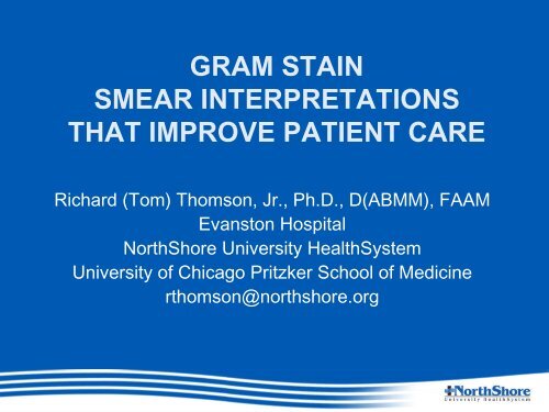
GRAM STAIN<br />
SMEAR INTERPRETATIONS<br />
THAT IMPROVE PATIENT CARE<br />
Richard (Tom) Thomson, Jr., Ph.D., D(ABMM), FAAM<br />
Evanston Hospital<br />
NorthShore University HealthSystem<br />
University of Chicago Pritzker School of Medicine<br />
rthomson@northshore.org
Multiple Urinary Tract Infections in<br />
and Elderly Male<br />
• Emergency Department Visit<br />
– 70 yo male with fever 101.6 o F<br />
– PMH includes 3 admissions <strong>for</strong> UTI in past 2 months<br />
– WBC 12,000 (87% PMN’s)<br />
– UA positive LE and Nitrites<br />
– Blood and midstream urine <strong>for</strong> culture collected<br />
– Admitted with <strong>the</strong> diagnosis of UTI<br />
– Levofloxacin IV
Urinary Tract Infection<br />
in an Elderly Male<br />
• Culture Report:<br />
50,000-100,000 CFU/ml E. coli<br />
• Gram Stain Report:<br />
4+ PMN’s<br />
4+ Mixed Urogenital Flora<br />
Gram stain reviewed and verified
Urinary Tract Infection<br />
in an Elderly Male<br />
• Culture Report:<br />
50,000-100,000 CFU/ml E. coli<br />
• Gram Stain Report:<br />
4+ PMN’s<br />
4+ Mixed Urogenital Flora<br />
Gram stain reviewed and verified<br />
Comment: Gram stain reviewed by Dr Thomson. The<br />
presence of PMN’s, bacterial morphologies<br />
resembling mixed colon flora and fecal debris<br />
suggests a vesico-colic fistula.
Questions, Answers and Topics<br />
• How has <strong>the</strong> Gram stain changed?<br />
• Is it still working?<br />
• Should <strong>the</strong> Gram stain be standardized?<br />
• Level I reporting (minimum competency) needed by<br />
all who “read” Gram <strong>stains</strong><br />
• Level II reporting <strong>for</strong> senior technologists and<br />
supervisors<br />
• Level III Interpretations <strong>for</strong> laboratory directors<br />
(medical microbiologists)
Questions, Answers and Topics<br />
• Pattern recognition of specific diseases<br />
– Case studies in Gram Stain Interpretations<br />
– Examples of interpretations that make a difference in<br />
patient care<br />
• How to make Gram stain reporting relevant in <strong>the</strong><br />
future
HOW TO STANDARDIZE THE<br />
GRAM STAIN PROCEDURE<br />
• Selecting a portion of <strong>the</strong> specimen<br />
• Preparing <strong>the</strong> smear<br />
• Low power (10X) examination<br />
• High power (100X) examination<br />
• Quantitation of cells and microorganisms<br />
• Examination and reporting based on specimen type<br />
• Slides <strong>for</strong> review
Selecting a Portion of <strong>the</strong> Specimen
PREPARING THE SMEAR<br />
Make a monolayer of cells
PREPARING THE SMEAR<br />
Concentrating fluids using a cytospin
LOW POWER (10X) EXAMINATION<br />
10-20 fields, quantitate cells<br />
select area <strong>for</strong> high power examination
HIGH POWER (100X) EXAMINATION<br />
20-40 fields, confirm cells, quantitate microorganisms
GRAM STAIN QUANTITIES<br />
• 1+ (very rare)<br />
– Less than 10 in all fields examined<br />
• 2+ (few)<br />
– More than 10 in all fields but less than 1/field<br />
• 3+ (moderate)<br />
– More than 1/field but less than 25/field<br />
• 4+ (many)<br />
– More than 25 in one field<br />
• 1-3+ versus 1-4+ quantities??
Staining Procedure to Standardize<br />
• Standard <strong>gram</strong> stain reagents and method to<br />
establish a normal against which variations<br />
can be compared<br />
– Fixation (heat v alcohol)<br />
– Dyes and concentrations<br />
– Decolorizer<br />
– Times
GRAM STAIN REPORTING<br />
BASED ON SPECIMEN SOURCE<br />
• Sterile fluid or tissue (presumed)<br />
– Report PMN’s<br />
– Report all microorganisms<br />
– > 3 morphologically typical shapes be<strong>for</strong>e reporting<br />
• Non-Sterile specimen source<br />
– Report PMN’s<br />
– “Name” microorganisms only if potential pathogen<br />
– Quantitate and report “normal flora”
SLIDES FOR REVIEW
GRAM STAIN<br />
POLICY FOR SLIDE REVIEW<br />
• Automatic Review (director requested)<br />
– Sterile fluid or tissue<br />
• Microorganism reported<br />
• 3-4 + PMN’s with no bacteria seen<br />
– Non-sterile source<br />
• Report is diagnostic<br />
• Requested Review (technologist requested)<br />
– Unsure of finding<br />
• Physician requested review<br />
– Interpretation
BACTERIAL MORPHOLOGIES<br />
LEVEL I<br />
MINIMUM COMPETENCY<br />
MORPHOLOGY REPORTED ORGANISM IMPLIED<br />
• Gram-positive cocci clusters Staphylococcus<br />
• Gram-positive coccci pairs/chains Streptococcus<br />
• Gram-positive cocci Staphylococus/Streptococcus<br />
• Gram-positive rod Any Gram-positive rod<br />
• Gram-negative diplococci Neisseria/Moraxella<br />
• Gram-negative coccobacilli Haemophilus/Bacteroides<br />
• Gram-negative rod Any Gram-negative rod<br />
• Yeast cells Yeast, usually Candida<br />
• Yeast cells with pseudohyphae Candida, not C. glabrata<br />
• O<strong>the</strong>r findings-leave <strong>for</strong> review<br />
• Cell morphologies<br />
– PMNs<br />
– Squamous epi<strong>the</strong>lial cells<br />
– Sputum screens
BACTERIAL MORPHOLOGIES<br />
LEVEL II<br />
Senior Technologists/Supervisors<br />
MORPHOLOGY REPORTED ORGANISM IMPLIED<br />
• Level I plus…<br />
• Gram-positive diplococci lancet-shaped S. pneumoniae<br />
• Gram-positive rod diph<strong>the</strong>roid Corynebacterium, etc.<br />
• Gram-positive rod boxcar Bacillus/Clostridium<br />
• Gram-positive rod endospores Bacillus/Clostridium<br />
• Gram-positive rod filamentous/branching Nocardia/Actinomyces<br />
• Hyphae septate Aspergillus, etc. type<br />
• Hyphae nonseptate Rhizopus, etc. type
BACTERIAL MORPHOLOGIES<br />
Level II<br />
Senior Technologists/Supervisors<br />
MORPHOLOGY REPORTED ORGANISM IMPLIED<br />
Continued<br />
• Gram-negative rod thick Enterobacteriaceae-type<br />
• Gram-negative rod thin Nonfermenter-type<br />
• Gram-negative rod pleomorphic Bacteroides-type<br />
• Gram-negative diplobacilli Acinetobacter-type<br />
• Gram-negative rod fusi<strong>for</strong>m Fusobacterium-<br />
/Capnocytophagia-type<br />
• Gram-negative rod curved Campylobacter/Vibrio<br />
• Gram-negative cocci tiny Brucella/Francisella
Interpretations<br />
Level III<br />
Laboratory Directors/Medical Microbiologists<br />
• Level I and Level II plus<br />
• Additional cells<br />
• Indicators of <strong>pathology</strong><br />
• Disease pattern recognition (Cases)<br />
– Vesico-colic fistula
Interpretations<br />
Level III<br />
Laboratory Directors/Medical Microbiologists<br />
• Disease pattern recognition<br />
– Respiratory tract<br />
• Pneumonia/bronchitis<br />
• Aspiration pneumonia<br />
• Chronic lung disease/COPD<br />
– Urinary tract<br />
• UTI<br />
• Vesico-colic fistula<br />
– Meningitis
Interpretations<br />
Level III<br />
Laboratory Directors/Medical Microbiologists<br />
• Skin-Soft Tissue-Closed Space Abscess<br />
– Staphylococcal<br />
– Mixed aerobic/anaerobic<br />
– Streptococcus milleri/anginosis<br />
– Nocardia<br />
• Toxemia<br />
– Streptococcal necortizing fasciitis<br />
– Clostridium gas gangrene<br />
• Miscellaneous<br />
– BV (bacterial vaginosis)<br />
– Lemierre’s disease (jugular vein thrombosis)<br />
– Gonococcal urethritis (male)<br />
– Crystalline joint disease
How To Make Gram Stain Reporting<br />
Relevant in <strong>the</strong> Future<br />
• Standardize smear preparation<br />
• Standardize staining procedures<br />
• Standardize reporting<br />
• Maintain levels of competency<br />
– Level I<br />
– Level II<br />
– Medical Microbiologist Interpretations<br />
• Standardize Gram stain review
Radiology Model
How To Make Gram Stain<br />
Reporting Relevant in <strong>the</strong> Future<br />
• Technologist <strong>gram</strong> stain report with digital<br />
images inserted in <strong>the</strong> report and an<br />
accompanying interpretation by <strong>the</strong> medical<br />
microbiologist<br />
• MICROBIOLOGY MODEL
Comment: Gram stain<br />
reviewed by Dr. Thomson.<br />
Branching septate hyphae<br />
also present among PMNs.<br />
Morphology suggests<br />
Aspergillus or<br />
morphologically similar<br />
mold. See attached image.


