

Phytochemical investigations on 'black glumed' Njavara (Oryza ...
Phytochemical investigations on 'black glumed' Njavara (Oryza ...
Phytochemical investigations on 'black glumed' Njavara (Oryza ...

You also want an ePaper? Increase the reach of your titles
YUMPU automatically turns print PDFs into web optimized ePapers that Google loves.
PHYTOCHEMICAL INVESTIGATIONS ON ‘BLACK GLUMED’<br />
NJAVARA (<strong>Oryza</strong> sativa L.), THE MEDICINAL RICE, AS<br />
COMPARED TO STAPLE VARIETIES AND EVALUATION OF<br />
THEIR ANTIOXIDANT, ANTI-INFLAMMATORY AND<br />
ANTICANCER EFFECTS<br />
THESIS SUBMITTED TO COCHIN UNIVERISTY OF SCIENCE AND TECHNOLOGY IN<br />
PARTIAL FULFILMENT OF THE REQUIREMENTS<br />
FOR THE DEGREE OF<br />
DOCTOR OF PHILOSOPHY IN CHEMISTRY UNDER<br />
THE FACULTY OF SCIENCE<br />
By<br />
SMITHA MOHANLAL<br />
(Reg. No. 3452)<br />
Under the supervisi<strong>on</strong> of<br />
Dr. A. JAYALEKSHMY<br />
ORGANIC CHEMISTRY SECTION<br />
CHEMICAL SCIENCES AND TECHNOLOGY DIVISION<br />
NATIONAL INSTITUTE FOR INTERDISCIPLINARY<br />
SCIENCE AND TECHNOLOGY, (NIIST)<br />
CSIR<br />
THIRUVANANTHAPURAM-695 019<br />
DECEMBER 2011
DECLARATION<br />
I hereby declare that the matter embodied in the thesis entitled,<br />
“<str<strong>on</strong>g>Phytochemical</str<strong>on</strong>g> <str<strong>on</strong>g>investigati<strong>on</strong>s</str<strong>on</strong>g> <strong>on</strong> ‘black glumed’ <strong>Njavara</strong> (<strong>Oryza</strong> sativa L.), the<br />
medicinal rice, as compared to staple varieties and evaluati<strong>on</strong> of their<br />
antioxidant, anti-inflammatory and anticancer effects” are results of<br />
<str<strong>on</strong>g>investigati<strong>on</strong>s</str<strong>on</strong>g> carried out by me at the Organic Chemistry Secti<strong>on</strong>, Chemical<br />
Sciences and Technology Divisi<strong>on</strong> of the Nati<strong>on</strong>al Institute for Interdisciplinary<br />
Science and Technology (CSIR), Thiruvananthapuram, under the supervisi<strong>on</strong> of Dr.<br />
A. Jayalekshmy and the same has not been submitted elsewhere for a degree.<br />
In keeping with the general practice of reporting scientific observati<strong>on</strong>s, due<br />
acknowledgement has been made wherever the work described is based <strong>on</strong> the<br />
findings of other investigators.<br />
19-12-2011<br />
Thiruvanthapuram<br />
i<br />
Smitha Mohanlal
ACKNOWLEDGEMENTS<br />
I express my deep sense of gratitude to my research supervisor Dr. A. Jayalekshmy,<br />
for suggesting the research topic and for her guidance, c<strong>on</strong>stant support and<br />
encouragement that led to the successful completi<strong>on</strong> of this work.<br />
I wish to thank Dr. Suresh Das, Dr. B. C. Pai and Professor T. K. Chandrashekar,<br />
present and former Directors, NIIST, Trivandrum, for providing necessary facilities<br />
for carrying out this work.<br />
My sincere thanks are also due to:<br />
Dr. Vijay Nair, Dr. Mangalam S. Nair, Dr. Luxmi Varma and Dr. K. V.<br />
Radhakrishnan, scientists of Organic Chemistry Secti<strong>on</strong>, for all their help and<br />
support.<br />
Dr. D. Ramaiah, Head, Chemical Sciences and Technology Divisi<strong>on</strong>.<br />
Dr. T. Prasad Rao and Dr. A. Ajayaghosh, Chemical Sciences and Technology<br />
Divisi<strong>on</strong>.<br />
Dr. C.H. Suresh, scientist of Computati<strong>on</strong>al Modeling and Simulati<strong>on</strong> Secti<strong>on</strong><br />
for computati<strong>on</strong>al studies.<br />
Dr. A. Helen (Kerala University), Dr. T. R. Santhoshkumar (RGCB,<br />
Trivandrum), for collaborati<strong>on</strong>.<br />
Ms. Soumini Mathew for NMR analysis, Ms. S. Viji for HRMS analysis, Mrs.<br />
Prasannakumari L. for GC-MS analysis, Ms. M. J. Ajitha for computati<strong>on</strong>al<br />
studies, Mr. B. Adarsh and Mr. Aby Mathew and Mr. Preethanuj P. for 500<br />
MHz NMR analysis.<br />
Ms. V. Shalini, Mr. Sathish Kumar M. and Mr. M. Ratheesh for all support<br />
and help during collaborati<strong>on</strong>.<br />
M Sc. project students, Mr. Sreejith, Ms. Divya, Ms. Jeeja, Ms. Laxmi, Ms.<br />
Aishwarya, Ms. Ramya, Ms. Anju, Ms. Shar<strong>on</strong> for their help, support and<br />
affecti<strong>on</strong>.<br />
iii
My colleagues Ms. T. Sreeja, Ms. P. S. Hema, Ms. Alan Sheeja, D. B, Ms.<br />
Priya Rani, A, Ms. Mini, V, Ms. Dhanya S., Mr. N. Adarsh, Mr. Sajin F., Ms.<br />
Sarika S., Ms. Rajsree K. P., Ms. Dhanya T. J., Ms. Shimi M. and all other<br />
members of Organic Chemistry Secti<strong>on</strong> for their valuable help and affecti<strong>on</strong>.<br />
I am thankful to library, administrative and technical staff of NIIST,<br />
Thiruvananthapuram for their help.<br />
All friends in other divisi<strong>on</strong>s of NIIST, Trivandrum for their help and support.<br />
Council of Scientific and Industrial Research (CSIR), Government of India, for<br />
fellowship and Kerala State Council for Science, Technology and Envir<strong>on</strong>ment<br />
(KSCSTE) for financial assistance.<br />
I am most grateful to my junior, Ms. R. Parvathy for her sincere help, support,<br />
affecti<strong>on</strong> and unforgettable compani<strong>on</strong>ship throughout the research career.<br />
I express my deep sense of gratitude to my teacher Mr. K. J. Jerly, for imparting me<br />
with the basics and knowledge in chemistry, and also his encouragement and<br />
support for pursuing research further.<br />
Words are less though I am deeply indebted to my parents and brother for their<br />
immense, invaluable love. Without their understanding and patience it would have<br />
been difficult for me to complete my journey of research for PhD.<br />
I express my deep love towards my better half ‘Rudy’, who c<strong>on</strong>stantly gave love,<br />
courage and c<strong>on</strong>fidence. I thank him for his guidance and admirati<strong>on</strong> which<br />
motivated me throughout the difficult times in the development of this thesis.<br />
Finally, I thank Almighty for everything.<br />
iv<br />
Smitha Mohanlal
ABBREVIATIONS<br />
A.D. : Anno Domini<br />
A° :Armstr<strong>on</strong>g<br />
AFLPs : Amplified fragment length polymorphisms<br />
AlCl3<br />
: Aluminium chloride<br />
ANR : Anthocyanidin reductase<br />
ANS : Anthocyanidin synthase<br />
ArO . : Aroxyl radical<br />
ArOH : Flav<strong>on</strong>oid /Phenol<br />
ArOH +. : Flav<strong>on</strong>oid/Phenol cati<strong>on</strong> radical<br />
ARP : Antiradical power<br />
ATCC :American type culture collecti<strong>on</strong><br />
ATP : Adenosine triphosphate<br />
B.C. : Before Christ<br />
B3LYP/6-31G* : Becke 3-parameter level density functi<strong>on</strong>al<br />
theory<br />
BDE : B<strong>on</strong>d dissociati<strong>on</strong> enthalpy<br />
BF3<br />
br : broad<br />
: Bor<strong>on</strong> trifluoride<br />
C H O : Carb<strong>on</strong> Hrdogen Oxygen<br />
CAT : catalase<br />
CB-A : Chain-breaking acceptor<br />
CB-D : Chain-breaking electr<strong>on</strong> d<strong>on</strong>or<br />
CCl3 . : trichloromethyl radical<br />
CCl3COOH : Trichloroacetic acid<br />
CD3OCD3<br />
: deuterated acet<strong>on</strong>e<br />
CH3CN : Acet<strong>on</strong>itrile<br />
v
CHI : Chalc<strong>on</strong>e isomerise<br />
CHKR : Chalc<strong>on</strong>e polyketide reductase<br />
CHO . : Formyl radical<br />
CHS : Chalc<strong>on</strong>e synthase<br />
cm -1 : Centimeter inverse<br />
Co. : Company<br />
CO2<br />
: Carb<strong>on</strong> dioxide<br />
CoA : Co-enzyme A<br />
COSY : Correlati<strong>on</strong> spectroscopy<br />
COX : Cyclooxygenase<br />
COX-1 : Cyclooxygenase-1<br />
COX-2 : Cyclooxygenase-2<br />
cP :Centipoise<br />
CPT : Camptothecin<br />
CPT-II : Camptosar<br />
Cu 2+ : Cupric i<strong>on</strong><br />
d : doublet<br />
dd : quartet<br />
DEPT :Distorti<strong>on</strong>less enhancement by polarizati<strong>on</strong><br />
transfer<br />
DFR : Dihydroflav<strong>on</strong>ol-4-reductase<br />
DFT : Density functi<strong>on</strong>al theory<br />
DMEM : Dulbecco modified Eagle’s minimal essential<br />
medium<br />
DMSO : Dimethyl sulphoxide<br />
DNA : Deoxyrib<strong>on</strong>ucleic acid<br />
DPPH : 2, 2-diphenyl-1-picrylhydrazyl<br />
DPPH · : 2, 2-dipheny-1-picryl hydrazil radical<br />
vi
DTT : Dithiothreitol<br />
e - : electr<strong>on</strong><br />
EC50<br />
:Effective c<strong>on</strong>centrati<strong>on</strong> for 50% activity<br />
ESR : Electr<strong>on</strong> spin res<strong>on</strong>ance<br />
F3’, 5’H : Flav<strong>on</strong>oid 3’, 5’ hydroxylase<br />
F3’H : Flavan<strong>on</strong>e 3’-hydroxylase<br />
FAB : Fast atom bombardment<br />
FACS : Fluorescence activated cell sorting<br />
FBS : Fetal bovine serum<br />
Fe 2+ : Ferrous i<strong>on</strong><br />
Fe 3+ : Ferric i<strong>on</strong><br />
FeCl3<br />
: Ferric chloride<br />
FGT : Flav<strong>on</strong>oid glycosyltransferase<br />
FHT : Flavan<strong>on</strong>e 3-β-hydroxylase<br />
FID : Flame I<strong>on</strong>izati<strong>on</strong> Detector<br />
FITC : Fluorescein isothiocyanate<br />
FLAP : 5- Lipoxygenase activating protein<br />
FLS : Flav<strong>on</strong>ol synthase<br />
FNS : Flav<strong>on</strong>es synthase<br />
FOMT : Flav<strong>on</strong>es O-methyltransferase<br />
FTIR : Fourier transform infrared<br />
g : gram<br />
GAE : Gallic acid equivalents<br />
GC : Gas chromatography<br />
GCMS : Gas chromatography mass spectrograph<br />
GFP :Green fluorescent protein<br />
GPX : Glutathi<strong>on</strong>e peroxidase<br />
vii
GSSG : Glutathi<strong>on</strong>e disulphide<br />
h : hour<br />
H + : Hydrogen i<strong>on</strong><br />
H2O : Water<br />
H2SO4<br />
: Sulphuric acid<br />
H3O + :Hydr<strong>on</strong>ium i<strong>on</strong><br />
HAT : Hydrogen atom theory<br />
Hb : hemoglobin<br />
HClO : Hypochlorous acid<br />
HCT 116 : Col<strong>on</strong> cancer cells<br />
HMQC : Heter<strong>on</strong>uclear multiple quantum coherence<br />
hpBMCs : Human peripheral blood m<strong>on</strong><strong>on</strong>uclear cells<br />
HPETE : Hydroperoxy eicosatetranoic acid<br />
HPLC : High performance liquid chromatography<br />
HRMS : High resoluti<strong>on</strong> mass spectrometer<br />
Hz : Hertz<br />
i.d. : inner diameter<br />
IC50<br />
:Inhibiti<strong>on</strong> c<strong>on</strong>centrati<strong>on</strong> for 50% activity<br />
IFD : Isoflav<strong>on</strong>e dehydratase<br />
IFS : Isoflav<strong>on</strong>e synthase<br />
iNOS : inducible Nitric oxide synthase<br />
IP : I<strong>on</strong>izati<strong>on</strong> potential<br />
IR : Infra red<br />
IR-64 : a rice variety<br />
J : coupling c<strong>on</strong>stant<br />
JC1 : 5, 5’, 6, 6’-tetrachloro-1,1’,3, 3’-tetraethylbenzamidazolocarbocyanin<br />
iodide<br />
viii
KBr : potassium bromide<br />
Kcal : Kilocalorie<br />
KCl : Potassium chloride<br />
Kg : Kilogram<br />
KJ : Kilo Joule<br />
L . : Lipid radical<br />
LAR : Leucoanthocyanidin reductase<br />
LH : Unsaturated fatty acid<br />
LOO . : Lipid peroxy radical<br />
LOOH : Lipid hydroperoxide<br />
LOX : Lipoxygenase<br />
LPS : Lipopolysaccharide<br />
LTB4<br />
: Leukotrienes B4<br />
Ltd. : Limited<br />
LTs : Leukotrienes<br />
m : multiplet<br />
m.p :Melting point<br />
m/z : mass to charge ratio<br />
M + : molecular i<strong>on</strong><br />
MCF-7 : Breast cancer cells<br />
MDA : mal<strong>on</strong>dialdehyde<br />
MeOH : Methanol<br />
min : minute(s)<br />
ml : millilitre<br />
mm : millimetre<br />
MMP : Mitoch<strong>on</strong>drial membrane potential<br />
Mn : Manganese<br />
ix
MnCl2<br />
mol : mole<br />
: Manganese chloride<br />
MPT : Mitoch<strong>on</strong>drial permeability transiti<strong>on</strong><br />
MS : Mass spectral<br />
n : number<br />
Na2CO3<br />
: Sodium carb<strong>on</strong>ate<br />
Na2HPO4.2H2O : Sodium phosphate dibasic dihydrate<br />
NADH : Nicotinamide adenine dinucleotide<br />
NADPH : Nicotinamide adenine dinucleotide phosphate<br />
NaH2PO4.2H2O : Sodium phosphate m<strong>on</strong>obasic dihydrate<br />
NaNO2<br />
: Sodium nitrite<br />
NaOAc : Sodium acetate<br />
NaOH : Sodium hydroxide<br />
NaOMe : Sodium methoxide<br />
NB : <strong>Njavara</strong> black<br />
NBb : Bran of <strong>Njavara</strong> Black<br />
NBPGR : Nati<strong>on</strong>al Bureau of Plant Genetic Resources<br />
NBr : Rice of <strong>Njavara</strong> Black<br />
NBT : Nitroblue tetrazolium dihydrochloride<br />
NF-kB : Nuclear factor kappaB<br />
NIST : Nati<strong>on</strong>al institute of standard and technology<br />
NMR : Nuclear magnetic res<strong>on</strong>ance<br />
NO : Nitric Oxide<br />
NO . : Nitric oxide radical<br />
NOS : Nitric oxide synthase<br />
NSAI : N<strong>on</strong>steroidal anti-inflammatory<br />
NSAID : N<strong>on</strong>steroidal anti-inflammatory drugs<br />
x
O2<br />
: Oxygen molecule<br />
O2 -. : Superoxide ani<strong>on</strong> radical<br />
O3<br />
: oz<strong>on</strong>e<br />
ODS : Octa decyl silyl<br />
OH : Hydroxyl<br />
OH . : hydroxyl radical<br />
p : Probability<br />
PA : Proanthocyanidin<br />
PCD : Programmed cell death<br />
PCM : Polarizable c<strong>on</strong>tinuum model<br />
PDA : Photodiode array<br />
PE : Phycoerythrin<br />
PGD2 : Prostaglandin D2<br />
PGF2 : Prostaglandin F2<br />
PGF2α<br />
PGG2<br />
: Prostaglandin2α<br />
: Postaglandin G2<br />
PGH2 : Prostaglandin H2<br />
PGHS : Prostaglandin H synthase<br />
PGI2 : Prostaglandin I2<br />
pH : Power of hydrogen<br />
PLA2<br />
: Phospholipase A2<br />
PM : Palakkadan Matta<br />
PMb : Bran of Palakkadan Matta<br />
PMr : Rice of Palakkadan Matta<br />
PMS : Phenazine methosulphate<br />
PS : Phosphatidyl serine<br />
PTFE : Polytetrafluoroethylene<br />
xi
PUFA : Polyunsaturated fatty acids<br />
q : quartet<br />
Q : Quercetin<br />
QE : Quercetin equivalent<br />
R & D : Research and development<br />
R 2 : Correlati<strong>on</strong> coefficient<br />
RA : Rheumatoid arthritis<br />
RAW 264.7 : cell line of mouse macrophages<br />
RNS : Reactive nitrogen species<br />
RO . : alkoxyl radicals<br />
RO2 - : peroxyl ani<strong>on</strong><br />
ROO . : peroxyl radicals<br />
ROOH : Alkyl peroxide<br />
ROS : Reactive oxygen species<br />
rpm : revoluti<strong>on</strong> per minute<br />
RPMI : Roswell Park Memorial Institute medium<br />
RS . : thiyl radical<br />
RT : Retenti<strong>on</strong> time<br />
s : singlet<br />
SD : Standard deviati<strong>on</strong><br />
SDS : Sodium dodecyl sulphate<br />
Se : Selenium<br />
SEM : Standard error mean<br />
SET : Single electr<strong>on</strong> transfer<br />
sh : shoulder<br />
SJ : Sujatha<br />
SJb : Bran of Sujatha<br />
xii
SJr : Rice of Sujatha<br />
SKOV3 : Ovarian cancer cells<br />
SOD : Superoxide dismutase<br />
SOD-U : enzyme c<strong>on</strong>centrati<strong>on</strong> required to inhibit<br />
chromogen producti<strong>on</strong> by 50 % in 1 min<br />
SPSS/PC+ : Statistical programme for social sciences for<br />
pers<strong>on</strong>al computer<br />
T1<br />
T2<br />
T3<br />
: LPS+2 µg/ml of compound<br />
: LPS+5 µg/ml of compound<br />
: LPS+10 µg/ml of compound<br />
TaOMT2 : Flav<strong>on</strong>e O-methyltransferase<br />
TBA : Thiobarbituric acid<br />
TCA : Tricholoroacetic acid<br />
TEGE : Tricin-4’-O-(erythro-β-guaiacylglyceryl)ether<br />
TFC : Total Flav<strong>on</strong>oid C<strong>on</strong>tent<br />
TLC : Thin layer chromatography<br />
TMRM : Tetramethyl rhodamine methylester<br />
TMS : Tetramethyl silane<br />
TNF-α : Tumor necrosis factor-alpha<br />
TPC : Total phenolic c<strong>on</strong>tent<br />
TTGE : Tricin-4’-O-(threo-β-guaiacylglycery)ether<br />
TXA2 : Thromboxane A2<br />
UV-A : Ultra violet between 320-400 nm (320-400nm)<br />
UV-B : Ultra violet between 290-320 nm(290-320nm)<br />
UV-C : Ultra violet below 290 (
Vc : Paw volume of c<strong>on</strong>trol rats<br />
Vitamin C : Ascorbic acid<br />
Vitamin E : Tocopherol<br />
Vt : Paw volume of test rats<br />
w/v : weight by volume<br />
w/v : weight by volume<br />
WHO : World Health Organizati<strong>on</strong><br />
ZPE : Zero point energy<br />
α : alpha<br />
β : beta<br />
δ : delta<br />
λmax<br />
: maximum wavelength<br />
µm : micro metre<br />
νmax<br />
: maximum frequency<br />
% : Percentage<br />
% [DPPH . ]REM<br />
:Percentage of DPPH radical remaining<br />
(K3[Fe(CN)6]) : Potassium ferricyanide<br />
[DPPH . ]0<br />
[DPPH . ]t<br />
>C=O : Carb<strong>on</strong>yl<br />
: DPPH radical at zero time<br />
: DPPH radical at any time<br />
°C : degree celsius<br />
µg : micro gram<br />
µl : microlitre<br />
µM : micro molar<br />
12-LOX : 12-Lipoxygenase<br />
13 C : Carb<strong>on</strong>-13<br />
15-LOX : 15-Lipoxygenase<br />
xiv
1 H<br />
1 O2<br />
3 O2<br />
: Prot<strong>on</strong><br />
: Singlet oxygen<br />
: Triplet oxygen<br />
[α]D 26 : Specific rotati<strong>on</strong><br />
% : Percentage<br />
% [DPPH . ]REM<br />
:Percentage of DPPH radical remaining<br />
(K3[Fe(CN)6]) : Potassium ferricyanide<br />
[DPPH . ]0<br />
[DPPH . ]t<br />
: DPPH radical at zero time<br />
: DPPH radical at any time<br />
xv
CONTENTS<br />
xvi<br />
Smitha Mohanlal<br />
Page No:<br />
Statement i<br />
Certificate ii<br />
Acknowledgements iii<br />
Abbreviati<strong>on</strong>s v<br />
Chapter 1 Introducti<strong>on</strong> 1-42<br />
1.1 Free radicals 1<br />
1.2 Major types of radicals in living organisms 2<br />
1.3 Sources of ROS and their role in biological systems 3<br />
1.3.1 Exogenous sources 3<br />
1.3.2 Endogenous sources 4<br />
1.4 Beneficial effects of ROS 5<br />
1.5 Deleterious effects of ROS 6<br />
1.6 Oxidative stress 6<br />
1.7<br />
1.7.1<br />
1.7.2<br />
1.8<br />
1.8.1<br />
1.8.2<br />
1.9<br />
1.9.1<br />
1.9.1.1<br />
1.9.1.2<br />
1.9.1.3<br />
1.9.1.4<br />
1.9.1.5<br />
Pathogenesis of human diseases by ROS<br />
Cancer<br />
Inflammati<strong>on</strong><br />
Antioxidants<br />
Endogenous antioxidants<br />
Exogenous antioxidants<br />
Natural products as drugs<br />
Natural products as antioxidants<br />
Tocopherols<br />
Vitamin C<br />
Carotenes<br />
Phenolic Acids<br />
Flav<strong>on</strong>oids<br />
7<br />
8<br />
8<br />
9<br />
9<br />
10<br />
10<br />
12<br />
12<br />
15<br />
15<br />
16<br />
18
1.9.1.5.1<br />
1.9.1.5.2<br />
1.9.1.6<br />
1.9.2<br />
1.9.2.1<br />
1.9.2.2<br />
1.9.2.3<br />
1.9.3<br />
1.9.3.1<br />
1.9.3.2<br />
1.9.3.3<br />
1.10<br />
1.11<br />
1.11.1<br />
1.11.1.1<br />
1.11.1.2<br />
1.11.2<br />
1.11.2.1<br />
1.11.2.2<br />
1.12<br />
1.13<br />
1.14<br />
Biosynthesis of flav<strong>on</strong>oids<br />
Antioxidant activity of flav<strong>on</strong>oids and their structure<br />
activity relati<strong>on</strong>ship<br />
Lignans<br />
Anticancer compounds from plants<br />
Podophyllotoxin<br />
Taxol<br />
Camptothecin<br />
Anti-inflammatory agents from terrestrial plants<br />
White willow bark<br />
Curcumin from turmeric<br />
Uncaria tomentosa (Cat’s Claw)<br />
Ayurveda<br />
<strong>Njavara</strong><br />
Properties of <strong>Njavara</strong><br />
Njavra Kizhi<br />
<strong>Njavara</strong> Theppu<br />
Types of <strong>Njavara</strong><br />
Black glumed <strong>Njavara</strong><br />
Golden yellow glumed<br />
Literature survey<br />
Objectives and organisati<strong>on</strong> of present work<br />
References<br />
Chapter 2 Materials and Methods 43-60<br />
2.1 Plant material 43<br />
2.2 Extracti<strong>on</strong> 43<br />
2.3 Determinati<strong>on</strong> of chemical indices and antioxidant<br />
activity<br />
2.3.1 Chemicals 44<br />
2.3.2 Determinati<strong>on</strong> of total phenolic c<strong>on</strong>tent (TPC) 44<br />
xvii<br />
19<br />
20<br />
22<br />
23<br />
23<br />
24<br />
25<br />
26<br />
26<br />
26<br />
27<br />
27<br />
29<br />
30<br />
30<br />
30<br />
31<br />
31<br />
31<br />
32<br />
34<br />
34<br />
44
2.3.3<br />
2.3.4<br />
2.3.5<br />
2.3.6<br />
2.3.7<br />
2.3.8<br />
2.4<br />
2.4.1<br />
2.4.2<br />
2.4.3<br />
2.4.3.1<br />
Determinati<strong>on</strong> of total flav<strong>on</strong>oid c<strong>on</strong>tent (TFC)<br />
Determinati<strong>on</strong> of total proanthocyanidin c<strong>on</strong>tent<br />
(TPAC)<br />
DPPH radical scavenging assay<br />
Superoxide ani<strong>on</strong> radical (O2 - • ) scavenging assay<br />
Hydrogen peroxide (H2O2) scavenging assay<br />
Reducing power assay<br />
Isolati<strong>on</strong> and quantificati<strong>on</strong> of compounds<br />
Chemicals<br />
Analysis and quantificati<strong>on</strong> of petroleum ether extract<br />
by HPLC<br />
Isolati<strong>on</strong> of compounds from diethyl ether extract<br />
GCMS analysis of fracti<strong>on</strong> B3<br />
2.4.3.2 UV-Visible spectroscopy of flav<strong>on</strong>oids 50<br />
2.4.4 Qunatificati<strong>on</strong> of compounds in diethyl ether extract by<br />
HPLC-PDA anlysis<br />
2.5 Anti-inflammatory assays 51<br />
2.5.1 Chemicals 51<br />
2.5.2 Animals 52<br />
2.5.3 Isolati<strong>on</strong> of m<strong>on</strong>ocytes 52<br />
2.5.4 Culture of m<strong>on</strong>ocytes 52<br />
2.5.5 Assay of inflammatory mediators 53<br />
2.5.5.1 Assay of 5-lippoxygenase (5-LOX) 53<br />
2.5.5.2 Assay of cyclooxygenase (COX) 53<br />
2.5.5.3 Assay of superoxide dismutase (SOD) 54<br />
2.5.5.4 Assay of mal<strong>on</strong>dialdehyde (MDA) 54<br />
2.5.5.5 Assay of nitric oxide synthase (NOS) 55<br />
2.5.5.6 Estimati<strong>on</strong> of protein 55<br />
2.5.6 In vivo anti-inflammatory activity 55<br />
2.5.6.1 In vivo anti-inflammatory activity of methanolic<br />
extracts<br />
xviii<br />
44<br />
45<br />
45<br />
46<br />
47<br />
47<br />
48<br />
48<br />
48<br />
49<br />
50<br />
51<br />
55
2.5.6.2 In vivo anti-inflammatory activity of compounds 56<br />
2.6 Cytotoxicity and apoptosis measurment 56<br />
2.6.1 Cell culture materials 56<br />
2.6.2 Chromatin c<strong>on</strong>densati<strong>on</strong> 56<br />
2.6.3<br />
2.6.4<br />
Mitoch<strong>on</strong>drial membrane potential loss<br />
Annexin V staining<br />
2.7 Statistical analysis 57<br />
2.8 Computati<strong>on</strong>al details for density functi<strong>on</strong>al theory<br />
(DFT) studies<br />
2.9 References 58<br />
Chapter 3 Chemical Indices, Antioxidant and Anti-<br />
inflammatory Activity of <strong>Njavara</strong> Extracts as<br />
Compared to Staple Varieties<br />
3.1 Total phenolic, flav<strong>on</strong>oid and proanthocyanidin c<strong>on</strong>tent<br />
(TPC, TFC and TPAC)<br />
xix<br />
57<br />
57<br />
58<br />
61-75<br />
3.2 DPPH radical scavenging activty 62<br />
3.3 Superoxide ani<strong>on</strong> radical (O2 -. ) scavenging activity<br />
3.4 Hydrogen peroxide (H2O2) scavenging activity 67<br />
3.5 Reducing power 69<br />
3.6 In vivo anti-inflammatory activity 70<br />
3.7 References 74<br />
Chapter 4<br />
<str<strong>on</strong>g>Phytochemical</str<strong>on</strong>g> Investigati<strong>on</strong>s and Quantificati<strong>on</strong> of<br />
Bioactive Compounds of ‘<strong>Njavara</strong>’ as Compared to<br />
Staple Varieties<br />
4.1 <str<strong>on</strong>g>Phytochemical</str<strong>on</strong>g> <str<strong>on</strong>g>investigati<strong>on</strong>s</str<strong>on</strong>g> and quantificati<strong>on</strong> of<br />
bioactive compounds from petroleum ether extracts<br />
61<br />
65<br />
76-116<br />
4.1.1 Plant material and extracti<strong>on</strong> 76<br />
4.1.2 Identificati<strong>on</strong> of oryzanols 77<br />
4.1.3 Quantificati<strong>on</strong> of oryzanols in petroleum ether extract 77<br />
76
4.2 <str<strong>on</strong>g>Phytochemical</str<strong>on</strong>g> investigati<strong>on</strong> and quantificati<strong>on</strong> of<br />
bioactive compounds in methanolic extract of NBb<br />
4.2.1 Plant material and extracti<strong>on</strong> 81<br />
4.2.2 Isolati<strong>on</strong> and characteristi<strong>on</strong> of major compounds from<br />
diethyl ether residue<br />
4.2.3 DPPH radical scavenging activity of diethyl ether<br />
4.2.4<br />
4.3<br />
Chapter 5:<br />
Secti<strong>on</strong> A<br />
extract of methanolic extract of <strong>Njavara</strong> and staple<br />
varieties<br />
Quantificati<strong>on</strong> of compounds in the diethyl ether extract<br />
by HPLC-PDAnalysis<br />
References<br />
Antioxidant Activity of the Compounds and their<br />
Density functi<strong>on</strong>al theory (DFT) Studies<br />
xx<br />
81<br />
81<br />
107<br />
111<br />
115<br />
117-132<br />
5.1 Antioxidant activity of compounds 118<br />
5.1.1 DPPH radical scavenging activity 118<br />
5.1.2 Superoxide ani<strong>on</strong> radical scavenging activity 123<br />
5.1.3 Hydrogen peroxide (H2O2) scavenging activity 124<br />
5.2<br />
5.3<br />
Chapter 5:<br />
Secti<strong>on</strong> B<br />
Density functi<strong>on</strong>al theory (DFT) studies of the<br />
compounds<br />
References<br />
126<br />
132<br />
Anti-infalmmaotory Activity of the Compounds 133-150<br />
5.4 Biochemical markers of inflammati<strong>on</strong> 133<br />
5.4.1<br />
5.4.1.1<br />
5.4.1.2<br />
5.4.2<br />
5.4.3<br />
5.5<br />
Arachid<strong>on</strong>ic acid metabolites: prostaglandins and<br />
Leukotrienes<br />
Prostaglandins<br />
Leukotrienes (LTs)<br />
ROS and scavenging enzymes<br />
Nitric oxide and nitric oxide synthase<br />
In vitro and in vivo anti-inflammatory activity of<br />
133<br />
134<br />
135<br />
136<br />
136<br />
137
5.5.1<br />
5.5.2<br />
5.5.3<br />
5.5.4<br />
5.5.5<br />
5.5.6<br />
5.6<br />
Chapter 5:<br />
Secti<strong>on</strong> C<br />
5.7<br />
5.7.1<br />
5.7.2<br />
5.8<br />
5.8.1<br />
5.8.2<br />
5.8.3<br />
5.9<br />
compounds<br />
5-Lipoxygenase activity<br />
Cyclooxygenase activity<br />
Superoxide dismutase activity<br />
Mal<strong>on</strong>dialdehyde (MDA) level<br />
Nitric oxide synthase (NOS) activity<br />
Anti-inflammatory activity (in vivo)<br />
References<br />
Anticancer Activity of Flav<strong>on</strong>olignans<br />
Apoptosis<br />
The biology of cancer<br />
Apoptosis<br />
Cytotoxic studies of flav<strong>on</strong>olignans <strong>on</strong> cancer cells<br />
Chromatin c<strong>on</strong>densati<strong>on</strong><br />
Mitoch<strong>on</strong>drial membrane potential loss (MMP) loss<br />
Annexin V staining<br />
References<br />
Summary and C<strong>on</strong>clusi<strong>on</strong>s 167<br />
List of Publicati<strong>on</strong>s 172<br />
Posters Presented 173<br />
xxi<br />
137<br />
139<br />
141<br />
143<br />
145<br />
147<br />
149<br />
151-166<br />
152<br />
152<br />
152<br />
153<br />
153<br />
154<br />
163<br />
165
Chapter 1<br />
1.1 Free radicals<br />
Introducti<strong>on</strong><br />
Free radicals are known in chemistry since the beginning of the 20 th century and<br />
were initially used to describe intermediate compounds in organic and inorganic<br />
chemistry and several chemical definiti<strong>on</strong>s were suggested for them. A free radical can<br />
be explained in simple terms as any species capable of independent existence that<br />
c<strong>on</strong>tains <strong>on</strong>e or more unpaired electr<strong>on</strong>s (an unpaired electr<strong>on</strong> being <strong>on</strong>e that singly<br />
occupies an orbital). It may be superoxide (O2 –• , an oxygen centred radical), thiyl (RS • ,<br />
a sulphur-centred radical), trichloromethyl (CCl3 • , a carb<strong>on</strong> centred radical) or nitric<br />
oxide (NO • ) radical in which the unpaired electr<strong>on</strong> is delocalized between both atoms.<br />
Only in 1954 when the pi<strong>on</strong>eering work of Daniel Gilbert and Rebecca Gersham was<br />
published 1 were these radicals suggested as important players in biological<br />
envir<strong>on</strong>ments and resp<strong>on</strong>sible for deleterious processes in the cell. So<strong>on</strong> after, in 1956,<br />
Herman Denham 2 suggested that these species might play a role in physiological events<br />
particularly, in the aging process. 3 His hypothesis, the free-radical theory of aging,<br />
inspired numerous studies and research efforts and c<strong>on</strong>tributed significantly to our<br />
knowledge of radicals and more specifically, oxygen-derived radicals and other oxygen<br />
derived, n<strong>on</strong>-racial reactive species. These metabolites are now c<strong>on</strong>sidered major<br />
players in biochemical reacti<strong>on</strong>s, cellular resp<strong>on</strong>ses and in clinical outcome. 4,5 Free<br />
radicals can be defined as molecules or molecular fragments c<strong>on</strong>taining <strong>on</strong>e or more<br />
unpaired electr<strong>on</strong>s in atomic or molecular orbitals. 6 This unpaired electr<strong>on</strong>(s) usually<br />
gives a c<strong>on</strong>siderable degree of reactivity to the species. Free radicals are less stable than<br />
n<strong>on</strong>-radical entities, although they are more reactive. Free radicals are formed from<br />
molecules via (i) the breakage of a chemical b<strong>on</strong>d such that each fragment keeps <strong>on</strong>e<br />
electr<strong>on</strong> (ii) by cleavage of a radical to give another radical and also via (iii) redox<br />
reacti<strong>on</strong>s. 7<br />
Oxygen is an element indispensable for life. Antoine Lavoisier, a pi<strong>on</strong>eer<br />
oxygen chemist, had pointed out, about 150 years ago that animals that respire are true<br />
1
combustible bodies that burn and c<strong>on</strong>sume themselves. 8 Today we know that when cells<br />
use oxygen to generate energy, free radicals are created as a c<strong>on</strong>sequence of adenosine<br />
triphosphate (ATP) producti<strong>on</strong> by the mitoch<strong>on</strong>dria. 6 These by-products are generally<br />
reactive oxygen species (ROS) and reactive nitrogen species (RNS) that result from the<br />
cellular redox reacti<strong>on</strong>s. Thus, the biological combusti<strong>on</strong> produces harmful<br />
intermediates called free radicals that are fundamental to any biochemical process and<br />
represent an essential part of aerobic life and our metabolism. They are c<strong>on</strong>tinuously<br />
produced by the body’s normal use of oxygen such as respirati<strong>on</strong> and some cell-<br />
mediated immune functi<strong>on</strong>s. These species play a dual role - as both toxic and<br />
beneficial compounds.<br />
1.2 Major types of radicals in living organisms<br />
Reactive Oxygen Species (ROS) and Reactive Nitrogen Species (RNS) are the<br />
terms collectively used for describing free radicals and other n<strong>on</strong>-radical reactive<br />
derivatives which are also called oxidants. Radicals derived from oxygen represent the<br />
most important class of radical species generated in living systems. 9 These oxygen-<br />
derived pro-oxidants, in general are referred to as ROS that can be classified into two<br />
groups - radicals and n<strong>on</strong>radicals. The radical group c<strong>on</strong>tains molecules such as nitric<br />
oxide radical (NO • ), superoxide i<strong>on</strong> radical (O2‾ • ), hydroxyl radical (OH • ), peroxyl<br />
(ROO • ) and alkoxyl radicals (RO • ) and singlet oxygen ( 1 O2). These species are radicals,<br />
because they c<strong>on</strong>tain at least <strong>on</strong>e unpaired electr<strong>on</strong> in the shells around the atomic<br />
nucleus and are capable of independent existence. 10,11 The occurrence of <strong>on</strong>e unpaired<br />
electr<strong>on</strong> results in high reactivity of these species by their tendency to d<strong>on</strong>ate or obtain<br />
another electr<strong>on</strong> to attain stability. By definiti<strong>on</strong>, the oxygen molecule itself is a radical,<br />
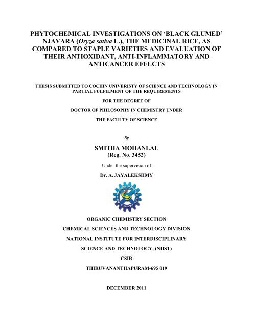
due to its unique electr<strong>on</strong>ic c<strong>on</strong>figurati<strong>on</strong>, as it c<strong>on</strong>tains two unpaired electr<strong>on</strong>s in two<br />
different orbitals (Figure 1.1) and thus, is biradical. 10 The oxygen radical is not a<br />
reactive <strong>on</strong>e due to the so-called spin restricti<strong>on</strong>, which does not allow the d<strong>on</strong>ati<strong>on</strong> or<br />
acceptance of another electr<strong>on</strong>, before the rearrangement of the spin directi<strong>on</strong>s around<br />
the atom. The group of n<strong>on</strong>radical compounds c<strong>on</strong>tains a large variety of substances,<br />
some of which are extremely reactive although not radical by definiti<strong>on</strong>. Am<strong>on</strong>g these<br />
compounds produced in high c<strong>on</strong>centrati<strong>on</strong>s in the living cells are hypochlorous acid<br />
2
(HClO), hydrogen peroxide (H2O2), organic peroxides, aldehydes, oz<strong>on</strong>e (O3), and<br />
singlet oxygen ( 1 O2), that can easily lead to free radical reacti<strong>on</strong>s in living organisms.<br />
Figure 1.1: Molecular orbital diagram of oxygen molecule (O2)<br />
1.3 Sources of ROS and their role in biological systems<br />
The cell is exposed to a large variety of ROS and RNS from two sources<br />
namely, exogenous and endogenous sources. 12<br />
1.3.1 Exogenous Sources<br />
Exogenous sources include, first, exposure to O2 molecule, which, although a<br />
n<strong>on</strong>reactive biradical, can independently cause oxidati<strong>on</strong> and damage to proteins and<br />
enzymes. 10 Oz<strong>on</strong>e which is not a radical like oxygen can damage lungs and can serve as<br />
a powerful oxidizing agent that can oxidize biological comp<strong>on</strong>ents directly. 13 Exposure<br />
of living organisms to i<strong>on</strong>izing and n<strong>on</strong>i<strong>on</strong>izing irradiati<strong>on</strong> c<strong>on</strong>stitutes another major<br />
exogenous source of ROS. 14 Even exposure to n<strong>on</strong>i<strong>on</strong>izing irradiati<strong>on</strong> such as UV-C (<<br />
3
290 nm), UV-B (290–320 nm) and UV-A (320–400 nm) can indirectly produce a<br />
variety of ROS including 1 O2, H2O2 and O2‾ • radical; homolytic cleavage of H2O2 by<br />
UV radiati<strong>on</strong> yields OH • radicals. Air pollutants such as car exhaust, cigarette smoke<br />
and industrial c<strong>on</strong>taminants encompassing many types of NO derivatives c<strong>on</strong>stitute<br />
major sources of ROS that attack and damage the organism either by direct interacti<strong>on</strong><br />
with skin or following inhalati<strong>on</strong> into the lung. 15 Drugs are also a major source of<br />
ROS. 16 There are drugs, such as belomycin and adreamicine whose mechanism of<br />
activity is mediated via producti<strong>on</strong> of ROS. Narcotic drugs and anesthetizing gases are<br />
c<strong>on</strong>sidered major c<strong>on</strong>tributors to the producti<strong>on</strong> of ROS. 17 A large variety of<br />
xenobiotics (eg, toxins, pesticides and herbicides such as paraquat) and chemicals (eg,<br />
mustard gas, alcohol) 18,19 produce ROS as a by-product of their metabolism in vivo<br />
(Table 1.1).<br />
1.3.2 Endogenous sources<br />
Although the exposure of the organism to ROS is extremely high from<br />
exogenous sources, the exposure to endogenous sources is much more important and<br />
extensive, because it is a c<strong>on</strong>tinuous process during the life span of every cell in the<br />
organism (Table 1.1). 20 The reducti<strong>on</strong> of oxygen to water in the mitoch<strong>on</strong>dria for ATP<br />
producti<strong>on</strong> occurs through the d<strong>on</strong>ati<strong>on</strong> of four electr<strong>on</strong>s to oxygen to produce water.<br />
During this process several major oxygen derivatives are formed. 21 In many cases there<br />
is a leakage of ROS from the mitoch<strong>on</strong>dria into the intracellular envir<strong>on</strong>ment. 21 The<br />
mitoch<strong>on</strong>dri<strong>on</strong> serves as the major organelle resp<strong>on</strong>sible for ROS producti<strong>on</strong> and events<br />
that follow throughout the cell cycle. 22 The massive producti<strong>on</strong> of mitoch<strong>on</strong>drial ROS is<br />
increased further in the aging cell where by the functi<strong>on</strong> of the mitoch<strong>on</strong>dri<strong>on</strong> is<br />
impaired and its membrane integrity gets damaged. 23 Enzymes comprise another<br />
endogenous source of ROS. While most enzymes produce ROS as a by-product of their<br />
activity, exemplified by the formati<strong>on</strong> of superoxide radicals by xanthine oxidase, there<br />
are some enzymes designed to produce ROS, such as nitric oxide synthase that yields<br />
NO radicals, those that produce H2O2 and those resp<strong>on</strong>sible for hydroxylati<strong>on</strong>. 24-26<br />
White blood cells, including neutrophils, eosinophils, basophils and m<strong>on</strong><strong>on</strong>uclear cells<br />
(m<strong>on</strong>ocytes) and lymphocytes, with their mechanisms to combat bacteria and other<br />
invaders, 27,28 are major producers of endogenous ROS and other factors that act<br />
synergistically with ROS. 29,30 Nicotinamide adenine dinucleotide phosphate (NADPH)<br />
4
serves as a d<strong>on</strong>or of electr<strong>on</strong>s to an activated enzyme complex in the plasma membrane.<br />
This NADPHoxidase complex utilizes electr<strong>on</strong>s to produce superoxide radicals from the<br />
oxygen molecule. Following dismutati<strong>on</strong>, the producti<strong>on</strong> of H2O2 leads to the formati<strong>on</strong><br />
of OH • by the metal-mediated, Haber-Weiss reacti<strong>on</strong>. The presence of the enzyme<br />
myeloperoxidase leads to the producti<strong>on</strong> of HClO by interacti<strong>on</strong> between hydrogen<br />
peroxides and chlorides. 31,32<br />
Table 1.1: Sources of ROS<br />
Reactive Oxygen Species (ROS)<br />
Exogenous Sources Endogenous sources<br />
γ irradiati<strong>on</strong><br />
UV irradiati<strong>on</strong><br />
Ultrasound<br />
Food<br />
Drugs<br />
Pollutants<br />
Xenobiotics<br />
Toxins<br />
1.4 Beneficial effects of ROS<br />
Cells (e.g., neutrophils)<br />
Direct-producing ROS Enzymes<br />
(e.g., NO synthase)<br />
Indirect-producing ROS enzymes<br />
(e.g., xanthin oxidase)<br />
Metabolism (e.g., mitoch<strong>on</strong>dria)<br />
Diseases (e.g., metal disorders, ischemic<br />
processes)<br />
At low or moderate c<strong>on</strong>centrati<strong>on</strong>s, ROS and RNS are necessary for the<br />
maturati<strong>on</strong> process of cellular structures and can act as weap<strong>on</strong>s for the host defense<br />
system. Beneficial effects of ROS occur at low/moderate c<strong>on</strong>centrati<strong>on</strong>s and involve<br />
physiological roles in cellular resp<strong>on</strong>ses to noxia, for example, in defense against<br />
infectious agents and in the functi<strong>on</strong> of a number of cellular signalling systems. At<br />
low/moderate c<strong>on</strong>centrati<strong>on</strong>s, ROS invokes inducti<strong>on</strong> of a mitogenic resp<strong>on</strong>se too. 33<br />
Indeed, phagocytes (neutrophils, macrophages, m<strong>on</strong>ocytes) release free radicals to<br />
destroy invading pathogenic microbes as part of the body’s defense mechanism against<br />
disease. 34 The importance of ROS producti<strong>on</strong> by the immune system is clearly<br />
exemplified by patients with granulomatous disease. These patients have defective<br />
membrane-bound NADPH oxidase system which makes them unable to produce the<br />
superoxide ani<strong>on</strong> radical (O2¯ • ), thereby resulting in multiple and persistent<br />
5
infecti<strong>on</strong>. 35,36 Other beneficial effects of ROS and RNS involve their physiological roles<br />
in the functi<strong>on</strong> of a number of cellular signalling systems. 33,6 Their producti<strong>on</strong> by n<strong>on</strong><br />
phagocytic NADPH oxidase isoforms play a key role in the regulati<strong>on</strong> of intracellular<br />
signalling cascades in various types of n<strong>on</strong>phagocytic cells including fibroblasts,<br />
endothelial cells, vascular smooth muscle cells, cardiac myocytes and thyroid tissue.<br />
For example, nitric oxide (NO • ) is an intercellular messenger for modulating blood<br />
flow, thrombosis and neural activity. 33 NO • is also important for n<strong>on</strong>specific host<br />
defense and for killing intracellular pathogens and tumours. In brief, ROS/RNS at low<br />
or moderate levels are vital to human health.<br />
1.5 Deleterious effects of ROS<br />
Free radicals and oxidants produced in excess can give rise to deleterious<br />
process which can seriously alter the cell membranes and other structures such as<br />
proteins, lipids, lipoproteins and deoxyrib<strong>on</strong>ucleic acid (DNA). For example, hydroxyl<br />
radical and peroxynitrite in excess can damage cell membranes and lipoproteins by a<br />
process called lipid peroxidati<strong>on</strong>. This reacti<strong>on</strong> leads to the formati<strong>on</strong> of<br />
mal<strong>on</strong>dialdehyde (MDA) and c<strong>on</strong>jugated diene compounds that are cytotoxic and<br />
mutagenic. Lipid peroxidati<strong>on</strong> occurs by a radical chain reacti<strong>on</strong>, i.e. <strong>on</strong>ce started, it<br />
spreads rapidly and affects a great number of lipid molecules. 37 Proteins may also be<br />
damaged by ROS/RNS, leading to structural changes and loss of enzyme activity. 37,38<br />
Oxidative damage to DNA leads to the formati<strong>on</strong> of different oxidative DNA lesi<strong>on</strong>s<br />
which can cause mutati<strong>on</strong>s.<br />
1.6 Oxidative stress<br />
Protective mechanisms present in our body, under normal physiological<br />
c<strong>on</strong>diti<strong>on</strong>s, are sufficient <strong>on</strong>ly to cope up with the normal, and threshold level of free-<br />
radical generati<strong>on</strong>. Any additi<strong>on</strong>al burden of free radicals , either from indigenous or<br />
exogenous sources, <strong>on</strong> the animal (human) physiological system, can tilt the balance<br />
between free radical (prooxidant) and anti-free radical (antioxidant) leading to oxidative<br />
stress. 39 Oxidative stress can thus arise when cells cannot adequately destroy the excess<br />
of free radicals formed. The oxidative stress, defined as the imbalance between oxidants<br />
and antioxidants, in favour of the former, potentially leading to damage has been<br />
6
suggested to be the cause of aging and various human diseases depending up<strong>on</strong> the<br />
sensitivity and susceptibility of the organ. 40<br />
1.7 Pathogenesis of human diseases by ROS<br />
Numerous pathologies and disease states serve as sources for the c<strong>on</strong>tinuous<br />
producti<strong>on</strong> of ROS. 41 More than 200 clinical disorders have been described in the<br />
literature in which ROS were important in the initiati<strong>on</strong> stage of the disease or produced<br />
during its course (Figure 1.2). ROS may be important initiators and mediators in many<br />
types of cancer, heart diseases, endothelial dysfuncti<strong>on</strong>, atherosclerosis, cardiovascular<br />
disorders, chr<strong>on</strong>ic inflammati<strong>on</strong>, intestinal tract diseases, brain degenerative<br />
impairments, diabetes, eye diseases and ischemic and post-ischemic pathologies, such<br />
as damage to skin, heart, brain, kidney, liver and intestinal tract. 42-48<br />
Figure 1.2: Pathogenesis of diseases in human by ROS<br />
ROS is also produced under many normal c<strong>on</strong>diti<strong>on</strong>s and play a role in the<br />
pathogenesis of the physiological c<strong>on</strong>diti<strong>on</strong>. These are exemplified during the aging<br />
process where ROS producti<strong>on</strong> significantly increases as a result of impaired<br />
mitoch<strong>on</strong>drial functi<strong>on</strong> and in the early stages of embry<strong>on</strong>ic development. 49 Other<br />
7
pathological disorders that are associated with impaired metal metabolism such as<br />
hemochromatosis, Wils<strong>on</strong> disease and thalassemia are known to increase the<br />
c<strong>on</strong>centrati<strong>on</strong> of ROS significantly. 50-52<br />
1.7.1 Cancer<br />
Carcinogenesis is a multistage disease process that has been classified into<br />
initiati<strong>on</strong>, promoti<strong>on</strong> and progressi<strong>on</strong> stages and each stage probably involves both<br />
genetic and epigenetic changes. 53 These observati<strong>on</strong>s have been substantiated<br />
experimentally by external administrati<strong>on</strong> of carcinogens. 54 Metabolic activati<strong>on</strong> of<br />
carcinogen is a free radical-dependent reacti<strong>on</strong>. DNA damage mediated by free radicals<br />
plays a critical role in carcinogenesis. 55,56 In biological systems, damaged DNA is<br />
repaired enzymatically and cells regain their normal functi<strong>on</strong>s. However, misrepair of<br />
DNA damage may result in mutati<strong>on</strong>s such as base substituti<strong>on</strong> and deleti<strong>on</strong>, leading to<br />
carcinogenesis. 57 Sequence specificity of DNA damage plays a key role in the<br />
mutagenic process. Endogenous DNA damage arises from a variety of intermediates of<br />
oxygen reducti<strong>on</strong> and several free radicals have been reviewed to take part in this<br />
process by various mechanisms. 58 These reactive species have different redox potentials<br />
and redox potentials of these free radical species may play an important role in<br />
sequence-specific DNA damage. 59 Apart from redox-potential of free-radical species,<br />
oxidati<strong>on</strong> potential of DNA bases also c<strong>on</strong>tributes to the determinati<strong>on</strong> of sequence<br />
specificity of DNA damage. Guanine is most easily oxidized am<strong>on</strong>g the four DNA<br />
bases, as its oxidati<strong>on</strong> potential is lowest (1.29 V vs normal hydrogen electrode) am<strong>on</strong>g<br />
others (adenine 1.42 V, cytosine 1.6 V and thiamin 1.7 V). 60,61 Though the most<br />
comm<strong>on</strong> hydroxyl radical causes DNA damage with no marked site specificity,<br />
Kawanishi et al. (2001) have explained the mechanism of guanine-specific DNA<br />
damage by different free-radical entities and their role in carcinogenesis. 58 Apart from a<br />
variety of free radicals, n<strong>on</strong>-radical oxidant like H2O2 also plays an important role in<br />
DNA damage. Hence, ROS plays a pivotal role in formati<strong>on</strong> of cancer in human.<br />
1.7.2 Inflammati<strong>on</strong><br />
Inflammati<strong>on</strong> involves a complex series of intra- and extracellular events.<br />
Cell-cell communicati<strong>on</strong> is critical and is accomplished by the release of numerous cell<br />
communicator substances (cytokines) from injured tissue and subsequent resp<strong>on</strong>ser<br />
cells. Evidence, supporting a role for oxidants and radicals in this process, is<br />
8
overwhelming. However, despite the beneficial effects of the inflammatory resp<strong>on</strong>ses in<br />
destroying the invading organisms and generating chemotactic factors, the resp<strong>on</strong>ses<br />
can also aggravate existing tissue damage. Thus, inflammati<strong>on</strong> represents a normal<br />
resp<strong>on</strong>se of injured tissue and despite the additi<strong>on</strong>al injury that may develop, is<br />
generally not pathologic because the producti<strong>on</strong> of reactive species is c<strong>on</strong>trolled and<br />
targeted at invading organisms and is reas<strong>on</strong>ably well localized. However, when<br />
unc<strong>on</strong>trolled, initiated by an abnormal stimulus or occurring for prol<strong>on</strong>ged durati<strong>on</strong> of<br />
time, inflammati<strong>on</strong> may become disease process. This appears to be the underlying<br />
basis of inflammati<strong>on</strong> mediated diseases. 62,63<br />
1.8 Antioxidants<br />
An antioxidant works by retarding the oxidati<strong>on</strong>. Literally, antioxidant is defined<br />
as “a substance that opposes oxidati<strong>on</strong> or inhibits reacti<strong>on</strong>s promoted by oxygen or<br />
peroxides”. In biology, oxidati<strong>on</strong> is often started by free radicals. The role of an<br />
antioxidant is to intercept a free radical before it can react with the substrate. The most<br />
important and widely accepted explanati<strong>on</strong> of an antioxidant is that defined by Halliwell<br />
and Gutteridge (2007), as “any substance that when present at low c<strong>on</strong>centrati<strong>on</strong>s<br />
compared with those of an oxidizable substrate significantly delays or prevents<br />
oxidati<strong>on</strong> of that substrate”. 6 Antioxidants are enzymes or other substances such as<br />
vitamin E or β-carotene that is capable of counteracting the damaging effects of<br />
oxidati<strong>on</strong> in animal tissues. 6 Antioxidants may exert their effects by different<br />
mechanisms, such as suppressing the formati<strong>on</strong> of active species by reducing<br />
hydroperoxides (ROO • ) and H2O2 and also by sequestering metal i<strong>on</strong>s, scavenging<br />
active free radicals, repairing and/or clearing damage. Similarly, some antioxidants also<br />
induce the biosynthesis of other antioxidants or defence enzymes.<br />
1.8.1 Endogenous antioxidants<br />
Antioxidants that are produced within the body for defence as a result of normal<br />
metabolic processes are called endogenous antioxidants. Catalase c<strong>on</strong>verts H2O2 to O2<br />
and H2O while superoxide dismutase (SOD) c<strong>on</strong>verts the superoxide radical to H2O2<br />
and O2. Some of the antioxidant enzymes exist in several forms. For example,<br />
membrane, cytosolic and plasma forms of glutathi<strong>on</strong>e peroxidase have been isolated and<br />
SOD has membrane, cytosolic and extracellular forms. The levels and locati<strong>on</strong>s of these<br />
9
antioxidants must be tightly regulated for cell survival. The antioxidant enzymes SOD,<br />
glutathi<strong>on</strong>e peroxidase (GPX) and catalase (CAT) work within the cells to remove most<br />
superoxides and peroxides, before they react with metal i<strong>on</strong>s to form more reactive free<br />
radicals. Peroxidative chain reacti<strong>on</strong>s initiated by free radicals that escaped the<br />
antioxidant defenses are terminated by chain-breaking water or lipid soluble<br />
antioxidants. 64 Hence, there is a vast network of intracellular and extracellular<br />
antioxidants with diverse roles within each area of defence.<br />
1.8.2 Exogenous antioxidants<br />
Antioxidant compounds supplied through diet are termed exogenous<br />
antioxidants. Diet plays a vital role in the producti<strong>on</strong> of the antioxidant defence system<br />
by providing essential nutrient antioxidants such as vitamin E, C and β-carotene, other<br />
antioxidant plant phenols including flav<strong>on</strong>oids and essential minerals (eg. selenium, Se)<br />
that are vital to important antioxidant enzymes. Diet also plays an important role in the<br />
oxidati<strong>on</strong> process by affecting the substrates that are subject to oxidati<strong>on</strong>. The best<br />
example is the oxidati<strong>on</strong> of lipids. Polyunsaturated fatty acids (PUFA) having two or<br />
more double b<strong>on</strong>ds are increasingly susceptible to free radical attack as the number of<br />
double b<strong>on</strong>ds increases. Antioxidants available at the site of radical attack break the<br />
chain of oxidati<strong>on</strong> by being preferentially oxidized by the attacking radical, thereby<br />
preventing oxidati<strong>on</strong> of the adjacent fatty acid.<br />
1.9 Natural products as drugs<br />
Natural products are chemical entities produced by living organisms. Sources of<br />
natural products are terrestrial plants, microorganisms, vertebrates, invertebrates and<br />
marine organisms etc. Nature is unravelled in its ability to craft small organic molecules<br />
endowed with structural and biological complexities. All living organisms are made up<br />
of these complex organic molecules known as primary and sec<strong>on</strong>dry metabolites. Of<br />
these, sec<strong>on</strong>dary metabolites are complex organic natural products which are not<br />
directly necessary for host survival. Sec<strong>on</strong>dary metabolites may be alkaloids,<br />
terpenoids, flav<strong>on</strong>oids, coumarins, steroids, peptides, glycosides, phenolics etc. 65 They<br />
are typically produced by organisms such as bacteria, plants or various marine<br />
intermediates and are usually used as “chemical warfare” to protect parent organisms<br />
from predators or as a means of attack. To efficiently fulfil this role, the natural<br />
10
products have been optimised in a very l<strong>on</strong>g natural selecti<strong>on</strong> process for optimal<br />
interacti<strong>on</strong>s with biological macromolecules. Natural products are therefore an excellent<br />
source of validated substructures for the design of new drugs. In early 1800s, an<br />
emerging fascinati<strong>on</strong> with such molecules, later known as natural products or sec<strong>on</strong>dry<br />
metabolites, gave rise to the field of organic chemistry of living things.<br />
The Greek physician Galen (AD 129–200) devised the first pharmacopoeia<br />
describing the appearance, properties and use of many plants of his time. 66 In the early<br />
1500s, the bark of cinch<strong>on</strong>a tree (Cinch<strong>on</strong>a officinalis) known as Indian fever bark was<br />
<strong>on</strong>e of the first medicinal plants to find appreciative c<strong>on</strong>sumers in Europe. Natural<br />
products chemistry actually began with the work of F. W. Serturner, who first isolated<br />
morphine from opium poppy (Papaver somniferum) in 1815 that had been used for over<br />
5000 years. 67 In 1860, a German chemist Carl Koler isolated cocaine and found its<br />
biological activity as a local anaesthetic in eye surgery. Later, scientists observed that<br />
cocaine paralysed nerve endings resp<strong>on</strong>sible for transmitting pain. As a local<br />
anaesthetic, it revoluti<strong>on</strong>ized several surgical and dental procedures. Tube curare in the<br />
West Amaz<strong>on</strong> is from Chr<strong>on</strong>drodendr<strong>on</strong> tomentosum; curare in modern medicine is<br />
made from this and named as tubocurarine. 66 Alkaloid rich aromatic oil extracted from<br />
jaborandi tree (Pilocarpus jaborandi) is a c<strong>on</strong>stituent of an alkaloid pilocarpine which<br />
acts against the blinding disease, glaucoma. American Indians used pineapple (Ananas<br />
comosos) poultices to reduce inflammati<strong>on</strong> in wounds and stomach ache. In 1891,<br />
bromelain, an enzyme was isolated from the fresh juice of pineapple that broke down<br />
proteins and was found to break down blood clots. 66 Many such similar developments<br />
followed. Other pharmaceuticals that have their origin in botanicals include atropine,<br />
hyoscine, digoxin, colchicine, reserpine and emetine. 66<br />
The above discoveries initiated an era wherein drugs from plants could be<br />
purified, studied and administered in precise dosages that did not vary with the source<br />
or age of the material. The basis of development of currently accepted modern medicine<br />
or allopathy remains rooted in traditi<strong>on</strong>al medicine and therapies from Natural products,<br />
especially plants. The emergence of today’s pharmaceutical industry, in large part, has<br />
been based <strong>on</strong> natural products. Drugs such as digoxin, taxol, artemisinin and scores<br />
more have been developed from phytochemicals. 68 Not <strong>on</strong>ly have many medical<br />
breakthroughs been based <strong>on</strong> compounds of natural origin, but these also represent a<br />
11
large share of the drug market. In 1999, close to 50% of the 20 best-selling drugs were<br />
derived from natural products and their sales amounted to approximately $16 billi<strong>on</strong>. 69<br />
According to a survey by the Nati<strong>on</strong>al Cancer Institute, 61% of the 877 small<br />
molecules, which are new chemical entities introduced as drugs worldwide from 1981<br />
to 2002, were inspired by natural products. 69<br />
1.9.1 Natural products as antioxidants<br />
Terrestrial plants provide a rich source of natural antioxidants. 70 These include<br />
tocopherols, vitamin C, carotenoids and phenolic compounds. Plant phenolics are<br />
thought to protect the plants against tissue injuries as they oxidize and combine with<br />
proteins and other comp<strong>on</strong>ents. In additi<strong>on</strong>, phenolic compounds in plants may serve as<br />
defence systems against herbivory. 71 By-products of photosynthesis may also produce<br />
high levels of oxygen, free radicals and reactive oxygen species (ROS) in profusi<strong>on</strong>;<br />
thus, plants use a myriad of antioxidant compounds to deal with these, in order to<br />
survive. Many of these compounds have basic molecular similarities in that most of<br />
them have at least <strong>on</strong>e aromatic ring and a hydroxyl group. These include phenolic<br />
acids, flav<strong>on</strong>oids and isoflav<strong>on</strong>es, gallate esters (hydrolysable tannins), lignans,<br />
coumarins, stilbenes, flav<strong>on</strong><strong>on</strong>es and oligomeric proanthocyanidins.<br />
1.9.1.1 Tocopherols<br />
Tocopherols and tocotrienols are grouped as chromanols; within each of these<br />
two classes, there are four isomers (α, β, γ, δ), making a total of eight tocophehrols<br />
(Figure 1.3).<br />
Figure 1.3: Structure of tocopherols<br />
Tocopherols can act as antioxidant by two primary mechanisms: (i) a chain–<br />
breaking electr<strong>on</strong> d<strong>on</strong>or (CB-D) mechanism and (ii) a chain-breaking acceptor (CB-A)<br />
mechanism. 72 The sec<strong>on</strong>d major mechanism, CB-A, includes singlet oxygen scavenging<br />
12
or quenching. CB-D antioxidants compete with the unsaturated fatty acid (LH) for the<br />
lipid peroxy radical (LOO • ) and as a result, minimise the formati<strong>on</strong> of the lipid radical<br />
(L • ) that occurs when LH reacts with LOO • ; this competiti<strong>on</strong> ultimately slows the<br />
propagati<strong>on</strong> stage of autoxidati<strong>on</strong>. Tocopherols can effectively compete with LH for the<br />
LOO • by readily transferring a hydrogen atom to the LOO • to produce the more stable<br />
LOOH. 73 In the case of tocopherols, a hydrogen transfer occurs, resulting in a<br />
semiquin<strong>on</strong>e intermediate (Figure 1.4) which c<strong>on</strong>verts to tocopheryl quin<strong>on</strong>e (Figure<br />
1.4) as the final stable end product.<br />
HO<br />
H 3C<br />
CH 3<br />
CH 3<br />
.<br />
O<br />
H 3 C<br />
A<br />
O<br />
CH 3<br />
CH 3<br />
C16H33 LOO . .<br />
O<br />
H 2 O 2[H]<br />
H 2O 2[H]<br />
LOOH<br />
C 16 H 33<br />
O<br />
H 3 C<br />
LOOH<br />
LOO .<br />
HO<br />
H 3C<br />
.<br />
CH 3<br />
CH 3<br />
CH 3<br />
CH 3<br />
C D<br />
B<br />
O<br />
O C16H33 HO<br />
C 16H 33<br />
Figure 1.4: α-tocopherol oxidati<strong>on</strong> to (B) α-tocopheryl quin<strong>on</strong>e through semiquin<strong>on</strong>e<br />
intermediates (C, D)<br />
The tocopheryl quin<strong>on</strong>e and possible semiquin<strong>on</strong>es can react with the L • to<br />
produce LH, thus slowing the chain propagati<strong>on</strong> stage. This type of mechanism is<br />
defined as the CB-A mechanism and in this mechanism tocopheryl quin<strong>on</strong>e competes<br />
with oxygen (triplet) for the L • . 73 As the reacti<strong>on</strong>s between carb<strong>on</strong>-centered radicals (L • )<br />
and oxygen ( 3 O2) are favoured over those of the quin<strong>on</strong>e oxygen and L • , it is likely that<br />
this type of antioxidant activity occurs primarily in biological systems or other low<br />
oxygen pressure systems. 73<br />
Tocopherols can also act <strong>on</strong> singlet oxygen ( 1 O2) as a means to c<strong>on</strong>trol oxidati<strong>on</strong><br />
processes. The oxygen- scavenging ability of tocopherols occurs through two primary<br />
methods (i) singlet oxygen quenching and (ii) irreversible reacti<strong>on</strong> with singlet oxygen<br />
to form a variety of products. The quenching mechanism is more predominant 74 and is<br />
dependent <strong>on</strong> the rate c<strong>on</strong>stants of the reacti<strong>on</strong> between 1 O2 and tocopherol vs the rate<br />
c<strong>on</strong>stant of 1 O2 being quenched to 3 O2. 75<br />
13
HO<br />
H 3C<br />
CH 3<br />
CH 3<br />
A<br />
O<br />
1 O2<br />
C 16H 33<br />
O<br />
H 3C<br />
CH 3<br />
CH 3<br />
B<br />
O<br />
OOH<br />
C 16H 33<br />
Figure 1.5: Singlet oxidati<strong>on</strong> of (A) α-tocopherol to (B) hydroperoxydien<strong>on</strong>e as given<br />
by Clough et al. 74<br />
However, it is not very clear how tocopherols and 1 O2 react. Clough et al. (1979)<br />
proposed that the irreversible reacti<strong>on</strong> of singlet oxygen with tocopherol results in the<br />
formati<strong>on</strong> of hydroperoxydien<strong>on</strong>e (Figure 1.5) as the major end product. 74 Grams et al.<br />
(1972) found a variety of products and proposed that the 1, 4-cycloadditi<strong>on</strong> of oxygen to<br />
form the endoperoxides (Figure 1.6) was the first stage of 1 O2 reacti<strong>on</strong> with<br />
tocopherol. 76 Clough et al. (1979) also agreed that endoperoxides could be<br />
intermediates for the hydroperoxydien<strong>on</strong>e compounds found in their study. 74<br />
HO<br />
H 3C<br />
CH 3<br />
CH 3<br />
O<br />
A<br />
O<br />
H 3C<br />
C 16H 33<br />
CH 3<br />
CH 3<br />
O<br />
1 O2<br />
C<br />
OH<br />
HO<br />
H 3C<br />
+<br />
C 16H 33<br />
CH 3<br />
CH 3<br />
H 3C<br />
O<br />
B<br />
H3C O<br />
O<br />
CH 3<br />
C 16H 33<br />
O<br />
D<br />
C 16H 33<br />
Figure 1.6: Singlet oxidati<strong>on</strong> of (A) α-tocopherol to (C) α-tocopheryl quin<strong>on</strong>e and (D)<br />
quin<strong>on</strong>e epoxide through (B) α-tocopherol endoperoxide as presented by Gram et al.<br />
(1972). 76<br />
Vitamin E acts as a primary antioxidant, whereas vitamin C reductively<br />
regenerates oxidized viramin E. Finckh and Kunert (1985) found that peroxidative cell<br />
damage was highly c<strong>on</strong>trolled by an antioxidative system of vitamins C and E. 77 The<br />
lipophilic antioxidants α-tocopherol and ascorbyl palmitate were more effective in an<br />
o o<br />
14
oil-in-water emulsi<strong>on</strong> system than in bulk oil, whereas the opposite was found for the<br />
hydrophilic antioxidants, Trolox (6-hydroxy-2, 5, 7, 8-tetramethylchroman-2-carboxylic<br />
acid, a hydrophilic carboxylic acid derivative of α-tocopherol) and ascorbic acid.<br />
Vitamin E has been proposed for the preventi<strong>on</strong> against col<strong>on</strong>, prostate and breast<br />
cancers, some cardiovascular diseases, ischemia, cataract, arthritis and certain<br />
neurological disorders. The dietary sources of vitamin E are vegetable oils, wheat germ<br />
oil, whole grains, nuts, cereals, fruits, eggs, poultry, meat. 78<br />
1.9.1.2 Vitamin C<br />
Vitamin C, also known as ascorbic acid, is a water-soluble vitamin (Figure 1.7).<br />
It is essential for collagen, carnitine and neurotransmitters biosynthesis. 79 Health<br />
benefits of vitamin C are as antioxidant, anti-atherogenic, anti-carcinogenic and as an<br />
immunomodulator. The positive effect of vitamin C resides in reducing the incidence of<br />
stomach cancer and in preventing lung and colorectal cancer. Vitamin C works<br />
synergistically with vitamin E to quench free radicals and also regenerates the reduced<br />
form of vitamin E. Natural sources of vitamin C are acidic fruits such as lem<strong>on</strong>, orange,<br />
green vegetables, gooseberry, tomatoes etc. 80<br />
1.9.1.3 Carotenes<br />
HO<br />
HO<br />
HO<br />
O<br />
O<br />
OH<br />
Figure 1.7: Structure of Vitamin C<br />
Like tocopherols, carotenes (in particular β–carotene) are also effective<br />
1 O2 quenchers. β-Carotene is a fat soluble member of the carotenoids which are<br />
c<strong>on</strong>sidered pro-vitamins because they can be c<strong>on</strong>verted to active vitamin A. β -Carotene<br />
is present in many fruits, grains, oil and vegetables. 81<br />
Figure 1.8: Structure of β–carotene<br />
15
Foote et al. (1968, 1971) 82,83 and Farmilo and Wilkens<strong>on</strong> 84 found that the<br />
quenching of 1 O2 by β–carotene was due to an energy transfer from 1 O2 to β–carotene as<br />
shown in equati<strong>on</strong> 1.1. Foote et al. (1970) also found that the rate of quenching was<br />
dependent <strong>on</strong> the number of c<strong>on</strong>jugated double b<strong>on</strong>ds. 85 The presence of nine or more<br />
double b<strong>on</strong>ds in the carotene structure greatly enhanced the quenching ability (Figure<br />
1.8). Carotenoids with seven or fewer double b<strong>on</strong>ds were not as effective due to the<br />
inability of the c<strong>on</strong>jugated chain to delocalize the unpaired electr<strong>on</strong>s gained from the<br />
1 O2. 85 A radical trapping mechanism has also been proposed. 86,87 This mechanism relies<br />
<strong>on</strong> the delocalizati<strong>on</strong> of the unpaired electr<strong>on</strong>s of the peroxy and free radical species<br />
over the carotenoid c<strong>on</strong>jugated polyene system. 86,87<br />
1 O2 + β – carotene 3 O2 + 3 β– carotene* (excited state) -----(1.1)<br />
1.9.1.4 Phenolic Acids<br />
Phenolic compounds c<strong>on</strong>stitute <strong>on</strong>e of the most widespread and diverse groups<br />
of sec<strong>on</strong>dary plant metabolites. Phenolic compounds are widely distributed in cereals<br />
and legumes. Ferulic, caffeic, protocatechuic, p-hydroxybenzoic, vanillic, syringic and<br />
p-coumaric acids are the most comm<strong>on</strong> phenolic acids found in cereals and legumes. 88<br />
Figure 1.9 shows the structures of these phenolic caids. Phenolic antioxidants are<br />
primary antioxidants which act as free radical terminators. The positi<strong>on</strong> and the degree<br />
of hydroxylati<strong>on</strong> are of primary importance in determining antioxidant activity. 89 The o-<br />
dipheols, such as caffeic acid, hydroxytyrosol and oleuropein exhibit str<strong>on</strong>g antioxidant<br />
activity compared to less sterically hindered phenolic acids such as tyrosol. 90 In<br />
additi<strong>on</strong>, phenolic acids that are hydroxyl derivatives of cinamic acid such as caffeic<br />
acid, ferulic, sinapic and p-coumaric acids, are more active antioxidants than hydroxy<br />
derivatives of benzoic acid (Figure 1.9), i.e., p-hydroxybenzoic, vanillic, syringic and<br />
3,4-dihydroxy benzoic acid. 91<br />
16
O<br />
OH<br />
HO HO<br />
p-Hydroxy benzoic acid p-Coumaric acid<br />
O<br />
OH<br />
HO HO<br />
H 3CO<br />
OCH 3<br />
O<br />
OH<br />
HO HO<br />
O<br />
OH<br />
HO HO<br />
OCH 3<br />
Vanillic acid Ferulic acid<br />
OCH 3<br />
H 3CO<br />
OCH 3<br />
Syringic acid Sinapic acid<br />
OH OH<br />
Dihydrobenzoic acid Caffeic acid<br />
Figure 1.9: Structure of phenolic acids<br />
Phenolic antioxidants act to inhibit lipid oxidati<strong>on</strong> by trapping the peroxy<br />
radical. This can be accomplished in <strong>on</strong>e of the two ways. In the first mechanism, the<br />
peroxy radical (LOO•) abstracts a (H • ) from the antioxidant (ArOH) to yield an aroxyl<br />
radical (ArO•) and the hydroperoxide (LOOH) (equati<strong>on</strong> 1.2). In the sec<strong>on</strong>d<br />
mechanism, a peroxy and an aroxyl radical react by radical-radical coupling to form a<br />
n<strong>on</strong>-radical products (equati<strong>on</strong> 1.3). 90 The aroxyl radicals formed from the oxidati<strong>on</strong> of<br />
an antioxidant can further react and can, in some instances, c<strong>on</strong>tribute to the producti<strong>on</strong><br />
of free radicals. 90<br />
LOO • + ArOH LOOH + ArO • -------(1.2)<br />
LOO • + ArO • N<strong>on</strong>-radical products -------(1.3)<br />
O<br />
O<br />
O<br />
O<br />
OH<br />
OH<br />
OH<br />
OH<br />
17
ArO • + LOOH LOO • + ArOH -------(1.4)<br />
2 ArO • N<strong>on</strong>-radical products -------(1.5)<br />
ArO • + LH ArOH+ L • -------(1.6)<br />
Chimi et al. (1991) reported that for sterically hindered phenols, the rates of<br />
reacti<strong>on</strong>s 1.3 and 1.5, which produce n<strong>on</strong>-radical products, exceed the rates of reacti<strong>on</strong><br />
1.4 and 1.6, which produce free radicals, resulting in an overall inhibiti<strong>on</strong> of lipid<br />
oxidati<strong>on</strong>. 90 Lack of hindrance favours reacti<strong>on</strong>s 1.4 and 1.6, which produce free<br />
radicals, thus decreasing the overall antioxidant activity of the phenol. Dziedzic and<br />
Huds<strong>on</strong> (1984) 92 evaluated a series of hydroxy aromatic acids (phenolic acids), their<br />
esters and lact<strong>on</strong>es for antioxidant activity and tried to correlate with their structures.<br />
Based <strong>on</strong> the results of their studies, several structure-activity c<strong>on</strong>clusi<strong>on</strong>s were drawn.<br />
High antioxidant activity was related to the molecule c<strong>on</strong>taining at least, two<br />
neighboring phenolic hydroxyl groups; three such groups were even more desirable. In<br />
additi<strong>on</strong>, a carb<strong>on</strong>yl group such as in an aromatic acid, an ester or a lact<strong>on</strong>e, enhanced<br />
activity. Activity also increased when the carb<strong>on</strong>yl group was separated from the<br />
aromatic ring. Cinnamic acid derivatives were more effective than corresp<strong>on</strong>ding<br />
benzoic acids and the most effective were the phenyl acetic and phenylpropi<strong>on</strong>ic acids.<br />
Steric hindrance of the phenolic hydroxyls by a neighboring inert group such as<br />
methoxyl group, enhanced activity.<br />
1.9.1.5 Flav<strong>on</strong>oids<br />
Flav<strong>on</strong>oids are a group of plant phenols characterized by the carb<strong>on</strong> skelet<strong>on</strong> C6-<br />
C3-C6. The basic structure of these compounds c<strong>on</strong>sists of two aromatic rings linked by<br />
a three – carb<strong>on</strong> aliphatic chain which normally has been c<strong>on</strong>densed to form a pyran or<br />
less comm<strong>on</strong>ly, a furan ring. 93 The basic flav<strong>on</strong>oid structure is the flavan nucleus,<br />
which c<strong>on</strong>sists of 15 carb<strong>on</strong> atoms arranged in three rings (C6-C3-C6), which are labeled<br />
A, B, and C (Figure 1.10).<br />
18
7<br />
6<br />
8 1<br />
O<br />
2'<br />
1'<br />
A C 2<br />
3<br />
5 4<br />
Figure 1.10: Basic flav<strong>on</strong>oid structure<br />
The different classes of flav<strong>on</strong>oids differ in the level of oxidati<strong>on</strong> and pattern of<br />
substituti<strong>on</strong> in the C ring while individual compounds within a class differ in the pattern<br />
of substituti<strong>on</strong> of the A and B rings. Am<strong>on</strong>g the many classes of flav<strong>on</strong>oids, those of<br />
particular interest are flav<strong>on</strong>es, flavan<strong>on</strong>es, isoflav<strong>on</strong>es, flav<strong>on</strong>ols, flavan<strong>on</strong>ols, flavan-<br />
3-ols and anthocyanidins. The structures of important subclasses of flav<strong>on</strong>oids are<br />
shown in Figure 1.11. Flav<strong>on</strong>es and flav<strong>on</strong>ols are found in almost every plant,<br />
particularly in the leaves and petals, with flav<strong>on</strong>ols occurring more frequently than<br />
94, 95<br />
flav<strong>on</strong>es.<br />
O<br />
O<br />
O<br />
Flav<strong>on</strong>es Flav<strong>on</strong>ols<br />
O<br />
O<br />
O<br />
OH<br />
OH<br />
O<br />
O<br />
Flavan<strong>on</strong>es Flavan<strong>on</strong>ols<br />
Flavanols<br />
3'<br />
B<br />
6'<br />
4'<br />
5'<br />
O<br />
O<br />
Isoflav<strong>on</strong>es<br />
Figure 1.11: Typical chemical structures of the flav<strong>on</strong>oid family<br />
1.9.1.5.1 Biosynthesis of flav<strong>on</strong>oids<br />
All flav<strong>on</strong>oids derive their 15-carb<strong>on</strong> skelet<strong>on</strong>s from two basic metabolites,<br />
mal<strong>on</strong>yl-CoA and p-coumaroyl-CoA. 96 Basically, flav<strong>on</strong>oids are derivatives of 1, 3-<br />
diphenylpropan-1-<strong>on</strong>e (C6-C3-C6). The crucial biosynthetic reacti<strong>on</strong> is the c<strong>on</strong>densati<strong>on</strong><br />
of three molecules of mal<strong>on</strong>yl-CoA with <strong>on</strong>e molecule p-coumaroyl-CoA to a chalc<strong>on</strong>e<br />
intermediate. Chalc<strong>on</strong>es and dihydrochalc<strong>on</strong>es are classes of flav<strong>on</strong>oids that c<strong>on</strong>sist of<br />
O<br />
OH<br />
19
two phenolic groups which are c<strong>on</strong>nected by an open three carb<strong>on</strong> bridge. Derived from<br />
the chalc<strong>on</strong>e structure, a flav<strong>on</strong>oid-class c<strong>on</strong>taining three rings, the flavan<strong>on</strong>es, can be<br />
formed. Here, the three-carb<strong>on</strong> bridge is part of an additi<strong>on</strong>al heterocyclic six-<br />
membered ring that involves <strong>on</strong>e of the phenolic groups <strong>on</strong> the adjacent ring. Based <strong>on</strong><br />
these flavan<strong>on</strong>es, all other flav<strong>on</strong>oid-classes are generated, including isoflav<strong>on</strong>es,<br />
flavanols, anthocyanidins, flav<strong>on</strong>ols and flav<strong>on</strong>es (Figure 1.12). 96<br />
3 x Mal<strong>on</strong>yl-<br />
CoA<br />
CHI<br />
6-Deoxychalc<strong>on</strong>es 5-Deoxyflavan<strong>on</strong>es 5-Deoxy-flav<strong>on</strong>oids<br />
CHS<br />
CHKR<br />
CHS<br />
Chalc<strong>on</strong>es<br />
CHI<br />
Flavan<strong>on</strong>es<br />
FHT<br />
Dihydroflav<strong>on</strong>ols<br />
DFR<br />
p-Coumaroyl-<br />
CoA<br />
O<br />
IFS + IFD<br />
O<br />
O<br />
O<br />
O<br />
O<br />
IFS + IFD<br />
Isoflav<strong>on</strong>es<br />
O<br />
FNS I<br />
or<br />
FNS II<br />
Flav<strong>on</strong>es<br />
O<br />
O<br />
O<br />
O<br />
OH<br />
FLS<br />
Flav<strong>on</strong>ols<br />
OH<br />
Flavan-4-ols 3-Deoxyflav<strong>on</strong>oids<br />
ANS<br />
Leucoanthocyanins<br />
O<br />
OH<br />
O<br />
OH<br />
LAR<br />
OH<br />
Anthocyanidins<br />
O<br />
ANR<br />
O<br />
OH<br />
trans Flavan-3-ols cis Flavan-3-ols<br />
OH<br />
FGT Anthocyanins<br />
Figure 1.12: Scheme of bioynthesis of flav<strong>on</strong>oid pathway. Enzymes are abbreviated as<br />
follows: CHS, chalc<strong>on</strong>e synthase; CHKR, chalc<strong>on</strong>e polyketide reductase; CHI, chalc<strong>on</strong>e<br />
isomerase; FHT, flavan<strong>on</strong>e 3-β-hydroxylase; DFR, dihydroflav<strong>on</strong>ol-4-reductase; ANS,<br />
anthocyanidin synthase; FGT, flav<strong>on</strong>oid glycosyltransferase; FNS, flav<strong>on</strong>e synthase;<br />
FLS, flav<strong>on</strong>ol synthase; LAR, leucoanthocyanidin reductase; ANR, anthocyanidin<br />
reductase; IFS, isoflav<strong>on</strong>e synthase; IFD, isoflav<strong>on</strong>e dehydratase<br />
1.9.1.5.2 Antioxidant activity of flav<strong>on</strong>oids and their structure activity relati<strong>on</strong>ship<br />
Flav<strong>on</strong>oids occur as glycosides which, during hydrolysis with acid and heat,<br />
produce an aglyc<strong>on</strong>e and a sugar moiety. 97 Many of the flav<strong>on</strong>oids and related<br />
compounds have str<strong>on</strong>g antioxidant characteristics. According to Halliwell and<br />
Gutteridge (2007), mechanisms of antioxidant acti<strong>on</strong> can include (1) suppressing<br />
O<br />
OGlc<br />
20
eactive oxygen species formati<strong>on</strong> either by inhibiti<strong>on</strong> of enzymes or chelating trace<br />
elements involved in free radical producti<strong>on</strong>; (2) scavenging reactive oxygen species<br />
and are free radical chain breakers; and (3) upregulating or protecting antioxidant<br />
defences. 6<br />
Huds<strong>on</strong> and Lewis 98 studied the antioxidant activity of a series of polyhydroxy<br />
flav<strong>on</strong>oids and related their activity to their structures (Figure 1.13). The results of their<br />
study led to several c<strong>on</strong>clusi<strong>on</strong>s relative to the structural features which affect the<br />
antioxidant activity of the flav<strong>on</strong>oids. It was noted that primarily, the molecule needed<br />
multiple hydroxyl groups: a 3’, 4’–dihydroxy c<strong>on</strong>figurati<strong>on</strong> gave str<strong>on</strong>g antioxidant<br />
activity and additi<strong>on</strong>ally the molecule must have a 4-carb<strong>on</strong>yl group for activity.<br />
R 7<br />
7<br />
1<br />
O 2<br />
6<br />
A<br />
5<br />
C 3<br />
4<br />
8<br />
R 5<br />
R 3<br />
2' 3'<br />
Figure 1.13: General chemical structure of flav<strong>on</strong>oid compounds.<br />
The presence of 3-hydroxy group opposite to 5-hydroxy group, makes the<br />
molecule effective. They projected that there was cooperati<strong>on</strong> between the 4-carb<strong>on</strong>yl<br />
and the 3- or 5- hydroxyl groups which acted to chelate the copper i<strong>on</strong> (Figure 1.14).<br />
Flav<strong>on</strong>es act as ligands with the cupric (Cu 2+ ) i<strong>on</strong> whereas the flavan<strong>on</strong>es complex with<br />
copper i<strong>on</strong>s through their enediol or enolic groups (Figure 1.14). 98 This mechanism of<br />
chelati<strong>on</strong> of metal i<strong>on</strong>s (copper i<strong>on</strong>s) has found to operate for c<strong>on</strong>trolling lipid<br />
oxidati<strong>on</strong>.<br />
Das and Pereira studied the antioxidant activity of flav<strong>on</strong>oids and related the<br />
observed antioxidant activity to their structure. 99 They found that the potency of<br />
flav<strong>on</strong>oids with polyhydroxylated substituti<strong>on</strong>s was affected by the locati<strong>on</strong> of the<br />
hydroxyl group <strong>on</strong> the B ring (Figure 1.13). Hydroxyl substituti<strong>on</strong> in the ortho posit<strong>on</strong><br />
in the B ring gave less antioxidant activity; however, hydroxyl substituti<strong>on</strong> in the ortho<br />
positi<strong>on</strong> accompanied by an additi<strong>on</strong>al hydroxyl group in the para positi<strong>on</strong> enhanced<br />
activity. Str<strong>on</strong>g antioxidant activity was found in compounds having hydroxylati<strong>on</strong> in<br />
the para positi<strong>on</strong> in the B ring. In additi<strong>on</strong>, it was found that a double b<strong>on</strong>d between the<br />
R 4<br />
B<br />
6'<br />
4'<br />
5'<br />
21
2 and 3 positi<strong>on</strong>s <strong>on</strong> the C ring c<strong>on</strong>tributed to antioxidant activity (Figure 1.13). Das and<br />
Pereira (1990) 99 also found that the aglyc<strong>on</strong>es were str<strong>on</strong>ger antioxidant than their<br />
corresp<strong>on</strong>ding glucosides, possibly due to the lack of a free 3-hydroxyl substituti<strong>on</strong> in<br />
the C ring. 99<br />
5<br />
5<br />
4 3<br />
OH 5<br />
O OH<br />
4 3<br />
O<br />
O<br />
Cu<br />
+<br />
4 3<br />
5<br />
O + O<br />
Cu<br />
5<br />
4 3<br />
4 3<br />
O<br />
O<br />
Cu<br />
4 3<br />
5<br />
O O<br />
Cu<br />
Figure 1.14: Formati<strong>on</strong> of metal chelates with flav<strong>on</strong>es and flav<strong>on</strong><strong>on</strong>es. 98<br />
Shahidi and Wanasundara 100 noted that the positi<strong>on</strong> and degree of hydroxylati<strong>on</strong><br />
was important in the antioxidant activity of flav<strong>on</strong>oids. They stated that o-<br />
dihydroxylati<strong>on</strong> of the B ring c<strong>on</strong>tributed to the antioxidant activity of the compound<br />
and a p-quinol structure <strong>on</strong> the B ring produces greater activity than the o-quinol.<br />
However, they also noted that p- and m- hydroxylati<strong>on</strong> of the B ring do not occur in<br />
nature. In short, criteria for effective radical-scavenging are: (i) the ortho-dihydroxyl<br />
structure in the B-ring, (ii) the 2, 3-double b<strong>on</strong>d, which allows p electr<strong>on</strong> delocalizati<strong>on</strong><br />
from the B-ring and (iii) the 3- and 5-OH with 4-oxo functi<strong>on</strong> in the A- and C-rings.<br />
Apart from antioxidant activity, flav<strong>on</strong>oids play remarkable spectrum of<br />
biological activties; antiallergic, anti-inflammatory, antimutagenic, anticarcinogenic and<br />
modulati<strong>on</strong> of enzymatic activities. 101<br />
1.9.1.6 Lignans<br />
Lignans are widely found in all parts of plants and include compounds with<br />
great chemical diversity. By definiti<strong>on</strong>, lignans are dimers of phenlypropanoid (C6-C3)<br />
units linked by the central carb<strong>on</strong>s of their side chains. 102 According to their<br />
oxygenati<strong>on</strong>, four major groups of linear lignans are found, namely, lignan or butane<br />
derivatives lignanolides or butanolide derivatives, m<strong>on</strong>oepoxylignans or tetrahydrofuran<br />
22
derivatives and bisepoxylignans or derivatives of 3,7-dioxabicyclo-(3,3,0)-octane.<br />
Cyclized lignans or cyclolignans occur as tetrahydr<strong>on</strong>aphthalene or naphthalene<br />
derivatives. 103 Lignan-type compounds, isolated from plants and oilseeds, have shown<br />
good antioxidant properties. 104<br />
1.9.2 Anticancer compounds from plants<br />
Cancer is a complex disease that involves unc<strong>on</strong>trolled multiplicati<strong>on</strong> and spread<br />
(metastasis) of abnormal form of body’s own cells. As per WHO 13% of world deaths,<br />
that is, about 7.6 milli<strong>on</strong> deaths accounted in 2005 are because of cancer, and this<br />
percentage is expected to increase in coming years. 105 Plant derived compounds have<br />
played an important role in treatment of cancers and some of the most promising and<br />
better drugs have come up from plant sources like Taxol, Camptothecin, Combrestatin,<br />
Epipodophyllotoxin and Vinca alkaloids (vinblastine, vincristine). These drugs have<br />
also been the major source of new drug candidates for the treatment of cancers.The<br />
chemistry and properties of few of them are discussed below.<br />
1.9.2.1 Podophyllotoxin<br />
O<br />
E<br />
O<br />
H 3CO<br />
OH<br />
A B<br />
C<br />
OCH 3<br />
D O<br />
O<br />
OCH 3<br />
Figure 1.13: Structure of Podophyllotoxin<br />
Podophyllotoxin is a well known naturally occurring aryltetralin lignan (Figure<br />
1.13). It was first isolated by Podwyssotzki in 1880 from the North American plant<br />
Podophyllum peltatum (May Apple). 106 Podophyllotoxin is effective in the treatment of<br />
Wilms’ tumours, various genital tumours in n<strong>on</strong>-Hodgkin, other lymphomas and lung<br />
cancer. 107,108 Chemically, podophyllotoxin is an aryltetralin lignan having a lact<strong>on</strong>e<br />
ring. The structure activity relati<strong>on</strong>ship studies reveal that <strong>on</strong>ly the A and E rings of this<br />
compound is essential for its activity and the D-ring in lact<strong>on</strong>e form enhances the<br />
23
activity. Also, introducti<strong>on</strong> of bulky groups at the C-4 positi<strong>on</strong> in ring C enhances the<br />
activity. Epipodophyllotoxin, etoposide and tenetoposide are synthetic analogues,<br />
having less toxic side effects than podophyllotoxin. Two of the semisynthetic<br />
derivatives of podophyllotoxin viz. etoposide and teniposide are currently used in<br />
fr<strong>on</strong>tline cancer chemotherapy against different types of cancers. 109<br />
1.9.2.2 Taxol<br />
O O<br />
N<br />
H<br />
OH<br />
O<br />
HO<br />
O<br />
O<br />
O<br />
Figure 1.14: Structure of Taxol<br />
Taxol (generic name paclitaxel, trade name Taxol) is a complex polyoxygenated<br />
diterpenoid isolated from the pacific yew (Figure 1.14), Taxus brevifolia. 110 Taxol has a<br />
basic [9.3.1.0] pentadecane, tetracyclic ring system. It has a N-benzoyl-β-<br />
phenylisoserine side chain attached at the C-13 hydroxyl through an ester linkage. This<br />
side chain is essentially required in taxol for anticancer activity and so is the C-2’-<br />
hydroxyl. Taxol is used for the treatment of refractory ovarian cancer, metastatic breast<br />
and lung cancer and Kaposi’s sarcoma. Taxol has a unique mode of acti<strong>on</strong>. 111 It acts as a<br />
microtubulin stabilizing agent. Tubulin polymerizes to microtubulin which in turn<br />
reverts back to tubulin. In a normal case, this process is in equilibrium. Taxol makes a<br />
microtubulin bundle larger in size than the normal bundle size required for the process<br />
of cell multiplicati<strong>on</strong>. Due to this, a defective polymerizati<strong>on</strong> occurs and the cells have<br />
unnatural bundles of microtubules with the absence of the mitotic spindle. The<br />
cancerous cells lack a check point to detect the absence of a spindle and attempt to<br />
c<strong>on</strong>tinue the cell cycle which eventually leads to cell death. Taxol is also referred to as a<br />
“spindle pois<strong>on</strong>” because of this reas<strong>on</strong>. 112 A major drawback of Taxol is that it has<br />
poor bio-availability due to its poor solubility in water. Taxol is a drug tolerated by its<br />
O<br />
H<br />
O<br />
OH<br />
O<br />
O<br />
O<br />
24
ecipients better than any other anticancer drugs used today. Taxotere (docetaxel), <strong>on</strong>e<br />
of its semisynthetic derivatives, is now known as a better anticancer drug than Taxol.<br />
Many derivatives of Taxol like Taxotere and Isotaxel having more advantages such as<br />
better potency, greater solubility and lesser side effects have been developed. 112<br />
1.9.2.3 Camptothecin<br />
A B<br />
4<br />
C N<br />
N 3 D<br />
O<br />
E<br />
20<br />
OH<br />
Figure 1.15: Structure of Camptothecin<br />
Camptothecin was first extracted from the stem wood of the Chinese ornamental<br />
tree Camptotheca acuminate. 113,114 It bel<strong>on</strong>gs to quinolinoalkaloid group which c<strong>on</strong>sists<br />
of a pentacyclic ring structure that includes a pyrrole (3,4β) quinoline moiety and <strong>on</strong>e<br />
asymmetric centre within the α-hydroxy lact<strong>on</strong>e ring with 20(S) c<strong>on</strong>figurati<strong>on</strong> (ring E).<br />
The planar pentacyclic ring structure (rings A–E) was suggested to be <strong>on</strong>e of the most<br />
important structural features of this type of compounds (Figure 1.15). The<br />
stereochemistry at C-20 of CPT is very crucial for its activity, as 20(S) hydroxyl is<br />
active while the corresp<strong>on</strong>ding 20(R) hydroxyl compound is inactive. 115 The discovery<br />
of Camptothecin by Wall and Wani (1993) as an anticancer drug in the early sixties<br />
added an entirely new dimensi<strong>on</strong> to the field of chemotherapy. 115 The molecule became<br />
so important and at present the first generati<strong>on</strong> analogues of Camptothecin, Hycamtin<br />
(topotecan) and Camptosar (CPT – II, irinotecan) are used for the treatment of ovarian<br />
and col<strong>on</strong> cancers. One of the major drawbacks observed in the use of CPT analogues in<br />
clinical studies was a marked loss of therapeutic activity due to their intrinsic<br />
instabilities resulting from the rapid hydrolysis of the lact<strong>on</strong>e ring in the body. Apart<br />
from the above drawback, it is a potent cytotoxic agent. It shows anticancer activity<br />
mainly for solid tumours. It shows anticancer activity mainly against ovarian, col<strong>on</strong> and<br />
pancreatic cancer cells.<br />
O<br />
O<br />
25
Analogues of Camptothecin showed anticancer activity in breast, liver and<br />
prostate cancers etc. Camptothecin inhibits DNA topoisomerase 1 116,117 thereby<br />
preventing DNA replicati<strong>on</strong>. The development of synthetic and semisynthetic strategies<br />
has facilitated the study of the CPT mechanism, as well as the identificati<strong>on</strong> of<br />
analogues with improved properties.The most successful derivatives of CPT have been<br />
obtained due to modificati<strong>on</strong>s of rings A and B. To date, the <strong>on</strong>ly CPT analogues<br />
approved for clinical use 118,119 are topotecan and irinotecan. All the anlogues of CPT<br />
have proved as potent cytotoxic agents by inhibiting cellular DNA topoisomerase 1 by a<br />
mechanism similar to CPT with comparable or better activity. C<strong>on</strong>tinued studies <strong>on</strong> the<br />
camptothecin-DNA topoisomerase 1 interacti<strong>on</strong> in additi<strong>on</strong> to its detailed mechanism of<br />
acti<strong>on</strong> may suggest new directi<strong>on</strong>s in the synthesis of new Camptothecins.<br />
1.9.3 Anti-inflammatory agents from terrestrial plants<br />
Plant derived preparati<strong>on</strong>s have been used for ages to obtain effective pain relief<br />
and herbal medicati<strong>on</strong>s are becoming increasingly popular because of their relatively<br />
few side effects. Since ancient times our ancestors have used phytochemicals found in<br />
plants to curtail the inflammatory process. For example, the bark of the willow tree was<br />
used as an analgesic and antipyretic medicati<strong>on</strong> even 2400 years ago, by the Greeks and<br />
Romans. 120 The discovery of aspirin in 1899 was based <strong>on</strong> this observati<strong>on</strong>. A few<br />
comm<strong>on</strong>ly used natural, anti-inflammatory agents and their mechanism of acti<strong>on</strong> are<br />
given below.<br />
1.9.3.1 White willow bark<br />
Bark from the white willow (Salix alba) tree is <strong>on</strong>e of the oldest herbal remedies<br />
for pain and inflammati<strong>on</strong>. It has been used by the ancient Egyptian, Roman, Greek and<br />
Indian civilizati<strong>on</strong>s as an analgesic and antipyretic agent. 120 The mechanism of acti<strong>on</strong> of<br />
white willow bark is similar to that of aspirin in that it is also a n<strong>on</strong>selective inhibitor of<br />
COX-1 and COX-2, thus reducing the release of inflammatory prostaglandins. 121 Salicin<br />
from white willow bark is c<strong>on</strong>verted to salicylic acid by the liver and is c<strong>on</strong>sidered to<br />
have fewer side effects than aspirin. 121<br />
1.9.3.2 Curcumin from turmeric<br />
Curcumin is a naturally occurring yellow pigment derived from turmeric<br />
(Curcuma l<strong>on</strong>ga L), a flowering plant in the ginger family. 122 Turmeric has traditi<strong>on</strong>ally<br />
been used for ages, as a coloring and flavoring spice in food products. 123 Curcumin has<br />
26
l<strong>on</strong>g been used in both Ayurveda and Chinese medicine as an anti-inflammatory agent,<br />
a treatment for digestive disorders and to enhance wound healing. Several clinical trials<br />
have dem<strong>on</strong>strated curcumin’s antioxidant, anti-inflammatory and antineoplastic<br />
effects. 122-125 Curcumin is known to inhibit inflammati<strong>on</strong> by suppressing nuclear factor<br />
kappaB (NF-kB), restricting various activators of NF-kB as well as stemming its<br />
expressi<strong>on</strong>. 126,127 In additi<strong>on</strong>, it regulates the activity of several enzymes and cytokines<br />
by inhibiting both COX-1 and -2. 126 Most of the studies are performed in experimental<br />
animals 128 but given the centuries of use of turmeric as well as its recently dem<strong>on</strong>strated<br />
activity in the NF-kB, COX-1 and COX-2 inflammatory pathways, it may be c<strong>on</strong>sidered<br />
a viable natural alternative to n<strong>on</strong>steroidal anti-inflammatory(NSAI) agents for the<br />
treatment of inflammati<strong>on</strong>. Curcumin’s therapeutic effects are c<strong>on</strong>sidered comparable to<br />
pharmaceutical n<strong>on</strong>steroidal medicati<strong>on</strong>s such as phenylbutaz<strong>on</strong>e, but with a major<br />
129, 126<br />
difference in that this compound is relatively n<strong>on</strong>toxic and free of side effects.<br />
1.9.3.3 Uncaria tomentosa (Cat’s Claw)<br />
Uncaria tomentosa and U. guianensis are Peruvian herbs with small clawlike<br />
thorns (hence the vernacular name, cat’s claw). 130-131 Traditi<strong>on</strong>ally, a decocti<strong>on</strong> of the<br />
bark of the cat’s claw is used to treat arthritis, bursitis and intestinal disorders. 120,132,133<br />
The active ingredients appear to be polyphenols (flav<strong>on</strong>oids, proanthocyanidins and<br />
tannins), alkaloids and sterols. 120,134 Studies indicate that this Peruvian herb induces a<br />
generalized reducti<strong>on</strong> in proinflammatory mediators . 120,135 This herb has been shown to<br />
prevent the activati<strong>on</strong> of the transcripti<strong>on</strong>al factor NF-kB and it directly inhibits Tumour<br />
necrosis factor- alpha (TNFα) producti<strong>on</strong> by up to 65 to 85%. 120,135 It inhibits the<br />
expressi<strong>on</strong> of inducible genes associated with inflammati<strong>on</strong>, specifically negating the<br />
expressi<strong>on</strong> of inducible nitric oxide synthase and hence attenuates nitrous oxide<br />
producti<strong>on</strong> . 135<br />
1.10 Ayurveda<br />
Ayurveda remains <strong>on</strong>e of the most ancient but still prevailing traditi<strong>on</strong>s practised<br />
extensively in India, Sri Lanka and other countries. This system of using natural<br />
resources for betterment of health was developed through experimentati<strong>on</strong> and<br />
experiences of day-to-day life style of Indian people and has a sound philosophical<br />
basis. The origin of Ayurveda is prehistoric, but its c<strong>on</strong>cepts and approaches have been<br />
27
perfected between 2500 and 500 B.C. in India. 136 Ayurveda is a Sanskrit word meaning<br />
"the complete knowledge for l<strong>on</strong>g life". In Sanskrit, words ayus, meaning "l<strong>on</strong>gevity",<br />
and veda, meaning "related to knowledge" or "science". The earliest literature <strong>on</strong> Indian<br />
medical practice appeared during the Vedic period in India. The Susruta Samhita and<br />
the Charaka Samhita were influential treatises <strong>on</strong> traditi<strong>on</strong>al medicine during this era.<br />
Over the centuries, ayurvedic practiti<strong>on</strong>ers developed a number of medicinal<br />
preparati<strong>on</strong>s and surgical procedures for the treatment of various ailments. The Charaka<br />
Samhita (900 B.C.) is c<strong>on</strong>sidered as the first recorded treatise, devoted to the c<strong>on</strong>cepts<br />
and practice of Ayurveda; its primary focus was therapeutics. 137 Charaka Samhita an<br />
ancient text in Ayurveda classified the plant drugs into 50 groups based <strong>on</strong> their<br />
Sanskrit name. This work listed 341 plants and plant products for use in medicine. The<br />
next landmark in Ayurvedic literature was the Susruta Samhita (600 B.C.), which<br />
placed special emphasis <strong>on</strong> surgery. It described 395 medicinal plants, 57 drugs of<br />
animal origin and 64 minerals and metals as therapeutic agents. Sushruta, the father of<br />
surgery, lived and practiced surgery in Varanasi, India, approximately 2,500 years ago.<br />
Another important authority in Ayurveda was Vagbhatta, who practised around<br />
700 A.D. His work Ashtanga Hridaya is c<strong>on</strong>sidered unrivalled for the principles and<br />
practice of medicine. The Madhava Nidana (800–900 A.D.) was the next important<br />
milest<strong>on</strong>e; it is the most famous Ayurvedic work <strong>on</strong> the diagnosis of diseases. The last<br />
celebrated writer <strong>on</strong> traditi<strong>on</strong>al Indian medicine was Bhava Mishra of Magadha, whose<br />
treatise Bhava Prakasha, written around 1550 A.D., is held in high esteem by modern<br />
Ayurvedic practiti<strong>on</strong>ers for its descripti<strong>on</strong>s of approximately 470 medicinal plants.<br />
Other than these m<strong>on</strong>umental treatises, more than 70 Nighantu Granthas (pharmacy<br />
lexic<strong>on</strong>s) were written, mostly between the seventh and sixteenth century. Raj Nighantu<br />
by Narhari Pandit and Madanpala Nighantu by Madanpala are c<strong>on</strong>sidered masterpieces<br />
<strong>on</strong> medicinal plants. 138 Thus, Ayurveda, from its origin, is a scientifically organized<br />
discipline. Ayurvedic texts are much respected in neighbouring countries and were<br />
translated into Greek (300 B.C.), Tibetan and Chinese (300 A.D.) and several other<br />
Asian languages. 139<br />
C<strong>on</strong>siderable research <strong>on</strong> pharmacognosy, chemistry, pharmacology and clinical<br />
therapeutics has been carried out <strong>on</strong> these ayurvedic medicinal plants. Numerous<br />
molecules have resulted from the experience of Ayurvedic practice. The first Ayurvedic<br />
28
herb which attracted internati<strong>on</strong>al attenti<strong>on</strong> was ‘Rauwolfia serpentina’ when it was<br />
found that its c<strong>on</strong>stituent alkaloid, reserpine, had the twin effect of lowering high blood<br />
pressure and can act as a tranquilizer. 140 Other examples include rauwolfia alkaloids for<br />
hypertensi<strong>on</strong>, psoralens in vitiligo, holarrhena alkaloids in amoebiasis, guggulster<strong>on</strong>s as<br />
hypolipidemic agents, Mucuna pruriens for Parkins<strong>on</strong>’s disease, piperidines as<br />
bioavailability enhancers, baccosides in mental retenti<strong>on</strong>, picrosides in hepatic<br />
protecti<strong>on</strong>, phyllanthins as antivirals, curcumin in inflammati<strong>on</strong>, withanolides, and<br />
many other steroidal lact<strong>on</strong>es and glycosides as immunomodulators. 141 Hence, newer<br />
and less studied indigenous plants utilised in Ayurveda, are being investigated for more<br />
potential ‘leads’ and drugs.<br />
1.11 <strong>Njavara</strong><br />
Although rice varieties with medicinal properties occur in different parts of the<br />
world, 142 n<strong>on</strong>e is used for medicinal applicati<strong>on</strong>s as extensively as <strong>Njavara</strong> (<strong>Oryza</strong><br />
sativa L., var. ‘njavara’), being used in Ayurveda. Descripti<strong>on</strong>s of the medicinal and<br />
nutritive qualities of <strong>Njavara</strong> rice can be found in various ancient treatises of Ayurveda<br />
such as Ashtanga Hrudayam (Vagbhatta, circa 400–500 A.D.) as cited by Murthy<br />
(2001). 143 This, short durati<strong>on</strong> variety is currently cultivated and utilized <strong>on</strong>ly in Kerala<br />
state. In Ayurveda, there is reference of a rice variety as “Shashtikovreehista<br />
shreshtaha” i.e., “red rice reaped in 60 days is greatest or superior”. The traditi<strong>on</strong>al<br />
Ayurveda scholars of Kerala, called ‘Vaidyas’ developed several health-c<strong>on</strong>ducive<br />
treatment systems in the past using <strong>Njavara</strong> grains. Black glumed <strong>Njavara</strong> has been<br />
used in Ayurveda from the age of Charaka i.e. BC.600. “<strong>Njavara</strong>” was recommended by<br />
saints to keep juvenile and to have l<strong>on</strong>g life. <strong>Njavara</strong> is a rice variety endemic to<br />
Kerala, mainly grown in the northern parts of Kerala (Figure 1.16). Due to many<br />
reas<strong>on</strong>s this species is <strong>on</strong> the verge of extincti<strong>on</strong>.<br />
29
1.11.1 Properties of <strong>Njavara</strong><br />
Figure 1.16: <strong>Njavara</strong> rice plant<br />
<strong>Njavara</strong> is a unique medicinal rice variety with extra short durati<strong>on</strong>, grown <strong>on</strong>ly<br />
in Kerala, from time immemorial. <strong>Njavara</strong> as a special cereal, have the properties to<br />
rectify the basic ills affecting our circulatory, respiratory, digestive systems and has the<br />
properties to rectify various disorders of nervous system. This is the <strong>on</strong>ly cultivar<br />
traditi<strong>on</strong>ally used effectively in the Ayurvedic system of medicine in certain specific<br />
treatments like Panchakarma in ‘<strong>Njavara</strong> Kizhi’ and ‘<strong>Njavara</strong> Theppu’. 144<br />
1.11.1.1 <strong>Njavara</strong> Kizhi<br />
It is a part of “Pindasweda” and in this method of healing; the body is forced to<br />
perspire using <strong>Njavara</strong> and other herbal medicines. In Panchakarma, <strong>Njavara</strong> Kizhi is an<br />
integral part for keeping the balance between “vatha”, “pitha” and “Kapha”. In<br />
“Pindasweda” the <strong>Njavara</strong> rice is boiled in “Kurunthottikazhayam”, which is a<br />
decocti<strong>on</strong> of Sida (Sida cordifolia) root and milk. It is taken in cloth bags (Kizhis) and<br />
is used for massaging.<br />
1.11.1.2 <strong>Njavara</strong> Theppu<br />
A paste of boiled <strong>Njavara</strong> rice with light warmth is applied <strong>on</strong> the body. It is<br />
recommended for patients unable to bear the <strong>Njavara</strong> Kizhi. Here the rice is boiled in<br />
“Kurunthottikazhayam”.<br />
These treatment protocols are suggested in Ayurveda, in c<strong>on</strong>diti<strong>on</strong>s of arthritis,<br />
paralysis and neurological complaints. The “Shashtikathailam” extracted from the bran<br />
of the rice is used for curing neural diseases and also used to cure body pain and eye<br />
disorders [www.njavara.com]. <strong>Njavara</strong> has applicati<strong>on</strong>s in many c<strong>on</strong>texts such as in the<br />
treatment for degenerati<strong>on</strong> of muscles, tuberculosis, children with anaemia, women<br />
during lactati<strong>on</strong>, certain types of ulcers, skin diseases etc. The rice with little sourness<br />
and sweetness is used for increasing immunity during m<strong>on</strong>so<strong>on</strong> especially during the<br />
30
m<strong>on</strong>th of “Karkkidakam”(July-August) in the form of “Marunnu kanji’(medicinal<br />
porridge). <strong>Njavara</strong> is a health food for people of all ages. It is revered as a sacred grain<br />
and is used in temples for cerem<strong>on</strong>ies.<br />
Edaphic, hydrological and atmospheric factors play an important role in quality<br />
and yield of <strong>Njavara</strong> rice and medicinal property may vary with envir<strong>on</strong>mental<br />
c<strong>on</strong>diti<strong>on</strong>s. This may be due to the variati<strong>on</strong> in the biosynthesis of active principles<br />
behind the medicinal property.<br />
1.11.2 Types of <strong>Njavara</strong><br />
Two types of <strong>Njavara</strong> are recognized based <strong>on</strong> glume color differences, the<br />
black and golden yellow glumed.<br />
‘Black glumed’ ‘Golden yellow glumed’<br />
1.11.2.1 Black glumed<br />
Figure 1.17: Types of <strong>Njavara</strong><br />
Black glumed type has black-shaded grains and the seed is red (Figure 1.17). It<br />
matures in about 60-90 days and reaches more than 1m height. This type is generally<br />
resistant to diseases and is highly resistant to drought c<strong>on</strong>diti<strong>on</strong>s. Black glumed type, an<br />
ecotype of “<strong>Njavara</strong>” rice is best for circulatory, respiratory as well as digestive<br />
problems. It is also used in Panchakarma for treating paralysis. The qualitative and<br />
quantitative medicinal properties vary with the habitat of this type. This is preferred in<br />
northern districts of Kerala. The Biochemical parameters like total free amino acids are<br />
higher for the black glumed <strong>Njavara</strong>. This plant type has a special capacity to absorb<br />
Manganese (Mn) and translocate it to the grain. 144<br />
1.11.2.2 Golden yellow glumed<br />
31
Grains of yellow type are golden yellow and the seed colour is red (Figure 1.17).<br />
It requires 60-90 days to mature depending <strong>on</strong> the seas<strong>on</strong> and the land for cultivati<strong>on</strong>.<br />
The plant will reach about 1m height. Generally this variety is grown in the sec<strong>on</strong>d<br />
cropping seas<strong>on</strong>. The crop is susceptible to lodging and diseases up<strong>on</strong> maturity. It is<br />
susceptible to drought. The Biochemical parameters like total soluble sugars showed<br />
that it was higher for the yellow glumed. The Golden yellow glumed plant was superior<br />
in yield but did not have the Mn preference. This variety is characterized by good seed<br />
bearing and high threshability. 144<br />
1.12 Literature survey<br />
<strong>Njavara</strong>-based healthcare practices and their health benefits are now gaining<br />
wider attenti<strong>on</strong>. Despite its distinctness am<strong>on</strong>g traditi<strong>on</strong>al rice strains as a medicinal<br />
plant, <strong>on</strong>ly recently did <strong>Njavara</strong> become the focus of research interest. The medicinal<br />
value of <strong>Njavara</strong> is being recognised increasingly which can be understood from the fact<br />
that recently a website <strong>on</strong> <strong>Njavara</strong> 144 has been launched by the agriculturists, scientists<br />
and producers of <strong>Njavara</strong> in Kerala. C<strong>on</strong>sequently, research <strong>on</strong>, different aspects of<br />
<strong>Njavara</strong> has progressed and is still <strong>on</strong> its way for substantiating its unique property<br />
compared to other native varieties. In the beginning, Men<strong>on</strong> and Potty (1998) had<br />
reported <strong>on</strong> the agr<strong>on</strong>omic evaluti<strong>on</strong> of <strong>Njavara</strong>. 145 Later, Sreejayan et al.(2005)<br />
evaluated <strong>Njavara</strong> morphologically and subsequently studied genomic DNA of <strong>Njavara</strong><br />
and other rice varieties with molecular marker-assisted characterizati<strong>on</strong> of germ plasm<br />
and reported <strong>Njavara</strong> as a distinct gene pool. 146 Their results showed three distinct<br />
varietal types of <strong>Njavara</strong>. Though it represented a composite of varietal types, its gene<br />
pool, as a group, is distinct from that of other rice varieties, including the traditi<strong>on</strong>al<br />
<strong>on</strong>es that have been grown with <strong>Njavara</strong> in Kerala since ancient times. According to<br />
these researchers, <strong>Njavara</strong> may represent either an ancient gene pool that remained<br />
unadulterated or a distinct lineage that had probably underg<strong>on</strong>e independent divergence<br />
after its separati<strong>on</strong> from the ancestral gene pool. 146 Further, studies by Deepa et al.<br />
(2009) have also c<strong>on</strong>firmed <strong>Njavara</strong> rice to be a distinct, unadulterated gene pool and<br />
found molecular markers resp<strong>on</strong>sible for properties such as higher protein c<strong>on</strong>tent and<br />
starch characteristics (for example distinct pasting and thermal properties) of <strong>Njavara</strong>. 147<br />
Very recently, Sreejayan et al.(2011) have reported the genetic diversity and populati<strong>on</strong><br />
32
genetic characteristics of <strong>Njavara</strong> germplasm, depicted by a combinati<strong>on</strong> of<br />
morphological traits and amplified fragment length polymorphisms (AFLPs). 148 Studies<br />
<strong>on</strong> nutriti<strong>on</strong>al attributes by Deepa et al.(2008) have shown that dehusked <strong>Njavara</strong> rice<br />
c<strong>on</strong>sisted of 73% carbohydrates, 9.5% protein, 2.5% fat, 1.4% ash and 1628 kJ per 100<br />
g of energy. 149 Physicochemical properties and nutritive comp<strong>on</strong>ents of dehusked rice<br />
of <strong>Njavara</strong> were also evaluated and compared with two comm<strong>on</strong>ly c<strong>on</strong>sumed n<strong>on</strong>-<br />
medicinal rice varieties – Jyothi (red coloured) and IR 64 (brown coloured). The<br />
carbohydrates, fats, apparent amylose equivalent, fatty acid profile and triglycerides of<br />
<strong>Njavara</strong> were comparable to Jyothi and IR 64. However, <strong>Njavara</strong> rice had 16.5% higher<br />
protein and c<strong>on</strong>tained higher amounts of thiamine (27–32%), riboflavin (4–25%) and<br />
niacin (2–36%) compared to the other two rice varieties. 149 The total dietary fibre<br />
c<strong>on</strong>tent in <strong>Njavara</strong> was found to be 34–44% higher than that of Jyothi and IR 64.<br />
Significantly higher levels of phosphorus, potassium, magnesium, sodium and calcium<br />
levels were found in <strong>Njavara</strong> rice, compared to the other two varieties. The cooking<br />
time of dehusked Jyothi and IR 64 varieties were found to be 30 min, while <strong>Njavara</strong><br />
needed l<strong>on</strong>ger time to cook (38 min). The cooked rice of <strong>Njavara</strong> was slimy in nature,<br />
probably due to the presence of n<strong>on</strong>-starch polysaccharides. 149 Simi and Abraham<br />
(2008) have reported <strong>Njavara</strong> rice starch for its morphological, physicochemical and<br />
thermal properties and were found to be different from the native ‘Chamba’ variety of<br />
rice. 150 <strong>Njavara</strong> rice starch has bigger granule and has a high (85°C) gelatinizati<strong>on</strong><br />
temperature and shows high thermal stability. The swelling power, solubility, water<br />
absorpti<strong>on</strong> capacity and enthalpy of gelatinizati<strong>on</strong> of <strong>Njavara</strong> starch were found to be<br />
high compared to the native rice starch. The 6% (w/v) <strong>Njavara</strong> rice starch gel had<br />
87.45% clarity and its pasting properties such as peak viscosity (957 cP), break down<br />
viscosity (324 cP) and set back values (421 cP) were also higher. It also had better<br />
freeze thaw stability, gel strength and high springiness against shear stress. Other<br />
properties like hardness, gumminess, adhesiveness, cohesiveness and chewiness of the<br />
gel are slightly higher than native rice starch. Their study c<strong>on</strong>cluded that the<br />
physicochemical, thermal, rheological and textural characteristics of <strong>Njavara</strong> starch to<br />
be unique compared to the native ‘Chamba’ rice starch. Recently Deepa et al. (2010)<br />
have also studied the in vitro starch digestibility and glycemic indices of the medicinal<br />
rice and found <strong>Njavara</strong> rice as easily digestible. 151<br />
33
1.13 Objectives and organisati<strong>on</strong> of present work<br />
Literature search <strong>on</strong> <strong>Njavara</strong>, as discussed above, shows that available scientific<br />
reports are <strong>on</strong> agr<strong>on</strong>omic aspects, genetic characteristics, proximate compositi<strong>on</strong>, lipid<br />
profile and starch characteristics. There are no detailed reports <strong>on</strong> the antioxidant<br />
activity, phytochemical <str<strong>on</strong>g>investigati<strong>on</strong>s</str<strong>on</strong>g> or bioactivity of the compounds of authentic<br />
samples of <strong>Njavara</strong> grown in Kerala. This is a major gap in the knowledge base <strong>on</strong><br />
<strong>Njavara</strong>. Hence, the present study <strong>on</strong> phytochemical <str<strong>on</strong>g>investigati<strong>on</strong>s</str<strong>on</strong>g> <strong>on</strong> <strong>Njavara</strong> for<br />
bioactive compounds was undertaken so as to explore the basis of its traditi<strong>on</strong>al use in<br />
Ayurveda, with the help of modern science.<br />
The study was planned to investigate the bioactive compounds in <strong>Njavara</strong><br />
compared to staple varieties and their bioactivity to substantiate the medicinal<br />
properties. Details of the materials and methods used in this study are included in<br />
Chapter 2. Results of the study <strong>on</strong> chemical indices, antioxidant activity and anti-<br />
inflammatory activity (in vivo) of <strong>Njavara</strong> black rice bran and rice in comparis<strong>on</strong> with<br />
n<strong>on</strong>-medicinal varieties like Sujatha and Palakkadan Matta rice bran and rice are given<br />
in Chapter 3. The phytochemical investigati<strong>on</strong> and quantificati<strong>on</strong> of <strong>Njavara</strong> extracts in<br />
comparis<strong>on</strong> with staple varieties are detailed in Chapter 4. The last chapter (Chapter 5)<br />
is divided in three secti<strong>on</strong>s (A, B and C). Secti<strong>on</strong> A comprises the antioxidant activity<br />
by in vitro assays like DPPH, superoxide ani<strong>on</strong> radical and hydrogen peroxide<br />
scavenging activity of the compounds. Also, theoretical studies using DFT were carried<br />
out based <strong>on</strong> DPPH radical scavenging activity for understanding the radical stability<br />
and mechanism of antioxidant activity. Secti<strong>on</strong> B comprises the anti-inflammatory<br />
activity of the identified compounds namely tricin and two flav<strong>on</strong>olignans in both in<br />
vivo and in vitro models. Secti<strong>on</strong> C describes the cytotoxicity of the rare flav<strong>on</strong>olignans,<br />
tricin 4’-O-(erythro-β-guaiacylglyceryl) ether and tricin 4’-O-(threo-β-guaiacylglyceryl)<br />
ether towards multiple cancer cells bel<strong>on</strong>ging to col<strong>on</strong>, ovarian and breast tumours.<br />
Finally, summary and c<strong>on</strong>clusi<strong>on</strong>s are also enclosed.<br />
1.14 References<br />
1. Gilbert, D. L. Perspective <strong>on</strong> the history of oxygen and life. In Oxygen and the<br />
Living Process: An Inter-disciplinary Approach. Gilbert, D. L. (Ed.) Springer<br />
Verlag, New York, 1981, p.1–43.<br />
34
2. Harman, D. J. Ger<strong>on</strong>tol. 1956, 11, 298–300.<br />
3. Harman, D. Proc. Natl. Acad. Sci. USA 1981, 78, 7124– 7128.<br />
4. Allen, R. G.; Tresini, M. Free Radic. Biol. Med. 2000, 28, 463–499.<br />
5. Beckman, K. B.; Ames, B. N. Physiol. Rev. 1998, 78, 547–581.<br />
6. Halliwell, B.; Gutteridge, J. M. C. Free Radicals in Biology and Medicine. 4 th<br />
editi<strong>on</strong>, Clarendr<strong>on</strong> Press, Oxford, UK, 2007.<br />
7. Bahorun, T.; Soorbrattee, M. A.; Luxim<strong>on</strong>-Ramma, V.; Aruoma, O. I. Internet J.<br />
Med. Update 2006, 1, 1-17.<br />
8. Lehninger, A. L.; Nels<strong>on</strong>, D. L.; Cox, M. M. Principle of Biochemistry, Worth<br />
Publishers, New York, 1990, 2nd edn, p. 359.<br />
9. Miller, D. M.; Buettner, G. R.; Aust, S. D. Free Rad. Biol. Med. 1990, 8, 95-<br />
108.<br />
10. Halliwell, B. Mutat. Res. 1999, 443, 37–52.<br />
11. Rice-Evans, C. A.; Burd<strong>on</strong>, R. H. New Comprehensive Biochemistry, Free<br />
Radical Damage and its C<strong>on</strong>trol. Rice-Evans, C. A.; Burd<strong>on</strong>, R. H. (Eds.)<br />
Elsevier Science, Amsterdam. 28, 1994.<br />
12. Kohen, R.; Vellaichamay, E.; Hrbac, L.; Gati, I.; Tirosh, O. Free Radic. Biol.<br />
Med. 2000, 28, 871–879.<br />
13. Rao, M. V.; Davis, K. R. Planta 2001, 213, 682–690.<br />
14. Pentland, A. P. Adv. Exp. Med. Biol. 1994, 366, 87–97.<br />
15. Koren, H. S. Envir<strong>on</strong>. Health. Perspect. 1995, 103, 235–242.<br />
16. Rav, R. S.; Mehrotra, S.; Shanker, U.; Babu, G. S.; Joshi, P. C., Hanss, R. K.<br />
Drug Chem. Toxicol. 2001, 24, 191–200.<br />
17. Chinev, S.; Bakalova, R.; Kovacheva, S.; Ribarov, S. R. Eur. J. Anaesthesiol.<br />
1998, 15, 686–694.<br />
18. Elsayed, N. M.; Omaye, S. T.; Klain, G. J.; Korte, D. W. Jr. Toxicology 1992,<br />
72, 153–165.<br />
19. J<strong>on</strong>es, D. P.; Carls<strong>on</strong>, J. L.; Mody, V. C.; Cai, J.; Lynn, M. J.; Sternberg, P. Free<br />
Radic. Biol. Med. 2000, 28, 625–635.<br />
20. Kohen, R.; Fanberstein, D.; Zelkowicz, A.; Tirosh, O.; Farfouri, S. Methods<br />
Enzymol. 1999, 300, 428–437.<br />
35
21. Ames, B. N.; Shigenaga, M. K.; Hagen, T. M. Biochim. Biophys. Acta 1995,<br />
1271, 165–170.<br />
22. Richter, C.; Gogvadze, V.; Laffranchi, R.; Schlapbach, R.; Schwezer, M.; Suter,<br />
M.; Walter, P.; Yaffee, M. Biochim. Biophy. Acta 1995, 1271, 67–74.<br />
23. Brunk, U. T.; Terman, A. Eur. J. Biochem. 2002, 269, 1996–2002.<br />
24. Canas, P. E. Acta Physiol. Pharmacol. Ther. Latinoam. 1999, 49, 13–20.<br />
25. Lewen, A.; Matz, P.; Chan, P. H. J. Neurotrauma 2000, 17, 871–890<br />
26. Shaul, P. W. Annu. Rev. Physiol. 2002, 64, 749–774.<br />
27. Dhalla, N. S.; Temsah, R. M.; Netticadan, T. J. Hypertens. 2000, 18, 655–<br />
673.<br />
28. Forman, H. J.; Torres, M. Mol. Aspects. Med. 2001, 22, 189–216.<br />
29. Ginsburg, I. Med. Hypotheses 1998, 51, 337–346.<br />
30. Ginsburg, I.; Kohen, R. Inflammati<strong>on</strong> 1995, 19, 101–118.<br />
31. Hawkins, C. L.; Brown, B. E.; Davies, M. J. Arch Biochem Biophys 2001, 395,<br />
137–145.<br />
32. Rodrigues, M. R.; Rodriguez, D.; Russo, M.; Campa, A. Biochem. Biophys. Res.<br />
Commun. 2002, 292, 869–873.<br />
33. Pacher, P.; Beckman, J. S.; Liaudet, L. Physiol. Rev. 2007, 87, 315-424.<br />
34. Pham-Huy, L. A.; He, H.; Pham-Huy, C. Int. J. Biomed. Sci. 2008, 4, 89-96<br />
35. Valko, M.; Leibfritz, D.; M<strong>on</strong>col, J.; Cr<strong>on</strong>in, M. D.; Mazur, M.; Telser, J. Int. J.<br />
Biochem. Cell Biol. 2007, 39, 44-84.<br />
36. Droge. W. Physiol. Rev. 2002, 82, 47-95.<br />
37. Frei, B. Free radicals in biology: sources, reactivities and roles in the etiology<br />
of human diseases. In Natural Antioxidants in Human Health and Disease. Frei,<br />
B. (Ed.) Academic press, New York, 1994, p. 25-40.<br />
38. Parthasarathy, S.; Santanam, N.; Ramachandran, S.; Meilhac, O. J. Lipid Res.<br />
1999, 40, 2143-2157.<br />
39. Tiwari, A. K. Curr. Sci. 2001, 81, 1179–1187.<br />
40. Sies, H. In Oxidative Stress. Sies, H. (Ed.) Academic Press, L<strong>on</strong>d<strong>on</strong>, 1982, p. 1–<br />
8.<br />
41. Gutteridge, J. M. Free Radic. Res. Commun. 1993, 19, 141–158.<br />
42. Droge, W. Physiol. Rev. 2002, 82, 47–95.<br />
36
43. Farre, L. F.; Casado, S. Hypertensi<strong>on</strong>. 2001, 38, 1400–1405.<br />
44. Laroux, F. S.; Pavlick, K. P.; Hines, I. N.; Kawachi, S.; Harada, H.; Bharwani,<br />
S.; Hoffman, J. M.; Grisham, M. B. Acta. Physiol. Scand. 2001, 173, 113–118.<br />
45. Kohen, R.; Kakunda, A.; Rubinstein, A. J. Biol. Chem. 1992, 267, 21349–<br />
21354.<br />
46. Halliwell, B. Drugs Aging 2001, 18, 685–716.<br />
47. Chiou, G. C. J. Ocul. Pharmacol. Ther. 2001, 17, 189–198.<br />
48. Tabakman, R.; Lazarovici, P.; Kohen, R. J. Neurosci. Res. 2002, 68, 463- 469.<br />
49. Lee, H. C.; Wei, Y. H. Bioger<strong>on</strong>tology. 2001, 2, 231–244.<br />
50. Lesnefsky, E. J. Adv. Exp. Med. Biol. 1994, 366, 129–146.<br />
51. Strausak, D.; Mercer, J. F.; Dieter, H. H.; Stremmel, W.; Multhaup, G. Brain<br />
Res. Bull. 2001, 55, 175–185.<br />
52. Meral, A.; Tuncel, P.; Surmen-Gur, E.; Ozbek, R.; Ozturk, E.; Gunay, U.<br />
Pediatr. Hematol. Oncol. 2000, 17, 687–693.<br />
53. Bishop, J. M. Cell, 1991, 64, 235–248.<br />
54. Wattenberg, L. W. Ann. N.Y. Acad. Sci. 1995, 768, 73–81.<br />
55. Guyt<strong>on</strong>, K. Z.; Kensler, T. W. Br. Med. Bull. 1993, 49, 523–544.<br />
56. Feig, D. I.; Reid, T. M.; Loeb, L. A. Cancer Res. 1994, 54, 1890–1894.<br />
57. Poulsen, H. E.; Prieme, H.; Loft, S. Eur. J. Cancer Prev. 1998, 7, 9–16.<br />
58. Kawanishi, S.; Hiraku, Y. Oikawa, S. Mutat. Res., 2001, 488, 65–76.<br />
59. Wright, J. S., Searching for fountain of youth. Chem. Br. 2003, 39, 25–27.<br />
60. Burrows, C. J.; Muller, J. G. Chem. Rev. 1998, 98, 1109–1151.<br />
61. Steenken, S.; Jovanovic, S. J. Am. Chem. Soc. 1997, 119, 617–618.<br />
62. Flohe, L.; Beckmann, R.; Giertz, H.; Loschen, G. In “Oxidative Stress” Sies, H.<br />
(Ed.) Academic Press, Orlando, Florida, 1985, p. 403-435<br />
63. Hurst, J. K.; Barrette, W. C. J. Crit. Rev. Biochem. Mol. Biol. 1989, 24, 271-328.<br />
64. Mates, J. M.; Perez-Gomez, C.; DeCastro, N. Clin. Biochem. 1999, 32, 595-603.<br />
65. Hans<strong>on</strong>, J. R. The classes of natural product and their isolati<strong>on</strong>. In Natural<br />
Products: The Sec<strong>on</strong>dry Metabolites. RSC, Cambridge UK, 2003, p 1-2.<br />
66. Patwardhan, B.; Vaidya, A. D. B.; Chorghade, M. Curr. Sci. 2004, 86, 789-799.<br />
67. Hamilt<strong>on</strong>, G. R.; Baskett, T. F. Can. J. Anaesth. 2000, 47, 367.<br />
37
68. Rotblatt, M.; Ziment, I. In Evidence-Based Herbal Medicine. Hanley & Belfus,<br />
Philadelphia, 2002, p. 114–118.<br />
69. Karch, S. B. In The C<strong>on</strong>sumer’s Guide to Herbal Medicine. Advanced Research<br />
Press, Hauppauge, New York, 1999, p. 55–56.<br />
70. Caragay, A. B. Food Technolog. 1992, 45, 65.<br />
71. Harb<strong>on</strong>e, J.B. In Polyphenols 94, Proceedings of the 17the Internati<strong>on</strong>al<br />
C<strong>on</strong>ference <strong>on</strong> Polyphenols. Brouillard, R.; Scalbert, A. (Eds.). INRA Edit<strong>on</strong>s,<br />
Paris, France, 1994, p. 19-26.<br />
72. Scott, G. Chem. Br. 1985, 21, 648.<br />
73. Frankel, E.N. J. Sci. Food Agric. 1991, 54, 495.<br />
74. Clough, R.L.; Yee, B.G.; Foote, C.S. J. Am. Chem. Soc. 1979, 101, 683-686.<br />
75. Yamauchi, R.; Matsushita, S. Agric. Biol. Chem. 1977, 41, 1425.<br />
76. Grams, G. W.; Eskin, K.; Inglett, G.H. J. Am. Chem. Soc. 1972, 94, 866.<br />
77. Finckh, B. F.; Kunert, K. J. J. Afric. Food Chem. 1985, 33, 574.<br />
78. Mayo Clinic Medical Informati<strong>on</strong>. Drugs and Supplements. Vitamin E. 2010,<br />
http://mayoclinic.com/health/vitamin-e/NS_patient-vitamin-e.<br />
79. Li, Y.; Schellhorn, H. E. J. Nutr. 2007, 137, 2171-2184.<br />
80. Naidu, A. K. Nutr. J. 2003, 2, 1-10.<br />
81. Willcox, J. K.; Ash, S. L.; Catignani, G. L. Crit. Rev. Food Sci. Nutr. 2004, 44,<br />
275-295.<br />
82. Foote, C.S.; Denny, R. W. J. Am. Chem. Soc. 1968, 90, 6233.<br />
83. Foote, C.S.; Denny, R. W.; Weaver, L.; Chang, Y.; Peters, J. Ann. N.Y. Acad.<br />
Sci. 1971, 1714, 139.<br />
84. Farmilo, A.; Wilkins<strong>on</strong>, F. Photochem. Photobiol. 1973,18, 447<br />
85. Foote, C.S.; Chang, Y.C.; Denny, R.W. J. Am. Chem. Soc. 1970, 92, 5216.<br />
86. Burt<strong>on</strong>, G. W.; Ingold, K.U. Science 1984, 224, 569-573.<br />
87. Terao, J. Lipids 1989, 24, 659-661.<br />
88. Pratt, D. E. In Flavor Chemistry in Fats and Oils. Min, D. B.; Smouse, T.H.<br />
(Eds.) Americal Oil Chemists Society, Champaig, 1985, p.145-153.<br />
89. Pratt, D. E. In Phenolic Compounds in Food and Their Effects <strong>on</strong> Health H,<br />
Antioxidants and Canceer Preventi<strong>on</strong>. Huang, M. T.; Ho, C. T.; Lee, C.Y. (Eds.)<br />
American Chemical Society, Washingt<strong>on</strong>, D.C., 1992, p. 54-71.<br />
38
90. Chimi, H.; Cillard, J.; Cillard, P.; Rahmani, M. J. Am. Oil Chem. Soc. 1991, 68,<br />
307-312.<br />
91. Marinova, E. M.; Yanishieva, N. V. J. Am. Oil Chem. Soc. 1994, 71, 427-434.<br />
92. Dziedzic, S. Z.; Huds<strong>on</strong>, B. J. F. Food Chem. 1984, 14, 45-51.<br />
93. Shahidi, F. Using natural antioxidants in foods. In Natural Antioxidants:<br />
Chemistry, Health effects and Applicati<strong>on</strong>s. Shahidi, F. (Ed.) AOCS press,<br />
Champaign, Illinois, 1997, p. 13-23.<br />
94. Hermann, K. J. Food Technol. 1976, 11, 433-448.<br />
95. Harborne, J. B. In The Flav<strong>on</strong>oids, Advances in Research since 1980. Harborne,<br />
J.B. (Ed.). Chapman & Hall, L<strong>on</strong>d<strong>on</strong>, 1988.<br />
96. Martens, S.; Mithofer, A. Phytochemistry 2005, 66, 2399-2407.<br />
97. Pratt, D. E.; Miller, E. E. J. Am. Oil Chem. Soc. 1984, 61, 1064.<br />
98. Huds<strong>on</strong>, B. J. F.; Lewis, J. I. Food Chem. 1983, 10, 47-55.<br />
99. Das, N. P.; Pereira, T. A. J. Am.Oil Chem. Soc. 1990, 67, 255-258.<br />
100. Shahidi, F.; Wanasundra, P. K. J. CRC Rev. Food Sci. Nutr. 1992, 32, 67-103.<br />
101. Middlet<strong>on</strong>, E. Jr.; Kandaswami, C.; Theoharides, T. C. Pharmacol. Rev. 2000,<br />
52, 673-751.<br />
102. Haworth, R. D. Ann. Rep. Prog. Chem. 1936, 33, 266-279.<br />
103. McRae, W. D.; Towers, G. H.n. Phytochem. 1984, 23, 1207-1220.<br />
104. Ayres, D. C.; Loike, J. D. A registry of natural lignans. In Lignans Chemical,<br />
Biological and Chemical Properties. Cambridge University Press, Cambridge,<br />
1990, p. 12-17.<br />
105. (2006) World health statistics 2006. pp. 1–80, WHO (http://www.who.int/)<br />
106. Podwyssotzki, V. Arch. Exp. Pathol. Pharmakol. 1880, 13, 29-52.<br />
107. Utsugi, T.; Shibata, J.; Sugimoto, Y.; Aoyagi, K.; Wierzba, K.; Kobunami, T.;<br />
Terada, T.; Oh-hara, T.; Tsuruo, T.; Yamada, Y. Cancer Res. 1996, 56, 2809-<br />
2814.<br />
108. Subrahmanyam, D.; Renuka, B.; Rao, C. B.; Sagar, P. S.; Deevi, D. S.; Babu, J.<br />
M.; Vyas, K. Bioorg. Med. Chem. Lett. 1998, 8, 1391-1396.<br />
109. O’Dwyer, P. J.; Leyland-J<strong>on</strong>es, B.; Al<strong>on</strong>so, M. T.; Mars<strong>on</strong>i, S.; Wittes, R. E. N.<br />
Engl. J. Med. 1985, 312, 692-700.<br />
39
110. Wani, M. C.; Taylor, H. L.; Wall, M. E.; Cogg<strong>on</strong>, P.; Mc Phail, A. T. J. Am.<br />
Chem. Soc. 1971, 93, 2325-2327.<br />
111. Fuchs, D. A.; Johns<strong>on</strong>, R. K. Cancer treat. Rep. 1978, 62, 1219-1222.<br />
112. Nicolaou, K. C.; Dai. W. M.; Guy, R. K. Angew. Chem. Int. Ed. Engl. 1994, 33,<br />
15-44.<br />
113. Wall, M. E.; Wani, M. C.; Cook, C, E.; Palmer, K. H.; McPhail, A. T.; Sim, G.<br />
A. J. Am. Chem. Soc. 1966, 88, 3888-3890.<br />
114. Wall, M. E. Med. Res. Rev. 1998, 18, 299-314.<br />
115. Wall, M. E.; Wani, M. C.; Nicholas, A. W.; Manikumar, G.; Tele, C.; Moore, L.;<br />
Truesdale, A.; Leitner, P.; Besterman, J. M. J. Med. Chem. 1993, 36, 2689-2700.<br />
116. Redinbo, M. R.; Stewart, L.; Kuhn, P.; Champouse, J. J.; Hol, W. G. J. Science<br />
1998, 279, 1504-1513.<br />
117. Staker, B. L.; Hjerrild, K.; Feese, M. D.; Behnke, C. A.; Burgin, A. B., Jr.;<br />
Stewart, L. Proc. Natl. Acad, Sci. USA. 2002, 99, 15387-15392.<br />
118. Ormrod, D.; Spencer, C. M. Drugs 1999, 58, 533-551.<br />
119. Herzog, T. J. Oncologist 2002, 7, 3-10.<br />
120. Setty, A. R.; Sigal, L. H. Semin Arthritis Rheum 2005, 34,773–784.<br />
121. Khayyal, M. T.; El-Ghazaly, M. A.; Abdallah, D. M.; Okpanyi, S. N.; Kelber,<br />
P.; Weiser, D. Arzneimittelforschung 2005, 55, 677–687.<br />
122. Araujo, C. C.; Le<strong>on</strong>, L. L. Mem. Inst. Osawaldo Cruz. 2001, 96, 723–728.<br />
123. Natarajan, C.; Bright, J. J. J. Immunol. 2002, 168, 6506–6513.<br />
124. Claes<strong>on</strong>, P.; Panth<strong>on</strong>g, A.; Tuchinda, P.; Reutrakul, V.; Kanjanapothi, D.;<br />
Taylor, W. C.; Santisuk, T. Planta Med. 1993, 59,451–454.<br />
125. Talalay, P. Acad. Med. 2001, 76, 238–247.<br />
126. Bengmark, S. J. Parenter Enteral Nutr. 2006, 30,45–51<br />
127. Plummer, S. M.; Holloway, K. A.; Mans<strong>on</strong>, M. M.; M<strong>on</strong>ks, R. J. L.; Kaptein,<br />
A.; Farrow, S.; Hubbard, A. Oncogene 1999, 18, 6013–6020.<br />
128. Jachak, S. M. Curr. Med. Chem. 2006, 13, 659–678.<br />
129. Badria, F. A.; El-Farahaty, T.; Shabana, A. A.; Hawas, S. A.; El-Batoty, M. F.<br />
Alt. Complement. Ther. 2002, 8, 341–348.<br />
130. Aquino, R.; De Feo, V.; De Sim<strong>on</strong>e, F.; Pizza, C.; Cirino G. J. Nat. Prod. 1991,<br />
54, 453–459.<br />
40
131. Rizzi, R.; Re, F.; Bianchi, A.; De Feo, V.; de Sim<strong>on</strong>e, F.; Bianchi, L.; Stivala, L.<br />
A. J. Ethnopharmacol. 1993, 38, 63–77.<br />
132. Foster, S.; Tyler, V. E. In Tyler’s H<strong>on</strong>est Herbal: A Sensible Guide to the Use of<br />
Herbs and Related Remedies, The Haworth Press, New York: 1999, p. 97–99.<br />
133. Piscoya, J.; Rodriquez, Z.; Bustamante, S. A.; Okuhama, N. N.; Miller, M. J. S.;<br />
Sandoval, M. Inflamm. Res. 2001, 50, 442–448.<br />
134. Aguilar, J.L.; Rojas, P.; Marcelo, A.; Plaza, A.; Bauer, R.; Reininger, E.; Klaas,<br />
C. A.; Merfort, I. J Ethnopharmacol. 2002, 81, 271–276.<br />
135. Sandoval-Chacón, M.; Thomps<strong>on</strong>, J. H.; Zhang, X. J.; Liu, X.; Mannick, E. E.;<br />
Sadowska-Krowicka, H.; Charb<strong>on</strong>net, R. M.; Clark, D. A.; Miller, M. S. J.<br />
Aliment Pharmacol. Ther. 1998, 12,1279–1289.<br />
136. Chopra, A.; Doiphode, V. Med. Clin. North Am., 2002, 86, 75–89.<br />
137. Valiathan, M. S. The Legacy of Caraka, Orient L<strong>on</strong>gman, Chennai, 2003.<br />
138. Dev, S. Ancient-modern c<strong>on</strong>cordance in Ayurvedic plants: some examples.<br />
Envir<strong>on</strong>mental Health Perspectives. 1999, 107, 783–789.<br />
139. Mukherjee, P. K. Evaluati<strong>on</strong> of Indian traditi<strong>on</strong>al medicine. Drug Informati<strong>on</strong><br />
Journal 2001, 35, 631–640.<br />
140. Waxler-Morris<strong>on</strong>, N. E. Soc. Sci. Med. 1988, 27, 531-544.<br />
141. Patwardhan, B., Ayurveda: The designer medicine. Indian Drugs, 2000, 37,<br />
213–227.<br />
142. Singh, R. K.; Singh, U. S. A treatise <strong>on</strong> the scented rices of India. Kalyani<br />
Publishers, Ludhiana, 2003.<br />
143. Murthy, K. R. S. Vagbatta’s Ashtanga Hrdayam (Text, English translati<strong>on</strong>,<br />
notes, appendix, indices), 5 th edn. Krishna Das Academy, Varanasi, 2001.<br />
144. <strong>Njavara</strong>.Org. http://www.njavara.com (accessed 2004).<br />
145. Men<strong>on</strong>, M.V.; Potty, N. N. <strong>Oryza</strong> 1998, 35, 208-210.<br />
146. Sreejayan, N. P.; Thomas, G.; Kumar, V. R.; Kumari, L. I. Rice Res. Notes.<br />
2005, 30, 22-23.<br />
147. Deepa, G.; Venkatachalam, L.; Bhagyalakshmi, N.; Shashidhar, H. E.; Singh,<br />
V.; Naidu, K. A. J. Agric. Food Chem. 2009, 57, 11476-11483.<br />
148. Sreejayan, Suresh Kumar, S.; Varghese, G.; Jacob, T. M.; Thomas, G. Genet.<br />
Resour. Crop Evol. 2011, 58, 697-711.<br />
41
149. Deepa, G.; Singh, V.; Naidu, K. A. Food Chem. 2008, 106, 165–171.<br />
150. Simi, C. K.; Abraham, T. E. J. Agric. Food Chem. 2008, 56, 12105-12113.<br />
151. Deepa, G.; Singh, V.; Naidu, K. A. J. Food Sci. Technol. 2010, 47, 644–649.<br />
42
Chapter 2<br />
2.1 Plant material<br />
Materials and Methods<br />
Authentic ‘<strong>Njavara</strong> black’ (NB) samples were collected directly from a certified<br />
farm namely, the ‘ECO FARM’ Karukamanikalam at Chittoor, Palakkad, Kerala. Plant<br />
specimens were verified and c<strong>on</strong>firmed by Dr. Maya C. Nair, Department of Botany,<br />
Government Victoria College, Palakkad- 678 001, Kerala, India as identical with the<br />
specimen sample IC 539968 deposited at Nati<strong>on</strong>al Bureau of Plant Genetic Resources,<br />
(NBPGR), New Delhi, India, against collecti<strong>on</strong> (voucher) No.MS004/05. Samples of<br />
staple varieties ‘Sujatha’ (SJ) and ‘Palakkadan Matta’ (PM) were also collected from<br />
the same farmer, for comparis<strong>on</strong>. Freshly milled bran samples were stabilized by<br />
heating 100 g lots of bran, spread <strong>on</strong> a petri dish, at 100°C for 30 min in air oven (Sri<br />
Rudran Instruments Co., Chennai, India). 1<br />
2.2 Extracti<strong>on</strong><br />
100 g lots of stabilised rice bran of ‘<strong>Njavara</strong> black’ (NBb), ‘Sujatha’(SJb) and<br />
‘Palakkadan Matta’(PMb) and rice of ‘<strong>Njavara</strong> black’ (NBr), ‘Sujatha’(SJr) and<br />
‘Palakkadan Matta’(PMr) were defatted using 800 ml of petroleum ether solvent for<br />
about 16 h in a Soxhlet extractor. The solvent from the extract was evaporated at 30°C<br />
by using rotary evaporator (Laborota 4000-Heidolph, Germany). The petroleum ether<br />
extract residue of NBb (8.61 g), SJb (6.79 g) and PMb (5.52 g) rice bran and NBr (1.80<br />
g), SJr (1.57 g) and PMr (1.98 g) rice were made up to a definite volume in chloroform<br />
solvent and stored in refrigerator until further analysis for oryzanol c<strong>on</strong>tent. The<br />
residual rice bran and rice were further extracted with 800 ml of methanol as described<br />
above. Methanolic extract residue of NBb (6.71 g), SJb (3.91g) and PMb (5.59 g) rice<br />
bran and NBr (1.41 g), SJr (1.60 g) and PMr (1.53 g) rice were made up to a definite<br />
volume in methanol solvent and stored in refrigerator until different assays for<br />
antioxidant activity and chemical indices were carried out.<br />
43
2.3 Determinati<strong>on</strong> of chemical indices and antioxidant activity<br />
2.3.1 Chemicals<br />
2,2-diphenyl-1-picrylhydrazyl radical (DPPH . ), nitroblue tetrazolium<br />
dihydrochloride (NBT), phenazine methosulphate (PMS), nicotinamide adenine<br />
dinucleotide (NADH), sodium phosphate m<strong>on</strong>obasic dihydrate (NaH2PO4.2H2O),<br />
sodium phosphate dibasic dihydrate (Na2HPO4.2H2O), gallic acid, (+)-catechin, vanillin<br />
and quercetin were procured from Sigma-Aldrich Co.,USA. Folin-Ciocalteu reagent,<br />
sodium carb<strong>on</strong>ate (Na2CO3), sodium nitrite (NaNO2), aluminium chloride (AlCl3),<br />
sodium hydroxide (NaOH), potassium ferricyanide (K3[Fe(CN)6]) and ferric chloride<br />
(FeCl3), hydrogen peroxide (H2O2), trichloroacetic acid (CCl3COOH), sodium<br />
hydroxide (NaOH), methanol and sulphuric acid (H2SO4) were purchased from Merck<br />
(India).<br />
2.3.2 Determinati<strong>on</strong> of total phenolic c<strong>on</strong>tent (TPC)<br />
The total phenolic c<strong>on</strong>tent of the extracts were determined<br />
spectrophotometrically. About 0.5 ml of Folin-Ciocalteu reagent, previously diluted<br />
with water (1:2), was added to 1.0 ml extract of different c<strong>on</strong>centrati<strong>on</strong>s (200-1000<br />
µg/ml) and mixed thoroughly. To the mixture, 1 ml saturated sodium carb<strong>on</strong>ate was<br />
added and made up to 10 ml with distilled water. The mixture was allowed to stand at<br />
(30 ± 1ºC) for 45 min. C<strong>on</strong>tents were centrifuged if a precipitate was noticed. The<br />
absorbance of the supernatant was read at 760 nmusing spectrophotometer (Shimadzu<br />
UV-1601 UV-VIS). 2 The total phenolic c<strong>on</strong>tent was assessed by plotting the gallic acid<br />
calibrati<strong>on</strong> curve (20 to100 µg/ml) and expressed as mg of gallic acid equivalents<br />
(GAE) per g of dry weight of sample (bran or rice). The equati<strong>on</strong> of the gallic acid<br />
calibrati<strong>on</strong> curve was Y = 0.0097 X + 0.0120 (where X was c<strong>on</strong>centrati<strong>on</strong> of gallic acid<br />
in µg/ml and Y was measured absorbance) and the correlati<strong>on</strong> coefficient was R 2 =<br />
0.9982.<br />
2.3.3 Determinati<strong>on</strong> of total flav<strong>on</strong>oid c<strong>on</strong>tent (TFC)<br />
Total flav<strong>on</strong>oid c<strong>on</strong>tent was determined by colorimetric method 3 employing<br />
quercetin as the standard. To appropriately diluted samples (1 ml), distilled water was<br />
added to make up to 5 ml and 0.3 ml of 5% (w/v) NaNO2 was added to it and placed for<br />
5 min, followed by reacti<strong>on</strong> with 0.3 ml of 10% (w/v) AlCl3 to form a flav<strong>on</strong>oid-<br />
44
aluminium complex. At the sixth min, 2 ml of 1M NaOH was added and the total<br />
volume was made up to 10 ml with distilled water. The soluti<strong>on</strong> was mixed well again<br />
and the absorbance was measured against a reagent blank, at 510 nm. Blank was<br />
prepared by adding all reagents except AlCl3. Distilled water was added in place of<br />
AlCl3 in the blank. The TFC was assessed by plotting the quercetin calibrati<strong>on</strong> curve<br />
(20 to100 µg/ml) and expressed as mg of quercetin equivalents (QE) per g of dry weight<br />
of sample (bran or rice). The equati<strong>on</strong> of the quercetin calibrati<strong>on</strong> curve was Y = 0.0064<br />
X + 0.0200 (where X was c<strong>on</strong>centrati<strong>on</strong> of quercetin in µg/ml and Y was measured<br />
absorbance) and the correlati<strong>on</strong> coefficient was R 2 = 0.9997.<br />
2.3.4 Determinati<strong>on</strong> of total proanthocyanidin c<strong>on</strong>tent (TPAC)<br />
This assay was performed with vanillin and H2SO4 according to the method<br />
described by Sun et al. (1998) 100 µl of 1% (w/v) vanillin in MeOH and 100 µl of 9 M<br />
H2SO4 in MeOH were added to 40 µl of the sample soluti<strong>on</strong> dissolved in MeOH. 4 The<br />
mixture was allowed to stand for 15 min at (30±1ºC) and the absorbance was measured<br />
at 500 nm. The interference from the compounds such as anthocyanins, was eliminated<br />
by running a blank determinati<strong>on</strong> of the proanthocyanidin c<strong>on</strong>tent. The total<br />
proanthocyanidin c<strong>on</strong>tent was assessed by plotting the (+)-catechin calibrati<strong>on</strong> curves<br />
(20 to100 µg/ml) and expressed as mg of (+)-catechin equivalents (GAE) per g of dry<br />
weight of sample (bran or rice). The equati<strong>on</strong> of the (+)-catechin calibrati<strong>on</strong> curve was<br />
Y = 0.0190 X + 1.4300 (where X was c<strong>on</strong>centrati<strong>on</strong> of (+)-catechin in µg/ml and Y was<br />
measured absorbance), and the correlati<strong>on</strong> coefficient was R 2 = 0.9995.<br />
2.3.5 DPPH radical scavenging assay<br />
DPPH • soluti<strong>on</strong> of 0.1 mM was prepared in methanol. Different c<strong>on</strong>centrati<strong>on</strong>s<br />
(50-1000 µg/ml) of the extracts, pure compounds and standard were prepared and for<br />
the scavenging experiments, the sample volume was fixed to be 0.5 ml. 5 ml of DPPH •<br />
soluti<strong>on</strong> was added to each test tube and shaken well. The test tubes were kept at<br />
ambient temperature (30 ± 1ºC) in the absence of light for 30 min. A c<strong>on</strong>trol was<br />
prepared with 0.5 ml MeOH and DPPH • soluti<strong>on</strong> (5 ml). Absorbance was read after 30<br />
min at 517 nm. 5 Percentage of radical scavenging was calculated using the formula:<br />
% of radical scavenging activity = [(A0-At)/A0] x 100 ------(2.1)<br />
45
where A0 and At are the absorbance of c<strong>on</strong>trol and sample respectively. IC50<br />
values (c<strong>on</strong>centrati<strong>on</strong> of extract required to scavenge 50% of free radicals) were<br />
calculated from the regressi<strong>on</strong> equati<strong>on</strong>, derived from the graph plotted with<br />
c<strong>on</strong>centrati<strong>on</strong> of the samples against percentage inhibiti<strong>on</strong> of the free radical formed in<br />
the assay system. 6 Quercetin was used as standard.<br />
The antioxidant capacity of compounds was expressed in EC50 values. The remaining<br />
c<strong>on</strong>centrati<strong>on</strong> of (DPPH • ) in the reacti<strong>on</strong> medium was calculated using a calibrati<strong>on</strong><br />
curve, determined by linear regressi<strong>on</strong>:<br />
A517nm = 0.01326 + 14.07831(DPPH • ) -----(2.2)<br />
The percentage of remaining DPPH • (%DPPH • REM) was calculated using the following<br />
equati<strong>on</strong>:<br />
% (DPPH • ) REM = ([DPPH • ]t / [DPPH • ]o ) x 100 -----(2.3)<br />
where [DPPH • ]t is the radical c<strong>on</strong>centrati<strong>on</strong> at any time (t) and [DPPH • ]0 is the<br />
c<strong>on</strong>centrati<strong>on</strong> at zero time. The % [DPPH • ]REM was found to be proporti<strong>on</strong>al to the<br />
antioxidant c<strong>on</strong>centrati<strong>on</strong> that caused a decrease in the initial [DPPH • ] c<strong>on</strong>centrati<strong>on</strong> by<br />
50%, defined as EC50 and was calculated graphically. The antiradical power (ARP) of<br />
the extract was calculated as: 5<br />
ARP = 1/EC50<br />
2.3.6 Superoxide ani<strong>on</strong> radical (O2 - • ) scavenging assay<br />
The extracts of <strong>Njavara</strong> and staple varieties, pure compounds and standard<br />
(quercetin) were evaluated for the efficacy to scavenge superoxide ani<strong>on</strong> (O2 -• ) radical<br />
also. The PMS-NADH–NBT system was giving c<strong>on</strong>sistent results and was followed in<br />
this study. 7 About 1 ml of nitro blue tetrazolium (NBT) soluti<strong>on</strong> (156 µM/L of NBT in<br />
100 mM/L phosphate buffer, pH 7.4) and 1 ml NADH soluti<strong>on</strong> (468 µM/L of NADH in<br />
100 mM/L phosphate buffer, pH 7.4) was added to 0.1ml of sample soluti<strong>on</strong> of extract<br />
(with c<strong>on</strong>centrati<strong>on</strong>s in the range 50–1000 µg/ml). The reacti<strong>on</strong> was started by adding<br />
100 µl of phenazine methosulphate (PMS) soluti<strong>on</strong> (60 µM/L of PMS in 100 mM/L<br />
phosphate buffer, pH 7.4) to the mixture, incubated at 25ºC for 5 min and the<br />
absorbance was measured at 560 nm against blank. Decrease in absorbance of the<br />
46
eacti<strong>on</strong> mixture indicated increased scavenging activity. The percentage inhibiti<strong>on</strong> of<br />
superoxide ani<strong>on</strong> was calculated using the following formula:<br />
% Inhibiti<strong>on</strong> = [(A0-At)/A0] x 100 -----(2.4)<br />
where A0 and At are the absorbance of c<strong>on</strong>trol and sample respectively. IC50<br />
values were calculated from the regressi<strong>on</strong> equati<strong>on</strong>, derived from the graph plotted<br />
with c<strong>on</strong>centrati<strong>on</strong> of the samples and against percentage inhibiti<strong>on</strong> of the free radical<br />
formed in the assay system. Quercetin was used as standard.<br />
2.3.7 Hydrogen peroxide (H2O2) scavenging assay<br />
About 0.5 ml aliquots of the extract, pure compounds, standards (with<br />
c<strong>on</strong>centrati<strong>on</strong>s 50-1000 µg/ml) were dissolved in 3.4 ml of 0.1 M phosphate buffer (pH<br />
7.4) and mixed with 600 µl of 43 mM soluti<strong>on</strong> of hydrogen peroxide. The absorbance<br />
value at 230 nm of the reacti<strong>on</strong> mixture was recorded after 10 min. For each<br />
c<strong>on</strong>centrati<strong>on</strong>, a separate ‘sample blank’ was used for background subtracti<strong>on</strong>. 8 The %<br />
inhibiti<strong>on</strong> was calculated according to the equati<strong>on</strong>:<br />
% Inhibiti<strong>on</strong> = [(A0-At)/A0] x 100 -----(2.5)<br />
where A0 and At are the absorbance of c<strong>on</strong>trol and sample respectively. IC50<br />
values were calculated from the regressi<strong>on</strong> equati<strong>on</strong>, derived from the graph plotted<br />
with c<strong>on</strong>centrati<strong>on</strong> of the samples against percentage inhibiti<strong>on</strong> of the free radical<br />
formed in the assay system. Quercetin was used as standard.<br />
2.3.8 Reducing power assay<br />
Total reducti<strong>on</strong> capability of extracts was estimated by the method of Oyaizu<br />
(1986). 9 1 ml aliquots of extract and standard (quercetin) having c<strong>on</strong>centrati<strong>on</strong>s in the<br />
range of (200-1000 µg/ml) was pipetted and 2.5 ml phosphate buffer (0.2 M, pH 6.6)<br />
and potassium ferricyanide (2.5 ml, 1%) were added. The mixture was incubated at<br />
50ºC for 20 min followed by additi<strong>on</strong> of 2.5 ml of (10%) trichloroacetic acid. It was<br />
centrifuged at 3000 g for 10 min. The upper layer of soluti<strong>on</strong> (2.5 ml) was mixed with<br />
distilled water (2.5 ml) and FeCl3 (0.5 ml, 0.1%) and the absorbance was measured at<br />
700 nm. Higher absorbance of the mixture indicates greater reducing power.<br />
47
2.4 Isolati<strong>on</strong> and quantificati<strong>on</strong> of compounds<br />
2.4.1 Chemicals<br />
HPLC grade acet<strong>on</strong>itrile, dichloromethane, acetic acid, methanol, n-butyl<br />
alcohol, water, silica gel for column chromatography, bor<strong>on</strong> trifluoride (BF3) etherate,<br />
sodium methoxide, sodium acetate, aluminium chloride, magnesium ribb<strong>on</strong>, ferric<br />
chloride, silica gel GF254 TLC aluminium sheets and deuterated solvents for NMR, were<br />
purchased from Merck (India). Sephadex LH-20 (particle size 25-100 µm) was<br />
purchased from Pharmacia Fine chemicals (Pharmacia Fine Chemicals AB Uppsala,<br />
Sweden). Standard compounds of oryzanol and 24-methylene cycloartanyl ferulate were<br />
a gift from M/s. Tsuno Rice Fine Chemicals, Wakayama, Japan.<br />
2.4.2 Analysis and quantificati<strong>on</strong> of petroleum ether extract by HPLC<br />
The petroleum ether residue of NBb, SJb, PMb, NBr, SJr and PMr obtained after<br />
Soxhlet extracti<strong>on</strong> as explained in secti<strong>on</strong> 2.2 was anlaysed and quantified for oryzanols<br />
using analytical HPLC. The analysis was performed using a Shimadzu high<br />
performance liquid chromatography (HPLC) system c<strong>on</strong>sisting of an SCL-10Avp<br />
system c<strong>on</strong>troller, two LC-8A solvent delivery pumps, Rheodyne injector, an SPD-<br />
M20A ultra violet-visible photo diode array (UV-VIS-PDA) detector, and equipped<br />
with Multi-PDA LC Soluti<strong>on</strong> (software). A YMC-Pack R&D ODS analytical column<br />
(YMC Co., Ltd. Japan) of 250 mm x 4.6 mm i.d., 5µm was used in the reverse phase<br />
with the solvent system of acet<strong>on</strong>itrile, dichloromethane, and acetic acid (42:3:5,<br />
v/v/v): methanol, n-butyl alcohol, water (45:1:4, v/v/v) in the ratio of 75:25 (v/v), at a<br />
flow rate of 1ml/min. The UV detector was set at 325 nm. All extracts were filtered<br />
through a 0.2 µm pore size syringe-driven polytetrafluoroethylene (PTFE) membrane<br />
filter (Millipore Corporati<strong>on</strong>, Billerica, MA) before HPLC analysis. The petroleum<br />
ether extract of NBb, SJb, PMb, NBr, SJr and PMr were analysed. All samples were<br />
diluted with the mobile phase in 0.5 mg/ml c<strong>on</strong>centrati<strong>on</strong> and analysed by injecting 20<br />
µl. Peak identificati<strong>on</strong> was based <strong>on</strong> comparis<strong>on</strong> of retenti<strong>on</strong> time (RT) values with<br />
authentic standard of 24-methylene cycloartanyl ferulate and mixture of oryzanol<br />
standards. The compounds (I, II, III, IV, V) corresp<strong>on</strong>ding to the peaks (with the same<br />
number) were further c<strong>on</strong>firmed by mass spectral (MS) analysis in FAB/HRMS at 5000<br />
resoluti<strong>on</strong> using JMS 600H (JEOL) mass spectrometer. For mass spectral analysis, the<br />
analytical HPLC was repeated ten times to collect eluting solvent corresp<strong>on</strong>ding to each<br />
48
peak, pooled and c<strong>on</strong>centrated in rotary evaporator. The different peaks were quantified<br />
based <strong>on</strong> the peak area of 24-methylene cycloartanyl ferulate al<strong>on</strong>e (since the molar<br />
extincti<strong>on</strong> coefficient (ε) is comparable for all the oryzanols), the major oryzanol<br />
comp<strong>on</strong>ent in rice bran 10 and rice.<br />
2.4.3 Isolati<strong>on</strong> of compounds from diethyl ether extract<br />
About 250 g of bran was collected and stabilized as explained in secti<strong>on</strong> 2.1.The<br />
bran was extracted with 2 L of petroleum ether followed by 2 L of methanol with<br />
Soxhlet extractor as explained in secti<strong>on</strong> 2.2. The methanolic residue of NBb (16.77 g)<br />
obtained was suspended in 200 ml water and partiti<strong>on</strong>ed with (5x100) ml of diethyl<br />
ether. The diethyl ether extract was c<strong>on</strong>centrated and the dried residue of NBb (2.30 g)<br />
was obtained. About 2 g of diethyl ether soluble residue was chromatographed over<br />
silica gel open column using petroleum ether:ethyl acetate gradient to collect A1-A118<br />
fracti<strong>on</strong>s. The TLC plates were kept in an air oven for 15 min at 100 °C prior to use.<br />
The spots were visualized under UV lamp or in an iodine chamber. Similar fracti<strong>on</strong>s<br />
were pooled, based <strong>on</strong> silica gel GF254 tlc, to get B1-B12 fracti<strong>on</strong>s and subjected to<br />
DPPH activity. Fracti<strong>on</strong> B3 (500 mg) which was DPPH • inactive was subjected to<br />
spectroscopic characterisati<strong>on</strong> and was esterified for GCMS analysis. Fracti<strong>on</strong> B11 (300<br />
mg), active towards DPPH • , was further chromatographed <strong>on</strong> a Sephadex LH-20<br />
column by eluti<strong>on</strong> with MeOH to give C1-C20 fracti<strong>on</strong>s. The C18 fracti<strong>on</strong> gave a yellow<br />
powder which was purified by re-crystallizati<strong>on</strong> with MeOH to give pure compound VI<br />
(24 mg). C5-C6 were subjected to preparative HPLC <strong>on</strong> a 250 mm x 20 mm i.d., 5µm,<br />
YMS-Pack R&D ODS column (YMC Co., Ltd. Japan) with (28:72) CH3CN:H2O at a<br />
flow rate of 20 ml/min at 330 nm (λmax) giving impure compounds VII and VIII, 11<br />
which were then recrystallised in HPLC grade methanol to afford VII (3.1 mg) and<br />
VIII (3 mg), respectively. 1 H (500MHz), 13 C (125 MHz), DEPT-135, 1 H- 1 H correlati<strong>on</strong><br />
spectroscopy (COSY) and heter<strong>on</strong>uclear multiple quantum coherence (HMQC) of the<br />
compounds, were recorded in were recorded in CD3COCD3 using a Bruker AMX 500<br />
spectrometer (Bruker Avance II 500) with TMS as the internal standard. Chemical shifts<br />
are given in δ scale with TMS as internal reference. Abbreviati<strong>on</strong>s used in 1 H NMR are:<br />
s - singlet, d - doublet, dd - doublet of a doublet, q - quartet and m - multiplet. Melting<br />
points were recorded <strong>on</strong> Aldrich Meltemp-II apparatus. Mass spectrum was recorded<br />
under FAB/HRMS at 5000 resoluti<strong>on</strong> using JMS 600H (JEOL) mass spectrometer. IR<br />
49
spectra were recorded using Spectrum <strong>on</strong>e FT-IR One spectrometer (Perkin Elmer).<br />
Melting points were recorded <strong>on</strong> Aldrich Meltemp-II apparatus. Specific rotati<strong>on</strong>s were<br />
recorded <strong>on</strong> Rudolph Research Analytical Autopol I automatic polarimeter.<br />
This typical method of isolati<strong>on</strong> of compounds from <strong>Njavara</strong> bran was followed<br />
for further isolati<strong>on</strong> of pure compounds for different assays like antioxidant, anti-<br />
inflammatory and anticancer activity. In total 1-2 kg of bran was collected from the<br />
same farm for carrying out rest of the studies.<br />
2.4.3.1 GCMS analysis of fracti<strong>on</strong> B3<br />
About 20 mg of dried reisdue of B3 fracti<strong>on</strong> was dissolved in 2 ml of toluene in a<br />
stoppered test tube. About 2 ml of dry methanol and two drops of BF3-etherate was<br />
added to the mixture. The mixture was boiled in steam bath for 2 min. When the<br />
reacti<strong>on</strong> was complete, the mixture was cooled and water (5 ml) was added. The esters<br />
were extracted using diethyl ether (10 ml) and c<strong>on</strong>centrated. 12 The residue obtained was<br />
stored in hexane and was analysed for GCMS analysis. Esters were analysed in a<br />
Hewlett Packard 5890 Gas chromatograph. The column used was fused silica capillary<br />
column (50 m x 0.2 mm id, film thickness 0.25 µm), coated with cross linked methyl<br />
silic<strong>on</strong>e. The oven temperature was 80-200ºC at the rate of 2ºC/min. Flame I<strong>on</strong>izati<strong>on</strong><br />
Detector (FID) was used as detector at 300ºC and injecti<strong>on</strong> temperature was 250ºC.<br />
GC-MS analysis was carried out using Shimadzu, GC-MS, QP5050A mass<br />
spectrometer with the same column and GC c<strong>on</strong>diti<strong>on</strong>s. Helium was used as the carrier<br />
gas and i<strong>on</strong>izati<strong>on</strong> voltage used was 70eV. Identificati<strong>on</strong> of the comp<strong>on</strong>ents was<br />
achieved by comparing the retenti<strong>on</strong> time of authentic samples, c<strong>on</strong>firmed by<br />
comparing the retenti<strong>on</strong> indices and the mass spectra of the samples with standard<br />
library (NIST) and library generated in the laboratory. 13-16<br />
2.4.3.2 UV-Visible spectroscopy of flav<strong>on</strong>oids<br />
After the measurement of the spectrum of the flav<strong>on</strong>oids in methanol, for<br />
recording sodium methoxide spectrum, 3 drops of sodium methoxide were added to the<br />
cuvette and after mixing, the spectrum was recorded and the sample discarded. For<br />
AlCl3/HCl spectrum, to the fresh flav<strong>on</strong>oid soluti<strong>on</strong>, six drops of AlCl3 (5g dry<br />
AlCl3/100 ml methanol) and three drops of HCl were added to fresh flav<strong>on</strong>oid soluti<strong>on</strong>,<br />
mixed and spectrum recorded. Powdered sodium acetate was added to fresh flav<strong>on</strong>oid<br />
soluti<strong>on</strong> and mixed thoroughly to record the sodium acetate spectrum. Two drops<br />
50
sodium hydroxide soluti<strong>on</strong> added to fresh flav<strong>on</strong>oid and mixed to record NaOH<br />
spectrum. 17<br />
2.4.4 Quantificati<strong>on</strong> of compounds in diethyl ether extract by HPLC-PDA analysis<br />
Isolated compounds were quantitated in NBb, SJb and PMb by analytical,<br />
reverse phase HPLC. 250 g bran of PMb and SJb were subjected to Soxhlet extracti<strong>on</strong><br />
with petroleum ether and methanol as explained in secti<strong>on</strong> 2.2. Methanolic extract<br />
residue of PMb (13.98 g) and SJb (9.78 g) were extracted with diethyl ether with same<br />
methodology applied to NBb in secti<strong>on</strong> 2.4.3. The diethyl ether extract was<br />
c<strong>on</strong>centrated and the dried residue of PMb (2.11 g) and SJb (976 mg) were made up to a<br />
definite volume in methanol solvent and stored in refrigerator. All samples (NBb, PMb<br />
and SJb) were diluted with the mobile phase in 0.5 mg/ml c<strong>on</strong>centrati<strong>on</strong> and were<br />
filtered through a 0.2 µm pore size syringe- driven filter (Millipore Corporati<strong>on</strong>,<br />
Billerica, MA) before injecti<strong>on</strong>. A 20-µl aliquot of sample soluti<strong>on</strong> was separated using<br />
a Shimadzu HPLC system c<strong>on</strong>sisting of an SCL-10Avp system c<strong>on</strong>troller, two LC-8A<br />
solvent delivery units, an SPD-M20A UV-vis photo diode array (PDA) detector, and<br />
equipped with Multi-PDA LC Soluti<strong>on</strong> (software) <strong>on</strong> a 250 mm x 4.6 mm i.d., 5µm,<br />
YMC-Pack R&D ODS analytical column (YMC Co., Ltd. Japan). The mobile phase<br />
c<strong>on</strong>sisted of acet<strong>on</strong>itrile and water (28:72) at a flow rate of 1 ml/min. The separated<br />
compounds were quantified using detector resp<strong>on</strong>se at 330 nm.<br />
2.5 Anti-inflammatory assays<br />
In vitro and in vivo anti-inflammatory assays were determined in collaborati<strong>on</strong><br />
with Department of Biochemistry, University of Kerala, Thiruvananthapuram.<br />
2.5.1 Chemicals<br />
Histopaque, Dithiothreitol (DTT), hemoglobin (Hb), RPMI (Roswell Park<br />
Memorial Institute medium) 1640 were purchased from Sigma chemicals Co, USA.<br />
Tween 20, linoleic acid, haemoglobin, GSH, arachid<strong>on</strong>ic acid, thiobarbituric acid<br />
(TBA), tricholoroacetic acid (TCA), sodium phosphate m<strong>on</strong>obasic dihydrate<br />
(NaH2PO4.2H2O), sodium phosphate dibasic dihydrate (Na2HPO4.2H2O), phenazine<br />
methosulphate (PMS), nicotinamide adenine dinucleotide (NADH), nitroblue<br />
tetrazolium dihydrochloride (NBT), glacial acetic acid, n-butanol, potassium chloride<br />
(KCl), sodium dodecyl sulfate (SDS), L-arginine, manganese chloride (MnCl2),<br />
51
nicotinamide adenine dinucleotide phosphate (NADPH), tetrahydrobiopterin, copper<br />
sulphate, sodium potassium tartarate, Tris HCl, sodium carb<strong>on</strong>ate and Folins phenol<br />
reagent from SRL chemicals, Mumbai, India. The inflammatory agent used for the<br />
study was Type IV Lamda carrageenan from Spectrochem Pvt. Ltd. India.<br />
2.5.2 Animals<br />
Female rats (Sprague Dawley strain) (120-200 g) bred in the Biochemistry<br />
department animal house were used for this study. They were kept in an envir<strong>on</strong>ment<br />
with c<strong>on</strong>trolled temperature (24–26 0 C), humidity (55- 60%) and photoperiod (12:12 h)<br />
light–dark cycle. A commercially balanced diet (Amrut laboratory Animal feeds,<br />
Maharashtra, India) and tap water were provided ad libitum. The animals received<br />
humane care, in compliance with the present instituti<strong>on</strong>al guidelines. All experiments<br />
were c<strong>on</strong>ducted as per the guidelines of the Animal Ethics Committee [IA EC-KU-<br />
12/08-09-BC-AH (13)] according to Government of India accepted principles for<br />
laboratory animals’ use and care.<br />
2.5.3 Isolati<strong>on</strong> of m<strong>on</strong>ocytes<br />
Human peripheral blood m<strong>on</strong><strong>on</strong>uclear cells (hPBMCs) were used as the in vitro<br />
experimental model. For isolati<strong>on</strong> of m<strong>on</strong>ocytes, blood was collected from healthy<br />
individuals and the method of isolati<strong>on</strong> was by Radhika et al. (2007). 18 Isolati<strong>on</strong> of<br />
m<strong>on</strong>ocytes was carried out in highly sterile c<strong>on</strong>diti<strong>on</strong> i.e., in laminar air chamber. The<br />
blood for the isolati<strong>on</strong> of m<strong>on</strong>ocytes was collected in heparin coated tubes. The cells<br />
were isolated by layering 3 ml blood over 3 ml histopaque soluti<strong>on</strong> in a 15 ml tars<strong>on</strong><br />
tube. The tubes were centrifuged at 2500 rpm for 30 min. After centrifugati<strong>on</strong> four<br />
layers were formed. The outer layer was yellow colored plasma, beneath that there was<br />
a ring of m<strong>on</strong><strong>on</strong>uclear cells; the next two layer was histopaque soluti<strong>on</strong> and sedimented<br />
red cells. The m<strong>on</strong>ocytes were pipetted out into a separate tubes and PBS was added<br />
and centrifuged at 2500 rpm for 30 min. This was d<strong>on</strong>e twice. The supernatant was<br />
removed and resuspended in RPMI 1640 media. This was then centrifuged at 2500 rpm<br />
for 30 min. Removed the supernatant and resuspended the cells in fresh medium.<br />
2.5.4 Culture of m<strong>on</strong>ocytes<br />
M<strong>on</strong>ocytes after isolati<strong>on</strong> were seeded <strong>on</strong>to 35 mm collagen coated culture<br />
plates. After giving 4 h for attachment, the medium and unattached cells were removed<br />
and supplemented with fresh medium. The cells were maintained at 95% air and 5%<br />
52
CO2 <strong>on</strong> a Sanyo CO2 incubator maintained at 37 0 C. The medium was removed and<br />
supplemented with fresh medium after 24 h and subjected to further treatment. The cells<br />
were divided five groups for each compound with six cells in each group. Groups 3-5<br />
were treated with three different c<strong>on</strong>centrati<strong>on</strong>s (2, 5 and 10 µg/ml) of each compound<br />
(VI, VII and VIII) 1 h prior to LPS priming. After 1 h groups 2-5 were primed with<br />
LPS. Group 1 was treated as normal c<strong>on</strong>trol and the durati<strong>on</strong> of exposure to various<br />
agents was 24 h.<br />
2.5.5 Assay of inflammatory mediators<br />
For the biochemical evaluati<strong>on</strong> of inflammatory enzymes like cyclooxygenase<br />
(COX), 5-lypoxygenase (5-LOX) and nitric oxide synthase (NOS), the lysed cellular<br />
fracti<strong>on</strong> of each group (1-5), by performing three freeze thaw cycles using liquid<br />
nitrogen was used as the enzyme source. Subsequently media were collected to measure<br />
the activity of superoxide dismutase (SOD) and level of mal<strong>on</strong>dialdehyde (MDA).<br />
2.5.5.1 Assay of 5-lipoxygenase (5-LOX)<br />
The assay of lipoxygenase was measured by Axelrod et al. (1981) 19 Reagents<br />
required: 0.2 M sodium phosphate buffer (pH-6.5), 0.2M Tris HCl (pH-7.4) and Sodium<br />
linoleate. 70 mg of linoleic acid and equal weight of Tween 20 was dissolved in 4 ml<br />
oxygen free water, mixed back and forth with a pipette avoiding air bubbles. Sufficient<br />
amount of 0.5 N NaOH was added to yield a clear soluti<strong>on</strong> (0.55 ml) and made up to 25<br />
ml using oxygen free water. This was divided into 0.5 ml porti<strong>on</strong> and flushed with<br />
nitrogen gas before closing and it is kept frozen until needed. The reacti<strong>on</strong> was carried<br />
out in a quartz cuvette at 25 0 C with 1 cm light path. The assay mixture c<strong>on</strong>tains 2.75 ml<br />
Tris HCl buffer (pH-7.4), 0.1 ml sodium linoleate and 50 µl of the enzyme source (cells<br />
(m<strong>on</strong>ocytes) of each group is the enzyme source). The increase in OD was measured at<br />
234 nm.<br />
2.5.5.2 Assay of cyclooxygenase (COX)<br />
The assay of cycloxygenase was d<strong>on</strong>e by TBA method. 20 Reagents required: 100<br />
mM Tris HCl (pH-8), 5 mM GSH, 5 mM Hemoglobin, 200 µM Arachid<strong>on</strong>ic acid, 10%<br />
TCA in HCl and 1% Thiobarbituric acid. The assay mixture c<strong>on</strong>tains 1 ml Tris HCl, 50<br />
µl hemoglobin and 50 µl enzyme (cultured cells (m<strong>on</strong>ocytes) of each group is the<br />
enzyme source). The reacti<strong>on</strong> was started by the additi<strong>on</strong> of the 100 µl arachid<strong>on</strong>ic acid<br />
and incubated at 37 0 C for 20 min. The reacti<strong>on</strong> was terminated by the additi<strong>on</strong> of 10%<br />
53
TCA in HCl mixed well and 200 µl thiobarbituric acid was added and c<strong>on</strong>densed,<br />
heated in a boiling water bath for 20 min, cooled and centrifuged at 1000 rpm for 3 min.<br />
The supernatant was measured at 532 nm for COX activity.<br />
2.5.5.3 Assay of superoxide dismutase (SOD)<br />
Superoxide dismutase (SOD) activity was determined by the method described<br />
by Kakkar et al. (1984) 21 The reagents required: Sodium pyrophosphate buffer:<br />
0.052mM (pH-8.3), Tris HCl buffer: 0.025 M (pH-7.4), Phenazine methoulphate: 186<br />
µM, Nitroblue tetrazolium (NBT): 300 µM, NADH: 780 µM, Glacial acetic acid and n-<br />
butanol. The assay mixture c<strong>on</strong>tained 1.2 ml sodium pyrophosphate buffer, 0.1 ml<br />
phenazine methosulphate, 0.3 ml nitroblue tetrazolium, appropriately diluted enzyme<br />
preparati<strong>on</strong> (0.2 ml cultured media (cultured m<strong>on</strong>ocytes) of each group) and water in a<br />
total volume of 3 ml. The reacti<strong>on</strong> was started by the additi<strong>on</strong> of 0.2 ml NADH. After<br />
incubati<strong>on</strong> at 30 0 C for 90 s, the reacti<strong>on</strong> was stopped by the additi<strong>on</strong> of 1 ml glacial<br />
acetic acid. The reacti<strong>on</strong> mixture was stirred vigorously and shaken with 4 ml of n-<br />
butanol. The mixture was allowed to stand for 10 min. Centrifuged and butanol layer<br />
taken out. Colour intensity of chromogen in the butanol was measured at 560 nm<br />
against butanol. The colour developed was stable up to 48 h. A system devoid of<br />
enzyme served as c<strong>on</strong>trol. One unit of enzyme activity is defined as the enzyme<br />
c<strong>on</strong>centrati<strong>on</strong> required to inhibit the OD at 560 nm of chromogen produced by 50% in<br />
<strong>on</strong>e minute under the assay c<strong>on</strong>diti<strong>on</strong> and expressed as specific activity in units/mg<br />
protein.<br />
2.5.5.4 Assay of mal<strong>on</strong>dialdehyde (MDA)<br />
Mal<strong>on</strong>dialdehyde was estimated by the method of Ohkawa et al (1979). 22<br />
Reagents required for experiment are 1.15% KCl, 8.1% SDS, 20% Acetic acid (pH-3.5),<br />
0.8% Thiobarbituric acid and n-butanol – pyridine reagent (15:1). 0.2 ml enzyme source<br />
(cultured media (cultured m<strong>on</strong>ocytes) of each group) was taken and mixed with 0.2 ml<br />
8.1% SDS, 1.5 ml acetic acid (pH-3.5), 1.5 ml of 0.8% thiobarbituric acid. The volume<br />
was made up to 4 ml by adding distilled water. Heated in a boiling water bath at 95 0 C<br />
for 1 h, capped with glass ball. After incubati<strong>on</strong> it was cooled under tap water. Added 1<br />
ml of distilled water and 5 ml of butanol- pyridine reagent and shaken vigorously. Then<br />
centrifuged at 4000 rpm for 10 min. The organic layer was taken and absorbance was<br />
read at 532 nm against blank.<br />
54
2.5.5.5 Assay of nitric oxide synthase (NOS)<br />
Nitric oxide synthase was determined by the method described by Salter et al.<br />
(1997) 23 The freeze fractured cells were used as the enzyme source (cultured media<br />
(cultured m<strong>on</strong>ocytes) of each group). The assay system c<strong>on</strong>tained substrate, 0.1ml L-<br />
arginine, 0.1ml MnCl2, 0.1ml DTT, 0.1ml NADPH, 0.1ml tetrahydrobiopterin, 0.1ml<br />
oxygenated haemoglobin (Hb) and 0.1ml enzyme preparati<strong>on</strong>. Increase in absorbance<br />
was recorded at 401nm.<br />
2.5.5.6 Estimati<strong>on</strong> of protein<br />
Protein was estimated by Lowry’s method. 24 The reagents required: Alkaline<br />
copper reagent: Reagent A: 1% copper sulphate+2%sodium potassium tartarate(1:1),<br />
Reagent B: sodium carb<strong>on</strong>ate (2% in 0.1N NaOH) : Added A and B in the ratio 1:50<br />
and Folins phenol reagent (1%). Different volumes of sample and standard BSA<br />
1mg/ml were taken and made upto 500µl. To this added 2.5ml alkaline copper reagent<br />
and kept for 15 min. After adding 250 µl of Folins reagent, the soluti<strong>on</strong> was allowed to<br />
stand for 30 min before measuring the optical density at 650nm.<br />
2.5.6 In vivo anti-inflammatory activity<br />
2.5.6.1 In vivo anti-inflammatory activity of methanolic extracts<br />
A dose resp<strong>on</strong>se study for the anti-inflammatory activity, against carrageenan<br />
induced rat paw edema, was carried out in the range of 1-100 mg/kg bodyweight. From<br />
this, a dose of 5 mg/kg bodyweight was found to be the minimal c<strong>on</strong>centrati<strong>on</strong> for<br />
maximum edema inhibiti<strong>on</strong> in case of NBb and NBr. The dose of 5 mg/kg was selected<br />
for all the extracts for further comparis<strong>on</strong> with staple varieties. For subsequent<br />
experiments, the rats were divided into nine groups (1-9) with six rats in each group (n<br />
= 6). Group 1 received saline. Acute inflammati<strong>on</strong> was produced by the sub-plantar<br />
administrati<strong>on</strong> of 0.1 ml of carrageenan (1% in normal saline) from group 2-9 into the<br />
right hind paw of the rats. The animals were pre-treated intraperit<strong>on</strong>eally (i.p.) in groups<br />
3-9, with methanolic extracts of NBb, SJb, PMb, NBr, SJr, PMr (dose 5 mg/kg) and<br />
diclofenac (dose 20 mg/kg) in saline respectively, 30 min before the administrati<strong>on</strong> of<br />
carrageenan. The volume of each paw was measured by means of a plethysmograph 25<br />
at 0, 3 rd and 5 th after carrageenan injecti<strong>on</strong>. The percentage of paw oedema was<br />
calculated according to Winter C.A et al. (1962). 26<br />
Percentage inhibiti<strong>on</strong> = [(Vc-Vt)/Vc] x 100 -----(2.6)<br />
55
Vc and Vt are the average paw volume of c<strong>on</strong>trol and test respectively.<br />
2.5.6.2 In vivo anti-inflammatory activity of compounds<br />
For the anti-inflammatory activity of compounds, the rats were divided into six<br />
groups (1-6) with six rats in each group (n = 6). The first group received saline. Sec<strong>on</strong>d<br />
group to fifth group were administered with compounds VI, VII and VIII in saline (2<br />
mg/kg, respectively). Acute inflammati<strong>on</strong> was produced by the subplantar<br />
administrati<strong>on</strong> of 0.1 ml of carrageenan (1% in normal saline) from group two to six<br />
into the right hind paw of the rats. The animals were pre-treated intra-perit<strong>on</strong>eally to the<br />
rats in Groups two to five, with the compound VI, VII, VIII and diclofenac (Voveran)<br />
(Novartis India Ltd.) respectively, 30 min before the administrati<strong>on</strong> of carrageenan. The<br />
percentage of paw oedema was calculated according to above discussed method in<br />
secti<strong>on</strong> 2.5.6.1.<br />
2.6 Cytotoxicity and apoptosis measurement<br />
Anticancer studies were determined in collaborati<strong>on</strong> with Department of<br />
Integrated cancer research programme, Rajiv Gandhi Centre for Biotechnology<br />
(RGCB), Thiruvananthapuram, Kerala, India.<br />
2.6.1 Cell culture materials<br />
The compounds VII and VIII were dissolved in DMSO to get 50 mg/ml stock<br />
soluti<strong>on</strong>s and stored at – 20 o C. Further diluti<strong>on</strong>s were made in complete medium.<br />
Hoechst 33342 and Alex Fluor 488 c<strong>on</strong>jugated Annexin V reagent and the<br />
mitoch<strong>on</strong>drial membrane potential specific fluorescent dye 5,5',6,6'-tetrachloro-1,1',3,3'-<br />
tetraethyl-benzamidazolocarbocyanin Iodide (JC1) and Tetramethyl rhodamine methyl<br />
ester (TMRM) were procured from Molecular Probes (USA). Dulbecco modified<br />
Eagle’s minimal essential medium (DMEM), fetal bovine serum (FBS) and trypsin were<br />
purchased from Invitrogen (USA). The human col<strong>on</strong> adenocarcinoma cell line HCT<br />
116, Ovarian Cancer cell line SKOV3 and Breast cancer cell line MCF-7 were obtained<br />
from ATCC, USA. SKOV3 and MCF-7 cells were maintained in DMEM medium<br />
supplemented with 10% fetal bovine serum. HCT 116 cells were maintained in<br />
Mc.Coys Medium supplemented with 10% FBS.<br />
2.6.2 Chromatin c<strong>on</strong>densati<strong>on</strong><br />
56
The compounds VII and VIII were dissolved in DMSO to get 1 mg/ml stock<br />
soluti<strong>on</strong>s and stored at –20 o C. Further diluti<strong>on</strong>s were made in complete medium. HCT<br />
116 cells were maintained in Mc. Coy’s medium supplemented with 10% FBS, SKOV3<br />
and MCF-7 cells were maintained in (DMEM) medium supplemented with 10% FBS.<br />
For analyzing chromatin c<strong>on</strong>densati<strong>on</strong> by Hoechst 33342 staining, the cells were grown<br />
<strong>on</strong> 96 well plates, after indicated treatment, were stained with 0.5 µg/ml of Hoechst<br />
33342 for 10 min and viewed under UV filter sets using Nik<strong>on</strong> fluorescent microscope<br />
(TE2000E). Apoptotic nucleus with c<strong>on</strong>densed chromatin was scored by two<br />
investigators, in percentage, from 200 to 300 cells/sample.<br />
2.6.3 Mitoch<strong>on</strong>drial membrane potential loss<br />
The lipophilic and weakly acidic dye that acts as a voltage sensitive probe, JC-1<br />
was used to analyze mitoch<strong>on</strong>drial membrane potential. The cells after treatment were<br />
re-suspended in serum free Opti-MEM c<strong>on</strong>taining 25 nm of JC-1 for 30 min. The cells<br />
were washed with fresh Opti-MEM and immediately observed under fluorescent<br />
microscope using l<strong>on</strong>g pass GFP filter, as per published protocol. 27 For qauntitating<br />
mitoch<strong>on</strong>drial membrane potential, another membrane potential specific dye TMRM<br />
was employed. After treatment with compounds VII and VIII, cells were trypsinized<br />
and centrifuged at 3000 rpm for 5 min and <strong>on</strong>ce again washed with Opti-MEM medium.<br />
Cells were then incubated for 10 min at 37ºC after adding Opti-MEM medium<br />
c<strong>on</strong>taining TMRM dye at 50 nM and analyzed by FACS Aria1 in PE (Phycoerythrin)<br />
channel.<br />
2.6.4 Annexin V staining<br />
The cells were treated with 40 µg/ml of the compound VII or 30 µg/ml of<br />
compound VIII for 24 h and 48 h. The dying cells and attached cells were collected by<br />
trypsinisati<strong>on</strong> and washed with Annexin binding buffer two times. Then the cells were<br />
stained with Annexin V reagent in the binding buffer for 30 min as per the instructi<strong>on</strong><br />
from the manufacturer. The cells were analyzed by FACS Aria II. The green<br />
fluorescence from at least 10, 000 cells were collected.<br />
2.7. Statistical analysis<br />
Values were represented as mean ± standard deviati<strong>on</strong> (SD) and standard error<br />
mean (SEM) from two analyses of three replicati<strong>on</strong>s (n = 6). Analysis of variance was<br />
57
performed by using the statistical program software (SPSS/ PC+), versi<strong>on</strong> 11.0 (SPSS,<br />
Chicago, IL, USA). Duncan’s multiple range tests was c<strong>on</strong>ducted for comparis<strong>on</strong> of<br />
means at P < 0.05.<br />
2.8 Computati<strong>on</strong>al details for density functi<strong>on</strong>al theory (DFT) studies<br />
The structures of compound VI, VII, VIII and standard (quercetin) were<br />
optimized using the B3LYP/6-31G* level density functi<strong>on</strong>al theory. 28,29 For all the<br />
radical systems, the unrestricted UB3LYP/6-31G* method is used. All the calculati<strong>on</strong>s<br />
were performed using Gaussian03 suite of programs. 30 Total enthalpy at 298 o K was<br />
taken to obtain the b<strong>on</strong>d dissociati<strong>on</strong> enthalpy (BDE) for the homolytic cleavage of O-H<br />
b<strong>on</strong>ds to generate the radicals. 31 The i<strong>on</strong>izati<strong>on</strong> potential (IP) is calculated from the zero<br />
point energy (ZPE) corrected electr<strong>on</strong>ic energy difference between the parent molecule<br />
and its radical cati<strong>on</strong>. The effect of solvati<strong>on</strong> <strong>on</strong> IP is taken into account by single point<br />
energy calculati<strong>on</strong> using self c<strong>on</strong>sistent reacti<strong>on</strong> field polarizable c<strong>on</strong>tinuum model<br />
(PCM) <strong>on</strong> the optimized geometries. 32,33 The solvent used for PCM calculati<strong>on</strong> is<br />
methanol (dielectric c<strong>on</strong>stant = 32.63).<br />
2.9 References<br />
1. Devi, R. R.; Jayalekshmy, A.; & Arumughan, C. In Proceedings of the 55 th<br />
Annual C<strong>on</strong>venti<strong>on</strong> and Seminar <strong>on</strong> Indian Oilseeds and Edible Oil Sector<br />
challenges Ahead , Hyderabad, IICT. 2000, p.85.<br />
2. Slinkard, K.; Singlet<strong>on</strong>, V. L. Am. J. Enol. Vitic. 1977, 28, 49-55.<br />
3. Kim, D. O.; Je<strong>on</strong>g, S. W.; Lee, C. Y. Food Chem. 2003, 81, 321-326.<br />
4. Sun, B. S.; Ricardo-da-silva, J. M.; Spranger, I. J. Agric. Food Chem. 1998,<br />
46, 4267-4274.<br />
5. Sanchez-moreno, C.; Larrauri, J. A.; Sauro-calixto, F. J. Sci. Food Agric.<br />
1998, 76, 270-276.<br />
6. Revanappa, S. B.; Salimath, P. V. J. Food Biochem. 2011, 35, 759-775<br />
7. Gulcin, I.; Oktay, M.; Kirecci, E.; Kufrevioglu, O. Food Chem. 2003, 83,<br />
371-382.<br />
8. Oktay, M.; Gulcin, I.; Kufrevioglu, O. I. Lebensm.-Wiss. Technol. 2003, 36,<br />
263-271.<br />
58
9. Oyaizu, M. Jap. J. Nutr. 1986, 44, 307-315.<br />
10. Devi, R. R.; Arumughan, C. Food Chem. Toxicol. 2007, 45, 2014-2021.<br />
11. Nakajima, Y.; Yun, Y. S.; Kunugi, A. Tetrahedr<strong>on</strong>. 2003, 59, 8011-8015.<br />
12. Christie, W.W. The preparati<strong>on</strong> of derivatives of lipids. In Lipid Analysis.<br />
W.W. Christie (Ed.) Pergam<strong>on</strong> Press, Oxford. 1982, p. 52-56.<br />
13. Jennings, W.G.; Shibamoto, T. In Qualitative analysis of flavour and<br />
fragrance volatiles by glass capillary gas chromatography. Academic Press,<br />
New York, 1980.<br />
14. Ramaswami, S. K.; Briscese, P.; Gargiullo, R. J.; Geldern, T. V. In<br />
Proceedings of the 10th 335 Internati<strong>on</strong>al C<strong>on</strong>gress of Essential Oils,<br />
Fragrances and Flavours, Washingt<strong>on</strong>, DC, U.S.A. 1986.<br />
15. Davies, N.W. J. Chromatography A 1990, 503, 1-24.<br />
16. Adams, R.P. In Identificati<strong>on</strong> of essential oil compounds by gas<br />
chromatography/mass spectroscopy. Allure, USA, 1995, p. 469.<br />
17. Markham, K. R. In Techniques of Flav<strong>on</strong>oid identificati<strong>on</strong>, Academic Press,<br />
New York, 1982.<br />
18. Radhika, A.; Jacob, S. S.; Sudhakaran, P. R. Mol. Cell Biochem. 2007, 305,<br />
133-43.<br />
19. Axelrod, B.; Cheesebrough, T. M.; Laakso, S. Methods. Enzymology. 1981,<br />
71, 441–445.<br />
20. Shimizu, T.; K<strong>on</strong>do, K.; Hayaishi, O. Arch. Biochem. Biophys. 1981, 206,<br />
271-276.<br />
21. Kakkar, P.; Das, B.; Viswanathan, P.N. Ind J Biochem Biophys. 1984, 21,<br />
130-132.<br />
22. Ohkawa, H.; Ohishi, Ν.; Yagi, Κ. Anal. Biochem. 1979, 95, 351-358.<br />
23. Salter, M.; Knowles, R.G. Humana press. 1997, 100, 61-65.<br />
24. Lowry, O.H.; Rosebrough, N.J.; Farr, A.L.; Randel, R.J. J. Biol. Chem.<br />
1951, 193, 265-275.<br />
25. Ghosh, M. N.; Singh, H. Br. J. Pharmac. 1974, 51, 503-508.<br />
26. Winter, C. A.; Risley, E. A.; Nuss, G. V. Proc. Soc. Exp. Biol. Med. 1962,<br />
111, 544-547.<br />
59
27. Karunagaran, D.; Rashmi, R.; Santhosh Kumar, T. R. Current Cancer Drug<br />
Targets 2005, 5,117-129.<br />
28. Becke, A. D. J. Chem. Phys. 1993, 98, 5648-5652.<br />
29. Lee, C.; Yang, W.; Parr, R. G. Phys. Rev. B 1988, 37, 785-789.<br />
30. Frisch, M. J.; Trucks, G. W.; Schlegel, H. B.; Scuseria, G. E.; Robb, M.<br />
A.; Cheeseman, J. R.; M<strong>on</strong>tgomery, J. A.; Jr.,T. V.; Kudin, N., Burant, J.<br />
C.; Millam, J. M.; Iyengar, S. S. ; Tomasi, J.; Bar<strong>on</strong>e, V.; Mennucci, B.;<br />
Cossi, M.; Scalmani, G.; Rega, N.;. Peterss<strong>on</strong>, G. A ; Nakatsuji, H.; Hada,<br />
M.; Ehara, M.; Toyota, K.; Fukuda, R.; Hasegawa, J.; Ishida, M.;<br />
Nakajima, T.; H<strong>on</strong>da, Y.; Kitao, O.; Nakai, H.; Klene, M.; Li, X.; Knox, J.<br />
E. ; Hratchian, H. P.; Cross, J. B.; Adamo, C.; Jaramillo, J.; Gomperts, R.;<br />
Stratmann, R. E. ; Yazyev, O.; Austin, A. J.; Cammi, R.; Pomelli, C.;<br />
Ochterski, J. W.; Ayala, P. Y.; Morokuma, K.; Voth, G. A.; Salvador, P.;<br />
Dannenberg, J. J .; Zakrzewski, V. G. ; Dapprich, S.; Daniels, A. D.; Strain,<br />
M. C.; Farkas, O.; Malick, D. K.; Rabuck, A. D.; Raghavachari, K.;<br />
Foresman, J. B.; Ortiz, J. V.; Cui, Q.; Baboul, A. G.; Clifford, S.;<br />
Cioslowski, J.; Stefanov, B. B.; Liu, G.; Liashenko, A.; Piskorz, P.;<br />
Komaromi, I.; Martin, R. L.; Fox, D. J.; Keith, T.; Al-Laham, M. A.; Peng,<br />
C. Y.; Nanayakkara, A.; Challacombe, M.; Gill, P. M. W.; Johns<strong>on</strong>, B.;<br />
Chen, W.; W<strong>on</strong>g, M. W.; G<strong>on</strong>zalez, C.; Pople J. A. Gaussian 03, Revisi<strong>on</strong><br />
D.02, Gaussian, Inc., Wallingford CT, 2004.<br />
31. Lucarini, M.; Pedulli, G. F.; Guerra, M. Chem. Eur. J. 2004, 10, 933-939.<br />
32. Mennucci, B.; Cammi, R.; Tomasi, J. J. Chem. Phys. 1999, 110, 6858.<br />
33. Cossi, M.; Scalmani, G.; Rega, N.; Bar<strong>on</strong>e, V. J. Chem. Phys. 2002, 117, 43-<br />
54.<br />
60
Chapter 3<br />
Chemical Indices, Antioxidant and Anti-inflammatory<br />
Activity of <strong>Njavara</strong> Extracts as Compared to Staple Varieties<br />
The petroleum ether extract residue of NBb (8.61 g), SJb (6.79 g), PMb (5.52 g)<br />
rice bran and NBr (1.80 g), SJr (1.57 g), PMr (1.98 g) rice and methanolic extract<br />
residue of NBb (6.71 g), SJb (3.91g), PMb (5.59 g) rice bran and NBr (1.41 g), SJr<br />
(1.60 g), PMr (1.53 g) rice, obtained from 100 g lots of stabilized bran and rice as<br />
explained in Chapter 2 under secti<strong>on</strong> 2.2, were evaluated by DPPH radical scavenging<br />
assay over a range of c<strong>on</strong>centrati<strong>on</strong>s. The petroleum ether extract of NBb showed<br />
PMb > NBr > SJr > PMr. Total<br />
flav<strong>on</strong>oid c<strong>on</strong>tent, expressed in terms of quercetin, is found to be highest in NBb,<br />
followed by PMb > SJb > NBr > PMr > SJr. Total proanthocyanidin c<strong>on</strong>tent,<br />
determined by vanillin assay and expressed as catechin equivalent, is highest in NBb,<br />
followed by PMb > SJb > NBr ≈ PMr > SJr.<br />
61
Plant phenolics c<strong>on</strong>stitute <strong>on</strong>e of the major groups of compounds acting as<br />
primary antioxidants or free radical terminators. Therefore, it is beneficial to determine<br />
their total amount in the food samples. Flav<strong>on</strong>oids, as <strong>on</strong>e of the most diverse and<br />
widespread group of natural compounds, are likely to be the most important natural<br />
phenolics. 1 Proanthocyanidins (PA) are oligomeric and polymeric flavans or flavan-3-<br />
ols. Recently catechins and low molecular weight PA have received c<strong>on</strong>siderable<br />
attenti<strong>on</strong> owing to their different biological activities, in particular their effects <strong>on</strong><br />
arteriosclerosis and their oxygen free radical scavenging ability. 2 Chotimarkorn et al.<br />
(2008) 3 had reported that the total phenolic and flav<strong>on</strong>oid c<strong>on</strong>tent in a staple variety CV.<br />
Khao Pathum Thani 60, was 3.2 mg/g and 0.06 mg/g respectively whereas in NBb, we<br />
find that the corresp<strong>on</strong>ding values are 27.15 mg/g and 4.495 mg/g. K<strong>on</strong>g and Lee<br />
(2010) 4 have reported the total phenolic c<strong>on</strong>tent in black rice of two cultivars<br />
(Heugjinjubyeo and Heugkwangbyeo) as 2.73 mg/g and 1.73 mg/g where as NBr<br />
showed 2.28 mg/g which is comparable. Similarly, in the same report, total flav<strong>on</strong>oid<br />
c<strong>on</strong>tent of the above two cultivars of black rice, were 0.32 mg/g and 0.15 mg/g<br />
respectively and NBr showed 0.31 mg/g of flav<strong>on</strong>oid c<strong>on</strong>tent. Laokuldilok et al. (2011) 5<br />
have reported the total phenolic c<strong>on</strong>tents in red, normal and black rice bran varieties as<br />
1.53, 2.1 and (2.7-3.2) mg/g respectively which are significantly low compared to NBb.<br />
Ragace et al. (2006) 6 have reported total phenolic c<strong>on</strong>tent of other whole cereals like<br />
wheat, barley, millet and rye as 0.562, 0.879, 1.387, and 1.026 respectively and these<br />
are significantly low compared to NBr and NBb. Thus, it can be seen that NBb is a rich<br />
source of phenols, flav<strong>on</strong>oids and proanthocyanidins compared to bran of staple<br />
varieties and corresp<strong>on</strong>ding rice samples, based <strong>on</strong> literature reports as well as our own<br />
results in this study (Table 3.1).<br />
3.2 DPPH radical scavenging activity<br />
DPPH is a stable free radical and accepts <strong>on</strong>e electr<strong>on</strong> or hydrogen radical to<br />
become a diamagnetic molecule. 7 The reducti<strong>on</strong> of DPPH radicals, as induced by<br />
antioxidants, can be followed by measuring the decrease in absorbance of the system at<br />
517 nm. Due to the simplicity and reliability of this method, DPPH is generally used as<br />
a substrate to evaluate radical scavenging activity of plant extracts and antioxidant<br />
compounds.<br />
62
TABLE 3.1: Total Phenolic C<strong>on</strong>tent (TPC), Total Flav<strong>on</strong>oid C<strong>on</strong>tent (TFC) and Total<br />
Proanthocyanidin C<strong>on</strong>tent (TPAC) of <strong>Njavara</strong> Black bran (NBb), Sujatha bran (SJb),<br />
Palakkadan Matta bran (PMb), <strong>Njavara</strong> Black rice (NBr), Sujatha rice (SJr) and<br />
Palakkadan Matta rice (PMr)<br />
Sample<br />
a-c Mean ± Standard Deviati<strong>on</strong> (SD) of two analyses from three replicate (n=6)<br />
determinati<strong>on</strong>s followed by different letters in a column are significantly different in<br />
the Duncan’s test at P<br />
PMb > NBr > SJr > PMr and is significantly different. Quercetin was used as<br />
antioxidant standard. The IC50 values are as follows: SJr (1700 µg/ml) > PMr (1680<br />
µg/ml) > NBr (287.67µg/ml) > PMb (268.67µg/ml) > SJb (166.73 µg/ml) > NBb (84.66<br />
µg/ml) > quercetin (10 µg/ml). At 200-1000 µg/ml NBb showed a comparable activity<br />
as that of standard quercetin compared to the staple varieties.<br />
TPAC<br />
(mg of catechin/g<br />
dry weight of<br />
sample)<br />
NBb 27.16±0.06 a 4.50±0.16 a 0.98 ± 0.03 a<br />
SJb 5.27± 0.06 b 0.82±0.01 c 0.22 ± 0.00 c<br />
PMb<br />
NBr<br />
SJr<br />
PMr<br />
3.11±0.03 c<br />
2.28±0.10 d<br />
1.93±0.09 e<br />
0.56± 0.03 f<br />
1.04±0.03 b<br />
0.31±0.02 d<br />
0.03±0.00 f<br />
0.14±0.01 e<br />
0.37 ± 0.03 b<br />
0.07±0.00 d<br />
0.05±0.00 d<br />
0.07±0.00 d<br />
63
Figure 3.1: DPPH radical scavenging activity of different c<strong>on</strong>centrati<strong>on</strong>s of methanolic<br />
extracts of NBb: <strong>Njavara</strong> black bran; SJb: Sujatha bran; PMb: Palakkadan Matta bran;<br />
NBr: <strong>Njavara</strong> black rice; SJr: Sujatha rice; PMr: palakkadan Matta rice; Q: Quercetin.<br />
Values are expressed as the mean±SD of 3 x 2 (n = 6) determinati<strong>on</strong>s at all<br />
c<strong>on</strong>centrati<strong>on</strong>s (50-1000 µg/ml).<br />
64
Lower IC50 value indicates higher activity. Results c<strong>on</strong>firm that NBb is more<br />
efficient as a radical scavenger than SJb, PMb, NBr, SJr and PMr. Muntana and Pras<strong>on</strong>g<br />
(2010) 8 have reported <strong>on</strong> the DPPH activity of bran extracts of 21 Thai rice varieties.<br />
According to them, the order of EC 50 is red > black > white varieties. The numerical<br />
values are not compared here with <strong>Njavara</strong> results as there is difference in methodology<br />
especially, reagent c<strong>on</strong>centrati<strong>on</strong>. In another study by Laokuldilok et al. (2011) 5 black<br />
varieties from H<strong>on</strong>gk<strong>on</strong>g and Lundburg are compared for antioxidant activity with<br />
aromatic red variety (Jap<strong>on</strong>ica) and normal white variety. In this study, they report that<br />
black > red > white variety with respect to DPPH activity. These literature reports also<br />
support our observati<strong>on</strong> that red variety is better than white types, in terms of radical<br />
scavenging effect. Choi et al.(2007) have reported that pigmented red sorghum (92%)<br />
and black rice (87%) showed relatively higher activity than n<strong>on</strong>-pigmented samples like<br />
white rice, millet, barley etc. 9 Miller et al. (2000) 10 have reported higher activity for<br />
highly pigmented berries, raisins, dates and prunes. These results are c<strong>on</strong>sistent with our<br />
results as <strong>Njavara</strong> bel<strong>on</strong>g to pigmented variety. Laloo and Sahu (2011) have reported<br />
activity (methodology being almost comparable) of the medicinal plants (Cinnamomum<br />
wightii, Ochrocarpus l<strong>on</strong>gifolius and Mesua ferrea known in Ayurveda) used for anti-<br />
inflammatory purposes with IC50 value in the range of 108-300 µg/ml, whereas NBb<br />
have shown lower IC50 value of 84.66 µg/ml compared to these medicinal plants. 11<br />
Hence, the DPPH radical scavenging activity of NBb is higher compared to these<br />
medicinal plants.<br />
3.3 Superoxide ani<strong>on</strong> radical (O2 -. ) scavenging activity<br />
Superoxide ani<strong>on</strong> radical is a biologically important active species since; it<br />
decomposes to form deleterious oxidative species such as singlet oxygen and hydroxyl<br />
radicals. 12 In the PMS/NADH-NBT system, superoxide ani<strong>on</strong> derived from dissolved<br />
oxygen by PMS/NADH coupling reacti<strong>on</strong>, reduces NBT, which absorbs at 560 nm<br />
(λmax). The decrease of absorbance in the presence of antioxidants indicates the<br />
scavenging of superoxide in the reacti<strong>on</strong> mixture. Figure 3.2 shows % inhibiti<strong>on</strong> of<br />
superoxide radical generati<strong>on</strong>, at c<strong>on</strong>centrati<strong>on</strong> levels of (200-1000 µg/ml) of<br />
methanolic extract of NBb and NBr in comparis<strong>on</strong> with SJb, PMb, SJr and PMr.<br />
65
Figure 3.2: Superoxide ani<strong>on</strong> scavenging activity of different c<strong>on</strong>centrati<strong>on</strong>s of<br />
methanolic extracts of NBb: <strong>Njavara</strong> black bran; SJb: Sujatha bran; PMb: Palakkadan<br />
matta bran; NBr: <strong>Njavara</strong> black rice; SJr: Sujatha rice; PMr: Palakkadan matta rice; Q:<br />
Quercetin. Values are expressed as the mean±SD of 3 x 2 (n = 6) determinati<strong>on</strong>s at all<br />
c<strong>on</strong>centrati<strong>on</strong>s (200-1000 µg/ml).*nd = not detected.<br />
66
It can be seen that, as c<strong>on</strong>centrati<strong>on</strong> increased superoxide scavenging activity<br />
also increased, with higher values for NBb than SJb; whereas PMb, NBr, SJr and PMr<br />
did not show any activity in this range. At lower c<strong>on</strong>centrati<strong>on</strong>s of 200 and 400 µg/ml<br />
NBb showed higher activity compared with standard quercetin. The IC50 value of NBb<br />
was found to be 203.16 µg/ml whereas for standard the value was 410 µg/ml<br />
(quercetin). NBb showed a higher decrease in absorbance compared to staple varieties<br />
and quercetin, indicating greater scavenging activity for superoxide radicals. Although<br />
reports are available <strong>on</strong> superoxide ani<strong>on</strong> scavenging activity of rice bran extracts, 13 the<br />
values cannot be compared with our results due to difference in methodologies<br />
followed. Laloo and Sahu (2011) have reported activity of the medicinal plant Mesua<br />
ferrea used in Ayurveda for anti-inflammatory purposes with IC50 value in the range of<br />
273.66 µg/ml, whereas NBb have shown lower IC50 value of 203.16 µg/ml, indicating<br />
higher activity for NBb extract. 11<br />
3.4 Hydrogen peroxide (H2O2) scavenging activity<br />
Hydrogen peroxide exhibits weak activity in initiating lipid peroxidati<strong>on</strong>.<br />
However, its potential to produce ROS such as hydroxyl radical through Fent<strong>on</strong><br />
reacti<strong>on</strong>, is very high causing damage to lipids. 14 Effective scavenging of H2O2 can<br />
therefore prevent oxidative damage. The decompositi<strong>on</strong> of H2O2 into water may depend<br />
<strong>on</strong> the antioxidant compounds present in the extract. As they are good d<strong>on</strong>ors, they can<br />
accelerate the c<strong>on</strong>versi<strong>on</strong> of H2O2 to H2O. Figure 3.3 depicts the hydrogen peroxide<br />
scavenging activity of NBb compared to SJb, PMb, NBr, SJr and PMr. NBb was<br />
capable of scavenging hydrogen peroxide in a dose-dependent manner and was tested in<br />
the range of 50-1000 µg/ml. Results indicate that it had str<strong>on</strong>ger hydrogen peroxide<br />
scavenging activity compared to SJb > PMb > NBr > SJr, where as PMr did not resp<strong>on</strong>d<br />
in the c<strong>on</strong>centrati<strong>on</strong> range menti<strong>on</strong>ed earlier. The order of IC50 values are: PMb (799.9<br />
µg/ml) > SJb (464.03 µg/ml) > NBb (73.55 µg/ml) > gallic acid (55 µg/ml) > quercetin<br />
(15 µg/ml). At higher c<strong>on</strong>centrati<strong>on</strong>s from 200-1000 µg/ml, NBb showed higher<br />
scavenging compared to standard quercetin.<br />
67
Figure 3.3: Hydrogen peroxide scavenging activity of different c<strong>on</strong>centrati<strong>on</strong>s of<br />
methanolic extracts of NBb: <strong>Njavara</strong> black bran; SJb: Sujatha bran; PMb: Palakkadan<br />
matta bran; NBr: <strong>Njavara</strong> black rice; SJr: Sujatha rice; PMr: Palakkadan matta rice; Q:<br />
Quercetin.. values are expressed as the mean±SD of 3 x 2 (n = 6) determinati<strong>on</strong>s at all<br />
c<strong>on</strong>centrati<strong>on</strong>s (50-1000 µg/ml). *nd = not detected.<br />
68
Greater scavenging of hydrogen peroxide by NBb may be attributed to the<br />
phenolics in it, which could d<strong>on</strong>ate electr<strong>on</strong>s to hydrogen peroxide, thus c<strong>on</strong>verting it to<br />
water 15 as shown below:<br />
H2O2+2H + + 2e- → 2H2O<br />
There are no reports in the literature <strong>on</strong> hydrogen peroxide scavenging effect of rice<br />
bran/rice varieties. However, hydroxyl radical scavenging effects of pigmented and<br />
n<strong>on</strong>pigmented rice varieties have been reported by Nam et al. (2006) 13 by ESR method.<br />
Gill et al. (2010) have reported activty of Benincasa hispida seed used in Ayurveda that<br />
showed 63.7% at a c<strong>on</strong>centrati<strong>on</strong> of 200 µg/ml, whereas NBb showed 93.3% of<br />
activity.This results shows significantly higher activity of NBb compared to ayurvedic<br />
seed. 16<br />
3.5 Reducing power<br />
For the measurement of the reductive ability, we investigated the Fe 3+ /Fe 2+<br />
redox system in the presence of the methanolic extracts of samples over the<br />
c<strong>on</strong>centrati<strong>on</strong> range of 200-1000 µg/ml. The reducing capacity of compounds serves as<br />
an indicator of its potential antioxidant activity. 17 Figure 3.4 shows the reductive ability<br />
of the NBb, which is significantly higher compared to NBr > PMb > SJb > SJr > PMr.<br />
The reducing potential of NBb is also higher compared to the standard quercetin. As<br />
with the free radical scavenging assays, the reducing potential of extracts increased in a<br />
dose-dependent manner with significant reducing potential for NBb. Based <strong>on</strong> the<br />
results, it can be c<strong>on</strong>cluded that NBb is an efficient electr<strong>on</strong> d<strong>on</strong>or capable of<br />
neutralizing free radicals compared with the staple varieties. This property would enable<br />
the antioxidant extract to c<strong>on</strong>vert free radicals to more stable products, terminating<br />
radical- initiated chain reacti<strong>on</strong>s. Chotimarkorn et al. (2008) 3 studied the antioxidant<br />
properties of bran extracts of five l<strong>on</strong>g-grained rice cultivars available in Thailand.<br />
According to them, at 100 µg/ml, the rice bran extracts showed reducing power in the<br />
range (0.12-0.55). NBb (Figure 3.4) showed reducing power of about 0.5 absorbance at<br />
this c<strong>on</strong>centrati<strong>on</strong>. In another study, Laokuldilok et al. (2011) 5 reported that pigmented<br />
rice varieties had higher reducing power. Choi et al. (2007) 9 have also reported that the<br />
highly pigmented red sorghum and black rice have relatively higher reducing power<br />
69
than white rice, millets and barley. These reports indirectly support our results that<br />
<strong>Njavara</strong> has higher reducing power.<br />
Figure 3.4: Reducing powers of different c<strong>on</strong>centrati<strong>on</strong>s of methanolic extracts of NBb:<br />
<strong>Njavara</strong> black bran; SJb: Sujatha bran; PMb: Palakkadan matta bran; NBr: <strong>Njavara</strong><br />
black rice; SJr: Sujatha rice; PMr: Palakkadan matta rice; Q: Quercetin. Values are<br />
expressed as the mean±SD of 3 x 2 (n = 6) determinati<strong>on</strong>s at all c<strong>on</strong>centrati<strong>on</strong>s (200-<br />
1000 µg/ml).<br />
3.6 In vivo Anti-inflammatory activity<br />
70
Since <strong>Njavara</strong> is used as medicinal rice especially, in the treatment for<br />
rheumatoid arthritis, the extract was checked for its anti-inflammatory activity in<br />
comparis<strong>on</strong> with staple varieties. The anti-inflammatory activity of extracts of NBb,<br />
SJb, PMb, NBr, SJr and PMr against acute edema (induced by carrageenan) <strong>on</strong> Sprague<br />
Dawley rats (Figure 3.5) is shown in Table 3.2 at a dose of 5 mg/kg body weight and<br />
the results are comparable to that of the standard drug diclofenac (Voveran). At 3 rd h,<br />
NBb showed 66.6% edema inhibiti<strong>on</strong> where as NBr, PMb, PMr, SJb, SJr showed low<br />
inhibiti<strong>on</strong>. At 5 th h, NBb inhibited edema formati<strong>on</strong> maximum, to an extent of 83.3%<br />
(Figure 3.6 and 3.7) whereas NBr, PMb, PMr, SJb and SJr showed in the range of 16-66<br />
% respectively. Compared with standard drug diclofenac as well as NBr, PMb, PMr,<br />
SJb and SJr, NBb showed significant inhibiti<strong>on</strong> at lower dose (5 mg/kg).<br />
Carrageenan-induced local inflammati<strong>on</strong> (paw edema or pleurisy) is a<br />
comm<strong>on</strong>ly used method to evaluate the efficacy of n<strong>on</strong>steroidal anti-inflammatory<br />
drugs (NSAID) and also in determining the role of mediators involved in vascular<br />
changes associated with acute inflammati<strong>on</strong>. 18,19 In the present study, the edema<br />
inhibiti<strong>on</strong> by NBb reached maximum and it was more pr<strong>on</strong>ounced in the sec<strong>on</strong>d phase,<br />
suggesting its inhibitory effect <strong>on</strong> prostaglandin producti<strong>on</strong> as a major mechanism by<br />
which the extract exerts anti-inflammatory effect. There are no previous reports <strong>on</strong><br />
comparis<strong>on</strong> of in vivo anti-inflammatory effect of NBb and NBr extracts, with staple<br />
varieties.<br />
Gorzalczany et al. (2011) have reported the in vivo anti-inflammatory activity of<br />
Lithrea molleoides which is traditi<strong>on</strong>ally used as a folk medicine for the treatment of<br />
various diseases related to inflammati<strong>on</strong>. 20 The methanolic extract of leaves of this plant<br />
has shown <strong>on</strong>ly 46% and 37% inhibiti<strong>on</strong> at 3 rd h and 5 th h respectively at a dose of 100<br />
mg/kg. The inhibiti<strong>on</strong> by NBb is highly significant compared to above results as it<br />
showed above 50% inhibiti<strong>on</strong> at 3 rd and 5 th h at a lower dose of 5 mg/kg. Similarly, Viji<br />
and Helen (2008) have reported the anti-inflammatory activity of Bacopa m<strong>on</strong>niera<br />
used for the treatment of inflammati<strong>on</strong> as 75% and 82% at 3 rd and 5 th h inhibiti<strong>on</strong>, at a<br />
higher dose of 100 mg/kg. 21<br />
71
Figure 3.5: Sprague Dawley rats<br />
Figure 3.6: Photograph showing rat paw edema at 0 h after treating with <strong>Njavara</strong> rice<br />
bran extract (NBb)<br />
Figure 3.7: Photograph showing rat paw edema at 5 th h after treating with <strong>Njavara</strong> rice<br />
bran extract (NBb)<br />
72
Table 3.2: Anti-inflammatory effects (in vivo) of the <strong>Njavara</strong> Black bran (NBb),<br />
Sujatha bran (SJb), Palakkadan Matta bran (PMb), <strong>Njavara</strong> Black rice (NBr), Sujatha<br />
rice (SJr), Palakkadan Matta rice (PMr) and diclofenac.<br />
Treatment group Dose Inhibiti<strong>on</strong> (%)<br />
3 rd h 5 th h<br />
NBb 5 mg/kg 66.60 ± 1.22 b 83.30 ± 1.52 a<br />
SJb 5 mg/kg 33.30 ± 0.61 f 33.30 ± 1.22 d<br />
PMb<br />
NBr<br />
SJr<br />
PMr<br />
5 mg/kg<br />
5 mg/kg<br />
5 mg/kg<br />
5 mg/kg<br />
45.50 ± 0.83 d<br />
55.50 ± 1.00 c<br />
11.10 ± 0.20 g<br />
36.40 ± 0.66 e<br />
53.80 ± 0.98 c<br />
66.69 ± 0.44 b<br />
16.60 ± 0.30 e<br />
53.80 ± 0.39 c<br />
Diclofenac 20 mg/kg 72.00 ± 2.08 a 86.00 ± 2.48 a<br />
a-g Mean±SD of two analyses from three replicate (n = 6)<br />
determinati<strong>on</strong>s followed by different letters in a column are significantly<br />
different in the Duncan’s test at P < 0.05.<br />
Very recently Yodmanee et al. (2011) 22 have published their results <strong>on</strong> colour,<br />
phenolic c<strong>on</strong>tent and antioxidant properties of pigmented rice varieties grown in<br />
southern Thailand. The increase in number of research papers <strong>on</strong> rice recently, shows<br />
the importance of antioxidant, sources through regular diet. The present study reports <strong>on</strong><br />
the antioxidant effects of <strong>Njavara</strong> rice bran and rice as compared with staple, pigmented<br />
(Palakkadan Matta) and white (Sujatha) varieties. Reports <strong>on</strong> antioxidant activity of<br />
rice extracts are very limited in the literature especially, <strong>on</strong> <strong>Njavara</strong> rice and as such, our<br />
study is the first report. In c<strong>on</strong>clusi<strong>on</strong>, we can say that NBb and NBr showed higher<br />
73
chemical indices, antioxidant activity and anti-inflammatory effect compared to<br />
corresp<strong>on</strong>ding rice and bran of the staple varieties.<br />
3.7 References<br />
1. Agarwal, P. K. Introducti<strong>on</strong>. In Carb<strong>on</strong>-13 NMR of flav<strong>on</strong>oids,<br />
(Agarwal, P.K. Ed.) Elsevier Science, New York, 1989, p. 1-3.<br />
2. Sun, B. S.; Ricardo-da-silva, J. M.; Spranger, I. J. Agric. Food Chem.<br />
1998, 46, 4267-4274.<br />
3. Chotimarkorn, C.; Benjakul, S.; Silalai, N. Food Chem. 2008, 111, 636-<br />
641.<br />
4. K<strong>on</strong>g, S.; Lee, J. Food Chem. 2010, 120, 278–281.<br />
5. Laokuldilok, T.; Shoemaker, C. F.; J<strong>on</strong>gkaewwattana, S. J. Agric. Food<br />
Chem. 2011, 59, 193-199.<br />
6. Ragace, S.; Abdel-aal, E. M.; Noaman, M. Food Chem. 2006, 98, 32-38.<br />
7. Soares, J. R.; Dinis, T. C. P.; Cunha, A. P.; Almeida, L. M. Free Radic.<br />
Res. 1997, 26, 469-478.<br />
8. Muntana, M.; Pras<strong>on</strong>g, S. J. Biol. Sci. 2010, 13, 170-174.<br />
9. Choi, Y.; Je<strong>on</strong>g, H.; Lee, J. Food Chem. 2007, 103,130-138.<br />
10. Miller, H. E.; Rigelhof, F.; Marquart, L.; Prakash, A.; Kanter, M. J. Am.<br />
Coll. Nutr. 2000, 19, 312S-319S.<br />
11. Laloo, D.; Sahu, A. N. J. Chem. Pharm. Res. 2011, 3, 277-283.<br />
12. Korycka-dahl, M.; Richards<strong>on</strong>, M. J. Diary Sci. 1978, 61, 400-407.<br />
13. Nam, S. H.; Choi, S. P.; Kang M. Y.; Koh, H. J.; Kozukue, N.;<br />
Friedman, M. Food Chem. 2006, 94, 613-620.<br />
14. Yoshikawa, T.; Naito, Y.; K<strong>on</strong>do, M. Free radicals and Diseases. In<br />
Food and free radicals. Hiramatsu, M.; Yoshukawa, T.; Inoue, M. (Eds.)<br />
Plenum press, New York. 1997, p. 11-19.<br />
15. Ruch, R. T.; Cheng, S. J.; Klauing, J. E. Methods. Enzymol. 1984, 105,<br />
198-209.<br />
16. Gill, N. S.; Dhiman, K.; Bajwa, J.; Sharma, P.; Sood, S. Int. J.<br />
Pharmacol. 2010, 6, 652-657.<br />
17. Hsu, B.; Coupar, I. M.; NG, K. Food Chem. 2006, 98, 317-328.<br />
74
18. Cuzzocrea, S.; Nocentini, G.; Di paola, R.; Agostini, M.; Mazz<strong>on</strong>, E.;<br />
R<strong>on</strong>chetti, S.; Crisafulli, C.; Esposito, E.; Caputi, A.P.; Riccardi, C. J.<br />
Immunol. 2006, 177, 631-641.<br />
19. Mazz<strong>on</strong>, E.; Esposito, E.; Di paola, R.; Muia, C.; Crisafulli, C.;<br />
Genovese, T.; Caminiti, R.; Meli, R.; Bramanti, P.; Cuzzocrea, S. Clin.<br />
Exp. Immunol. 2008, 153, 136-149.<br />
20. Gorzalczany, S.; López, P.; Acevedo, C.; Ferraro, G. J.<br />
Ethnopharmacology 2011,133 994–998.<br />
21. Viji, V.; Helen A.; J Ethnopharmacology 2008, 118, 305–311.<br />
22. Yodmanee, S.; Karrila, T.T.; Pakdeechanuan, P. Int. Food Res. J. 2011,<br />
18, 901-906.<br />
75
CHAPTER 4<br />
<str<strong>on</strong>g>Phytochemical</str<strong>on</strong>g> Investigati<strong>on</strong>s and Quantificati<strong>on</strong> of Bioactive<br />
Compounds of ‘<strong>Njavara</strong>’ as Compared to Staple Varieties<br />
The studies based <strong>on</strong> the activity of methanolic extract as discussed in Chapter 3<br />
c<strong>on</strong>firmed that the active part of <strong>Njavara</strong> is its rice bran. Hence we undertook the<br />
phytochemical investigati<strong>on</strong> (activity guided) of the methanolic extract and also the<br />
quantificati<strong>on</strong> of the bioactive compounds in comparis<strong>on</strong> with staple varieties. Even<br />
though the petroleum extracts showed weak radical scavenging activity, there are<br />
reports of bioactive compounds like oryzanols, being present in the bran of rice<br />
varieties. <strong>Oryza</strong>nols are reported to have physiological effects of lowering plasma<br />
cholesterol, reducing platelet aggregati<strong>on</strong>, decreasing hepatic cholesterol biosynthesis,<br />
increasing faecal bile acid excreti<strong>on</strong>, decreasing cholesterol absorpti<strong>on</strong>, nerve<br />
imbalances, menopausal disorders and aortic fatty streak formati<strong>on</strong>. 1 Hence the<br />
petroleum ether extracts of <strong>Njavara</strong> rice bran and rice were identified and analysed for<br />
oryzanols in comparis<strong>on</strong> with staple varieties by analytical HPLC and quantificati<strong>on</strong><br />
was also carried out.<br />
4.1 <str<strong>on</strong>g>Phytochemical</str<strong>on</strong>g> investigati<strong>on</strong> and quantificati<strong>on</strong> of bioactive<br />
compounds from petroleum ether extracts<br />
4.1.1 Plant material and extracti<strong>on</strong><br />
100 g lots of ‘<strong>Njavara</strong> black’ (NBb), ‘Sujatha’ (SJb) and ‘Palakkadan Matta’<br />
(PMb) and rice of ‘<strong>Njavara</strong> black’ (NBr), ‘Sujatha’ (SJr) and ‘Palakkadan Matta’(PMr)<br />
were collected from <strong>Njavara</strong> Eco farm . The rice bran was stabilized as explained in<br />
Chapter 2 secti<strong>on</strong> 2.1. The rice bran NBb, SJb and PMb and rice of NBr, SJr and PMr<br />
were extracted using petroleum ether and c<strong>on</strong>centrated as explained in chapter 2 secti<strong>on</strong><br />
2.2. The petroleum ether extract residue of NBb (8.61 g), SJb (6.79 g) and PMb (5.52 g)<br />
rice bran and NBr (1.80 g), SJr (1.57 g) and PMr (1.98 g) rice were made up to a<br />
definite volume in chloroform solvent and stored in refrigerator until further analysis for<br />
oryzanol c<strong>on</strong>tent.<br />
76
4.1.2 Identificati<strong>on</strong> of oryzanols<br />
For identificati<strong>on</strong> of oryzanols, reverse phase analytical HPLC with solvent<br />
system of acet<strong>on</strong>itrile, dichloromethane, and acetic acid (42:3:5, v/v/v): methanol, n-<br />
butyl alcohol, water (45:1:4, v/v/v) in the ratio of 75:25 (v/v), at a flow rate of 1ml/min<br />
was selected. 2 Samples of rice bran and rice were prepared in mobile phase with<br />
c<strong>on</strong>cemtrati<strong>on</strong> of 0.5 mg/ml. Figure 4.1 shows the HPLC profile of oryzanol in NBb.<br />
Similar profile were obtained for NBr, SJb, PMb, SJr and PMr. The identificati<strong>on</strong> of<br />
compound peaks were based <strong>on</strong> the c<strong>on</strong>gruence of retenti<strong>on</strong> times with standard<br />
oryzanol, 2 mass spectral (MS) analysis 3 and comparing with literature. Thus, the (M+1)<br />
peak and the peaks of major fragmentati<strong>on</strong> of the compounds are:<br />
Cycloartenyl ferulate (Compound I): (calculated for C40H59O4 (M+1), 603.43) 603.61<br />
(M+1), 409.37, 195.32, 177.26.<br />
24-methylene cylcoartanyl ferulate (Compound II): (calculated for C41H61O4 (M+1),<br />
617.45) 617.64 (M+1), 439.12, 195.28, 177.26.<br />
Campesteryl ferulate (Compound III): (calculated for C38H57O4 (M+1), 577.42) 577.63<br />
(M+1), 383.63, 195.28, 177.26.<br />
β-sitosteryl ferulate (Compound IV): (calculated for C39H59O4 (M+1), 591.43) 591.85<br />
(M+1), 397.52,195.35, 177.30.<br />
Cycloartanyl ferulate (Compound V): (calculated for C40H61O4 (M+1), 605.45) 605.56<br />
(M+1), 411.17, 195.28, 177.26.<br />
The peaks marked as I, II, III, IV and V were identified as cycloartenyl ferulate, 24-<br />
methylene cylcoartanyl ferulate, campesteryl ferulate, β-sitosteryl ferulate and<br />
cycloartanyl ferulate respectively (Figure 4.1). The chemical structures of the<br />
compounds I, II, III, IV and V identified in NBb petroleum ether extract are shown in<br />
Figure 4.2.<br />
4.1.3 Quatificati<strong>on</strong> of oryzanols in petroleum ether extract<br />
The peaks corresp<strong>on</strong>ding to the compounds I, II, III, IV and V were quantified using<br />
standard 24-methylene cylcoartanyl ferulate. Standard soluti<strong>on</strong>s of 24-methylene<br />
cylcoartanyl ferulate were prepared with c<strong>on</strong>centrati<strong>on</strong>s of 0.12, 0.14, 0.16, 0.18 and 0.2<br />
µg /20 µl (injecti<strong>on</strong> volume) and run by HPLC. The standard soluti<strong>on</strong>s exhibited linear<br />
relati<strong>on</strong>ship between area resp<strong>on</strong>se (Y) and c<strong>on</strong>centrati<strong>on</strong> of sample injected (X), with<br />
the correlati<strong>on</strong> coefficient (R 2 ) of 0.9982 and equati<strong>on</strong> of calibrati<strong>on</strong> curve was Y =<br />
77
23408500 X - 2469950. The quantificati<strong>on</strong> values of individual oryzanols and total<br />
oryzanol c<strong>on</strong>tent are shown in Table 4.1. Am<strong>on</strong>g the oryzanols, the c<strong>on</strong>tent of 24-<br />
methylene cylcoartanyl ferulate is 3 and 9 fold higher in NBb than in SJb and PMb. It is<br />
seen that the total oryzanol c<strong>on</strong>tent is 2.7 and 7 times higher in NBb than in SJb and<br />
PMb, whereas in rice extracts of NBr, SJr and PMr, the values were less. Hence, the<br />
c<strong>on</strong>tent of each of the oryzanols as well as the total oryzanols, are significantly higher in<br />
NBb (rice bran) compared to staple varieties and corresp<strong>on</strong>ding rice samples. There are<br />
<strong>on</strong>ly very few reports in the literature <strong>on</strong> oryzanol c<strong>on</strong>tent of rice extracts. Our study is<br />
the first attempt to determine oryzanols in <strong>Njavara</strong> rice samples. Iqbal et al. (2005) have<br />
reported the oryzanol c<strong>on</strong>tent to be 0.802 mg/g in rice bran ‘Super kernel’, whereas in<br />
NBb, it is 1.84 mg/g, the methodology being comparable in both cases. 4<br />
Figure 4.1 Reverse phase HPLC/PDA chromatographic profiles of oryzanols in NBb<br />
(<strong>Njavara</strong> ‘black’ rice bran) of petroleum ether extract. Peak identificati<strong>on</strong>: I.<br />
cycloartenyl ferulate; II. 24-methylene cycloartanyl ferulate; III. campesteryl ferulate;<br />
IV. β-sitosteryl ferulate; V. cycloartanyl ferulate.<br />
78
HO<br />
HO<br />
OCH 3<br />
OCH 3<br />
O<br />
O<br />
O<br />
O<br />
Figure 4.2: Chemical structures of the compounds I. cycloartenyl ferulate; II. 24-<br />
methylene cycloartanyl ferulate; III. campesteryl ferulate; IV. β-sitosteryl ferulate; V.<br />
cycloartanyl ferulate.<br />
K<strong>on</strong>g and Lee (2010) have reported the oryzanol c<strong>on</strong>tent of two black rice<br />
cultivars (Heugjinjubyeo and Heugkwangbyeo) as 0.07 mg/g and 0.03 mg/g which are<br />
lesser than NBr (0.15 mg/g). 5 Hence, the literature reports as well as the present study<br />
comparing <strong>Njavara</strong> with staple varieties, show that the total oryzanol c<strong>on</strong>tent of <strong>Njavara</strong><br />
is significantly higher.<br />
I<br />
HO<br />
III<br />
OCH 3<br />
O<br />
O<br />
HO<br />
HO<br />
V<br />
OCH 3<br />
OCH 3<br />
O<br />
O<br />
O<br />
O<br />
IV<br />
II<br />
79
letters in a row are significantly different in the Duncan’s test at P < 0.05<br />
a-f Mean ± SD of two analyses from three replicate (n = 6) determinati<strong>on</strong>s followed by different<br />
$ mg/g (dry weight of rice)<br />
# mg/g dry weight of bran<br />
SJr: Sujatha rice; PMr: Palakkadan Matta rice.<br />
*NBb: <strong>Njavara</strong> Black bran; SJb: Sujatha bran; PMb: Palakkadan Matta bran; NBr: <strong>Njavara</strong> Black rice;<br />
Total 1.84 ± 0.07 a 0.67 ± 0.03 b 0.25 ± 0.01 c 0.15± 0.00 d 0.09± 0.00 f 0.14± 0.00 e<br />
V 0.06 ± 0.00 a 0.02 ± 0.00 b 0.01 ± 0.00 c 0.01 ± 0.00 c 0.00 ± 0.00 0.01±0.00 c<br />
IV 0.07 ± 0.00 a 0.03 ± 0.00 c 0.02 ± 0.00 d 0.01± 0.00 f 0.01± 0.00 e 0.01± 0.00 e<br />
III 0.42 ± 0.01 a 0.19 ± 0.01 b 0.09 ± 0.00 c 0.05 ±0.00 b 0.02± 0.00 e 0.05± 0.00 d<br />
II 0.91 ± 0.04 a 0.30 ± 0.01 b 0.10 ± 0.01 c 0.05 ±0.00 d 0.04±0.00 e 0.05 ±0.00 d<br />
I 0.38 ± 0.02 a 0.13 ± 0.01 b 0.03 ± 0.00 c 0.03 ± 0.00 c 0.02± 0.00 d 0.02±0.00 d<br />
Compounds NBb # SJb #<br />
PMb #<br />
NBr $<br />
SJr $<br />
PMr $<br />
TABLE 4.1: <strong>Oryza</strong>nols in NBb, SJb, PMb, NBr, SJr and PMr * of petroleum ether extract<br />
80
4.2 <str<strong>on</strong>g>Phytochemical</str<strong>on</strong>g> investigati<strong>on</strong> and quantificati<strong>on</strong> of bioactive<br />
compounds in methanolic extract of NBb<br />
4.2.1 Plant material and extracti<strong>on</strong><br />
The medicinal rice, ‘<strong>Njavara</strong> black’ (NBb) was collected from the ‘ECO FARM’<br />
Karukamanikalam at Chittoor, Palakkad, Kerala as explained in Chapter 2, secti<strong>on</strong> 2.1.<br />
‘Activity-guided’ fracti<strong>on</strong>ati<strong>on</strong> and isolati<strong>on</strong> of compounds was followed. Methanolic<br />
extract residue of NBb (16.77 g) from 250 g of bran obtained after Soxhlet extracti<strong>on</strong> as<br />
explained in Chapter 2 was suspended in 500 ml water and partiti<strong>on</strong>ed with (5 x 250 ml)<br />
of diethyl ether. The diethyl ether extracts were combined and the removal of solvent in<br />
the rotary evaporator yielded 2.30 g of crude extract. The diethyl ether extract gave 91%<br />
DPPH radical scavenging activity at 1000 µg/ml. The extract showed 81% inhibiti<strong>on</strong><br />
towards rat paw edema, in the in vivo anti-inflammatory study at 5 mg/kg dose. It also<br />
gave a positive shinoda test and green colour with FeCl3 that indicated the presence of<br />
flav<strong>on</strong>oids. The higher antioxidant and anti-inflammatory effect of the diethyl ether<br />
extract made us to investigate further for the isolati<strong>on</strong> of bioactive flav<strong>on</strong>oid<br />
compounds.<br />
4.2.2 Isolati<strong>on</strong> and characterizati<strong>on</strong> of major compounds from diethyl ether<br />
residue<br />
The diethyl ether extract (2 g) was then subjected to careful column<br />
chromatography using silica gel (40 g, 100-200 mesh) starting with 100% hexane as<br />
eluent and thereafter gradually raising the polarity (hexane:ethyl acetate; 100:0 to<br />
0:100) based <strong>on</strong> the separati<strong>on</strong> observed, after examining the fracti<strong>on</strong>s, by TLC. A total<br />
of 118 fracti<strong>on</strong>s (A1-A118) of 40-50 ml each were collected. They were further pooled<br />
together according to similarities in TLC into twelve major fracti<strong>on</strong> (B1-B12) pools.<br />
These 12 major fracti<strong>on</strong>s were tested for DPPH radical scavenging actity. Fracti<strong>on</strong> B3<br />
(500 mg) which was DPPH inactive, UV inactive and iodine active appeared to be<br />
single spot in TLC analysis, was subjected to spectroscopic characterisati<strong>on</strong>. The prot<strong>on</strong><br />
1 H and 13 C analysis showed the fracti<strong>on</strong> to be mixture of fatty acids. This fracti<strong>on</strong> was<br />
further esterified for GCMS analysis. Up<strong>on</strong> GCMS analysis three major peaks were<br />
obtained (Figure 4.3). Identificati<strong>on</strong> of the comp<strong>on</strong>ents was achieved by comparing the<br />
retenti<strong>on</strong> time and the mass spectra of the samples with standard library available with<br />
81
instrument. 6-9 Three major peaks at RT values 27.6, 31.208 and 31.52 were identified as<br />
esters of a) hexadecanoic acid, b) 10- octadecenoic acid and c) octadecanoic acid<br />
respectively at m/z 270, 296 and 298 (Figure 4.3). These compounds may be occurring<br />
as derived products from glycerides in rice bran.<br />
Figure 4.3: GCMS profile of the esters<br />
a<br />
b<br />
c<br />
Figure 4.4: Structures of the fatty acids a) hexadecanoic acid, b) 10- octadecenoic acid<br />
and c) octadecanoic acid<br />
Fracti<strong>on</strong> B11 (300 mg), active towards DPPH, UV and iodine active was a polar<br />
mixtures based <strong>on</strong> TLC analysis, could not be further fracti<strong>on</strong>ated by silica column<br />
chromataograhy. This mixture was further chromatographed <strong>on</strong> a Sephadex LH-20<br />
column by eluti<strong>on</strong> with methanol (MeOH) to give twenty fracti<strong>on</strong>s (C1-C20) fracti<strong>on</strong>s.<br />
O<br />
OH<br />
O<br />
O<br />
OH<br />
OH<br />
82
The C18 fracti<strong>on</strong> gave a yellow powder which was purified by re-crystallizati<strong>on</strong> with<br />
MeOH to give pure compound VI (24 mg). It was analyzed by various spectroscopic<br />
techniques. Methanolic soluti<strong>on</strong> of compound VI resp<strong>on</strong>ded positively towards<br />
Shinoda test and imparted an intense green colour with ferric chloride soluti<strong>on</strong><br />
indicating that VI is a flav<strong>on</strong>oid derivative. The UV-visible spectrum of the compound<br />
showed str<strong>on</strong>g absorpti<strong>on</strong>s below 400 nm. Usually, the UV spectra of flav<strong>on</strong>oids will<br />
have two major absorpti<strong>on</strong> maxima, <strong>on</strong>e in the range 240-285 nm (band II absorpti<strong>on</strong><br />
due to A-ring benzoyl system) and the other in the range 300-400 nm (band I absorpti<strong>on</strong><br />
due to B ring cinnamoyl system) (Figure 4.5).<br />
7<br />
6<br />
8<br />
A<br />
5<br />
C<br />
1<br />
O<br />
4<br />
O<br />
Benzoyl Cinnamoyl<br />
Figure 4.5: Skelet<strong>on</strong> of flav<strong>on</strong>oid<br />
2<br />
3<br />
The UV spectra of flav<strong>on</strong>oids are readily distinguished from those of other<br />
flavan<strong>on</strong>es in that they exhibit a low intensity band II absorpti<strong>on</strong> which often appears as<br />
a shoulder to band I peak. 10 The UV spectrum of VI (Figure 4.6a) was suggestive of a<br />
flav<strong>on</strong>oid with a flav<strong>on</strong><strong>on</strong>e skelet<strong>on</strong> from the high intensity band I peak (352 nm) and a<br />
shoulder band II peak (269 nm). Further, the bathochromic shift of band I with NaOMe<br />
without a decrease in intensity showed the presence of a free 4’-OH group while the<br />
bathochromic shift of band II with NaOAc, indicated the presence of a free 7-hydroxyl<br />
group. The bathochromic shift of band I in the presence of AlCl3/HCl indicated the<br />
presence of a free 5-OH group (Figure 4.6b). To c<strong>on</strong>firm the structure of VI, other<br />
spectroscopic techniques were employed. The IR spectrum showed absorpti<strong>on</strong>s arising<br />
from –OH and >C=O groups at 3313 cm -1 and 1654 cm -1 respectively. The 1 H NMR<br />
spectrum (Figure 4.7) showed a sharp singlet at δ 13.04 indicating the presence of the<br />
phenolic -OH group at C5 positi<strong>on</strong>. 10 This sharp peak is the result of the str<strong>on</strong>g<br />
hydrogen b<strong>on</strong>ding between the carb<strong>on</strong>yl group at the C4 positi<strong>on</strong> and the 5-OH group.<br />
Prot<strong>on</strong>s at the 6 th and 8 th positi<strong>on</strong>s appeared separately as doublets at δ 6.27 (J = 2.5 Hz)<br />
and δ 6.57 (J = 2.0 Hz) respectively. The H-6 doublet c<strong>on</strong>sistently occurred upfield than<br />
2'<br />
3'<br />
B<br />
6'<br />
4'<br />
5'<br />
83
the H-8 doublet. These signals are clearly distinguished from each other because of their<br />
widely different paramagnetic induced shifts. 10 The two prot<strong>on</strong> singlet at δ 7.40<br />
indicated the B-ring aromatic prot<strong>on</strong>s. The sharp singlet at δ 3.89 integrating for six<br />
prot<strong>on</strong>s clearly indicated the presence of two methoxy group in the B ring. The 2’ and<br />
6’ prot<strong>on</strong>s appeared downfield due to the deshielding influence of C-ring functi<strong>on</strong>s <strong>on</strong><br />
them. Thus, it could be c<strong>on</strong>cluded that the B-ring of compound VI is substituted but<br />
symmetrically. Singlet at δ 6.76 could be attributed to the prot<strong>on</strong> at C3, adjacent to the<br />
carb<strong>on</strong>yl group. Furthermore, the 13 C NMR spectrum (Figure 4.8) clearly showed 13<br />
carb<strong>on</strong> signals (exclusive of 3 additi<strong>on</strong>al symmetric carb<strong>on</strong>s), including the<br />
characteristic signal of α, β unsaturated >C=O group at δ183.1 and the others at δ 164.8,<br />
163.4, 158.9, 149.1 (2C), 140.9, 122.3, 105.6 (2C), 105.2, 104.7, 99.7, 94.9, 56.9 (-<br />
OCH3 x 2). The mass spectrum of compound VI (Figure 4.9) gave the molecular i<strong>on</strong><br />
peak as the base peak at m/z 331 [M+H] + .The melting point of the compound was found<br />
to be 267-268°C.<br />
Figure 4.6a: UV spectra of compound VI in methanol<br />
84
Figure 4.6b: UV spectra of compound VI a) Methanol b) Sodium methoxide<br />
c) Aluminium chloride/HCl d) Sodium acetate<br />
85
Figure 4.7: 1 H NMR spectrum of compound VI<br />
86
Figure 4.8: 13 C NMR spectrum of compound VI<br />
87
Figure 4.9: Mass spectrum of compound VI<br />
88
From all the above spectral details and by comparing with the reported data, 12<br />
compound VI was c<strong>on</strong>firmed as 5, 7, 4’-trihydroxy-3’, 5’-dimethoxyflav<strong>on</strong>e known as<br />
tricin as shown below (Figure 4.10).<br />
Figure 4.10: Structure of compound VI<br />
Tricin (VI). Pale yellow crystal. m.p.°C: 267-268°C. UV: λmax (MeOH) 245 (sh), 269,<br />
305 (sh), 352; +NaOMe 262, 275 (sh), 323, 419; +AlCl3/HCl 277, 305, 365, 389;<br />
+NaOAc 277, 306, 363 (sh), 394 IR: υmax (KBr) 3313, 2928, 2851, 1654 cm -1 . HRFAB-<br />
MS: m/z 331.26, calcd for CHO [M+H] + , 331.07). 1 H NMR (CD3COCD3): δ 13.04<br />
(1H, s, 5-OH), 7.40 (2H, s, H-2’ and 6’), 6.76 (1H, s, H-3), 6.57 (1H, d, J = 2 Hz, H-8),<br />
6.27 (1H, d, J = 2.5 Hz, H-6), 3.99 (6H, s, -OCH3 x 2). 13 C NMR (CD3COCD3): δ 183.1<br />
(C-4), 164.8 (C-2), 163.4 (C-9), 158.9 (C-5), 149.1 (C-3’, C-5’), 140.9 (C-4’), 122.3 (C-<br />
1’), 105.6 (C-2’, C-6’), 105.2 (C-10), 104.7 (C-3), 99.7 (C-6), 94.9 (C-8), 56.9 (-OCH3<br />
x 2).<br />
Tricin is widely distributed in Gramineae family. Tricin was first isolated in the<br />
free form from rust infected wheat (Triticum dicoccum L.) leaves. 11 It was later found to<br />
be typically distributed in grasses, sedges and palms, 12-14 including many important<br />
cereal crop plants, such as wheat, rice, barley, sorghum, oat and maize. In rice tricin is<br />
reported in leaves, rice husk and bran. 12,15<br />
The early steps of flav<strong>on</strong>oid biosynthesis 16 involve the stepwise additi<strong>on</strong> of<br />
mal<strong>on</strong>yl CoA (polyketide pathway) and p-coumaroyl CoA (phenylpropanoid pathway),<br />
mediated by the sequential acti<strong>on</strong> of chalc<strong>on</strong>e synthase and chalc<strong>on</strong>e isomerase, to give<br />
rise to naringenin chalc<strong>on</strong>e and the flavan<strong>on</strong>e, naringenin, respectively (Figure 4.11). B-<br />
89
ing hydroxylati<strong>on</strong> is introduced by both the flavan<strong>on</strong>e 3’-hydroxylase (F3’H) and<br />
flav<strong>on</strong>oid 3, 5’ hydroxylase (F3’, 5’H) to give rise to their respective products,<br />
eriodictyol and 5’-hydroxyeriodictyol. Followed by the acti<strong>on</strong> of flav<strong>on</strong>e synthase<br />
(FNS) that introduces a double b<strong>on</strong>d between C-2 and C-3, and ultimately gives rise to<br />
tricetin. The later step in tricin biosynthesis involves the sequential O-methylati<strong>on</strong> of<br />
tricetin to its 3’-m<strong>on</strong>omethyl-(seigin) and 3’, 5’-dimethyl-(tricin), with small amounts<br />
of 3’, 4’, 5’-trimethyl ether derivatives (Figure 4.11). The stepwise methylati<strong>on</strong> of<br />
tricetin is catalyzed by flav<strong>on</strong>e O-methyltransferase (TaOMT2). 17<br />
O<br />
O O<br />
S CoA<br />
3<br />
OH<br />
+<br />
O<br />
S CoA<br />
Mal<strong>on</strong>yl CoA p-Coumaryl CoA<br />
OH<br />
OH<br />
7<br />
O<br />
5<br />
OH<br />
OH O<br />
Tricin<br />
Figure 4.11: Proposed biosynthetic pathway of tricin, F 3’H- flav<strong>on</strong>oid 3’-hydroylase,<br />
F 3’, 5’ H- flav<strong>on</strong>oid 3’, 5’-hydroxylases, FNS- flav<strong>on</strong>e synthase, FOMT- flav<strong>on</strong>e O-<br />
methyltransferase<br />
O<br />
3<br />
OCH 3<br />
OH<br />
4'<br />
OCH 3<br />
OH<br />
4'<br />
FOMT<br />
F 3'H<br />
OH<br />
OH<br />
OH<br />
OH<br />
O<br />
O<br />
The C5-C6 fracti<strong>on</strong> was subjected to preparative HPLC <strong>on</strong> a 250 mm x 20 mm<br />
i.d., 5µm, YMS-Pack R&D ODS column (YMC Co., Ltd. Japan) with (28:72)<br />
CH3CN:H2O at a flow rate of 20 ml/min at 330 nm (λmax) gave two fracti<strong>on</strong>s D1 and D2<br />
(Figure 4.27 a). D1 up<strong>on</strong> crystallisati<strong>on</strong> in HPLC grade methanol gave compound VII<br />
(3.1 mg). Methanolic soluti<strong>on</strong> of compound VII also resp<strong>on</strong>ded positively towards<br />
Shinoda test and imparted an intense green colour with ferric chloride soluti<strong>on</strong><br />
O<br />
O<br />
OH<br />
OH<br />
OH<br />
OH<br />
Eriodictoyl 5' -Dihroxyeriodictyol<br />
Seigin<br />
OCH 3<br />
F3', 5'H<br />
OH<br />
4'<br />
OH<br />
OH<br />
7<br />
FOMT<br />
5<br />
OH<br />
O<br />
O<br />
FNS I/II<br />
O<br />
O<br />
3<br />
Tricetin<br />
90<br />
OH<br />
OH<br />
4'<br />
OH<br />
OH<br />
OH<br />
4'<br />
OH
indicating that VII is a flav<strong>on</strong>oid derivative. The UV spectrum of VII (Figure 4.12) was<br />
suggestive of a flav<strong>on</strong>oid with a flav<strong>on</strong><strong>on</strong>e skelet<strong>on</strong> from the high intensity band I peak<br />
(335 nm) and a shoulder band II peak (272 nm). The bathochromic shift of band II with<br />
NaOAc and band I in the presence of AlCl3/HCl indicated the presence of a free 7-OH<br />
and 5-OH group, as observed in tricin. Absorpti<strong>on</strong>s in IR spectrum at 3367 cm -1 and<br />
1646 cm -1 c<strong>on</strong>firmed –OH and >C=O groups respectively. The bathochromic shift with<br />
a decrease in intensity to band I <strong>on</strong> additi<strong>on</strong> of NaOH indicated the absence of a free 4’-<br />
OH group and indicated a substituent at 4’-OH (Figure 4.12). The 1 H NMR spectrum<br />
(Figure 4.13) showed a sharp singlet at δ 12.78 c<strong>on</strong>firmed the presence of the phenolic<br />
free 5-OH group. Prot<strong>on</strong>s at the 6 th and 8 th positi<strong>on</strong>s appeared separately as doublets at δ<br />
6.15 (J = 2 Hz) and δ 6.45 (J = 2.0 Hz) respectively. The two prot<strong>on</strong> singlet at δ 7.29<br />
indicated the B-ring aromatic prot<strong>on</strong>s. The sharp singlet at δ 3.88 integrating for six<br />
prot<strong>on</strong>s clearly indicated the presence of two methoxy group in the B ring. Singlet at δ<br />
6.70 could be attributed to the two prot<strong>on</strong>s at C3, adjacent to the carb<strong>on</strong>yl group. This<br />
pattern of 1 H NMR spectrum indicated the presence of tricin moiety, which was<br />
c<strong>on</strong>firmed by a peak at m/z 331 in mass spectra. Furthermore, the signal at δ 183.1 in<br />
13 C NMR spectrum (Figure 4.14) clearly c<strong>on</strong>firmed the characteristic signal of α, β<br />
unsaturated >C=O group in the tricin moiety. One prot<strong>on</strong> doublet of doublet (J = 8, 2<br />
Hz) at δ 6.74, <strong>on</strong>e prot<strong>on</strong> doublet (J = 8 Hz) at δ 6.66, <strong>on</strong>e prot<strong>on</strong> singlet at 6.94 and<br />
three prot<strong>on</strong> singlet at δ 3.71 indicated the presence of a guaicyl moiety. One prot<strong>on</strong><br />
doublet at δ 4.90, multiplet at δ 4.25-4.22 integrating for <strong>on</strong>e prot<strong>on</strong> and two prot<strong>on</strong>s<br />
doublet of doublet at δ 3.78 (J = 12, 5.5 Hz) and δ 3.41 (J = 12, 3.5 Hz) indicated the<br />
presence of glyceryl moiety. Furthermore, the 13 C NMR spectrum (Figure 4.14) clearly<br />
showed 24 carb<strong>on</strong> signals (exclusive of 3 symmetrical carb<strong>on</strong> atoms), including the<br />
characteristic signal of α, β unsaturated >C=O group at δ183.1 and the others at δ 165.4,<br />
164.6, 163.3, 158.9 , 154.6 (2C), 148.0, 146.6, 140.1, 133.7, 127.6, 120.1, 115.3, 111.0,<br />
106.0, 105.3, 105.1 (2C), 99.9, 95.0, 88.1, 73.6, 61.2, 57.0 (2 –OCH3), 56.3 (–OCH3).<br />
DEPT-135 spectrum c<strong>on</strong>firmed the presence of <strong>on</strong>ly <strong>on</strong>e –CH2-OH in the glyceryl<br />
moiety and the remaining two are >CH-OH c<strong>on</strong>nected to tricin and guaicyl moiety<br />
(Figure 4.15). The Figure 4.16 shows the 1 H - 13 C correlati<strong>on</strong> by HMQC and is depicted<br />
in Table 4.2. The mass spectrum of compound VII (Fig. 4.17) gave the molecular i<strong>on</strong><br />
peak as the base peak, at m/z 527 [M+H] + .<br />
91
Figure 4.12: UV spectra of compound VII a) methanol b) NaOH<br />
92
Figure 4.13: 1 H NMR spectrum of compound VII<br />
93
Figure 4.14: 13 C NMR spectrum of compound VII<br />
94
Figure 4.15: DEPT-135 spectrum of compound VII<br />
95
Figure 4.16: HMQC spectrum of compound VII<br />
96
Figure 4.17: Mass spectrum of compound VII<br />
97
Table 4.2: 1 H and 13 C correlati<strong>on</strong> assignment of chemical shift by HMQC<br />
Atom positi<strong>on</strong> δ 1 H (J) δ 13 C<br />
3 6.70 s 106.0<br />
6 6.15 d (2) 99.9<br />
8 6.45 d (2) 95.0<br />
2’, 6’ 7.29 s 105.1<br />
3’, 5’ –OCH3 3.88 s 57.0<br />
15 6.94 d (2) 111.0<br />
18 6.66 d ( 8) 115.3<br />
19 6.74 dd (8, 2) 120.1<br />
13 4.90 d (5) 73.6<br />
12 4.23 m 88.1<br />
11 3.78 dd (12, 5.5) 61.2<br />
3.41dd (12, 3.5)<br />
16-OCH3 3.71 s 56.3<br />
Stereoisomerism arises due to two chiral centres at positi<strong>on</strong>s 12 and 13. The<br />
prot<strong>on</strong> at 13 th positi<strong>on</strong> give rise to the coupling c<strong>on</strong>stant J = 5 Hz (Table 4.2) and<br />
comparing with literature, the above data c<strong>on</strong>firms the compound VII as erythro form. 18<br />
From all the above spectral details and by comparing with the reported data 18-20<br />
compound VII was c<strong>on</strong>firmed as tricin-4’-O-(erythro-β-guaiacylglyceryl)ether as<br />
shown below (Figure 4.18).<br />
Figure 4.18: Structure of compound VII<br />
98
Tricin-4’-O-(erythro-β-guaiacylglyceryl) ether (Compound VII). Yellow amorphous<br />
powder. UV: λmax (MeOH) 272, 288 sh, 305 sh, 335; +NaOH 278, 298 sh, 371<br />
(decreased intensity); +AlCl3 and HCl 280, 303, 345, 394 sh; +NaOAc 278, 312 sh,<br />
367. [α]D 26 +14.8° (c about 0.05, MeOH). IR: υmax (KBr) 3367, 2921, 2851, 1646, 1610,<br />
1591 cm -1 . HR FAB-MS: m/z 527.42 calcd for CHO [M+H] + , 527.15. 1 H NMR<br />
(CD3COCD3): δ12.78 (1H, s, 5-OH), 7.29 ( 2H, s, H-2’, 6’), 6.94 (1H, d, J = 2 Hz, H-<br />
15), 6.74 (1H, dd, J = 8, 2 Hz, H-19), 6.70 (1H, s, H-3), 6.66 (1H, d, J = 8 Hz, H-18),<br />
6.45 (1H, d, J = 2 Hz, H-8), 6.15 (1H, d, J = 2 Hz, H-6), 4.90 (1H, d, J = 5 Hz, H-13),<br />
4.23 (1H, m, H-12), 3.88 (6H, s, 3’, 5’-OCH3), 3.78 (1H, dd, J = 12, 5.5 Hz, 11-CH2),<br />
3.71 (3H, s, H-16 -OCH3), 3.41(1H, dd, J = 12, 3.5 Hz, 11-CH2). 13 C NMR<br />
(CD3COCD3): δ183.1 (C-4), 165.4 (C-7), 164.6 (C-2), 163.3 (C-5), 158.9 (C-9), 154.6<br />
(C-3’, 5’), 148.0 (C-16), 146.6 (C-17), 140.1 (C-4’), 133.7(C-14), 127.6 (C-1’), 120.1<br />
(C-19), 115.3 (C-18), 111.0 (C-15), 106.0 (C-3), 105.3 (C-10), 105.1 (C-2’, 6’), 99.9<br />
(C-6), 95.0 (C-8), 88.1 (C-12), 73.6 (C-13), 61.2 (C-11), 57.0 (C-3’, 5’–OCH3), 56.3<br />
(C-16 –OCH3).<br />
D2 fracti<strong>on</strong> up<strong>on</strong> crystallisati<strong>on</strong> in HPLC grade methanol gave compound VIII<br />
(3 mg). Methanolic soluti<strong>on</strong> of compound VIII also resp<strong>on</strong>ded positively towards<br />
Shinoda test and imparted an intense green colour with ferric chloride soluti<strong>on</strong><br />
indicating that VIII is a flav<strong>on</strong>oid derivative. The UV spectrum of compound VIII<br />
(Figure 4.19) in methanol and shift reagents gave almost similar UV pattern and λmax as<br />
that compound VII with high intensity band I peak (334 nm) and a shoulder band II<br />
peak (272 nm). The IR spectrum showed absorpti<strong>on</strong>s arising from –OH and >C=O<br />
groups at 3390 cm -1 and 1651 cm -1 respectively. The 1 H NMR spectrum (Figure 4.20)<br />
showed a sharp singlet at δ 12.78 indicating the presence of the phenolic 5-OH group.<br />
Prot<strong>on</strong>s at the 6 th and 8 th positi<strong>on</strong>s appeared doublet at δ 6.15 (J = 1 Hz) and singlet at δ<br />
6.46 respectively. The two prot<strong>on</strong> singlet at δ 7.29 indicated the B-ring aromatic<br />
prot<strong>on</strong>s. The sharp singlet at δ 3.90 integrating for six prot<strong>on</strong>s clearly indicated the<br />
presence of two methoxy group in the B ring. Singlet at δ 6.68 could be attributed to<br />
<strong>on</strong>e prot<strong>on</strong> at C3, adjacent to the carb<strong>on</strong>yl group. This pattern of 1 H NMR spectrum<br />
indicated the presence of tricin moiety, which was c<strong>on</strong>firmed by a peak at m/z 331 in<br />
mass spectra. Furthermore, the signal at δ 183.2 in 13 C NMR spectrum (Fig 4.21) clearly<br />
c<strong>on</strong>firmed the characteristic signal of α, β unsaturated >C=O group in the tricin moiety.<br />
99
One prot<strong>on</strong> doublet of doublet (J = 8.3, 1.8 Hz) at δ 6.80, <strong>on</strong>e prot<strong>on</strong> doublet (J = 8.5<br />
Hz) at δ 6.65, <strong>on</strong>e prot<strong>on</strong> doublet at δ 6.95 and three prot<strong>on</strong> singlet at δ 3.70 indicated<br />
the presence of guaicyl moiety. One prot<strong>on</strong> doublet at δ 4.92, multiplet at 4.04<br />
integrating for <strong>on</strong>e prot<strong>on</strong> and two prot<strong>on</strong>s doublet of doublet at δ 3.62 (J = 12.3, 3.3<br />
Hz) and δ 3.27 (J = 12, 2.3 Hz) indicated the presence of glyceryl moiety. Furthermore,<br />
the 13 C NMR spectrum (Fig 4.21) clearly showed 24 carb<strong>on</strong> signals (excluding 3<br />
symmetric carb<strong>on</strong> atoms), including the characteristic signal of α, β unsaturated >C=O<br />
group at δ183.2 and the others at δ 165.3,164.2, 163.3, 158.8, 154.3 (2C), 148.0, 146.8,<br />
140.5, 133.6, 127.7, 120.6, 115.2, 111.4, 106.0, 105.1, 105.0 (2C), 99.9, 95.1, 89.8,<br />
73.9, 61.6, 57.0 (2 –OCH3), 56.2 (16 –OCH3). DEPT-135 spectrum c<strong>on</strong>firmed the<br />
presence of <strong>on</strong>ly <strong>on</strong>e –CH2-OH in the glyceryl moiety and the remaining two are >CH-<br />
OH c<strong>on</strong>nected to tricin and guaicyl moiety (Figure 4.22). The Figure 4.23 shows the 1 H<br />
- 13 C correlati<strong>on</strong> by HMQC and is depicted in Table 4.3.The mass spectrum of<br />
compound VIII (Fig. 4.24) gave the molecular i<strong>on</strong> peak as the base peak at m/z 527<br />
[M+H] + .<br />
Figure 4.19: UV spectra of compound VIII in a) methanol b) sodium hydroxide<br />
100
Figure 4.20: 1 H NMR spectrum of compound VIII<br />
101
Figure 4.21: 13 C NMR spectrum of compound VIII<br />
102
Figure 4.22: DEPT-135 spectrum of compound VIII<br />
103
Figure 4.23: HMQC spectrum of compound VIII<br />
104
Figure 4.24: Mass spectrum of compound VIII<br />
105
Table 4.3: 1 H and 13 C correlati<strong>on</strong> assignment of chemical shift by HMQC<br />
Atom positi<strong>on</strong> δ 1 H (J) δ 13 C<br />
3 6.68 s 106.0<br />
6 6.15 d (1) 99.9<br />
8 6.46 s 95.1<br />
2’, 6’ 7.29 s 105.0<br />
3’, 5’ –OCH3 3.90 s 57.0<br />
15 6.95 d (1.5) 111.4<br />
18 6.65 d ( 8) 115.2<br />
19 6.80 dd (8.3,1.8) 120.6<br />
13 4.92 d (7) 73.9<br />
12 4.04 m 89.8<br />
11 3.62 dd (12.3, 3.3) 61.6<br />
3.27dd (12, 2.3)<br />
16-OCH3 3.71 s 56.2<br />
From all the above spectral details and by comparing with the reported data,<br />
compound VIII was c<strong>on</strong>firmed as stereoisomer of compound VII, the coupling c<strong>on</strong>stant<br />
of the prot<strong>on</strong> at 13 th positi<strong>on</strong> gave J = 7 Hz (Table 4.3), c<strong>on</strong>firmed compound VIII is<br />
threo form of compound VII, thus the compound is tricin-4’-O-(threo-β-<br />
guaiacylglyceryl) ether as shown below (Figure 4.25). 18<br />
Figure 4.25: Structure of compound VIII<br />
106
Tricin-4’-O-(threo-β-guaiacylglyceryl) ether (VIII). Yellow amorphous powder.<br />
UV: λmax (MeOH) 272, 287 sh, 300 sh, 334; +NaOH 278, 298 sh, 370 (decreased<br />
intensity, stable); +AlCl3 and HCl 280, 303, 347, 392 sh; +NaOAc 278, 312 sh, 367.<br />
[α]D 26 -10° (c about 0.05, MeOH). IR: υmax (KBr) 3390, 2920, 2851, 1651, 1613, 1590<br />
cm -1 . HRFAB-MS: m/z 527.19, calcd. for CHO [M+H] + , 527.15. 1 H NMR<br />
(CD3COCD3): δ12.78 (1H, s, 5-OH), 7.29 ( 2H, s, H-2’, 6’), 6.95 (1H, d, J = 1.5 Hz, H-<br />
15), 6.80 (1H, dd, J = 8.3, 1.8 Hz, H-19), 6.68 (1H, s, H-3), 6.65 (1H, d, J = 8.5 Hz, H-<br />
18), 6.46 (1H, br s, H-8), 6.15 (1H, d, J = 1 Hz, H-6), 4.92 (1H, d, J = 7 Hz, H-13), 4.04<br />
(1H, m, H-12), 3.90 (6H, s, 3’, 5’-OCH3), 3.70 (3H, s, H-16-OCH3), 3.62 (1H, dd, J =<br />
12.3, 3.3 Hz, 11-CH2), 3.27 (1H, J = 12, 2.3 Hz, 11-CH2). 13 C NMR (CD3COCD3): δ<br />
183.2 (C-4),165.3 (C-7),164.2 (C-2), 163.3 (C-5), 158.8 (C-9), 154.3 (C-3’, 5’), 148.0<br />
(C-16), 146.8 (C-17), 140.5 (C-4’), 133.6 (C-14), 127.7 (C-1’), 120.6 (C-19), 115.2 (C-<br />
18), 111.4 (C-15), 106.0 (C-3), 105.1 (C-10), 105.0 (C-2’, 6’), 99.9 (C-6), 95.1 (C-8),<br />
89.8 (C-12), 73.9 (C-13), 61.6 (C-11), 57.0 (C-3’, 5’–OCH3), 56.2 (16 –OCH3).<br />
The compounds VII and VIII bel<strong>on</strong>g to n<strong>on</strong>-c<strong>on</strong>venti<strong>on</strong>al category of lignans as<br />
flav<strong>on</strong>olignans, the molecular backb<strong>on</strong>e of which is rather unusual with a phenyl<br />
propanoid unit linked to the ‘A’ ring of a flav<strong>on</strong>oid moiety 21 and have a rare occurrence<br />
in the species. These were first isolated from the aerial parts of Salsola collina L.<br />
(Chenopodiaceae). 22 Later, these compounds were isolated from Poaceae family from<br />
Hyparrhenia hirta (L.) Stapf, Sasa veitchii (Carr.) Rehder and Avena sativa L 18,20 Tricin<br />
is previously reported in rice bran 15 but the rare flav<strong>on</strong>olignans (compound VII and<br />
VIII) are first time identified and reported in <strong>Njavara</strong> and in the <strong>Oryza</strong> sativa species.<br />
4.2.3 DPPH radical scavenging activity of diethyl ether extract of methanolic<br />
extract of <strong>Njavara</strong> and staple varieties<br />
Methanolic extract residue of NBb (6.71 g), SJb (3.91g) and PMb (5.59 g) were<br />
obtained from 100 g lots of bran after Soxhlet exracti<strong>on</strong> as explained in Chapter 2<br />
secti<strong>on</strong> 2.2. Each residue were suspended in 200 ml water and partiti<strong>on</strong>ed with (100 x 5)<br />
ml of diethyl ether separately. The diethyl ether residue gave a yield of 934.23 ± 7.47<br />
mg/100g, (390.32 ± 3.12 mg/100g) and (844.07 ± 6.75 mg/100g) from NBb, SJb and<br />
PMb bran respectively. The diethyl ether extracts were evaluated for DPPH radical<br />
scavenging activity. [DPPH˙], a stable free radical with a characteristic absorpti<strong>on</strong> at<br />
517 nm, was used to study the radical scavenging effects of extracts. As antioxidants<br />
107
d<strong>on</strong>ate prot<strong>on</strong>s to this radical, the absorpti<strong>on</strong> decreases. The decrease in absorpti<strong>on</strong> is<br />
taken as a measure of the extent of radical scavenging. At each c<strong>on</strong>centrati<strong>on</strong> of diethyl<br />
ether extract of NBb, SJb and PMb, graph was plotted with time versus % [DPPH . ]REM<br />
for 30 min (Figure 4.26 a, c and e). This graphic representati<strong>on</strong> showed the behaviour of<br />
each antioxidant compound. From this the amount of diethyl ether extract required to<br />
decrease the [DPPH . ] c<strong>on</strong>centrati<strong>on</strong> by 50% is obtained (Figure 4.26b, d and f). The<br />
EC50 values were taken as a measure of antiradical activity. Lower the EC50, higher the<br />
antioxidant potential. The EC50 values of the three extracts were in the order NBb<br />
(259.83 µg/ml) < PMb (340.26 µg/ml) < SJb (457.36 µg/ml). Lower the EC50 value of<br />
NBb showed that it had significantly higher radical scavenging activity compared to SJb<br />
and PMb. Hence diethyl ether extract of NBb was further taken for phytochemical<br />
investigati<strong>on</strong>.<br />
Figure 4.26a: % [DPPH˙]REM of diethyl ether extracts of NBb at different<br />
c<strong>on</strong>centrati<strong>on</strong>s<br />
108
Figure 4.26b: EC50 of diethyl ether extracts of NBb<br />
Figure 4.26c: % [DPPH˙]REM of diethyl ether extracts of SJb at different c<strong>on</strong>centrati<strong>on</strong>s<br />
109
Figure 4.26d: EC50 of diethyl ether extracts of SJb<br />
Figure 4.26e: % [DPPH˙]REM of diethyl ether extracts of PMb at different<br />
c<strong>on</strong>centrati<strong>on</strong>s.<br />
110
Figure 4.26f: EC50 of diethyl ether extracts of PMb.<br />
4.2.4 Quantificati<strong>on</strong> of compounds in the diethyl ether extract by HPLC-PDA<br />
analysis<br />
After identifying and characterising the compounds present in diethyl ether<br />
extract, we focussed our work <strong>on</strong> the occurrence of these compounds in staple varieties<br />
SJb and PMb and their quantificati<strong>on</strong>, as NBb showed higher antioxidant activity<br />
compared to staple varieties by DPPH radical scavenging activity (secti<strong>on</strong> 4.2.3).<br />
Reverse phase HPLC was used for the analysis of compounds in NBb, SJb and PMb.<br />
Figure 4.27 shows the HPLC-PDA chromatograms of NBb, SJb and PMb with<br />
corresp<strong>on</strong>ding ultra violet-visible (UV-VIS) spectra. Quantificati<strong>on</strong> of each compound<br />
in the diethyl ether extract was carried out using pure tricin and tricin 4’-O-(erythro-β-<br />
guaiacylglyceryl) ether (isolated from <strong>Njavara</strong>). The peaks corresp<strong>on</strong>ding to the<br />
compounds VI, VII and VIII were quantified using tricin and tricin 4’-O-(erythro-β-<br />
guaiacylglyceryl) ether as standards and their soluti<strong>on</strong>s were prepared in acet<strong>on</strong>itrile<br />
111
with the c<strong>on</strong>centrati<strong>on</strong>s 2, 3, 4, 5, 6, 7 and 8 µg/20 µl and run by HPLC. The standard<br />
soluti<strong>on</strong>s exhibited linear relati<strong>on</strong>ship between area resp<strong>on</strong>se and c<strong>on</strong>centrati<strong>on</strong> of<br />
sample injected, with the correlati<strong>on</strong> coefficient (R 2 ) of 0.9976 and 0.9997 respectively.<br />
The detecti<strong>on</strong> limit for tricin was 0.47 µg/20 µl and % recovery was between 99.05 and<br />
100.37, whereas for tricin 4’-O-(erythro-β-guaiacylglyceryl) ether, detecti<strong>on</strong> limit was<br />
0.23 µg/20 µl and % recovery was between 99.12 and 100.02. The identificati<strong>on</strong> of<br />
compound peaks in the experimental samples was based <strong>on</strong> the c<strong>on</strong>gruence of retenti<strong>on</strong><br />
times and UV-VIS spectra with those of pure compounds in literature. 18-20 Compounds<br />
VII and VIII were checked for their absorpti<strong>on</strong> characteristics and found comparable.<br />
Hence, compound VII which was available in more quantity was chosen for<br />
quantificati<strong>on</strong> purpose. Table 4.4 shows the quantificati<strong>on</strong> of the compounds VI, VII<br />
and VIII in NBb, PMb and SJb rice bran. Tricin is present 39.64 and 16.12 fold higher<br />
in NBb, compared to SJb and PMb respectively.<br />
112
Figure 4.27: HPLC-PDA chromatographic profiles of compounds: tricin (peak 1),<br />
tricin-4’-O-(erythro-β-guaiacylglyceryl) ether (peak 2) and tricin-4’-O-(threo-β-<br />
guaiacylglyceryl) ether (peak 3) with corresp<strong>on</strong>ding UV-VIS spectra of diethyl ether<br />
extracts of rice bran, at 330 nm in a) <strong>Njavara</strong> black (NBb) b) Sujatha (SJb) and c)<br />
Palakkadan Matta (PMb)<br />
113
Table 4.4 C<strong>on</strong>tent (mg/100g dry weight) of tricin (Compound V1), tricin-4’-O-<br />
(erythro-β-guaiacylglyceryl) ether (TEGE-Compound VII) and tricin-4’-O-(threo-β-<br />
guaiacylglyceryl) ether (TTGE-Compound VIII) in <strong>Njavara</strong> black (NBb), Sujatha (SJb)<br />
and Palakkadan Matta (PMb) rice bran<br />
C<strong>on</strong>tent<br />
(mg/100g)<br />
Compound NBb SJb PMb<br />
Tricin<br />
TEGE<br />
TTGE<br />
193.05±0.03 a<br />
57.15±0.09 a<br />
64.62±0.03 a<br />
4.86±0.01 c<br />
1.88±0.08 b<br />
2.71±0.01 b<br />
11.98±0.01 b<br />
a-c Mean±SD of two analyses from three replicate (n=6) determinati<strong>on</strong>s followed by<br />
different letters in a row are significantly different in the Duncan’s test at P
greater radical scavenging activity and the quantificati<strong>on</strong> of the compounds tricin, tricin-<br />
4’-O-(erythro-β-guaiacylglyceryl) ether and tricin-4’-O-(threo-β-guaiacylglyceryl) ether<br />
in the extracts showed that they are at higher c<strong>on</strong>centrati<strong>on</strong> in NBb. The higher activity<br />
of diethyl ether extract of NBb can be attributed to the higher c<strong>on</strong>centrati<strong>on</strong> of these<br />
compounds compared to SJb and PMb. The antioxidant activity and biological effects of<br />
these compounds are detailed in Chapter 5.<br />
4.3 References<br />
1. Kumar, R. R.; Tiku, P. K.; Prakash, V. J. Sci. Food Agric. 2009, 89, 195-200.<br />
2. Devi, R. R.; Arumughan, C. Food Chem.Toxicol. 2007, 45, 2014-2021.<br />
3. Rogers, E. J.; Rice, S. M.; Nicolosi, R. J.; Carpenter, D. R.; Mc Clelland, C. A.;<br />
Romanczyk, L. J. J. Am. Oil Chem. Soc. 1993, 70, 301-307.<br />
4. Iqbal, S.; Bhanger, M. I.; Anwar, F. Food Chem. 2005, 93, 265-272<br />
5. K<strong>on</strong>g, S.; Lee, J. Food Chem. 2010, 120, 278–281.<br />
6. Jennings, W.G.; Shibamoto, T. In Qualitative analysis of flavour and fragrance<br />
volatiles by glass capillary gas chromatography. Academic Press, New York,<br />
1980.<br />
7. Ramaswami, S. K.; Briscese, P.; Gargiullo, R. J.; Geldern, T. V. In Proceedings<br />
of the 10th 335 Internati<strong>on</strong>al C<strong>on</strong>gress of Essential Oils, Fragrances and<br />
Flavours, Washingt<strong>on</strong>, DC, U.S.A. 1986.<br />
8. Davies, N.W. J. Chromatography A 1990, 503, 1-24.<br />
9. Adams, R.P. In Identificati<strong>on</strong> of essential oil compounds by gas<br />
chromatography/mass spectroscopy. Allure, USA, 1995, p. 469.<br />
10. Markham, K. R.; Mabry, T. J. Ultraviolet-visible and prot<strong>on</strong> magnetic<br />
res<strong>on</strong>ance spectroscopy of flav<strong>on</strong>oids. In The Flav<strong>on</strong>oids, Harborne, J. B.;<br />
Mabry, T. J.; Mabry, H. (Eds.), Chapman and Hall, L<strong>on</strong>d<strong>on</strong>, 1975, p. 45.<br />
11. Anders<strong>on</strong>, J. A.; Perkin, A. G. I. J. Chem. Soc. 1931, 2624–2625<br />
12. Harborne, J. B.; Hall, E. Phytochemistry 1964, 3, 421–428.<br />
13. Harborne, J. B. Biochemical systematics of flav<strong>on</strong>oids. In The Flav<strong>on</strong>oids (part<br />
2), Harborne, J. B.; Mabry, T. J.; Mabry, H. (Eds.). Academic Press, New York,<br />
1975, p. 1064–1095<br />
115
14. Harborne, J. B.; Williams, C. Flav<strong>on</strong>oid patterns in leaves of the Gramineae.<br />
Biochem. Syst. Ecol. 1976, 4, 267–280<br />
15. Huds<strong>on</strong>, E. A.; Dinh, P. A.; Kokubun, T.; Simm<strong>on</strong>ds, M. S. J.; Gescher, A.<br />
Cancer Epidemiol. Biomarkers Prev. 2000, 9, 1163–70.<br />
16. Forkmann, G.; Heller, W. Biosynthesis of flav<strong>on</strong>oids. In Comprehensive Natural<br />
Products Chemistry, vol I. Sankawa, U. (Ed.) Elsevier, Amsterdam, 1999, p.<br />
713–748<br />
17. Zhou, J.; Ibrahim, R. K. Phytochem. Rev. 2010, 9, 413-424.<br />
18. Bouaziz, M.; Veitch, N. C.; Grayer, R. J.; Simm<strong>on</strong>ds, M. S. J.; Damak, M.<br />
Phytochemistry 2002, 60, 515-520.<br />
19. Nakajima, Y.; Yun, Y. S.; Kunugi, A. Tetrahedr<strong>on</strong> 2003, 59, 8011-8015.<br />
20. Wenzig, E.; Kunert, O.; Ferreira, D.; Schimid, M.; Schuhly, W.; Bauer, R.;<br />
Hiermann, A. J. Nat. Prod. 2005, 68, 289-292.<br />
21. Sahai, M.; Harle, J.; Begum, S. A.; Ray, A. B. Fortschritte der Chemie<br />
organischer Naturstoffe. Progress Chem. Organic Nat. Prod. Progrès dans la<br />
chimie des substances organiques naturelles 2010, 93, 1-70.<br />
22. Syrchina, A. I.; Gorshkov, A. G.; Shcherbakov, V. V.; Zinchenko, S. V.;<br />
Vereshchagin, A. L.; Zaikov, K. L.; Semenov, A. A. Khim. Prir. Soed. 1992, 2,<br />
182-186. (Engl. Trans., Chem. Nat. Compd. 1992, 28, 155-158).<br />
116
Secti<strong>on</strong> A<br />
Chapter 5<br />
Antioxidant Activity of the Compounds and their Density<br />
Functi<strong>on</strong>al Theory (DFT) Studies<br />
Secti<strong>on</strong> B<br />
Secti<strong>on</strong> C<br />
Anti-inflammatory Activity of the Compounds<br />
Anticancer Activity of Flav<strong>on</strong>olignans<br />
117
Chapter 5: Secti<strong>on</strong> A<br />
Antioxidant Activity of the Compounds and their Density<br />
Functi<strong>on</strong>al Theory (DFT) Studies<br />
The diethyl ether extract of <strong>Njavara</strong> showed higher scavenging activity<br />
compared to staple varieties due to the presence of higher c<strong>on</strong>centrati<strong>on</strong> of tricin, tricin-<br />
4’-O-(erythro-β-guaiacylglyceryl) ether (TEGE) and tricin-4’-O-(threo-β-<br />
guaiacylglyceryl) ether (TTGE), which was discussed in Chapter 4. Hence, the three<br />
compounds were subjected to antioxidant studies by scavenging of DPPH radical,<br />
superoxide radical ani<strong>on</strong> and hydrogen peroxide. We have discussed the structure<br />
activity relati<strong>on</strong>ship based <strong>on</strong> in vitro assays in Chapter 1 secti<strong>on</strong> 1.9.1.5.2. Theoretical<br />
studies based <strong>on</strong> quantum mechanical models can provide interesting informati<strong>on</strong><br />
c<strong>on</strong>cerning the relati<strong>on</strong>ship between the geometric and electr<strong>on</strong>ic structures and the<br />
antioxidant properties. A quantum-mechanical investigati<strong>on</strong> using DFT was thus<br />
undertaken in order to correlate experimental findings in DPPH radical scavenging with<br />
structural features of the compounds and to rati<strong>on</strong>alize the mechanism affecting the<br />
scavenging activity.<br />
5.1 Antioxidant activity of compounds<br />
5.1.1 DPPH radical scavenging activity<br />
In the present investigati<strong>on</strong>, the flav<strong>on</strong>oid and flav<strong>on</strong>olignans were initially<br />
tested for antioxidant capacity using the [DPPH • ] radical. As explained earlier in<br />
Chapter 4 the decrease in absorpti<strong>on</strong> is a measure of the extent of radical scavenging<br />
activity. Figure 5.1a, c, e and g gives % [DPPH • ]REM of tricin, TEGE, TTGE and<br />
quercetin at different c<strong>on</strong>centrati<strong>on</strong>s. The EC50 of the tricin, TEGE, TTGE and quercetin<br />
are read from the graph (Figure 5.1b, d, f and h) respectively and are tabulated in Table<br />
5.1. Lower the EC50 higher is the antioxidant activity. Hence, the antioxidant activity of<br />
the compounds are as follows: quercetin (standard) > tricin > TTGE > TEGE.<br />
118
Figure 5.1a: % [DPPH˙]REM of tricin at different c<strong>on</strong>centrati<strong>on</strong>s<br />
Figure 5.1b: EC50 of tricin from graph<br />
119
Figure 5.1c: % [DPPH˙]REM of TEGE at different c<strong>on</strong>centrati<strong>on</strong>s<br />
Figure 5.1d: EC50 of TEGE from graph<br />
120
Figure 5.1e: % [DPPH˙]REM of TTGE at different c<strong>on</strong>centrati<strong>on</strong>s<br />
Figure 5.1f: EC50 of TTGE from graph<br />
121
Figure 5.1g: % [DPPH˙]REM of quercetin at different c<strong>on</strong>centrati<strong>on</strong>s<br />
Figure 5.1h: EC50 of quercetin from graph<br />
122
5.1.2 Superoxide ani<strong>on</strong> radical scavenging activity<br />
These compounds were also tested for superoxide ani<strong>on</strong> radical scavenging<br />
properties. Figure 5.2 shows dose dependent (%) inhibiti<strong>on</strong> of superoxide radical<br />
generati<strong>on</strong>, at c<strong>on</strong>centrati<strong>on</strong> levels of (50-700 µg/ml) of all the compounds.<br />
Figure 5.2: Superoxide ani<strong>on</strong> scavenging activity of tricin, TEGE, TTGE and quercetin<br />
(standard). Values are expressed as the mean±SD of 3 x 2 (n = 6) determinati<strong>on</strong>s at all<br />
c<strong>on</strong>centrati<strong>on</strong>s (50-700 µg/ml).<br />
123
Am<strong>on</strong>g the compounds, tricin showed a higher scavenging power for the<br />
superoxide ani<strong>on</strong> radical, with 88% scavenging capacity, at a c<strong>on</strong>centrati<strong>on</strong> of 700<br />
µg/ml. Tricin also showed higher scavenging activity than the standard quercetin, at all<br />
c<strong>on</strong>centrati<strong>on</strong>s except at 600 µg/ml. But the flav<strong>on</strong>olignans showed a lower superoxide<br />
scavenging power compared to quercetin at all c<strong>on</strong>centrati<strong>on</strong>s tested. Between TEGE<br />
and TTGE isomer, TTGE showed higher scavenging power, at all c<strong>on</strong>centrati<strong>on</strong>s. At<br />
lower c<strong>on</strong>centrati<strong>on</strong> between 50 to 200 µg/ml, both TEGE and TTGE showed almost<br />
similar scavenging activity, with a tendency for higher activity for TTGE > 200 µg/ml.<br />
TTGE isomer showed a higher tendency of scavenging activity of 79% at higher<br />
c<strong>on</strong>centrati<strong>on</strong> (700 µg/ml) almost near to the standard quercetin (83%). The IC50 values<br />
are depicted in Table 5.1. The activity of the compounds follows the order as shown:<br />
tricin > quercetin (standard) > TTGE > TEGE. Am<strong>on</strong>g the compounds except standard,<br />
the same order of activity is observed as in DPPH radical scavenging activities as<br />
shown: tricin > TTGE > TEGE.<br />
5.1.3 Hydrogen peroxide scavenging activity<br />
The hydrogen peroxide scavenging ability of the compounds and standard is<br />
illustrated in the Figure 5.3a and 5.3b. All the compounds showed higher hydrogen<br />
peroxide scavenging activity at lower c<strong>on</strong>centrati<strong>on</strong> range 10-200 µg/ml compared to<br />
superoxide scavenging activity which showed more effect at higher c<strong>on</strong>centrati<strong>on</strong> range<br />
of 50-700 µg/ml. At lower c<strong>on</strong>centrati<strong>on</strong> range of 10-20 µg/ml, tricin showed <strong>on</strong>ly<br />
activity in the range of 23–44 % compared to standard (Figure 5.3 a) whereas at higher<br />
c<strong>on</strong>centrati<strong>on</strong>s (25-200 µg/ml), it showed higher activity in the range of 74-90 %<br />
(Figure 5.3b), compared to quercetin (66-89 %). Between TEGE and TTGE isomer,<br />
TTGE showed higher scavenging power, at all c<strong>on</strong>centrati<strong>on</strong>s. The IC50 values are given<br />
in Table 5.1. The activity of the compounds follow the order: quercetin (standard) ><br />
tricin > TTGE > TEGE. Am<strong>on</strong>g the compounds excluding standard the same order of<br />
activity is observed as in DPPH radical and superoxide radical ani<strong>on</strong> scavenging<br />
activities as shown: tricin > TTGE > TEGE.<br />
124
Figure 5.3: Hydrogen peroxide scavenging activity of tricin, TEGE, TTGE and<br />
Quercetin at a) 10-25 µg/ml b) 50-200 µg/ml. Values are expressed as the mean±SD of<br />
3 x 2 (n = 6) determinati<strong>on</strong>s at all c<strong>on</strong>centrati<strong>on</strong>s (10-200 µg/ml).<br />
125
Table 5.1: Antioxidant capacity of tricin, TEGE, TTGE and quercetin<br />
Compound<br />
Scavenging capacity<br />
(DPPH • ) (O2 -• ) H2O2<br />
EC50<br />
IC50<br />
Tricin 90.39±0.03 c 325.43±0.16 d 20.65±0.01 c<br />
TEGE 352.04±0.16 a 654.31±0.33 a 145.61±0.07 a<br />
TTGE 208.00±0.09 b 492.62±0.24 b 103.86±0.01 b<br />
Quercetin 42.98±0.02 d 406.27±0.20 c 14.31±0.01 d<br />
a-c Mean±SD of two analyses from three replicate<br />
(n=6) determinati<strong>on</strong>s followed by different letters in a column are<br />
significantly different in the Duncan’s test at P
(ArOH + ) whereas the HAT mechanism will be solvent independent as it passes through<br />
charge less radicals and neutral species. Radical scavenging activity of the antioxidants<br />
is also influenced by structural and envir<strong>on</strong>mental features of the compounds in the in<br />
vivo surroundings. 1-7<br />
All the compounds tricin, TEGE, TTGE and standard quercetin were optimised<br />
using the B3LYP/6-31G* level density functi<strong>on</strong>al theory. Figure 5.4 shows the<br />
optimized geometries of all the compounds with some important structural parameters.<br />
Tricin, TEGE and TTGE showed a twist angle (∠C3C2C1′C6) of 15.5, 19.3 and 19.0<br />
respectively with respect to the central C2-C1′ b<strong>on</strong>d whereas quercetin showed planar<br />
geometry (twist angle = 0.0). In all the compounds, the hydroxyl groups interact with<br />
nearby oxygen atoms for making intramolecular hydrogen b<strong>on</strong>ds (shown by dotted lines<br />
in Figure 5.4) and this will enhance the π-delocalisati<strong>on</strong> in the aromatic ring. 2,3,8,9<br />
Figure 5.4: The optimized geometries of the flav<strong>on</strong>oids with their structural features.<br />
B<strong>on</strong>d distances are given in Å and torsi<strong>on</strong> angles are in degrees<br />
The O-H b<strong>on</strong>ds present in these molecules are pr<strong>on</strong>e to undergo homolytic<br />
cleavage when they interact with a radical such as DPPH • . For tricin molecule, three<br />
127
different phenoxide type radicals are possible by the cleavage of O-H b<strong>on</strong>ds at C5, C7<br />
and C4’ positi<strong>on</strong>s, while the rest of the molecules have five different opti<strong>on</strong>s. The BDE<br />
values of all these possibilities are presented in Table 5.2. For a given system, the most<br />
stable radical formed by the O-H b<strong>on</strong>d cleavage is the <strong>on</strong>e which shows the lowest<br />
BDE. In the case of tricin, the most stable radical is formed by the cleavage of the C4’O-<br />
H b<strong>on</strong>d in the C ring as it shows the lowest BDE of 74.3 kcal/mol compared to the BDE<br />
for the C5O-H (97.0 kcal/mol) and the C7O-H (83.63 kcal/mol) b<strong>on</strong>ds. Similarly, for<br />
TEGE and TTGE, the O-H b<strong>on</strong>d cleavage is the easiest at C17 positi<strong>on</strong> in the D ring and<br />
their values are 78.9 kcal/mol and 76.9 kcal/mol, respectively. The O-H b<strong>on</strong>d cleavage<br />
of quercetin is favoured at C4′ positi<strong>on</strong> in the B ring and the corresp<strong>on</strong>ding BDE values<br />
is 68.03kcal/mol.<br />
Am<strong>on</strong>g the system molecules studied here, quercetin shows the lowest BDE for<br />
the C4′-OH b<strong>on</strong>d, where apart from <strong>on</strong>e hydrogen b<strong>on</strong>d <strong>on</strong> the C4′O • , the planarity of the<br />
whole molecule is helpful in delocalizing the unpaired electr<strong>on</strong>. In the case of tricin, the<br />
radical formed at C4′ positi<strong>on</strong> lacks stabilizati<strong>on</strong> by hydrogen b<strong>on</strong>d interacti<strong>on</strong>s and<br />
hence higher BDE is observed compared to quercetin. In general, a radical is more<br />
stabilized by the nearby hydrogen b<strong>on</strong>ds than the coplanar nature of the entire molecule.<br />
The compounds with increasing order of BDE values are as follows: quercetin < tricin <<br />
TTGE < TEGE. In Figure 5.5, BDE values are correlated with EC50 values which show<br />
that the DPPH radical scavenging activity increases almost exp<strong>on</strong>entially with increase<br />
in the BDE values (R 2 = 0.9403).<br />
The delocalizati<strong>on</strong> of the unpaired electr<strong>on</strong> and its c<strong>on</strong>jugati<strong>on</strong> effects in radical<br />
system can be evaluated by spin density distributi<strong>on</strong>. Figure 5.6 shows the relative spin<br />
density distributi<strong>on</strong> of the most favourable radicals formed from all the four molecules<br />
studied herein. In the case of quercetin and tricin, the spin density is delocalized<br />
significantly even up to the B ring whereas for the rest of the molecules, the spin density<br />
is c<strong>on</strong>fined within <strong>on</strong>ly <strong>on</strong>e aromatic ring. In general, lower values of spin density <strong>on</strong><br />
the oxygen atom is indicative of higher delocalizati<strong>on</strong> of the unpaired electr<strong>on</strong>.<br />
According to the SET mechanism, the radical scavenging acti<strong>on</strong> of the flav<strong>on</strong>oid<br />
antioxidants (ArOH) can be represented as follows:<br />
128<br />
ArOH + RO2 . → ArOH .+ + RO2 - -------------- (5.3)
129<br />
ArOH .+ + H2O → ArO . + H3O + -------------- (5.4)<br />
Table 5.2: The BDE (kcal/mol) and spin density values of the radicals of compounds<br />
given in Figure 5.4 (numbering is as given in Figure 5.4)<br />
Compound<br />
tricin<br />
Positi<strong>on</strong><br />
of<br />
cleavage<br />
BDE<br />
(kcal/mol)<br />
(B3LYP/6-<br />
31G*)<br />
Spin<br />
density<br />
C5-OH 97.0 0.395<br />
C7-OH 83.6 0.447<br />
C4′-OH 74.3 0.346<br />
C5-OH 103.1 0.615<br />
C7-OH 85.1 0.453<br />
TEGE C13-OH 100.2 0.892<br />
C17-OH 78.9 0.786<br />
C20-OH 98.9 0.386<br />
C5-OH 96.8 0.396<br />
C7-OH 83.7 0.454<br />
TTGE C13-OH 96.3 0.770<br />
C17-OH 76.9 0.814<br />
C20-OH 99.6 0.380<br />
C3-OH 76.7 0.322<br />
C5-OH 92.2 0.336<br />
quercetin C7-OH 83.0 0.403<br />
C3′-OH 70.5 0.357<br />
C4′-OH 68.0 0.295
Figure 5.5: Correlati<strong>on</strong> between BDE values and DPPH radical scavenging activity,<br />
EC50 values<br />
Figure 5.6: The spin density distributi<strong>on</strong> (blue surface) in the most stable radicals of<br />
tricin, TEGE, TTGE and quercetin. The b<strong>on</strong>d distances are given in Å and twist angles<br />
in degrees.<br />
EC 50 (μg/ml)<br />
400<br />
300<br />
200<br />
100<br />
0<br />
quercetin tricin<br />
65 70 75 80<br />
BDE (kcal/mol)<br />
TEGE-C 17O •<br />
TTGE-C 17O •<br />
130
Table 5.3: I<strong>on</strong>izati<strong>on</strong> potential (IP) of the compounds given in Figure 5.4<br />
Compound IP (kcal/mol)<br />
Tricin 125.50<br />
TEGE 121.60<br />
TTGE 121.29<br />
Quercetin 120.08<br />
Further, RO2 - can interact with H3O + to form ROOH and H2O. We have<br />
calculated the i<strong>on</strong>izati<strong>on</strong> potential (IP) of ArOH to generate ArOH .+ and used the IP<br />
values (Table 5.3) as a c<strong>on</strong>venient parameter to evaluate this mechanism. The IP values<br />
do not show any correlati<strong>on</strong> with the EC50 values which means that the HAT<br />
mechanism is more important than the SET mechanism for the study of the antioxidant<br />
activity of flav<strong>on</strong>oids. Hence, in this study the flav<strong>on</strong>oid–tricin, and its flav<strong>on</strong>olignan<br />
c<strong>on</strong>jugates – tricin-4′-O-(erythro-β-guaiacylglyceryl)ether (TEGE) and tricin-4′-O-<br />
(threo-β-guaiacylglyceryl)ether (TTGE) exhibited antioxidant activity in the order as<br />
tricin > tricin-4’-O-(threo-β-guaiacylglyceryl) ether > tricin-4’-O-(erythro-β-<br />
guaiacylglyceryl)ether. The DPPH • scavenging activity of the compounds were<br />
theoretically investigated using B3LYP/6-31G* DFT method. The experimental order<br />
of measured free radical scavenging activity of these compounds almost exp<strong>on</strong>entially<br />
increased with respect to the increase in the BDE values. Lower the BDE value easier is<br />
the O-H b<strong>on</strong>d cleavage and higher will be the radical scavenging activity. Analysis of<br />
spin density is useful to evaluate the delocalizati<strong>on</strong> of the unpaired electr<strong>on</strong> in the<br />
radical species. In general, a radical system with more delocalized spin density is more<br />
effective in free radical scavenging. Hydrogen b<strong>on</strong>d interacti<strong>on</strong> around the phenoxide<br />
radical centre increases the radical scavenging activity. Moreover, extended c<strong>on</strong>jugati<strong>on</strong><br />
of the π-electr<strong>on</strong>s to more than <strong>on</strong>e aromatic ring enhances the stability of the radical<br />
systems as well as the radical scavenging activity. In c<strong>on</strong>clusi<strong>on</strong>, the experimental<br />
scavenging activity based <strong>on</strong> DPPH radical activity of the compounds is in same order<br />
as that of DFT studies.<br />
131
5.3 References<br />
1. Wright, J. S.; Johns<strong>on</strong>, E. R.; DiLabio, G. A. J. Am. Chem. Soc. 2001, 123,<br />
1173-1183.<br />
2. Van Acker, S. A. B. E.; De Groot, M. J.; Van Berg, D. J. D.; Tromp, M. N. J.<br />
132<br />
L.; Kelder, G. D. O. D.; Van Der Vijgh, W. J. F.; Bast, A. Chem. Res.<br />
Toxicol. 1996, 9, 1305-1312.<br />
3. Trouillas, P.; Fagnère, C.; Lazzar<strong>on</strong>i, R.; Calliste, C.; Marfak, A.; Duroux, J.<br />
L. Food Chem. 2004, 88, 571-582.<br />
4. Leopoldini, M.; Marino, T.; Russo, N.; Toscano, M. J. Phys. Chem. A 2004a,<br />
108, 4916-4922.<br />
5. Leopoldini, M.; Prieto Pitarch, I.; Russo, N.; Toscano, M. J. Phys. Chem. A<br />
2004b, 108, 92-94.<br />
6. Pitchai, D.; Manikkam, R. Med. Chem. Res. 2011, 1.<br />
7. Fiorucci, S.; Golebiowski, J.; Cabrol-Bass, D.; Ant<strong>on</strong>czak, S. J. Agric. Food<br />
Chem. 2007, 55, 903-911.<br />
8. Lucarini, M.; Mugnaini, V.; Pedulli, G. F.; Guerra, M. J. Am. Chem. Soc.<br />
2003, 125, 8318-8329.<br />
9. Russo, N.; Toscano, M.; Uccella, N. J. Agric. Food Chem. 2000, 48, 3232
Chapter 5: Secti<strong>on</strong> B<br />
Anti-inflammatory Activity of the Compounds<br />
The three compounds tricin, tricin-4′-O-(erythro-β-guaiacylglyceryl)ether<br />
(TEGE) and tricin-4′-O-(threo-β-guaiacylglyceryl)ether (TTGE) showed significant<br />
antioxidant activity. As <strong>Njavara</strong> is used in inflammatory related diseases, we undertook<br />
the study of the anti-inflammatory activity of the compounds. Before discussing the<br />
results of the anti-inflammatory activity of the compounds, a brief descripti<strong>on</strong> of the<br />
inflammatory process is portrayed here.<br />
5.4 Biochemical markers of inflammati<strong>on</strong><br />
Inflammati<strong>on</strong> is a complex biological protective resp<strong>on</strong>se of the body against<br />
infectious agents and injury. Although it is a protective resp<strong>on</strong>se, c<strong>on</strong>tinous or chr<strong>on</strong>ic<br />
inflammati<strong>on</strong> is deleterious and paves path to the development of many chr<strong>on</strong>ic<br />
diseases. Inflammati<strong>on</strong> results in triggering signalling cascades, activati<strong>on</strong> of<br />
transcripti<strong>on</strong> factors, gene expressi<strong>on</strong>, increased levels of inflammatory enzymes, and<br />
release of various oxidants and proinflammatory molecules in immune or inflammatory<br />
cells. The biochemical markers linked to inflammati<strong>on</strong> have the potential to predict<br />
outcomes before, during and after progressi<strong>on</strong> of inflammatory diseases. The different<br />
biochemical markers of inflammati<strong>on</strong> include lipid derived mediators, redox status,<br />
cytokines, chemokines, adhesi<strong>on</strong> molecules and numerous transcripti<strong>on</strong> factors that<br />
have been linked to inflammati<strong>on</strong>. 1<br />
5.4.1 Arachid<strong>on</strong>ic acid metabolites: prostaglandins and leukotrienes<br />
Lipid-derived mediators are well positi<strong>on</strong>ed to play key roles as signalling<br />
molecules in inflammati<strong>on</strong> because they are small molecules, locally acting, rapidly<br />
generated and locally inactivated. Arachid<strong>on</strong>ic acid derivatives are important<br />
inflammatory mediators in several models of acute and chr<strong>on</strong>ic inflammati<strong>on</strong>. 2 Products<br />
derived from the metabolism of arachid<strong>on</strong>ic acid, also called eicosanoids, affect a<br />
variety of biologic processes, including inflammati<strong>on</strong> and platelet functi<strong>on</strong>. Arachid<strong>on</strong>ic<br />
acid is derived from cell membrane phospholipids of certain cells in resp<strong>on</strong>se to some<br />
stimulus which activates phospholipase A2 (PLA2). Possible stimuli are many and<br />
133
include antigen-antibody complexes, oxygen free radicals, bradykinin and thrombin.<br />
Many enzyme systems are involved in this process including cyclooxygenases (COX)<br />
and lipoxygenases (LOX), to name a few. The arachid<strong>on</strong>ic acid cascade generates a<br />
family of bioactive lipids, including prostaglandins, thromboxanes and leukotrienes that<br />
modulate diverse physiological and pathophysiological resp<strong>on</strong>ses in the bodyas shown<br />
in Figure 5.7. 3<br />
PGD 2<br />
PGF 2<br />
6-keto-<br />
PGF 2α<br />
PGD synthase<br />
Diacylglycerol or phospholipid<br />
Phospho-<br />
Lipase C<br />
Phospholipase<br />
A 2<br />
Arachid<strong>on</strong>ic acid HPETE (hydroperoxy-<br />
Eicosatetraenoic acid)<br />
PGH 2 synthase<br />
(COX-1 or -2 and<br />
peroxidase)<br />
PGE<br />
synthase<br />
Lipoxygenase<br />
(FLAP, Alox5)<br />
LTB 4<br />
Prostaglandin H 2 (PGH 2)<br />
PGE 2<br />
Prostacyclin synthase<br />
Prostacyclin<br />
endothelium (PGI2) Thromboxane synthase<br />
Thromboxane<br />
(TXA 2)<br />
H2O Leukotriene A4 Glutathi<strong>on</strong>e Glutathi<strong>on</strong>e –<br />
S-transferase<br />
Leukotriene C 4<br />
Glutarric acid<br />
Leukotriene D4 platelets<br />
Leukotriene E 4<br />
Thromboxane<br />
(TXA 2)<br />
Figure 5.7: COX and LOX pathways of arachid<strong>on</strong>ic acid metabolism<br />
5.4.1.1 Prostaglandins<br />
Prostaglandins are bioactive lipids produced from arachid<strong>on</strong>ic acid by<br />
cyclooxygenase (COX) enzymes and specific terminal prostanoid synthase enzymes. 4<br />
Prostanoids are a group of lipid mediators that regulate numerous processes in the body.<br />
These processes include regulati<strong>on</strong> of blood pressure, blood clotting, sleep, labour and<br />
inflammati<strong>on</strong>. When tissues are exposed to diverse physiological and pathological<br />
stimuli, arachid<strong>on</strong>ic acid is liberated from membrane phospholipids by phospholipase<br />
134
A2 and is c<strong>on</strong>verted to PGH2 by prostaglandin H synthase (PGHS) or cyclooxygenase<br />
(COX).<br />
Three isoforms of COXs have been identified: COX-1, COX-2 and COX-3.<br />
COX-1 and -2 are 60% homologous. These isozymes are encoded by two different<br />
genes. Different mechanisms stimulate these two different types of cyclooxygenase.<br />
COX-1 is c<strong>on</strong>stitutively expressed in most tissues and appears to be resp<strong>on</strong>sible for<br />
maintaining normal physiological functi<strong>on</strong>s whereas COX-2 has been shown to be<br />
involved in cutaneous inflammati<strong>on</strong>, cell proliferati<strong>on</strong> and skin tumor promoti<strong>on</strong>. 5 The<br />
locati<strong>on</strong> of the COX-1 enzyme dictates the functi<strong>on</strong>s of the prostaglandins it releases.<br />
Prostaglandins produced by COX-1 in turn stimulate body functi<strong>on</strong>s, such as stomach<br />
mucous producti<strong>on</strong> and kidney water excreti<strong>on</strong> as well as platelet formati<strong>on</strong>. This<br />
isoform was associated with many of the side effects related to NSAIDs therapy such as<br />
gastro-intestinal irritati<strong>on</strong> and ulcerati<strong>on</strong>. 6 COX-2 is not normally present in all cells but<br />
its expressi<strong>on</strong> can be increased dramatically by the acti<strong>on</strong> of macrophages, the<br />
scavenger cells of the immune system. COX-2 is expressed mainly in inflammatory<br />
disorders, such as atherosclerosis and c<strong>on</strong>verts arachid<strong>on</strong>ic acid to (prostaglandin G2)<br />
PGG2. 7<br />
5.4.1.2 Leukotrienes (LTs)<br />
The LTs are a family of lipid mediators derived from arachid<strong>on</strong>ic acid via the 5-<br />
lipoxygenase pathway. Leukotrienes together with prostaglandins, thromboxanes and<br />
lipoxins, are the major c<strong>on</strong>stituents of a group of biologically active oxygenated fatty<br />
acids known as eicosanoids. 8 They have been shown to be present at a variety of<br />
inflammatory sites and to be generated by cells like polymorph<strong>on</strong>uclear leukocytes,<br />
activated macrophages and mast cells involved in inflammatory resp<strong>on</strong>se following an<br />
inflammatory stimulus. 9,10 The LTs play a major role in the inflammatory resp<strong>on</strong>se to<br />
injury; they have been implicated in the pathogenesis of several inflammatory diseases,<br />
most notably asthma, psoriasis, rheumatoid arthritis and inflammatory bowel disease. 11<br />
Lipoxygenases (LOX) are a family of n<strong>on</strong> heme ir<strong>on</strong>-c<strong>on</strong>taining enzymes that<br />
insert molecular oxygen into polyunsaturated fatty acids such as arachid<strong>on</strong>ic and<br />
linoleic acids. 12 Mammalian lipoxygenases are classified into three types: 5, 12 and 15-<br />
lipoxygenase <strong>on</strong> the basis of the carb<strong>on</strong> atom of arachid<strong>on</strong>ic acid at which oxygen is<br />
inserted. 5-Lipoxygenase and 5- Lipoxygenase activating protein (FLAP), followed by<br />
135
leukotriene A4 hydrolase, are the enzymes resp<strong>on</strong>sible for the sequential formati<strong>on</strong> of<br />
Leukotriene B4 (LTB4) from arachid<strong>on</strong>ic acid. 5-LOX is expressed in m<strong>on</strong>ocytes and<br />
macrophages and c<strong>on</strong>tributes to the development and rupture of atherosclerotic<br />
plaques. 13 Oxygenati<strong>on</strong> of arachid<strong>on</strong>ic acid at C-12 and C-15 by 12-LOX and 15-LOX<br />
generates products which can modulate various neutrophil functi<strong>on</strong>s. The 15-LOX is<br />
expressed in atherosclerotic lesi<strong>on</strong>s of various species. 14,15<br />
5.4.2 ROS and scavenging enzymes<br />
The human body has several mechanisms to counteract damage by free radicals<br />
and other reactive oxygen species. One important line of defence is a system of<br />
enzymes, including glutathi<strong>on</strong>e peroxidases, superoxide dismutases and catalase; and<br />
n<strong>on</strong>-enzymes like glutathi<strong>on</strong>e and thioredoxin which decrease c<strong>on</strong>centrati<strong>on</strong>s of the<br />
most harmful oxidants in the tissues. Catalase is involved in the detoxificati<strong>on</strong> of H2O2<br />
and glutathi<strong>on</strong>e peroxidase catalyses the reacti<strong>on</strong> of hydroxides with reduced<br />
glutathi<strong>on</strong>e to form glutathi<strong>on</strong>e disulphide (GSSG) and the reducti<strong>on</strong> product of H2O2.<br />
Under the c<strong>on</strong>diti<strong>on</strong>s of oxidative stress, superoxide dismutase (SOD) degrades<br />
superoxide (O2 -• ) into oxygen and hydrogen peroxide 16 and protects cells and tissues<br />
from superoxide radicals and other peroxides such as lipid peroxides in vivo 17 Thus,<br />
SOD is a major endogenous cellular defence system in our body, evidenced by its anti-<br />
inflammatory activity. 18<br />
5.4.3 Nitric oxide and nitric oxide synthase<br />
Nitric oxide (NO) is an important signaling molecule regulating the severity of<br />
inflammati<strong>on</strong> and has major role in host defence and homeostasis. This free radical is<br />
generated in many cells and tissues including neur<strong>on</strong>s, macrophages, neutrophils,<br />
endothelial cells, smooth muscle cells, lungs, respiratory tract etc. NO synthesized by<br />
activated inflammatory cells regulates the functi<strong>on</strong>s of the other cells involved in the<br />
inflammatory process. NO is produced in mammalian cells by a group of isoenymes<br />
collectively termed NO synthase (NOS). All forms of NOS catalyse the c<strong>on</strong>versi<strong>on</strong> of<br />
L-arginine to L-citrulline in nicotinamide adenine dinucleotide phosphate (NADPH)<br />
dependent reacti<strong>on</strong>, producing NO from the N-guanidino group of L-arginine. 19<br />
Lipopolysaccharide (LPS) is a gram-negative bacterial endotoxin and a major<br />
factor that c<strong>on</strong>tributes to multiple organ failure, including various inflammatory<br />
disorders. Progressi<strong>on</strong> of inflammati<strong>on</strong> mostly depends <strong>on</strong> the presence of<br />
136
proinflammatory cytokines and reactive oxygen/nitrogen species. It is well documented<br />
that LPS causes oxidative stress by intensificati<strong>on</strong> of proinflammatory cytokines<br />
producti<strong>on</strong> and by inducing the generati<strong>on</strong> of ROS by different mechanisms. 20,21<br />
5.5 In vitro and in vivo anti-inflammatory activity of compounds<br />
For understanding the mechanism of anti-inflammatory acti<strong>on</strong> of the compounds<br />
isolated, inflammatory resp<strong>on</strong>se and oxidative stress was induced in the human<br />
peripheral blood m<strong>on</strong><strong>on</strong>uclear cells (hpBMCs) by LPS. Oxidti<strong>on</strong> products like MDA,<br />
activity of SOD and the activity of inflammatory enzymes like NOS, COX and 5-LOX<br />
resp<strong>on</strong>sible for inflammati<strong>on</strong> were evaluated using different biochemical assays. The<br />
compounds were subjected to anti-inflammatory activity like 5-lipoxygenase,<br />
cyclooxygenase activity, superoxide dismutase activity, mal<strong>on</strong>dialdehyde level and<br />
nitric oxide synthase actity carrageenan induced rat paw edema.There were five groups<br />
of cultured hpBMCs for each compound with group 1 treated as normal, group 2 treated<br />
as LPS c<strong>on</strong>trol and rest three groups T1 (LPS + 2 µg/ml), T2 (LPS + 5 µg/ml)and T3<br />
(LPS + 10 µg/ml)as compound treatment groups with three c<strong>on</strong>centrati<strong>on</strong> of 2, 5 and 10<br />
µg/ml respectively al<strong>on</strong>g with LPS.<br />
5.5.1 5-Lipoxygenase activity<br />
Leukotrienes, the end products of lipoxygenase pathway play a major role in the<br />
inflammatory resp<strong>on</strong>se injury; they have been implicated in the pathogenesis of<br />
inflammatory diseases. 22 Pre-treatment of cells with tricin, TEGE and TTGE<br />
significantly decreased 5-LOX activity in a dose-dependent manner (Figure 5.8a, b and<br />
c). Tricin treated cells were normalised at 5 µg/ml, whereas tricin-4′-O-(threo-β-<br />
guaiacylglyceryl) ether and tricin-4′-O-(erythro-β-guaiacylglyceryl) ether did not<br />
normalise at these three c<strong>on</strong>centrati<strong>on</strong>s (2, 5 and 10 µg/ml) but significantly lowered the<br />
activity of 5- LOX compared to LPS c<strong>on</strong>trol.<br />
137
Figure 5.8a: Effect of tricin <strong>on</strong> level of 5-LOX in LPS induced hPBMCs<br />
Figure 5.8b: Effect of TEGE <strong>on</strong> level of 5-LOX in LPS induced hPBMCs<br />
138
Figure 5.8c: Effect of TTGE <strong>on</strong> level of 5-LOX in LPS induced hPBMCs<br />
(Human peripheral blood m<strong>on</strong>ocytes).<br />
Groups treated: 1) Normal 2) LPS c<strong>on</strong>trol 3) T1- (LPS+2 µg/ml of compound) 4) T2 -<br />
(LPS+5 µg/ml of compound) 5) T3- (LPS+10 µg/ml of compound).Values are expressed<br />
as average of 6 samples ±SEM in each group<br />
*Significantly different from normal c<strong>on</strong>trol (p < 0.05).<br />
# Significantly different from LPS c<strong>on</strong>trol (p < 0.05).<br />
5.5.2 Cyclooxygenase activity<br />
COX is <strong>on</strong>e of the important enzymes which catalyse the formati<strong>on</strong> of mediators<br />
involved in the inflammatory process.Inhibitors of COXs are the main focus of current<br />
therapy aimed to modulate pain, inflammati<strong>on</strong> and to c<strong>on</strong>trol fever. 23 In the present<br />
study, tricin, TEGE and TTGE were found to be significantly inhibiting (p>0.05)<br />
cyclooxygenase enzymes of the arachid<strong>on</strong>ic acid metabolism (Fig 5.9a, b and c). The<br />
inhibiti<strong>on</strong> was found to be in dose dependent manner in three treatments (2, 5 and 10<br />
µg/ml) of each compound. Tricin treated cells were normalised at 2 µg/ml, whereas<br />
TTGE almost normalised at 10 µg/ml and TEGE did not normalise at these three<br />
c<strong>on</strong>centrati<strong>on</strong>s but significantly lowered the activity of SOD.<br />
139
Figure 5.9a: Effect of tricin <strong>on</strong> level of COX in LPS induced hPBMCs<br />
Figure 5.9b: Effect of TEGE <strong>on</strong> level of COX in LPS induced hPBMCs<br />
140
Figure 5.9c: Effect of TTGE <strong>on</strong> level of COX in LPS induced hPBMCs (human<br />
peripheral blood m<strong>on</strong>ocytes).<br />
Groups treated: 1) Normal 2) LPS c<strong>on</strong>trol 3) T1- (LPS+2 µg/ml of compound) 4) T2-<br />
(LPS+5 µg/ml of compound) 5) T3- (LPS+10 µg/ml of compound). Values are<br />
expressed as average of 6 samples ±SEM in each group<br />
*Significantly different from normal c<strong>on</strong>trol (p < 0.05).<br />
# Significantly different from LPS c<strong>on</strong>trol (p < 0.05).<br />
5.5.3 Superoxide dismutase activity<br />
Cytoplasmic enzyme and superoxide dismutase (SOD) protect the cell c<strong>on</strong>tents<br />
against oxidizing activity by destroying superoxide and hydrogen peroxide,<br />
respectively. 24 SOD is an important protective system that accelerates the dismutati<strong>on</strong><br />
of superoxide ani<strong>on</strong> radicals to hydrogen peroxide and acts as a primary defence, as it<br />
prevents further generati<strong>on</strong> of free radicals. SOD, a primary defence, could reduce the<br />
oxidative stress and the activati<strong>on</strong> of inflammatory mediators. SOD activity is<br />
significantly lowered in the LPS primed group compared to normal group (p < 0.05)<br />
(Fig. 5.10 a, b and c). Furthermore, when LPS and three treatment groups (T1, T2 and<br />
T3) of each compounds tricin, TEGE and TTGE were compared by means of SOD<br />
activities, it was found significantly higher in treatment groups in dose dependent<br />
manner. Tricin treated cells were normalised at 2 µg/ml, whereas TTGE almost<br />
141
normalised at 10 µg/ml and TEGE did not normalise at three c<strong>on</strong>centrati<strong>on</strong>s (2, 5 and 10<br />
µg/ml) but significantly lowered the activity of SOD.<br />
Figure 5.10a: Effect of tricin <strong>on</strong> level of SOD in LPS induced hPBMCs<br />
Figure 5.10b: Effect of TEGE <strong>on</strong> level of SOD in LPS induced hPBMCs<br />
142
Figure 5.10c: Effect of TTGE <strong>on</strong> level of SOD in LPS induced hPBMCs (human<br />
peripheral blood m<strong>on</strong>ocytes). Groups treated: 1) Normal 2) LPS c<strong>on</strong>trol 3) T1- (LPS+2<br />
µg/ml of compound) 4) T2- (LPS+5 µg/ml of compound) 5) T3- (LPS+10 µg/ml of<br />
compound).Values are expressed as average of 6 samples ±SEM in each group<br />
*Significantly different from normal c<strong>on</strong>trol (p < 0.05).<br />
# Significantly different from LPS c<strong>on</strong>trol (p < 0.05).<br />
$ SOD-U=enzyme c<strong>on</strong>centrati<strong>on</strong> required to inhibit chromogen producti<strong>on</strong> by 50 % in 1<br />
min.<br />
5.5.4 Mal<strong>on</strong>dialdehyde (MDA) level<br />
The levels of MDA were often used as an indicati<strong>on</strong> of oxidative damage and as<br />
a marker for free radicals-induced lipid peroxidati<strong>on</strong>. Previous studies have shown that<br />
the level of mal<strong>on</strong>dialdehyde (MDA) was decreased by antioxidant and anti-<br />
inflammatory agents. 25 LPS primed hpBMC cells were pretreated with three different<br />
c<strong>on</strong>centrati<strong>on</strong>s of 2, 5 and 10 µg/ml (T1, T2 and T3 treatment groups respectively) of<br />
tricin, tricin-4′-O-(erythro-β-guaiacylglyceryl)ether (TEGE) and tricin-4′-O-(threo-β-<br />
guaiacylglyceryl)ether (TTGE) (Figure 5.11 a, b and c). In present study, compared with<br />
the normal group, the level of MDA c<strong>on</strong>tents were increased remarkably in the LPS<br />
primed hPBMCs. This stressful c<strong>on</strong>diti<strong>on</strong> was reversed by treatment with the<br />
compounds. The three compounds provided protective effects in LPS primed oxidati<strong>on</strong><br />
probably by the radical scavenging and antioxidant properties in a dose dependent<br />
143
manner and were significantly different from LPS c<strong>on</strong>trol. Tricin treated cells were<br />
normalised at 5µg/ml, whereas TTGE normalised at 10 µg/ml and TEGE did not<br />
normalise at these three c<strong>on</strong>centrati<strong>on</strong>s but significantly lowered the level of MDA.<br />
Figure 5.11a: Effect of tricin <strong>on</strong> level of MDA in LPS induced hPBMCs<br />
Figure 5.11b: Effect of TEGE <strong>on</strong> level of MDA in LPS induced hPBMCs<br />
144
Figure 5.11c: Effect of tricin <strong>on</strong> level of MDA in LPS induced hPBMCs (human<br />
peripheral blood m<strong>on</strong>ocytes. Groups treated: 1) Normal 2) LPS c<strong>on</strong>trol 3) T1- (LPS+2<br />
µg/ml of compound) 4) T2- (LPS+5 µg/ml of compound) 5) T3- (LPS+10 µg/ml of<br />
compound).Values are expressed as average of 6 samples ±SEM in each group<br />
*Significantly different from normal c<strong>on</strong>trol (p < 0.05).<br />
# Significantly different from LPS c<strong>on</strong>trol (p < 0.05).<br />
5.5.5 Nitric oxide synthase (NOS) activity<br />
Another mediator reported to play an important role in inflammati<strong>on</strong> is NO. 26<br />
Studies have dem<strong>on</strong>strated that the suppressi<strong>on</strong> of biological activities of iNOS by<br />
neutralizing antibodies, selective inhibitors or gene targeting have led to a dramatic<br />
improvement in the local inflammati<strong>on</strong> and progressi<strong>on</strong> of RA and atherosclerosis. 27,28<br />
The three compounds: tricin, TEGE and TTGE significantly inhibited total NOS<br />
activity and there by blocked the inflammatory changes exerted by nitric oxide,<br />
compared to LPS induced inflammati<strong>on</strong> c<strong>on</strong>trol (Figure 5.12a, b and c). These results<br />
support the ability of the three compounds in blocking the activati<strong>on</strong> of inducible<br />
enzymes of inflammati<strong>on</strong> which regulates chr<strong>on</strong>ic changes in pathogenesis.<br />
145
Figure 5.12a: Effect of tricin <strong>on</strong> level of NOS in LPS induced hPBMCs<br />
Figure 5.12b: Effect of TEGE <strong>on</strong> level of NOS in LPS induced hPBMCs (human<br />
peripheral blood m<strong>on</strong>ocytes.<br />
146
Figure 5.12c: Effect of TTGE <strong>on</strong> level of NOS in LPS induced hPBMCs (human<br />
peripheral blood m<strong>on</strong>ocytes. Groups treated: 1) Normal 2) LPS c<strong>on</strong>trol 3) T1- (LPS+2<br />
µg/ml of compound) 4) T2- (LPS+5 µg/ml of compound) 5) T3- (LPS+10 µg/ml of<br />
compound).<br />
Values are expressed as average of 6 samples ±SEM in each group<br />
* Significantly different from normal c<strong>on</strong>trol (p < 0.05).<br />
#Significantly different from LPS c<strong>on</strong>trol (p < 0.05).<br />
5.5.6 Anti-inflammatory activity (in vivo)<br />
Studies c<strong>on</strong>ducted in hPBMCs showed the dose dependent, anti-inflammatory<br />
activity of the three compounds, as evidenced by normalizing the upregulated activity of<br />
various inflammatory enzymes like COX, 5-LOX, NOS and alleviating the oxidative<br />
stress. Induced paw edema is a working model in the search for new anti-inflammatory<br />
drug. 29 The in vivo anti-inflammatory activity of tricin, (TEGE), (TTGE) and a standard<br />
diclofenac was evaluated by carrageenan induced rat paw edema method. A<br />
c<strong>on</strong>siderable increase in the percentage of paw edema inhibiti<strong>on</strong> was observed in all<br />
groups <strong>on</strong> 3 rd and 5 th h of carrageenan inducti<strong>on</strong>. The edema was significantly (P
whereas TEGE showed <strong>on</strong>ly 20%. Of the three compounds, tricin inhibited edema<br />
formati<strong>on</strong> maximum, to an extent of 70%, followed by TTGE with 66.6% and TEGE<br />
with 44.4% at 5 th h. Compared to standard drug diclofenac, the three compounds<br />
showed better inhibiti<strong>on</strong> at lower c<strong>on</strong>centrati<strong>on</strong>.<br />
Table 5.4: Anti-inflammatory effect of tricin, TEGE, TTGE and diclofenac <strong>on</strong><br />
carrageenan induced paw edema in rats<br />
Inhibiti<strong>on</strong> (%) of different treatment groups Drug<br />
Time Tricin TEGE TTGE Diclofenac<br />
(Dose 2mg/Kg) (Dose 20mg/Kg)<br />
3 rd h 50.0±1.44 b 20.0±0.57 c 50.0±1.44 b 72.0±2.08 a<br />
5 th h 70.0±2.02 b 44.4±1.27 c 66.6±1.91 b 86.0±2.48 a<br />
a-c Mean±SEM of six determinati<strong>on</strong>s followed by different letters in a row are<br />
significantly different in the Duncan’s test at P
oxidative stress. These total effects were c<strong>on</strong>firmed by in vivo anti-inflammatory effect.<br />
Radical Scavenging, antioxidant efficacy of the compounds are exemplified by the<br />
results of different biochemical assays especially, SOD assay, that points towards better<br />
utilisati<strong>on</strong> of <strong>Njavara</strong> (as ‘brown rice’) as a cardioprotective medicinal food also.<br />
5.6 References<br />
1. Medzhitov, R. Nature, 2008, 454, 428.<br />
2. Simopoulos, A. P. J. Am. Coll. Nutr. 2002, 21, 495-505.<br />
3. Simm<strong>on</strong>s, D. L.; Botting, R. M.; Hla, T. Pharmacol. Rev. 2004, 56, 387-437.<br />
4. Sales, K. J.; Jabbour, H. N. Reproducti<strong>on</strong>. 2003, 126, 559-67.<br />
5. Prescott, S. M.; Fitzpatrick, F. A. Biochim Biophys Acta; 2000, 470, M69–78.<br />
6. Wallace, J. L.; Reuter, B.; Ciacala, C.; Mcknight, W.; Grisham, M. B.; Cirino,<br />
G. Gastroenterology 1994, 107, 173–179.<br />
7. Bailey, D. B.; Jane, A.; Mitchell, T. D.; Warner. Arterioscler. Thromb. Vasc.<br />
Biol. 2006, 26, 956-958.<br />
8. Henders<strong>on</strong>, W. R.; Jr. Am. Rev. Respir. Dis. 1991, 143, S86-90.<br />
9. Brock, T. G.; McNish, R. W.; Coffey, M. J.; Ojo, T. C.; Phare, S. M.; Peters-<br />
Golden, M. J. Immunol. 1996, 156, 2522–2527.<br />
10. Chensue, S. W.; Kunkel, S. L. Clin. Lab. Med. 1983, 3, 677–694.<br />
11. Kayama, Y.; Minamino, T.; Toko, H.; Sakamoto, M.; Shimizu, I.; Takahashi,<br />
H.; Okada, S.; Tateno, K.; Moriya, J.; Yokoyama, M.; Nojima, A.; Yoshimura,<br />
M.; Egashira, K.; Aburatani, H.; Komuro, I. The J. Exp. Med. 2008, 206,1565-<br />
1574.<br />
12. Funk, C. D. Biochim. Biophys. Acta 1996, 1304, 65–84.<br />
13. Steinhilber, D. Curr. Med. Chem. 1999, 6, 71–85.<br />
14. Yla Herttuala, S.; Rosenfeld, M. E.; Parthasarathy, S.; Glass, C. K.; Sigal, E.;<br />
Witztum, J. T.; Steinberg, D. Proc. Natl. Acad. Sci. 1990, 87, 6959–6963.<br />
15. Hiltunen, T.; Luoma, J.; Nikkari, T.; Yla-Herttuala, S. Circulati<strong>on</strong>, 1995, 92,<br />
3297–3303.<br />
16. Yasui. K.; Baba. A. Inflamm. Res. 2006, 55, 359-63.<br />
17. Okabe, T.; Hamaguchi, K.; Inafuku, T.; Hara, M. J. Neurol. Sci. 1996, 141, 100-<br />
104.<br />
149
18. McCord. J.M.; Fridovich. I. J. Biol. Chem. 1969, 244, 6049–6055.<br />
19. Knowles. R. G.; M<strong>on</strong>cada. S. Biochem. J. 1994, 298, 249.<br />
20. Zhao, L.; Chen, Y. H.; Wang, H. J. Toxicol. Sci. 2008, 103, 149-157.<br />
21. Jaworek, J.; K<strong>on</strong>turek, S. J.; Macko, M. J. Physiol. Pharmacol. 2007, 58, 131-<br />
147.<br />
22. Currie, G. P. ; Srivastava, P. ; Dempsey, O. J.; Lee, D. K. C. Q. J. M. 2005, 98,<br />
171-182.<br />
23. Kozak, W.; Ar<strong>on</strong>koff, D. M.; Bautaudo, O.; Kozak, A. Exp. Bio. Med. 2003,<br />
228, 786–794.<br />
24. Fridovich, I. Oxygen radicals, hydrogen peroxide, and oxygen toxicity. In Free<br />
Radicals in Biology. Pryor, W. A. (Ed.), Academic Press, New York, 1976, p.<br />
239–277.<br />
25. Kuralay, F.; Yildiz, C.; Ozutemiz, O. J. Tox. Envir<strong>on</strong>mental Health. 2003,<br />
66,169–179.<br />
26. Hens<strong>on</strong>, P. M.; Johnst<strong>on</strong>, R. B. Jr. J. Clin. Invest. 1987, 79, 669–674.<br />
27. Tak, P. P.; Gerlag, D. M.; Aupperle, K. R.; van de Geest, D. A.; Overbeek, M.;<br />
Bennett, B. L.; Boyle, D. L.; Manning, A. M.; Firestein, G. S. Arthritis Rheum.<br />
2001, 44, 1897-1907.<br />
28. Napoli, C.; Ignarro, L. J. Arterioscler. Thromb. Vasc. Biol. 2005, 25, 29.<br />
29. Winter, C. A.; Risley, E. A.; Nuss, G. V. Proc. Soc. Exp. Biol. Med. 1962, 111,<br />
544-547.<br />
30. Moscatelli, V.; Hnatyszyn, O.; Acevedo, C.; Megias, J.; Alcaraz, M. J.; Ferraro,<br />
G. Planta Med. 2006, 72,72-74.<br />
31. Chang, C.; Zhang, L.; Chen, R. Y.; Kuo, L. Y.; Huang, J.; Huang, H.; Lee, K.;<br />
Wu, Y.; Kuo, Y. J. Nat. Prod. 2010, 73, 1482-1488.<br />
150
Chapter 5: Secti<strong>on</strong> C<br />
Anticancer Activity of Flav<strong>on</strong>olignans<br />
Cancer c<strong>on</strong>tinues to be the sec<strong>on</strong>d major cause of death worldwide, even after<br />
significant progress in pharmaceuticals, which reminds that more research has to be<br />
undertaken still further. Many undesirable side effects occur during chemotherapy.<br />
Natural remedies, such as the use of plant-derived products in holistic cancer treatment,<br />
may reduce such side effects. About 60% of anticancer drugs developed during last 25<br />
years are from natural sources. 1 Apoptosis has always been a target in cancer therapy<br />
and plant sources are always negotiated for identifying novel bioactive compounds for<br />
inducti<strong>on</strong> of apoptosis especially, with minimal side effects. 2 Some of the anticancer<br />
agents isolated from plant sources have been discussed early in Chapter 1<br />
Tricin, the flav<strong>on</strong>oid found in some plant species, Tricin is already reported to<br />
be a chemopreventive agent 3 but the tricin c<strong>on</strong>jugates, tricin 4’-O-(erythro-β-<br />
guaiacylglyceryl) ether (TEGE) and tricin 4’-O-(threo-β-guaiacylglyceryl) ether<br />
(TTGE), are unexplored in terms of cytotoxic studies. Though, <strong>Njavara</strong> has been<br />
traditi<strong>on</strong>ally usedas a health food and in the treatment of rheumatism, inflammatory<br />
disorders ,rejuvenati<strong>on</strong> therapy etc. , its applicati<strong>on</strong> in cancer cure has not been directly<br />
verified so far. Recently an anticancer gene, Bowman-Brisk trypsin inhibitor protein,<br />
has been identified in <strong>Njavara</strong>. 4 Rao et al. (2010) have shown cell cytotoxic properties<br />
for methanolic extracts of <strong>Njavara</strong> rice bran. 5 Earlier studies show that flav<strong>on</strong>olignans<br />
have cytotoxic effects towards cancer cells. Silibinin, a flav<strong>on</strong>olignan isolated from<br />
‘milk thistle’ (Silybum marianum), was reported to have anticancer effects against<br />
human prostate adenocarcinoma, estrogen-dependent and independent breast carcinoma<br />
cells. 6 Hence based <strong>on</strong> above all reports the flav<strong>on</strong>olignans, were evaluated for the<br />
cytotoxic studies towards three cancer cell lines- col<strong>on</strong> adenocarcinoma cell line HCT<br />
116, ovarian cancer cell line SKOV3 and breast cancer cell line MCF-7. Before going<br />
to the details of results of the anticancer studies, a brief descripti<strong>on</strong> of apotosis is<br />
portrayed below.<br />
151
5.7 Apoptosis<br />
Cancer is <strong>on</strong>e of the most dreadful diseases of the 20 th century and is emerging<br />
as a major problem globally; both in more developed and in less developed countries.<br />
Cancer mortality in the world, as a whole is more than twice in developing countries.<br />
Studies shows that the increase in rate the of cancer is due to the earlier <strong>on</strong>set of the<br />
tobacco epidemic, earlier exposure to occupati<strong>on</strong>al carcinogens and the western diet and<br />
life style. The recent cancer report, released by WHO, observes that world cancer rates<br />
are set to double by 2020. 7<br />
5.7.1 The biology of cancer<br />
Cancer development is a dynamic and l<strong>on</strong>g term process, involves many<br />
complex factors with a stepwise progressi<strong>on</strong> that ultimately leads to metastasis, an<br />
unc<strong>on</strong>trolled spreading and growth of cancerous cells throughout the body. 8 Cancer cells<br />
manifest, to varying degrees, four characteristics that distinguish them from normal<br />
cells: (1) unc<strong>on</strong>trolled proliferati<strong>on</strong>, (2) differentiati<strong>on</strong> and loss of functi<strong>on</strong>, (3)<br />
invasiveness, and (4) metastasis. 9,10 An abnormal cell that proliferates unc<strong>on</strong>trollably<br />
eventually gives rise to a tumour, or neoplasm, a relentlessly growing mass of abnormal<br />
cells. A tumour is counted as a cancer <strong>on</strong>ly if it is malignant, that is, <strong>on</strong>ly if its cells<br />
have the ability to invade surrounding tissue. Invasiveness usually implies an ability to<br />
break loose, enter the blood stream or lymphatic vessels, and form sec<strong>on</strong>dary tumours,<br />
or metastases, at other sites in the body. 11<br />
5.7.2 Apoptosis<br />
Programmed cell death (PCD) is essential for the development and maintenance<br />
of multicellular organisms. PCD plays a central role in embryogenesis and normal adult<br />
tissue homeostasis by regulating the balance between cell death and cell proliferati<strong>on</strong>. It<br />
is also important for eliminating cells that are potentially harmful to the body, such as<br />
cancerous cells and cells harbouring viruses. 12 Many eukaryotic cells that die and are<br />
removed in a programmed way undergo an ast<strong>on</strong>ishingly stereotypical series of<br />
biochemical and morphological changes. The most defining features of this PCD are<br />
chromatin c<strong>on</strong>densati<strong>on</strong>, the display of phagocytosis markers <strong>on</strong> the cell surface,<br />
disrupti<strong>on</strong> of mitoch<strong>on</strong>dria membrane potential and the activati<strong>on</strong> of a family of<br />
proteases called caspases that are resp<strong>on</strong>sible for the systematic breakdown of the cell,<br />
culminating in “cellular suicide”. 13,14 The underlying death process has been called<br />
152
apoptosis to distinguish it clearly from other cell death programmes. 15 The earliest<br />
recognized morphological changes in apoptosis are c<strong>on</strong>densati<strong>on</strong> and segregati<strong>on</strong> of the<br />
nuclear chromatin, with the formati<strong>on</strong> of sharply delineated, finely granular masses that<br />
become marginalized against the nuclear envelope. C<strong>on</strong>densati<strong>on</strong> of the cytoplasm (cell<br />
shrinkage) translocati<strong>on</strong> of phosphatidylserine (PS) from the inner to the outer leaflet of<br />
the plasma membrane is known to be a very early event during the initial stages of<br />
apoptosis. 16<br />
5.8 Cytotoxic studies of flav<strong>on</strong>olignans <strong>on</strong> cancer cells<br />
Most anticancer drugs that are in current clinical use kill their target cells by<br />
apoptosis. 17-21 Morphology remains the definite criteri<strong>on</strong> for apoptosis and should<br />
always be used to c<strong>on</strong>firm any biochemical evidence of apoptosis. 12 Both<br />
morphological and biochemical methods were used to investigate the inducti<strong>on</strong> of<br />
apoptosis in this study. Fluorescence microscopy was used to study chromatin<br />
c<strong>on</strong>densati<strong>on</strong> and nuclear fragmentati<strong>on</strong> by fluorescent dyes like Hoechst stain.<br />
Biochemical methods of detecti<strong>on</strong> included annexin V binding to indicate<br />
phosphatidylserine externalizati<strong>on</strong> and mitoch<strong>on</strong>drial membrane potential loss.<br />
Morpholgical and biochemical methods used in present study are discussed in Chapter<br />
2, secti<strong>on</strong> 2.6 and their results are discussed below.<br />
5.8.1 Chromatin c<strong>on</strong>densati<strong>on</strong><br />
Chromatin undergoes c<strong>on</strong>densati<strong>on</strong>, subsequent to cleavage of structural<br />
elements of nucleus by activated caspases, the important enzymes triggered during<br />
programmed cell death. In order to understand whether the compounds tricin 4’-O-<br />
(erythro-β-guaiacylglyceryl) ether (TEGE) and tricin 4’-O-(threo-β-guaiacylglyceryl)<br />
ether (TTGE) induced apoptotic cell death, three different cell lines bel<strong>on</strong>ging to breast<br />
(MCF-7), ovarian (SKOV3) and col<strong>on</strong> tumours (HCT 116) cancer cells were exposed to<br />
different c<strong>on</strong>centrati<strong>on</strong>s of compounds for 24 and 48 h. The cells, after indicated period,<br />
were stained with the cell-permeable, DNA-specific stain, Hoechst 33342. The cells<br />
with intense localized fluorescence in the nucleus were scored as c<strong>on</strong>densed and<br />
apoptotic cells. Apoptosis was observed in all the three types of cells by TEGE and<br />
TTGE (c<strong>on</strong>centrati<strong>on</strong> from 1-100 µg/ml) for 24 and 48 h (Figure 5.13a and b).<br />
Treatment with 40 µg/ml of TEGE, after 24 h, increased the percentage of cells with<br />
153
c<strong>on</strong>densed nuclei to 48 % in HCT 116 cells, 52% in MCF-7 cells and 54% in SKOV3<br />
cells. By 48 h, cell death was increased to 55% in HCT 116, 58% in MCF-7 cells and<br />
59% in SKOV3 cells. Similarly, TTGE at a c<strong>on</strong>centrati<strong>on</strong> of 30 µg/ml, induced<br />
chromatin c<strong>on</strong>densati<strong>on</strong> in 53% of cells in HCT 116, 48% in MCF-7 cells, 63% in<br />
SKOV3 cells at 24 h of TEGE treatment (Figure 5.14a, b and c). More than 50% of<br />
apoptosis was shown by TEGE, TTGE and standard drug Camptothecin at<br />
c<strong>on</strong>centrati<strong>on</strong>s of 40 µg/ml, 30 µg/ml and 1 µg/ml respectively which is shown in<br />
Figure 5.15. In all the three cell lines, c<strong>on</strong>centrati<strong>on</strong> (1-100 µg/ml of TEGE and TTGE)<br />
and time dependent (24 and 48 h) increase in percentage of apoptosis, was observed.<br />
5.8.2 Mitoch<strong>on</strong>drial membrane potential (MMP) loss<br />
Decrease in the mitoch<strong>on</strong>drial membrane potential is an indicator of opening of<br />
mitoch<strong>on</strong>drial permeability transiti<strong>on</strong> (MPT) pore and is known to be induced by a<br />
variety of antitumor agents targeting mitoch<strong>on</strong>drial apoptosis. Most often, mitoch<strong>on</strong>dria<br />
up<strong>on</strong> loss of its membrane potential, release its apoptogenic intermembrane protein in to<br />
cytosol leading to caspase dependent or caspase independent cell death. 22 To<br />
substantiate the role of mitoch<strong>on</strong>dria, we have analyzed the status of mitoch<strong>on</strong>drial<br />
membrane potential after treatment with the compounds, by staining the cells with<br />
potential-specific dye JC-1, followed by analysis using fluorescent microscope. Figure<br />
5.16 shows that the mitoch<strong>on</strong>drial membrane potential loss was induced by both the<br />
compounds TEGE (40 µg/ml) and TTGE (30 µg/ml) in all the cells as indicated by the<br />
loss of red mitoch<strong>on</strong>drial fluorescence up<strong>on</strong> membrane potential loss. The study reveals<br />
that both the compounds are capable of targeting classical mitoch<strong>on</strong>dria mediated<br />
apoptotic cell death in cancer cell lines. Since significant mitoch<strong>on</strong>drial potential loss<br />
was observed in treated samples, flow cytometry - based quantitati<strong>on</strong> of MMP loss was<br />
carried out further using another dye, TMRM.<br />
154
chromatin c<strong>on</strong>densati<strong>on</strong> assay by Hoechst staining after 24 and 48 h at c<strong>on</strong>centrati<strong>on</strong>s 1, 10, 20, 25, 30, 40 50 and 100 µg/ml. The<br />
intensely stained nuclei represent cells with c<strong>on</strong>densed chromatin. The magnificati<strong>on</strong> is 20X.<br />
Figure 5.13 a: Images showing the morphology of HCT116, MCF7 and SKOV3 cells as c<strong>on</strong>trol and the cells treated with TEGE in the<br />
155
chromatin c<strong>on</strong>densati<strong>on</strong> assay by Hoechst staining after 24 and 48 h at c<strong>on</strong>centrati<strong>on</strong>s 1, 10, 20, 25, 30, 40 50 and 100 µg/ml. The intensely<br />
stained nuclei represent cells with c<strong>on</strong>densed chromatin. The magnificati<strong>on</strong> is 20X.<br />
Figure 5.13 b: Images showing the morphology of HCT116, MCF7 and SKOV3 cells as c<strong>on</strong>trol and the cells treated with TTGE in the<br />
156
compared with standard camptothecin (campt-1µg/ml) and a c<strong>on</strong>trol of cells in a) HCT 116. Results show mean values ± SD of two<br />
analyses of three replicati<strong>on</strong>s (n=6). *Significantly different from c<strong>on</strong>trol (P< 0.05).<br />
in the field and plotted graphically with percentage of apoptotic cells against different c<strong>on</strong>centrati<strong>on</strong>s of compound TEGE and TTGE<br />
Figure 5.14a: Cells with c<strong>on</strong>densed or fragmented nuclei were taken as the apoptotic populati<strong>on</strong> and counted against total number of cells<br />
157
compared with standard camptothecin (campt-1µg/ml) and a c<strong>on</strong>trol of cells in MCF-7. Results show mean values ± SD of two analyses of<br />
three replicati<strong>on</strong>s (n=6). *Significantly different from c<strong>on</strong>trol (P< 0.05).<br />
in the field and plotted graphically with percentage of apoptotic cells against different c<strong>on</strong>centrati<strong>on</strong>s of compound TEGE and TTGE<br />
Figure 5.14b: Cells with c<strong>on</strong>densed or fragmented nuclei were taken as the apoptotic populati<strong>on</strong> and counted against total number of cells<br />
158
compared with standard camptothecin (campt-1µg/ml) and a c<strong>on</strong>trol of cells in SKOV3. Results show mean values ± SD of two analyses of<br />
three replicati<strong>on</strong>s (n=6). *Significantly different from c<strong>on</strong>trol (P< 0.05).<br />
in the field and plotted graphically with percentage of apoptotic cells against different c<strong>on</strong>centrati<strong>on</strong>s of compound TEGE and TTGE<br />
Figure 5.14c: Cells with c<strong>on</strong>densed or fragmented nuclei were taken as the apoptotic populati<strong>on</strong> and counted against total number of cells<br />
159
Figure 5.15: Images showing the morphology of HCT116, MCF7 and SKOV3 cells as<br />
c<strong>on</strong>trol and images of the cells treated with TEGE (40 µg/ml) , TTGE (30 µg/ml) and<br />
camptothecin (1 µg/ml) in the chromatin c<strong>on</strong>densati<strong>on</strong> assay by Hoechst staining after<br />
48 h. The magnificati<strong>on</strong> is 20X.<br />
160
Figure 5.16: HCT 116, MCF-7 and SKOV3 cells pretreated with TEGE and TTGE<br />
were stained with lypophilic cati<strong>on</strong> dye- JC-1, which is a sensitive fluorescent probe, to<br />
assess mitoch<strong>on</strong>drial membrane potential (MMP) loss. In apoptotic cells the membrane<br />
potential is lost and the dye exists as m<strong>on</strong>omer there by showing green fluorescence.<br />
Images shown were taken using fluorescent microscopy at 20X magnificati<strong>on</strong>.<br />
161
Figure 5.17: HCT 116 cells were treated with the compound TEGE and TTGE for the indicated time point. Then the cells were<br />
trypsinized and stained with TMRM as described and analysed by Flow cytometer.<br />
162
The principle underlying this method was that TMRM cannot stay in<br />
mitoch<strong>on</strong>dria, up<strong>on</strong> loss of mitoch<strong>on</strong>drial membrane potential, resulting in the loss of<br />
cellular red fluorescence. Thus, TMRM fluorescence was followed after treating HCT<br />
116 cells with the two compounds, for 24h and 48h. Both the compounds significantly<br />
induced accumulati<strong>on</strong> of pale red cells in a time-dependent manner (Figure 5.17). In<br />
short, our results suggest that both the compounds target the cancer cells and induce<br />
apoptosis through mitoch<strong>on</strong>drial-dependent pathways.<br />
5.8.3 Annexin V staining<br />
During the early phase of apoptosis, exposure of phosphatidyl serine (PS) <strong>on</strong> to<br />
the membrane of cells has been observed. 23 One of the simple methods to detect the PS<br />
exposure is binding of fluorescent labelled Annexin V to the exposed PS group <strong>on</strong> to the<br />
cell membrane. ‘Alexa Fluor 488’ labelled Annexin V was employed to study the<br />
apoptosis induced by both compounds TEGE and TTGE in HCT 116 cells (that<br />
resp<strong>on</strong>ded maximum am<strong>on</strong>g the three cell lines), for 24 h and 48 h. The Annexin V<br />
binding was quantified by FACS Aria II. The results are represented in Figure 5.18. As<br />
seen from the data, both the compounds significantly increased the Annexin V binding<br />
to most of the treated cells compared to untreated c<strong>on</strong>trol.<br />
This study shows that both the compounds are capable of inducing significant<br />
cytotoxicity in all the tumor cells studied. Moreover, preliminary data suggests that the<br />
cytotoxicty is associated with apoptotic changes as dem<strong>on</strong>strated by c<strong>on</strong>densed<br />
chromatin and Annexin V exposure <strong>on</strong> to the surface of the treated cells. These results<br />
also show that these compounds target mitoch<strong>on</strong>dria and induce its potential loss<br />
thereby promoting mitoch<strong>on</strong>drial membrane permeability mediated cell death. Recent<br />
reports suggest that mitoch<strong>on</strong>dria are the prominent organelle targeted by most<br />
antitumor agents currently used in the clinic. 24<br />
163
described in material and methods. The fluorescent signals from 10,000 cells were collected and analysed using FACS ARIA II from<br />
Bect<strong>on</strong>, Dickins<strong>on</strong> and given as histogram.<br />
Figure 5.18: Treated cells were stained with fluorescent c<strong>on</strong>jugate Annexin V-Fluoroscein-isothiocynate (Annexin-FITC) c<strong>on</strong>jugate as<br />
164
In c<strong>on</strong>clusi<strong>on</strong>, the study establishes the antitumour effect of the rare<br />
flav<strong>on</strong>olignans namely, tricin 4’-O-(erythro-β- guaiacylglyceryl) ether and tricin 4’-O-<br />
(threo-β-guaiacylglyceryl) ether and their possible role as potential cytotoxic agents<br />
against multiple cancer cells, in future. At this juncture, it is to be remembered that<br />
tricin, a proven chemotherapeutic agent, occurs at higher c<strong>on</strong>centrati<strong>on</strong>s in <strong>Njavara</strong>.<br />
These results also indicate possibility of utilising <strong>Njavara</strong> in the holistic treatment<br />
regimes suggested by many medical experts in diseases like cancer, at least am<strong>on</strong>g<br />
populati<strong>on</strong>s c<strong>on</strong>suming rice.<br />
5.9 References<br />
1. Cragg, G. M.; Newman, D. J. Phytochem. Rev. 2009, 8, 313-331.<br />
2. Tansuwanw<strong>on</strong>g, S.; Yamamoto, H.; Imai, K.; Viniketkumnuen, U. Plant Foods<br />
Hum. Nutr. 2009, 64, 11-17.<br />
3. Huds<strong>on</strong>, E. A.; Dinh, P. A.; Kokubun, T.; Simm<strong>on</strong>ds, M. S. J.; Gescher, A.<br />
Cancer Epidemiol. Biomarkers Prev. 2000, 9, 1163–70.<br />
4. Scientists identify anti-cancer gene in <strong>Njavara</strong> rice. July 2007.<br />
http://www.newindpress.com/Newsltems.<br />
5. Rao, A. S.; Reddy, S. G.; Babu, P. P.; Reddy, A. R. BMC Complementary and<br />
Alternative Medicine. 2010, 10, 4.<br />
6. Duan, W.; Jin, X.; Li, Q.; Tashiro, S. I.; Onodera, S.; Ikejima, T. J. Pharmacol.<br />
Sci. 2010, 113, 48-56.<br />
7. Eat<strong>on</strong>, L. Br. Med. J. 2003, 326, 728.<br />
8. Pan, M. H.; Ho, C. T. Chem. Soc. Rev. 2008, 37, 2558-2574.<br />
9. Rang, H. P.; Dale, M. M.; Ritter, J. M. Pharmacology, 4 th editi<strong>on</strong>, Churchill<br />
Livingst<strong>on</strong>e, Edinburgh, 1999, p. 184-187.<br />
10. Alberts, B.; Bray, D.; Lewis, J.; Raff, M.; Roberts, K.; Wats<strong>on</strong>, J. D. Molecular<br />
Biology of the cell, 3 rd Editi<strong>on</strong>, Garland Publishing, New York, 1994, p. 1255-<br />
1291.<br />
11. Robbins, S. L.; Cotran, R. S.; Kumar, V. Pathologic basis of disease, 4 th editi<strong>on</strong>,<br />
Saunders, Philadelphia, 1989, p. 239-305.<br />
165
12. Kass, G. E. N.; J<strong>on</strong>es, R. A. Methods for assessing apoptosis. In Approaches to<br />
high throughput toxicity screening. Atterwill, C. K.; Goldfarb, P.; Purcell, W.<br />
(Eds.) Taylor and Francis, L<strong>on</strong>d<strong>on</strong>, 2000, p. 107-130.<br />
13. Hengartner, M. O. Nature, 2000, 407, 770-776.<br />
14. Kerr, J. F. R.; Wyllie, A. H.; Currie, A. R. Br. J. Cancer. 1972, 26, 239-257.<br />
15. Leist, M.; Jaattela, M. Nature Rev. (Molecular Cell Biology) 2001, 2, 589-598.<br />
16. Verhoven, B.; Schlegel, R. A.; Williams<strong>on</strong>, P. J. Exp. Med. 1995, 182, 1597-<br />
1601.<br />
17. Huschtscha, L. I.; Bartier, W. A.; Ross, C. E. A.; Tattersall, M. H. N. Br. J.<br />
Cancer 1996, 73, 54-60.<br />
18. Havrilesky, L. J.; Elbendary, A.; Hurteau, J. A.; Whitaker, R. S.; Rodriquez, G.<br />
C.; Berchuck, A. Obstet. Gynecol. 1995, 85, 1007-1010.<br />
19. Shinomiya, N.; Shinomiya, M.; Wakiyama, H.; Katsura, Y.; Rokutanda, M.<br />
Exp. Cell. Res. 1994, 210, 236-242.<br />
20. Walker, P. R.; Smith, C.; Youdale; Leblanc, J.; Whitfield, J. F.; Sikorska, M.<br />
Cancer Res. 1991, 51, 1078-1085.<br />
21. Kaufmann, S. Cancer Res. 1989, 49, 5870-5878.<br />
22. Crompt<strong>on</strong>, M. Biochem. J. 1999, 341, 233-249.<br />
23. Elmore, S. Toxicol. Pathol. 2007, 35,495-516.<br />
24. Karunagaran, D.; Rashmi, R.; Santhosh Kumar, T. R. Curr. Cancer Drug<br />
Targets 2005, 5,117-129.<br />
166
Summary and C<strong>on</strong>clusi<strong>on</strong>s<br />
C<strong>on</strong>temporary importance of free radicals and Reactive Oxygen Species (ROS)<br />
as causative agents in degenerative diseases and c<strong>on</strong>diti<strong>on</strong>s like atherosclerosis, cancer,<br />
inflammati<strong>on</strong>, rheumatism, aging etc. cannot be overlooked. Plants are a potent source<br />
of natural antioxidants. India, especially the southern state of Kerala, is well known for<br />
its rich biodiversity, medicinal plants and practice of Ayurveda. Recently, there is<br />
greater acceptance of ‘holistic’ treatment regimes to fight degenerative disease<br />
c<strong>on</strong>diti<strong>on</strong>s using herbal extracts that are rich in antioxidant phytochemicals. <strong>Njavara</strong> is<br />
an important medicinal rice variety endemic to Kerala and traditi<strong>on</strong>ally used in<br />
Ayurvedic treatment protocols of ‘Panchakarma’ such as ‘<strong>Njavara</strong>kizhi’ and ‘<strong>Njavara</strong><br />
Theppu’, for arthritis, paralysis, rejuvenati<strong>on</strong> therapy etc. There are two types of<br />
<strong>Njavara</strong> - ‘black glumed’ and ‘yellow glumed’, depending <strong>on</strong> colour of outer husk. The<br />
black type is more preferred by ayurvedic physicians. As there are no previous scientific<br />
reports <strong>on</strong> the bioactive compounds of <strong>Njavara</strong> and their activity, the present study was<br />
undertaken focussing <strong>on</strong> the antioxidant activity, identificati<strong>on</strong> and quantificati<strong>on</strong> of<br />
bioactive compounds in ‘black glumed’ <strong>Njavara</strong> (<strong>Njavara</strong> Black) as compared to staple<br />
varieties Sujatha and Palakkadan Matta, followed by evaluati<strong>on</strong> of antioxidant, anti-<br />
inflammatory and anticancer effects of bioactive compounds.<br />
The results of the studies <strong>on</strong> chemical indices, antioxidant activity and anti-<br />
inflammatory effect (in vivo) of ‘<strong>Njavara</strong> black’ rice bran (NBb) and rice (NBr), in<br />
comparis<strong>on</strong> with rice bran and rice of n<strong>on</strong>-medicinal, staple varieties Sujatha(SJ) and<br />
Palakkadan Matta(PM). For NBb, the phenolic, flav<strong>on</strong>oid and proanthocyanidin<br />
c<strong>on</strong>tents were equivalent to 27.16 mg of gallic acid/g dry weight of bran, 4.50 mg of<br />
quercetin/g dry weight of bran and 0.98 mg of catechin/g dry weight of bran,<br />
respectively and for <strong>Njavara</strong> rice and staple varieties (both bran and rice) the<br />
corresp<strong>on</strong>ding values varied from 0.56 to 5.27 mg of gallic acid/g, 0.03 to 1.04 mg of<br />
quercetin/g and 0.05 to 0.37mg of catechin/g dry weight of respective samples. Thus,<br />
the total phenolic, flav<strong>on</strong>oid and proanthocyanidin c<strong>on</strong>tents were higher in <strong>Njavara</strong> rice<br />
bran compared to its rice and staple varieties (both bran and rice). Scavenging of 2, 2-<br />
diphenyl-1-picryl hydrazyl radical (IC50: 84.66 µg/ml), superoxide ani<strong>on</strong> radical (IC50:<br />
203.16 µg/ml), hydrogen peroxide (IC50: 73.55µg/ml) and reducing power were<br />
167
significantly higher for NBb compared to <strong>Njavara</strong> rice extracts as well as bran and rice<br />
extracts of staple varieties. The anti-inflammatory effect (in vivo) of NBb and NBr were<br />
83.3% and 66.69% respectively whereas, staple varieties showed activity in the range<br />
16.60-53.80%. The anti-inflammatory effect was also significantly higher for <strong>Njavara</strong><br />
rice bran compared to its rice and other staple varieties.<br />
HO<br />
The phytochemical <str<strong>on</strong>g>investigati<strong>on</strong>s</str<strong>on</strong>g> and quantificati<strong>on</strong> of bioactive compounds of<br />
petroleum ether extracts of <strong>Njavara</strong> showed five major oryzanols namely, cycloartenyl<br />
ferulate (1), 24- methylene cylcoartanyl ferulate (II), campesteryl ferulate (III), β-<br />
sitosteryl ferulate (IV) and cycloartanyl ferulate (V). These compounds were identified<br />
and quantified from petroleum ether extracts of <strong>Njavara</strong> rice bran and rice, in a<br />
comparative study with staple varieties. For NBb, the total oryzanol c<strong>on</strong>tent was 1.84<br />
mg/g dry weight of bran and for <strong>Njavara</strong> rice and staple varieties (both bran and rice)<br />
the corresp<strong>on</strong>ding values varied from 0.67 to 0.09 mg/g dry weight of bran.<br />
Quantitatively, the total oryzanol c<strong>on</strong>tent was 2.7 and 7.3 times higher in NBb than in<br />
SJb (Sujatha bran) and PMb (Palakkadan Matta bran) whereas corresp<strong>on</strong>ding values<br />
were less in rice. <str<strong>on</strong>g>Phytochemical</str<strong>on</strong>g> <str<strong>on</strong>g>investigati<strong>on</strong>s</str<strong>on</strong>g> of the diethyl ether fracti<strong>on</strong> of<br />
methanolic extract of <strong>Njavara</strong> Black rice bran led to the identificati<strong>on</strong> of hexadecanoic<br />
acid, 10-octadecenoic acid and octadecanoic acid by GCMS and three very important<br />
bioactive compounds namely, tricin (VI) and two rare flav<strong>on</strong>olignans-tricin 4’-O-<br />
(erythro-β-guaiacylglyceryl) ether (VII) and tricin 4’-O-(threo-β-guaiacylglyceryl)<br />
ether (VIII). Tricin and two rare flav<strong>on</strong>olignans in diethyl ether extract of <strong>Njavara</strong> rice<br />
bran were quantified in comparis<strong>on</strong> with staple varieties by HPLC and showed that<br />
tricin (chemopreventive agent) is present 39.64 and 16.12 fold higher in <strong>Njavara</strong>,<br />
compared to Sujatha and Palakkadan Matta respectively. This is the first report <strong>on</strong> the<br />
occurrence of tricin at significantly higher levels in <strong>Njavara</strong> and occurrence of the two<br />
flav<strong>on</strong>olignans in <strong>Oryza</strong> sativa species.<br />
OCH 3<br />
O<br />
O<br />
I<br />
HO<br />
OCH 3<br />
O<br />
O<br />
II<br />
168
HO<br />
HO<br />
HO<br />
OCH 3<br />
OCH 3<br />
OH<br />
O<br />
O<br />
O<br />
O<br />
O<br />
O<br />
III<br />
V<br />
OH OH<br />
H<br />
CH3O H<br />
O<br />
VII<br />
OCH 3<br />
HO<br />
OCH 3<br />
OH<br />
OCH 3<br />
The bioactive compounds, tricin and the two flav<strong>on</strong>olignans, were studied for<br />
antioxidant and anti-inflammatory effect. The order of activity, based <strong>on</strong> these assays, is<br />
tricin > tricin 4’-O-(threo-β-guaiacylglyceryl) ether > tricin 4’-O-(erythro-β-<br />
guaiacylglyceryl) ether. In order to gain insight into the stability of the radicals formed<br />
and the ease with which it is formed from the above three active compounds, Density<br />
Functi<strong>on</strong>al Theory (DFT) was applied. Free-radical scavenging activity based <strong>on</strong> DPPH<br />
radical scavenging of tricin and the two flav<strong>on</strong>olignans (tricin c<strong>on</strong>jugates) were<br />
explained theoretically in c<strong>on</strong>juncti<strong>on</strong> with isodesmic approach for hydrogen transfer<br />
mechanism(HAT) and compared with standard quercetin by the critical evaluati<strong>on</strong> of<br />
the factors such as b<strong>on</strong>d dissociati<strong>on</strong> enthalpy (BDE), i<strong>on</strong>isati<strong>on</strong> potential (IP) and spin<br />
HO<br />
O<br />
OH<br />
O<br />
HO<br />
O<br />
O<br />
IV<br />
OH<br />
O<br />
O<br />
VI<br />
OH OH<br />
CH3O H H<br />
O<br />
OCH3 VIII<br />
OCH 3<br />
OH<br />
OCH 3<br />
169<br />
OCH 3<br />
OH
density distributi<strong>on</strong> of its radical. The theoretical studies agree with the results of DPPH<br />
radical scavenging activities of the three compounds, in the same order as menti<strong>on</strong>ed<br />
above.<br />
Results of in vitro anti-inflammatory activity of tricin and two flav<strong>on</strong>olignans<br />
showed that the three compounds substantially attenuated the levels of various<br />
inflammatory enzymes like COX, LOX, NOS and oxidative stress (in terms of SOD and<br />
MDA values), induced by LPS in cell culture of human peripheral blood m<strong>on</strong><strong>on</strong>uclear<br />
cells. In the in vivo model of the experiment, the three compounds significantly<br />
inhibited paw edema (carrageenan- induced) in rats, with tricin and the threo- form of<br />
flav<strong>on</strong>olignan showing anti-inflammatory effect of >65% after 5 h, at 2 mg/kg.<br />
Tricin, which is present in higher c<strong>on</strong>centrati<strong>on</strong>s in <strong>Njavara</strong>, has been<br />
highlighted as a multifuncti<strong>on</strong>al nutraceutical by reviewers, very recently. As it is<br />
already established as a known chemopreventive agent, <strong>on</strong>ly the flav<strong>on</strong>olignans were<br />
evaluated for anticancer effects, in this study. Chromatin c<strong>on</strong>densati<strong>on</strong> in the three<br />
cancer cell lines (col<strong>on</strong>, ovarian and breast tumours) by Hoechst staining, showed >50%<br />
of apoptosis by compounds tricin 4’-O-(erythro-β-guaiacylglyceryl) ether and tricin 4’-<br />
O-(threo-β-guaiacylglyceryl) ether at c<strong>on</strong>centrati<strong>on</strong> 40 µg/ml and 30 µg/ml respectively,<br />
after 48 h. The study also substantiates that both the compounds targeted cancer cells<br />
through mitoch<strong>on</strong>drial membrane potential loss and subsequent chromatin<br />
c<strong>on</strong>densati<strong>on</strong>. Both compounds significantly increased the Annexin V binding thus<br />
c<strong>on</strong>firming compounds to be potential apoptotic agents. As Huds<strong>on</strong> et al. (2000) has<br />
already established the anticancer effects of tricin, <strong>on</strong>ly the flav<strong>on</strong>olignans were<br />
evaluated in this thesis work.<br />
To sum up, higher values of chemical indices like phenols, flav<strong>on</strong>oids and<br />
proanthocyanidins and higher antioxidant activity for <strong>Njavara</strong> c<strong>on</strong>firms that it is a<br />
potential source of antioxidants. Our study gave a new insight in to the phytochemical<br />
c<strong>on</strong>stituents of <strong>Njavara</strong> and established the presence of three important bioactive<br />
compounds- tricin and two rare flav<strong>on</strong>olignans-tricin 4’-O-(erythro-β-guaiacylglyceryl)<br />
ether and tricin 4’-O-(threo-β-guaiacylglyceryl) ether exhibiting significant antioxidant<br />
and anti-inflammatory activity. Higher amount of bioactive oryzanols were also<br />
established in the present study. The report of the occurrence of the rare flav<strong>on</strong>olignans<br />
in <strong>Oryza</strong> sativa species, for the first time, forms <strong>on</strong>e of the highlights of our<br />
170
investigati<strong>on</strong> and throws light <strong>on</strong> the cytotoxic property of these compounds and<br />
<strong>Njavara</strong>, <strong>on</strong> cancer cells. Tricin which is already a known chemopreventive and<br />
bioactive compound, is present in higher c<strong>on</strong>centrati<strong>on</strong>s in <strong>Njavara</strong>, as evidenced in this<br />
work. These results, no doubt, points towards the possibility and better utilisati<strong>on</strong> of<br />
<strong>Njavara</strong> as a health food for cancer patients also. The study also c<strong>on</strong>firmed that the<br />
higher antioxidant activity and anti-inflammatory effect of <strong>Njavara</strong>, compared to staple<br />
varieties, were due to the presence of higher levels of tricin and flav<strong>on</strong>olignans in<br />
<strong>Njavara</strong> compared to staple varieties. The results <strong>on</strong> the antioxidant and anti-<br />
inflammatory activity of <strong>Njavara</strong> c<strong>on</strong>firm the preferential use of <strong>Njavara</strong> in indigenous<br />
medicine, over staple varieties.<br />
To c<strong>on</strong>clude, the results of this thesis work provides, to the extent possible, a<br />
scientific basis to the traditi<strong>on</strong>al use of <strong>Njavara</strong>.<br />
171
List of publicati<strong>on</strong>s<br />
1. Isoalti<strong>on</strong>, Characterisati<strong>on</strong> and Quantificati<strong>on</strong> of Tricin and Flav<strong>on</strong>olignans in<br />
the Medicinal Rice <strong>Njavara</strong> (<strong>Oryza</strong> sativa L.) as Compared to Staple Varieties.<br />
Smitha Mohanlal, Rathnam Parvathy, Vasantha Shalini, Ant<strong>on</strong>y Helen,<br />
Ananthasankaran Jayalekshmy, Plant Foods Hum Nutr 2011, 66, 91-96.<br />
2. Chemical Indices, Antioxidant Activity and Anti-inflammatory Effect of Extracts<br />
of the Medicinal Rice ‘<strong>Njavara</strong>’ and Staple Varieties- A Comparative Study.<br />
Smitha Mohanlal, Rathnam Parvathy, Vasantha Shalini, Ratheesh Mohanan,<br />
Ant<strong>on</strong>y Helen, Ananthasankaran Jayalekshmy. Accepted in Journal of Food<br />
Biochemistry (doi.10.1111/j.1745-4514.2011.00646.x).<br />
3. DPPH Radical Scavenging Activity of Tricin and its C<strong>on</strong>jugates Isolated from<br />
‘<strong>Njavara</strong>’ Rice Bran: Experimental and Theoretical Investigati<strong>on</strong>s <strong>on</strong> Structure-<br />
activity Relati<strong>on</strong>ships. Manjaly J. Ajitha, Smitha Mohanlal, Cherumuttathu H.<br />
Suresh and Ananthasankaran Jayalekshmy (Communicated to Journal of<br />
Agricultural Food and Chemistry).<br />
4. Tricin 4’-O-(erythro-β-guaiacylglyceryl) ether and Tricin 4’-O-(threo-β-<br />
guaiacylglyceryl) ether Isolated from <strong>Njavara</strong> (<strong>Oryza</strong> sativa L, var. <strong>Njavara</strong>),<br />
Induce Apoptosis in Multiple Tumor Cells by Mitoch<strong>on</strong>drial Pathway. Smitha<br />
Mohanlal, Sathish Kumar Maney, Thankayyan Retnabai Santhoshkumar,<br />
Ananthasankaran Jayalekshmy. (to be communicated shortly to Journal of<br />
Natural Medicine)<br />
5. Anti-inflammatory and Anti-oxidant Properties of a Flav<strong>on</strong>oid, Tricin Isolated<br />
from <strong>Njavara</strong> (<strong>Oryza</strong> sativa L.) Rice bran. Vasantha Shalini, Shobha Bhaskar,<br />
Smitha Mohanlal, Ananthasankaran Jayalekshmy, Ant<strong>on</strong>y Helen. (to be<br />
communicated shortly to Phytomedicine).<br />
172
Oral presentati<strong>on</strong><br />
Posters Presented<br />
1. Antioxidant studies and Bioactive compound profiles of <strong>Njavara</strong> (<strong>Oryza</strong> sativa<br />
L., var. <strong>Njavara</strong>). Smitha Mohanlal. Nati<strong>on</strong>al Seminar <strong>on</strong> Medicinal Plants.<br />
Rajiv Gandhi Centre for Biotechnology, Jagathy, Thiruvananthapuram. 24-25<br />
July, 2008.<br />
Posters presented<br />
2. <str<strong>on</strong>g>Phytochemical</str<strong>on</strong>g> Investigati<strong>on</strong> of ‘<strong>Njavara</strong>’ (<strong>Oryza</strong> sativa L, var.<br />
‘njavara’).Mohanlal Smitha, Jayalekshmy Ananthasankaran. In: Proceedings of<br />
5 th J-NOST C<strong>on</strong>ference for Research Scholars, Kanpur, IIT. 4-7 Dec, 2009, p.73.<br />
3. Antioxidant Activity of ‘<strong>Njavara</strong>’ (<strong>Oryza</strong> sativa L, var. ‘njavara’). Mohanlal, S.,<br />
Parvathy, R., Jayalekshmy, A. In: Proceedings of 97 th Indian Science C<strong>on</strong>gress.<br />
Thiruvananthapuram, University of Kerala. 3-7 Jan, 2010 p. 80.<br />
4. Antioxidant and Anti-inflammatory Studies of <strong>Njavara</strong>, the Medicinal Rice<br />
variety of Kerala. Parvathy R, Smitha Mohanlal and A. Jayalekshmy. In:<br />
proceedings of Nati<strong>on</strong>al Seminar <strong>on</strong> Recent Trends in Chemical Sciences:<br />
Fr<strong>on</strong>tiers and Challenges (RTCSFC-2011), Thiruvananthapuram, University of<br />
Kerala. 25-26 August, 2011.<br />
5. Bioactive Flav<strong>on</strong>oids of '<strong>Njavara</strong>' (oryza sativa l., variety 'njavara') and their<br />
Antioxidant, Anti-inflammatory Studies. Parvathy R, Smitha Mohanlal and A.<br />
Jayalekshmy. In: proceedings of 3 rd Internati<strong>on</strong>al C<strong>on</strong>ference <strong>on</strong> heterocyclic<br />
chemistry. University of Rajasthan, Jaipur, Rajasthan, India 10-13 Dec, 2011.<br />
173


