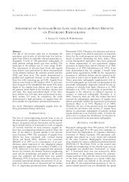
effect of calcium hydroxide and chlorhexidine based gutta-percha ...
effect of calcium hydroxide and chlorhexidine based gutta-percha ...
effect of calcium hydroxide and chlorhexidine based gutta-percha ...

Create successful ePaper yourself
Turn your PDF publications into a flip-book with our unique Google optimized e-Paper software.
July 30, 2004<br />
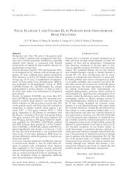
Abstract<br />
Aims <strong>and</strong> Methods: The aim <strong>of</strong> the present study was to<br />
demonstrate the possible <strong>effect</strong> <strong>of</strong> different endodontic<br />
<strong>calcium</strong> <strong>hydroxide</strong> <strong>and</strong> <strong>chlorhexidine</strong>-<strong>based</strong> <strong>gutta</strong><strong>percha</strong><br />
points, on two different human cell culture systems.<br />
Two different <strong>calcium</strong> <strong>hydroxide</strong> (Roeko, Langenau,<br />
Germany) <strong>and</strong> one <strong>chlorhexidine</strong> (Activ Point/ Roeko,<br />
Langenau, Germany) <strong>gutta</strong>-<strong>percha</strong> points were tested<br />
with gingival fibroblasts <strong>and</strong> epithelial tumor cells over<br />
a period <strong>of</strong> six days (n = 12). Conventional <strong>gutta</strong>-<strong>percha</strong><br />
points (VDW, Munich, Germany) <strong>and</strong> cells that<br />
were not exposed to any substances served as controls<br />
(n = 12). Study parameters included cell vitality, cell<br />
count, protein synthesis <strong>and</strong> cell proliferation.<br />
Results: All tested materials induced cell growth specific<br />
alterations. Chlorhexidine-<strong>based</strong> <strong>gutta</strong>-<strong>percha</strong> points<br />
showed a significant lower protein synthesis with both,<br />
gingival fibroblasts (0.013 ± 0.009 mg/ml) <strong>and</strong> epithelial<br />
tumor cells (0.07 ± 0.039 mg/ml), when compared<br />
with the controls (p > 0.05). Protein synthesis increase<br />
<strong>of</strong> the epithelial tumor cells (0.581 ± 0.013 mg/ml,<br />
control) was observed with the conventional <strong>gutta</strong><strong>percha</strong><br />
points (0.688 ± 0.078 mg/ml) <strong>and</strong> with both<br />
<strong>gutta</strong>-<strong>percha</strong> points containing different <strong>calcium</strong> <strong>hydroxide</strong>-<strong>based</strong><br />
formulations (0.776 ± 0.115 <strong>and</strong> 0.7 ±<br />
0.047mg/ ml).<br />
Conclusions: Under the conditions <strong>of</strong> this study, <strong>chlorhexidine</strong><br />
containing <strong>gutta</strong>-<strong>percha</strong> points showed the<br />
highest <strong>effect</strong> on cell growth inhibition. No significant<br />
differences were observed between the tested material<br />
<strong>and</strong> the two different cell culture types.<br />
Key words: Gutta-<strong>percha</strong> points; <strong>calcium</strong> <strong>hydroxide</strong>;<br />
<strong>chlorhexidine</strong>; cell culture<br />
INTRODUCTION<br />
Calcium <strong>hydroxide</strong> is the most commonly utilized, <strong>and</strong><br />
studied root canal medication. The first report about<br />
<strong>calcium</strong> <strong>hydroxide</strong> is attributed to Nygren (1838) for<br />
the treatment <strong>of</strong> the “fistula dentalis”, whilst Codman<br />
(1851) was the first to attempt to preserve an injuried<br />
dental pulp (Fava <strong>and</strong> Saunders 1999). Hermann<br />
(1920, 1936) demonstrated the biological properties<br />
<strong>and</strong> antimicrobial <strong>effect</strong> <strong>of</strong> <strong>calcium</strong> <strong>hydroxide</strong> through<br />
his pioneering work. Since then its clinical indications<br />
have exp<strong>and</strong>ed, <strong>and</strong> has been recommended to control<br />
EUROPEAN JOURNAL OF MEDICAL RESEARCH 345<br />
Eur J Med Res (2004) 9: 345-350 © I. Holzapfel Publishers 2004<br />
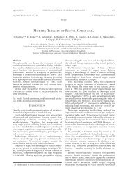
EFFECT OF CALCIUM HYDROXIDE AND CHLORHEXIDINE BASED<br />
GUTTA-PERCHA POINTS ON GINGIVAL FIBROBLASTS AND EPITHELIAL<br />
TUMOR CELLS<br />
B. Willershausen, B. Hagedorn, H. Tekyatan, B. Briseño Marroquín<br />
Department <strong>of</strong> Operative Dentistry, Johannes Gutenberg-University Mainz, Germany<br />
microbial growth, to enhance the healing process <strong>of</strong><br />
periapical lesions, to arrest inflammatory root resorption,<br />
to induce hard tissue deposition, as an inter-appointment<br />
root canal dressing material as well as to<br />
promote healing <strong>of</strong> vital pulps <strong>and</strong> periapical tissues<br />
(Andreasen 1981, Caliskan et al 1998). Calcium <strong>hydroxide</strong>’s<br />
alkaline pH, its ionic activity, diffusion<br />
through dentinal tubules, influence on apical microleakage<br />
<strong>and</strong> placement within the root canal are examples<br />
on how this material has been evaluated since<br />
its introduction. Its alkalizing <strong>effect</strong> (Economides et al<br />
1999, Larsen <strong>and</strong> Horsted-Bindslev 2000, Podbielski et<br />
al 2000, Schäfer <strong>and</strong> Al Behaissi 2000) is meant to support<br />
the mechanical <strong>and</strong> chemical preparation <strong>of</strong> the<br />
root canal through its antibacterial <strong>effect</strong> (Lado et al<br />
1986, Stuart et al 1991). An inter-appointment antimicrobial<br />
medication is also recommended in order to<br />
prevent recovery <strong>and</strong> multiplication <strong>of</strong> remanent microorganisms<br />
even after careful instrumentation <strong>and</strong><br />
debridement <strong>of</strong> the root canal system (Byström 1985).<br />
Thus, the use <strong>of</strong> various formulations <strong>and</strong> suggestions<br />
for mixing <strong>calcium</strong> <strong>hydroxide</strong> power with other substances<br />
which have antibacterial <strong>and</strong> radiopac properties<br />
have been also recommended. Conventionally, <strong>calcium</strong><br />
<strong>hydroxide</strong> is prepared by mixing the powder with<br />
a liquid <strong>and</strong> inserted into the root canal by means <strong>of</strong><br />
an injection, root canal instrument, <strong>and</strong> <strong>gutta</strong>-<strong>percha</strong><br />
or paper point. Sterile water or glycerine are recommended<br />
as vehicles to make a paste with Ca(OH) 2<br />
powder (Walton <strong>and</strong> Torabinejad 1989). Glycerine may<br />
be preferred as the vehicle for the placement <strong>of</strong> <strong>calcium</strong><br />
<strong>hydroxide</strong> into the root canal when depth <strong>of</strong> penetration<br />
<strong>and</strong> paste density (Riviere 1994) should be enhanced.<br />
In addition to placement, the removal <strong>of</strong> <strong>calcium</strong><br />
<strong>hydroxide</strong> from the root canal without leaving any<br />
residues behind is also a time-consuming <strong>and</strong> cumbersome<br />
procedure. Thus, a potential negative influence<br />
on the adhesive properties <strong>of</strong> the root canal sealer is<br />
possible (Lambrianidis et al 1999). These shortcomings<br />
prompted the development <strong>of</strong> <strong>gutta</strong>-<strong>percha</strong> points<br />
containing <strong>calcium</strong> <strong>hydroxide</strong> or <strong>chlorhexidine</strong>, which<br />
could be easily placed in the root canal especially in<br />
the apical area. Likewise, these types <strong>of</strong> <strong>gutta</strong>-<strong>percha</strong><br />
points can be easily removed without leaving material<br />
residuals behind. In addition to <strong>calcium</strong> <strong>hydroxide</strong>,<br />
<strong>gutta</strong>-<strong>percha</strong> points containing <strong>chlorhexidine</strong> diacetate<br />
have also been used to avoid re-infection <strong>of</strong> the root<br />
canal system. Chlorhexidine is <strong>effect</strong>ive against a
346 EUROPEAN JOURNAL OF MEDICAL RESEARCH<br />
July 30, 2004<br />
broad spectrum <strong>of</strong> microorganisms, including yeast<br />
<strong>and</strong> fungi (McDonell <strong>and</strong> Russell 1999). Chlorhexidine<br />
diacetate is soluble <strong>and</strong> releases cations binding to the<br />
anionic surface molecules <strong>of</strong> bacterial cell membranes,<br />
thereby disrupting the osmotic equilibrium <strong>of</strong> cells<br />
(McDonell <strong>and</strong> Russell 1999). However, the biocompatibility<br />
<strong>of</strong> these <strong>calcium</strong> <strong>hydroxide</strong> <strong>and</strong> <strong>chlorhexidine</strong>-<strong>based</strong><br />
<strong>gutta</strong>-<strong>percha</strong> points with regard to the surrounding<br />
tissue structures also needs to be considered.<br />
Tissue compatibility is decisive in the selection <strong>of</strong> the<br />
active ingredient, since the intra-canal medication may<br />
otherwise lead to irritation <strong>of</strong> the surrounding tissues;<br />
thus, a possible interference <strong>of</strong> the post-treatment<br />
healing process could be possible. The aim <strong>of</strong> the present<br />
study was to investigate <strong>and</strong> compare the <strong>effect</strong> <strong>of</strong><br />
<strong>gutta</strong>-<strong>percha</strong> points containing either <strong>calcium</strong> <strong>hydroxide</strong><br />
or <strong>chlorhexidine</strong> diacetate on human gingival fibroblasts<br />
<strong>and</strong> epithelial tumor cells.<br />
MATERIALS AND METHODS<br />
CELL CULTURES<br />
Gingival fibroblasts were obtained from biopsies <strong>of</strong><br />
healthy individuals <strong>of</strong> both genders (age range: 20–30<br />
years), derived from the lower molar region showing<br />
no signs <strong>of</strong> inflammation. Epithelial tumor cells were<br />
obtained from one male patient with T4 squamous cell<br />
carcinoma <strong>of</strong> the sinus piriformis. The cells were cultivated<br />
in Eagle’s Basal medium (Gr<strong>and</strong> Isl<strong>and</strong> Biological<br />
Co., Gr<strong>and</strong> Isl<strong>and</strong>, NY) with 10% fetal calf serum<br />
(Gr<strong>and</strong> Isl<strong>and</strong> Biological Co.), 50 IU/ml penicillin<br />
(Seromed) <strong>and</strong> 50 µg/ml streptomycin (Seromed). The<br />
cultures were kept in an environment <strong>of</strong> 95% air <strong>and</strong><br />
5% CO 2 <strong>and</strong> a temperature <strong>of</strong> 37 °C (Martin 1973).<br />
Following caryological characterization (Dutrillaux <strong>and</strong><br />
Couturier 1983), normal diploid gingival fibroblasts<br />
between the 5th <strong>and</strong> 7th passage <strong>and</strong> epithelial tumor<br />
cells between the 35th <strong>and</strong> 37th passage were used.<br />
INTRA-CANAL MATERIALS<br />
Two different intra-canal dressing materials in three<br />
different <strong>gutta</strong>-<strong>percha</strong> point types size ISO 30 (n = 12)<br />
were investigated: Roeko St<strong>and</strong>ard Ca(OH) 2 Points<br />
(58% <strong>calcium</strong> <strong>hydroxide</strong>, 42% <strong>gutta</strong>-<strong>percha</strong> <strong>and</strong>
July 30, 2004 EUROPEAN JOURNAL OF MEDICAL RESEARCH<br />
347<br />
Fig. 1 a, b. Gingival fibroblasts exposed to a) conventional<br />
<strong>gutta</strong>-<strong>percha</strong> point <strong>and</strong> b) Activ Point (x 125). An inhibition<br />
zone around the testing material (b) can be observed.<br />
The protein concentration <strong>of</strong> gingival fibroblasts in<br />
the control group was 0.1 mg/ml (±0.008) at the beginning<br />
<strong>and</strong> reached 0.115 mg/ml (± 0.014) at the 6th<br />
day <strong>of</strong> culturing. Similar protein concentration values at<br />
the 6th day were also found when the gingival fibroblasts<br />
were exposed to conventional <strong>gutta</strong>-<strong>percha</strong> <strong>and</strong><br />
to <strong>calcium</strong> <strong>hydroxide</strong>-<strong>based</strong> Plus points, while the <strong>calcium</strong><br />
<strong>hydroxide</strong>-<strong>based</strong> conventional points resulted in a<br />
Fig. 2 a, b. Epithelial tumor cells exposed to a) conventional<br />
<strong>gutta</strong>-<strong>percha</strong> point <strong>and</strong> b) Activ Point (x125). A direct contact<br />
between the testing material <strong>and</strong> cells can be observed in a)<br />
<strong>and</strong> an inhibition zone in the vicinity <strong>of</strong> the Activ point (b).<br />
Fig. 3. Effect <strong>of</strong> <strong>gutta</strong><strong>percha</strong><br />
containing intracanal<br />
dressing material<br />
on the proliferation rate<br />
(RFU) <strong>of</strong> gingival fibroblasts<br />
at different observation<br />
times (n = 12).<br />
statistically higher increase <strong>of</strong> protein concentration.<br />
The <strong>chlorhexidine</strong>-<strong>based</strong> Activ Points was the only material<br />
to yield a significant decrease in fibroblast protein<br />
concentration when compared with the controls (Fig.<br />
5). Likewise, the epithelial tumor cells exposed to Activ<br />
Points produced significantly less protein than the controls,<br />
whereas the other <strong>gutta</strong>-<strong>percha</strong> points showed<br />
higher protein concentration values than the controls
348 EUROPEAN JOURNAL OF MEDICAL RESEARCH<br />
July 30, 2004<br />
Fig. 4. Effect <strong>of</strong> <strong>gutta</strong><strong>percha</strong><br />
containing intracanal<br />
dressing material<br />
on the proliferation rate<br />
(RFU) <strong>of</strong> epithelial tumor<br />
cells at different<br />
observation times (n =<br />
12).<br />
Fig. 5. Protein concentration<br />
(mg/ml) <strong>of</strong> gingival<br />
fibroblasts (n = 12) after<br />
six days <strong>of</strong> incubation.<br />
Fig. 6. Protein concentration<br />
(mg/ml) <strong>of</strong> epithelial<br />
tumor cells (n =<br />
12) after six days <strong>of</strong> incubation.
July 30, 2004 EUROPEAN JOURNAL OF MEDICAL RESEARCH<br />
349<br />
(Fig. 6). Significantly higher protein concentration was<br />
observed only with regular <strong>calcium</strong> <strong>hydroxide</strong> <strong>based</strong><br />
<strong>gutta</strong>-<strong>percha</strong> points.<br />
DISCUSSION<br />
The main use indications <strong>of</strong> <strong>calcium</strong> <strong>hydroxide</strong> are the<br />
stimulation <strong>of</strong> re-mineralization, its antibacterial properties<br />
<strong>and</strong> the dissolution <strong>of</strong> necrotic tissue (Gomes<br />
2003, Byström 1985, Sjögren 1991, Foreman 1990).<br />
These properties are mostly correlated to its high pH<br />
<strong>and</strong> necrotizing capacity. Some authors described the<br />
destruction <strong>of</strong> epithelium present in periradicular lesions;<br />
thus, promoting connective tissue invagination<br />
<strong>and</strong> healing (Sahli 1990, Bruyne 2000). Yet, the <strong>effect</strong><br />
<strong>of</strong> such materials in the periapical tissue has to be considered.<br />
After root canal instrumentation different contaminated<br />
fluids such as extracellular fluid, exudates, pus or<br />
even saliva may seep into the canal. It is known that<br />
some bacteria can survive at pH 9; therefore, the application<br />
<strong>of</strong> an intra-canal dressing material that releases a<br />
considerable amount <strong>of</strong> hydroxyl <strong>and</strong> maintains a high<br />
pH in the root canal space is advantageous (Byström<br />
1985). The application <strong>of</strong> an intra-canal dressing material<br />
will enhance the reduction <strong>of</strong> the number <strong>of</strong> microorganisms<br />
<strong>and</strong> can penetrate in areas not reached by<br />
the instruments or irrigating solutions during root<br />
canal preparation (Barbosa 1997). It has been demonstrated<br />
that conventional <strong>gutta</strong>-<strong>percha</strong> due to zinc-oxide<br />
content inhibits the growth <strong>and</strong> vitality <strong>of</strong> certain<br />
bacteria known to be present in infected root canals<br />
(Moorer <strong>and</strong> Genet 1982, Moorer <strong>and</strong> Genet 1982,<br />
Weiger et al 1993). Likewise, <strong>calcium</strong> <strong>hydroxide</strong> <strong>and</strong><br />
<strong>chlorhexidine</strong> are also known to inhibit bacterial<br />
growth (Barbosa et al 1997, Heling et al 1992, Tchaou<br />
et al 1996). A microbiological study (Podbielski et al<br />
2000) has shown that some microorganisms are more<br />
<strong>effect</strong>ively reduced by <strong>calcium</strong> <strong>hydroxide</strong>, while others<br />
are more susceptible to <strong>chlorhexidine</strong>. Fuss et al (1996)<br />
used a freshly prepared <strong>calcium</strong> <strong>hydroxide</strong> solution<br />
whose bactericidal potential was slightly higher to that<br />
<strong>of</strong> premixed <strong>calcium</strong> <strong>hydroxide</strong> pastes. However, the<br />
bacterial advantages <strong>of</strong> <strong>calcium</strong><strong>hydroxide</strong> <strong>and</strong><br />
<strong>chlorhexidine</strong> could have a toxic <strong>effect</strong> in the periapical<br />
tissue. Cell culture studies can provide information on<br />
the behaviour <strong>of</strong> different materials against healthy tissues.<br />
Because the periapical tissues can be exposed to<br />
intra-canal dressing materials, the possible toxic <strong>effect</strong><br />
<strong>of</strong> commercial available <strong>calcium</strong> <strong>hydroxide</strong> <strong>and</strong><br />
<strong>chlorhexidine</strong>-<strong>based</strong> <strong>gutta</strong>-<strong>percha</strong> points also needs to<br />
be evaluated.<br />
Different studies have proven the <strong>effect</strong> <strong>of</strong> <strong>calcium</strong><br />
<strong>hydroxide</strong> (Nakamura et al 1986, Rappaport et al 1964).<br />
In a previous publication (Willershausen et al 2000) the<br />
biological compatibility <strong>of</strong> various root canal filling<br />
materials was investigated with similar research parameters.<br />
It was established that conventional <strong>gutta</strong>-<strong>percha</strong><br />
points had a non-significant <strong>effect</strong> on the<br />
prostagl<strong>and</strong>in release <strong>of</strong> gingival fibroblasts. These<br />
findings are supported by the comparison <strong>of</strong> cell proliferation<br />
results between conventional <strong>gutta</strong>-<strong>percha</strong><br />
points <strong>and</strong> the unexposed cell cultures. Our findings<br />
also indicate that <strong>calcium</strong> <strong>hydroxide</strong>-<strong>based</strong> <strong>gutta</strong>-<strong>percha</strong><br />
points have only a slight inhibitory <strong>effect</strong> on the cell<br />
proliferation <strong>of</strong> gingival fibroblasts <strong>and</strong> epithelial tumor<br />
cells. The only tested material that significantly inhibited<br />
the growth <strong>of</strong> gingival fibroblasts was the<br />
<strong>chlorhexidine</strong>-<strong>based</strong> Activ Points. The protein synthesis<br />
patterns were also significantly different from the<br />
controls. During root canal treatment, it is important<br />
to create an environment in the access cavity in order<br />
to prevent the entrance <strong>of</strong> microorganisms into the<br />
root canal system. The clinical advantages or disadvantages<br />
<strong>of</strong> the use <strong>of</strong> Activ Points need to be considered.<br />
The present study shows that the culture systems employed<br />
are sensitive <strong>and</strong> provide information on the<br />
possible <strong>effect</strong>s <strong>of</strong> intra-canal dressing materials on the<br />
periapical tissues. Thus, these results can facilitate employment<br />
decisions between various active ingredients<br />
<strong>of</strong> <strong>gutta</strong>-<strong>percha</strong> points <strong>and</strong> contribute to the success <strong>of</strong><br />
root canal treatment.<br />
REFERENCES<br />
1. Andreasen JO (1981) Relationship between the surface<br />
<strong>and</strong> inflammatory resorption <strong>and</strong> changes in the pulp<br />
after replantation <strong>of</strong> permanent incisors in monkeys. J<br />
Endodont 7: 294-301<br />
2. Barbosa CA, Goncalves RB, Siqueira Jr. JF, De Uzeda M<br />
(1997) Evaluation <strong>of</strong> the antibacterial activities <strong>of</strong><br />
<strong>calcium</strong> <strong>hydroxide</strong>, <strong>chlorhexidine</strong>, <strong>and</strong> camphorated<br />
paramonochlorophenol as intra-canal medicament.<br />
A clinical <strong>and</strong> laboratory study. J Endodont 23: 297-<br />
300<br />
3. Briseño MB, Willershausen B (1990) Root canal sealer<br />
cytotoxicity on human gingival fibroblasts. I. Zinc<br />
oxide-eugenol-<strong>based</strong> sealers. J Endodont 16: 383-386<br />
4. De Bruyne MAA, De Moor RJG, Raes FM (2000)<br />
Necrosis <strong>of</strong> the gingiva caused by <strong>calcium</strong> <strong>hydroxide</strong>. A<br />
case report. Int Endodont J 33: 67-71<br />
5. Byström A, Claesson R, Sundqvist G (1985) The antibacterial<br />
<strong>effect</strong> <strong>of</strong> camphorated paramonochlorophenol,<br />
camphorated phenol <strong>and</strong> <strong>calcium</strong> <strong>hydroxide</strong> in the<br />
treatment <strong>of</strong> infected root canals. Endodont Dent<br />
Traumatol 1: 170-175<br />
6. Caliskan MK, Türkün M, Türkün LS(1998) Effect <strong>of</strong> <strong>calcium</strong><br />
<strong>hydroxide</strong> as an intracanal dressing opn apical leakage.<br />
Int Endodont J 31: 173-177<br />
7. Codman WW (1951) Ossification <strong>of</strong> the pulp <strong>of</strong> a tooth.<br />
Newsletter IV, 90 (printed in Malo PRT, Kessler Nieto<br />
F, Vadillo MVM (1987) Hidroxido de calcio y apic<strong>of</strong>ormacion.<br />
Rev Espan Endodon 5: 41-61)<br />
8. Dutrillaux B, Couturier J (1983) Praktikum der Chromosomenanalyse.<br />
F. Enke, Stuttgart<br />
9. Economides N, Koulaouzidou EA, Beltes P, Kortsaris<br />
AH (1999) In vitro release <strong>of</strong> hydroxyl ions from <strong>calcium</strong><br />
<strong>hydroxide</strong> <strong>gutta</strong>-<strong>percha</strong> points. J Endodont 25: 481-<br />
482<br />
10. Fava LRG, Saunders WP (1999) Calcium <strong>hydroxide</strong><br />
pastes: classification <strong>and</strong> clinical indications. Int Endodont<br />
J 32: 257-282<br />
11. Foreman PC, Barnes IE (1990) A review <strong>of</strong> <strong>calcium</strong> <strong>hydroxide</strong>.<br />
Int Endodont J 23: 283-297<br />
12. Fuss Z, Rafael<strong>of</strong>f R, Tagger M, Szajkis S (1996) Intracanal<br />
pH changes <strong>of</strong> <strong>calcium</strong> <strong>hydroxide</strong> pastes exposed<br />
to carbon dioxide in vitro. J Endodont 22: 362-364<br />
13. Gomes BPFA, Sato E, Ferraz CCR, Teixeira FB, Zaia<br />
AA, Souza-Filho FJ (2003) Evaluation <strong>of</strong> time required<br />
for recontamination <strong>of</strong> coronally sealed canals mediated<br />
with <strong>calcium</strong> <strong>hydroxide</strong> <strong>and</strong> <strong>chlorhexidine</strong>. International<br />
EndodontJ 36: 604-609
350 EUROPEAN JOURNAL OF MEDICAL RESEARCH<br />
July 30, 2004<br />
14. Heling I, Sommer M, Steinberg D, Friedman M, Sela<br />
MN (1992) Microbiological evaluation <strong>of</strong> the efficacy <strong>of</strong><br />
<strong>chlorhexidine</strong> in a sustained-release device for dentine<br />
sterilization. Int Endodont J 25: 15-19<br />
15. Hermann BW (1920) Calciumhydroxid als Mittel zum<br />
beh<strong>and</strong>eln und füllen von Wurzelkanälen (Dissertation).<br />
Würzburg (printed in: Malo PRT, Kessler Nieto F,<br />
Vadillo MVM (1987) Hidroxido de calcio y apic<strong>of</strong>ormacion.<br />
Rev Espan Endodon 5: 41-61)<br />
16. Hermann BW (1952) On the reaction <strong>of</strong> the dental pulp<br />
to vital amputation <strong>and</strong> calxyl capping. Dtsch Zahnarztl<br />
Z 15: 1446-1447<br />
17. Lado EA, Pappas J, Tyler K, Stanley HR, Walker C<br />
(1986) In vitro antimicrobial activity <strong>of</strong> six pulp-capping<br />
agents. Oral Surg Oral Med Oral Pathol 61: 197-200<br />
18. Lambrianidis T, Margelos J, Beltes P (1999) Removal efficiency<br />
<strong>of</strong> <strong>calcium</strong> <strong>hydroxide</strong> dressing from the root<br />
canal. J Endodont 25: 85-88<br />
19. Larsen MJ, Horsted-Bindslev P (2000) A laboratory<br />
study evaluating the release <strong>of</strong> hydroxyl ions from various<br />
<strong>calcium</strong> <strong>hydroxide</strong> products in narrow root canallike<br />
tubes. Int Endodont J 33: 238-242<br />
20. Martin GM (1973) Human skin fibroblasts. In: Kurse PF<br />
<strong>and</strong> Patterson MK (eds) Tissue culture methods <strong>and</strong> applications.<br />
Academic Press, New York, p 39-43<br />
21. McDonell G, Russell AD (1999) Antiseptics <strong>and</strong> disinfectants:<br />
activity, action <strong>and</strong> resistance. Clin Microbiol<br />
Rev 12: 147-179<br />
22. Moorer WR, Genet JM (1982) Antibacterial activity <strong>of</strong><br />
<strong>gutta</strong>-<strong>percha</strong> cones attributed to the zinc oxide component.<br />
Oral Surg Oral Med Oral Pathol 53: 508-517<br />
23. Moorer WR, Genet JM (1982) Evidence for antibacterial<br />
activity <strong>of</strong> endodontic <strong>gutta</strong>-<strong>percha</strong> cones. Oral Surg<br />
Oral Med Oral Pathol 53: 503-507<br />
24. Nakamura H, Sakakibara F, Matsumoto Y et al (1986)<br />
Study on the cytotoxicity <strong>of</strong> root canal filling materials. J<br />
Endodont 12: 156-160<br />
25. Nygren J (1938) Radgivare Angaende Basta Sattet Att<br />
Varda Ah Bevara T<strong>and</strong>ernas Fuskhet, Osv. Stockholm<br />
26. Podbielski A, Boeckh C,Haller B (2000) Growth inhibitory<br />
activity <strong>of</strong> <strong>gutta</strong>-<strong>percha</strong> points containing root<br />
canal medications on common endodontic bacterial<br />
pathogens as determined by an optimized quantitative in<br />
vitro assay. J Endodont 26: 398-403<br />
27. Rappaport HM, Lilly GE, Kapsimalis P (1964) Toxicity<br />
<strong>of</strong> endodontic filling materials. Oral Surg Oral Med Oral<br />
Pathol 18: 758-802<br />
28. Rivera EM, Williams K (1994) Placement <strong>of</strong> <strong>calcium</strong> <strong>hydroxide</strong><br />
in simulated canals: comparison <strong>of</strong> glycerine vs<br />
water. J Endodont 20: 445-458<br />
29. Sahli CC (1990) Perspectivas actuales del tratmiento endodoncico<br />
en dientes con lesione periapicales cronicas.<br />
Endodoncia 8: 99-107<br />
30. Schäfer E, Al Behaissi A (2000) PH changes in root<br />
dentin after root canal dressing with <strong>gutta</strong>-<strong>percha</strong> points<br />
containing <strong>calcium</strong> <strong>hydroxide</strong>. J Endodont 26: 665-667<br />
31. Sjögren U, Figdor D, Spangberg L, Sundqvist G (1991)<br />
The antibacterial <strong>effect</strong> <strong>of</strong> <strong>calcium</strong> <strong>hydroxide</strong> as a shortterm<br />
intracanal dressing. Int Endodont J 24: 119-125<br />
32. Stuart KG, Miller CH, Brown Jr. CE, Newton CW<br />
(1991) The comparative antimicrobial <strong>effect</strong> <strong>of</strong> <strong>calcium</strong><br />
<strong>hydroxide</strong>. Oral Surgery, Oral Med Oral Pathol Oral<br />
Radiol Endodont 72: 101-104<br />
33. Tchaou WS, Turng BF, Minah GE, Coll JA (1996)<br />
Inhibition <strong>of</strong> pure cultures <strong>of</strong> oral bacteria by root canal<br />
filling materials. PediatrDentistry 18: 444-449<br />
34. Walton RE, Torabinejad M (1989) Claening <strong>and</strong> shaping.<br />
In: Pedersen P (ed) Principles <strong>and</strong> Practice <strong>of</strong> Endodontics.<br />
Saunders, Philadelphia<br />
35. Weiger R, Manncke B, Löst C (1993) Antibakterielle<br />
Wirkung von Gutta<strong>percha</strong>stiften auf verschiedene endopathogene<br />
Mikroorganismen. Dtsch Zahnärztl Z 48:<br />
658-660<br />
36. Willershausen B, Briseño Marroquín B, Schäfer D,<br />
Schulze R (2000) Cytotoxicity <strong>of</strong> root canal filling materials<br />
to three different human cell lines. J Endodont 26:<br />
703-707<br />
Received: February 23, 2004 / Accepted: May 3,2004<br />
Adress for correspondence:<br />
Pr<strong>of</strong>. Dr. B. Willershausen<br />
Department for Operative Dentistry<br />
Johannes Gutenberg University Mainz<br />
Augustusplatz 2<br />
D-55131 Mainz, Germany<br />
Tel: +49 – 6131 – 177247<br />
Fax: +49 – 6131 – 173406<br />
e-mail: willersh@mail.uni-mainz.de