CCR connections - National Cancer Institute
CCR connections - National Cancer Institute
CCR connections - National Cancer Institute

You also want an ePaper? Increase the reach of your titles
YUMPU automatically turns print PDFs into web optimized ePapers that Google loves.
<strong>National</strong> <strong>Cancer</strong> <strong>Institute</strong><br />
U.S. DEPARTMENT<br />
OF HEALTH AND<br />
HUMAN SERVICES<br />
<strong>National</strong> <strong>Institute</strong>s<br />
of Health<br />
Of Mice and Men:<br />
Tracking the Origins of Metastatic Prostate <strong>Cancer</strong><br />
V o l u m e 5, No. 1 | 2011<br />
<strong>CCR</strong> <strong>connections</strong><br />
CenTer fOr CanCer researCh ccr.cancer.gov
We invite your comments and suggestions about <strong>CCR</strong> <strong>connections</strong>.<br />
Please email your feedback to tellccr@mail.nih.gov.<br />
Center for <strong>Cancer</strong> Research<br />
<strong>National</strong> <strong>Cancer</strong> <strong>Institute</strong> | <strong>National</strong> <strong>Institute</strong>s of Health | Building 31 – Room 3A11 | 31 Center Drive | Bethesda, MD 20892<br />
Table of Contents<br />
e d i t o r i a l<br />
03 When Persistence Pays Off<br />
n e w s<br />
04 Setting the Sun on Skin <strong>Cancer</strong><br />
05 Hitting the Target<br />
06 Patent Pool Goes Global<br />
07 The Frontiers of Thymic Malignancy<br />
08 A Look at Rare Diseases, from Molecules to Patients<br />
09 Test Before You Treat<br />
09 Staff News at <strong>CCR</strong><br />
11 Recent <strong>CCR</strong> Awards<br />
12 In Conversation: Research Fellow Ram Savan, Ph.D.<br />
f e a t u r e s<br />
13 Of Mice and Men: Tracking the Origins of Metastatic Prostate <strong>Cancer</strong><br />
17 It’s All About the Client: The Development of Hsp90 Inhibitors as<br />
Anti-<strong>Cancer</strong> Agents<br />
22 Don’t Throw Out the Packing Materials<br />
c o m m e n t a r y<br />
26 Breast <strong>Cancer</strong>: The Triple-Negative Problem<br />
in t h e clinic<br />
28 One Molecule, Multiple <strong>Cancer</strong>s: The Devil is in the Details<br />
13 17 22 28<br />
f e a t u r e f e a t u r e f e a t u r e in t h e clinic<br />
Of Mice and Men:<br />
Tracking the Origins<br />
of Metastatic<br />
Prostate <strong>Cancer</strong><br />
It’s All About the Client:<br />
The Development of<br />
Hsp90 Inhibitors as<br />
Anti-<strong>Cancer</strong> Agents<br />
Don’t Throw Out the<br />
Packing Materials<br />
One Molecule,<br />
Multiple <strong>Cancer</strong>s: The<br />
Devil is in the Details<br />
V o l u m e 5, No. 1 | 2011<br />
ccr <strong>connections</strong> | Vo l u m e 5, No. 1 | 2011 1
The mission of <strong>CCR</strong> is:<br />
Contributors:<br />
D. Kerrigan, M.S.<br />
K. Martin, M.S.<br />
S. Fox, B.A., B.S.W.<br />
J. Crawford, Ph.D.<br />
N. Giannosa, M.P.H.<br />
To inform and empower the entire cancer research<br />
community by making breakthrough discoveries in<br />
basic and clinical cancer research and by developing<br />
them into novel therapeutic interventions for adults<br />
and children afflicted with cancer or infected with HIV.<br />
http://home.ccr.cancer.gov/<strong>connections</strong><br />
Some say that breakthroughs happen<br />
when people with a fresh, even naïve,<br />
perspective look at an old problem.<br />
But in science, particularly in the<br />
complex and detail-intense world<br />
of biomedicine, sustained work and<br />
investment in a particular set of<br />
problems, coupled with an open and<br />
creative mind, is often the most fruitful<br />
path to scientific advancement.<br />
In this issue of <strong>CCR</strong> <strong>connections</strong>,<br />
we see several examples of<br />
researchers that have tackled the<br />
hard problems of a particular field<br />
for decades and consistently reaped<br />
great rewards. In “It’s All About the<br />
Client,” we learn how Len Neckers,<br />
Ph.D., identified the ubiquitous<br />
heat shock protein Hsp90 as<br />
an anti-cancer target 15 years<br />
ago. Defying conventional wisdom,<br />
he worked within NCI to develop<br />
the first clinical trial of an Hsp90<br />
inhibitor and paved the way for<br />
the 13 Hsp90 inhibitors currently<br />
in clinical trials. Neckers and his<br />
team are continuing to establish the<br />
basic mechanisms of Hsp90 function<br />
and doing the preclinical work to<br />
optimize this therapeutic strategy.<br />
Along with other NIH intramural<br />
colleagues, Gordon Hager, Ph.D.,<br />
studied the role of chromatin<br />
structure in gene regulation long<br />
before most scientists believed it<br />
had any relevance and before the<br />
advent of research tools such as<br />
modern gene cloning technologies.<br />
Our feature, “Don’t Throw Out the<br />
Packing Materials,” explains how<br />
Hager’s work has led to fundamental<br />
insights into the dynamic nature<br />
of gene regulation that have direct<br />
implications for understanding and<br />
overcoming the gene dysregulation<br />
associated with cancer.<br />
One of our newest tenure-track<br />
Investigators, Christina Annunziata,<br />
M.D., Ph.D., has been studying the<br />
subtleties of a single molecular<br />
pathway—NF-kB—since she was a<br />
graduate student. As an inaugural<br />
participant in our Clinical Investigator<br />
Development Program, she had the<br />
opportunity to apply her insights and<br />
knowledge of this pathway from prior<br />
research in multiple myeloma and<br />
create a strong preclinical ovarian<br />
cancer program to evaluate NF-kB<br />
pathway inhibitors. “One Molecule,<br />
Multiple <strong>Cancer</strong>s: The Devil is in the<br />
Details,” describes her plans to<br />
translate this research into better<br />
treatment for patients.<br />
We are also pleased to have an<br />
article from Joyce O’Shaughnessy,<br />
M.D., who developed a passion for<br />
clinical research in breast cancer<br />
during her years at NCI in the late<br />
1980s. She brought that passion with<br />
her to Texas Oncology and Baylor<br />
College of Medicine, and in “Breast<br />
<strong>Cancer</strong>: The Triple-Negative Problem,”<br />
O’Shaughnessy talks about the most<br />
recent fruits of her research—results<br />
of a very exciting phase 2 trial recently<br />
editorial<br />
When Persistence<br />
Pays Off<br />
As important as innovation, depth of expertise and determination<br />
are often key to great advances in biomedicine.<br />
Robert Wiltrout, Ph.D.<br />
published in the New England<br />
Journal of Medicine, on the use of<br />
PARP inhibitors for the treatment of<br />
triple-negative breast cancer.<br />
At <strong>CCR</strong>, we are proud of the role<br />
we have played in selecting and<br />
supporting scientists who have the<br />
commitment and fortitude it takes<br />
to brave uncharted territories, and<br />
who stay the course and get answers<br />
from their scientific investigations.<br />
Without this kind of sustained effort<br />
and determination to grapple with the<br />
complex and sometimes very basic<br />
questions in biomedical research,<br />
clinical breakthroughs would simply<br />
not be possible.<br />
2 ccr <strong>connections</strong> | Vo l u m e 5, No. 1 | 2011<br />
ccr <strong>connections</strong> | Vo l u m e 5, No. 1 | 2011 3<br />
(Photo: B. Branson)
(Image: R. Zaidi, <strong>CCR</strong>)<br />
news<br />
Setting<br />
Skin <strong>Cancer</strong> the Sun on<br />
Researchers<br />
Interferon-γ has been found to promote UV-induced melanoma in a mouse model.<br />
A series of experiments designed<br />
to understand how solar ultraviolet<br />
(UV) radiation causes aggressive<br />
cutaneous melanoma has led to<br />
an unanticipated discovery that<br />
could upend assumptions about<br />
the relationship between interferon<br />
proteins and cancer. Interferon-γ<br />
(IFN-γ), which traditionally has been<br />
thought to contribute to an innate<br />
defense system against cancer,<br />
under some circumstances may<br />
promote melanoma and incite the<br />
development of tumors. This finding<br />
from Glenn Merlino, Ph.D., Co-Chief<br />
of <strong>CCR</strong>’s Laboratory of <strong>Cancer</strong> Biology<br />
and Genetics, and Research Fellow M.<br />
Raza Zaidi, Ph.D., was published in<br />
the January 27, 2011, issue of Nature.<br />
Over the past decade, these<br />
researchers used genetically engineered<br />
mice first to establish, and then to<br />
dissect, the connection between<br />
exposure to UV radiation and the<br />
initiation of melanoma. The current<br />
work—made possible through longterm<br />
collaborations with Edward De<br />
Fabo, Ph.D., and Frances Noonan,<br />
Ph.D., of George Washington<br />
University Medical Center—was their<br />
latest attempt to define the molecular<br />
mechanisms of this cause-and-effect<br />
relationship. The results of this study<br />
offer the possibility that the inhibition<br />
of IFN-γ immediately after sunburn<br />
might block the carcinogenic activation<br />
of melanocytes, the skin’s pigmentproducing<br />
cells, by UV radiation.<br />
Crucial to the experiments<br />
was the development of a unique<br />
genetically engineered mouse in<br />
which the melanocytes were labeled<br />
with a green fluorescent protein. This<br />
fluorescent tag allowed melanocytes<br />
to be visually tracked and isolated<br />
and enabled researchers to evaluate,<br />
for the first time, their response to<br />
UV radiation while in their natural<br />
environment in a living animal.<br />
The researchers observed that<br />
UV radiation doses equivalent to<br />
those causing sunburn in human<br />
skin triggered aberrant growth and<br />
migration of melanocytes in mouse<br />
skin. UV radiation exposure also<br />
persistently activated genes known<br />
to respond to IFN-γ, including genes<br />
that may help tumor cells evade<br />
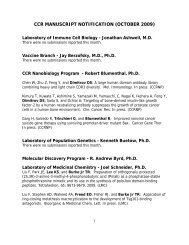
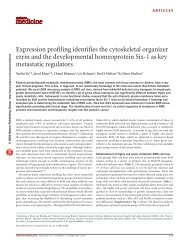
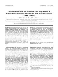
A human melanoma tumor, left, and a transplanted mouse melanoma tumor, right, show<br />
infiltration of macrophages. While a subset of mouse tumor macrophages display expression<br />
on interferon-gamma (IFN-γ), virtually all the macrophages in human melanoma express this<br />
protein (identified by the yellow colored overlap of the two antibodies).<br />
detection and attack by the immune<br />
system. When the activity of IFN-γ was<br />
inhibited, the growth and migration of<br />
melanocytes remained normal after<br />
exposure to UV radiation.<br />
“Interferons have long been touted<br />
as anti-tumorigenic and cytostatic—<br />
that they have an inhibitory effect<br />
on cellular growth,” said Dr. Zaidi.<br />
“The finding that IFN-γ can have a<br />
profoundly different effect—that it can<br />
exacerbate the growth of melanoma—<br />
is a paradigm-shifting discovery.”<br />
The team additionally showed<br />
that white blood cells known as<br />
macrophages were producing the<br />
IFN-γ. Macrophages significantly<br />
enhanced melanoma tumor growth<br />
when researchers injected them under<br />
the skin of healthy mice along with<br />
cultured mouse melanoma cells, and<br />
this effect was abolished by blocking<br />
IFN-γ activity. The researchers also<br />
identified IFN-γ-producing macrophages<br />
in 70 percent of 27 human melanomas<br />
they examined, supporting the<br />
possibility that IFN-γ plays a role in<br />
this type of cancer—not just in mice,<br />
but also in humans.<br />
Moreover, Dr. Zaidi noted,<br />
inhibiting IFN-γ immediately after<br />
sunburn, an approach that he and<br />
his colleagues are pursuing, may<br />
prove to be an effective preventive<br />
strategy against UV radiation-induced<br />
melanoma. The discovery could one<br />
day lead to drug treatments that block<br />
this mechanism and thus the cancer’s<br />
growth, potentially saving many from<br />
the lethal threat of skin cancer.<br />
To learn more about Dr. Merlino’s research,<br />
please visit his <strong>CCR</strong> Web site at http://ccr.<br />
nci.nih.gov/staff/staff.asp?Name=merlino.<br />
Hitting the Target<br />
Single genetic mutations and,<br />
more commonly, combinations of<br />
mutations lead to the development<br />
of cancers such as lymphoma—a<br />
cancer of the blood that arises<br />
from infection-fighting white blood<br />
cells. Diffuse large B cell lymphoma<br />
(DLBCL), a type of non-Hodgkin’s<br />
lymphoma, is the most common<br />
form of this disease and currently<br />
has a dismally low cure rate. There<br />
are three subtypes of DLBCL, of<br />
which the activated B cell-like (ABC)<br />
lymphoma has the worst outcome<br />
with a three-year survival rate of just<br />
40 percent.<br />
However, researchers have identified<br />
a recurring genetic mutation that<br />
could lead to targeted therapies for<br />
ABC lymphoma patients. Mutations of<br />
the MYD88 gene (normally involved<br />
in the immune response to invading<br />
microorganisms) are found in 39<br />
percent of patients with the ABC<br />
subtype of DLBCL and could drive the<br />
growth of some lymphoma tumors by<br />
activating multiple signaling pathways<br />
associated with cancer. A study<br />
published in the December 22, 2010,<br />
issue of Nature from the laboratory<br />
of Louis Staudt, M.D., Ph.D., Deputy<br />
Chief of <strong>CCR</strong>’s Metabolism Branch,<br />
reveals a mechanism whereby a single<br />
alteration in the MYD88 protein<br />
sequence can cause uncontrolled<br />
cellular signaling, leading to survival<br />
of malignant cells.<br />
Dr. Staudt and colleagues have<br />
worked to identify proteins that play<br />
a role in the development of the<br />
ABC subtype as potential targets to<br />
improve the treatment of patients with<br />
this form of lymphoma. To identify<br />
these critical proteins, the researchers<br />
performed a screen in which<br />
thousands of genes were inactivated.<br />
They found that ABC lymphoma cells<br />
were killed when they inactivated the<br />
genes encoding MYD88 and IRAK1,<br />
another cell signaling protein that<br />
works with MYD88.<br />
The scientists then looked for<br />
specific mutations in MYD88 that<br />
might explain the survival-dependence<br />
they observed. Sequencing of the<br />
MYD88 gene in 382 lymphoma biopsy<br />
samples revealed that 29 percent<br />
of ABC lymphoma samples had<br />
the same mutation, which altered<br />
a single amino acid in the MYD88<br />
protein, but this mutation was rare or<br />
absent in other lymphoma subtypes.<br />
The mutant form of MYD88 sustained<br />
the survival of the ABC lymphoma<br />
cells while the non-mutated version<br />
did not, suggesting that mutations<br />
in the MYD88 gene could play an<br />
important role in the development of<br />
ABC DLBCL.<br />
The researchers then examined<br />
proteins that interact with MYD88 in<br />
lymphoma cells. The mutant form of<br />
MYD88 spontaneously assembled a<br />
protein complex that included IRAK1,<br />
identified in the genetic screen,<br />
4 ccr <strong>connections</strong> | Vo l u m e 5, No. 1 | 2011 ccr <strong>connections</strong> | Vo l u m e 5, No. 1 | 2011 5<br />
news<br />
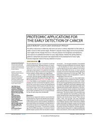
have identified a possible target for treating the most aggressive form of lymphoma.<br />
Biopsies of diffuse large B cell lymphoma (DLBCL) reveal varying gene expression levels in<br />
activated B cell-like DLBCL, germinal center B cell-like DLBCL, and primary mediastinal B<br />
cell lymphoma.<br />
and a related protein, IRAK4. In this<br />
protein complex, IRAK4 functioned<br />
as an enzyme to modify IRAK1, which<br />
was required for the mutant MYD88<br />
protein to promote lymphoma cell<br />
survival. This particular finding may<br />
hold promise for the treatment of<br />
lymphomas with MYD88 mutations<br />
since pharmaceutical companies are<br />
developing IRAK4 inhibitors for use<br />
in inflammatory and autoimmune<br />
diseases, noted Dr. Staudt.<br />
“The results of this study may<br />
provide a method to identify patients<br />
with the ABC subtype of diffuse large<br />
B cell lymphoma whose tumors may<br />
depend upon MYD88 signaling,”<br />
said Dr. Staudt. “And these patients<br />
may one day benefit from therapies<br />
targeting this and other regulatory<br />
pathways that sustain the survival of<br />
these lymphoma cells.”<br />
To learn more about Dr. Staudt’s research,<br />
please visit his <strong>CCR</strong> Web site at http://ccr.<br />
nci.nih.gov/staff/staff.asp?Name=staudt.<br />
(Image: L. Staudt, <strong>CCR</strong>)
(Photo: E. Branson)<br />
news<br />
Patent Pool Goes Global<br />
<strong>CCR</strong>-developed HIV drug darunavir is the first patent licensed to the Medicines Patent Pool.<br />
Hiroaki Mitsuya, M.D., Ph.D.<br />
In recent years, there have been<br />
heated debates on how to ensure that<br />
patents do not stand in the way of<br />
access to medicines—especially for<br />
the world’s poorest nations. According<br />
to UNITAID, an independent global<br />
health financing agency founded by<br />
the United Nations in 2006, treating<br />
a patient for one year with today’s<br />
recommended first-line AIDS treatment<br />
costs between $151 and $1,033—in<br />
part because certain products are<br />
patented in some countries. This,<br />
according to the agency, is an almost<br />
two- to 13-fold increase from the price<br />
of the most affordable and widely<br />
used older regimen.<br />
Enter the Medicines Patent Pool,<br />
a newly established UNITAID initiative<br />
that aims to find a route for expensive<br />
drugs to reach developing countries.<br />
By streamlining licensing processes<br />
for the production of generic versions<br />
of patented HIV/AIDS medicines, the<br />
6 ccr <strong>connections</strong> | Vo l u m e 5, No. 1 | 2011<br />
Pool serves as a one-stop shop that<br />
will accelerate the pace at which newer<br />
medicines reach patients and will help<br />
drive down prices to low, sustainable<br />
levels by encouraging competition<br />
among multiple generic producers.<br />
The Pool received its first,<br />
high-profile member when the NIH<br />
licensed a patent for the new AIDS<br />
drug darunavir in September 2010.<br />
The royalty-free license stipulates<br />
that this technology is to be available<br />
for the benefit of all low- and middleincome<br />
countries, as defined by the<br />
World Bank. Darunavir (Prezista ) is<br />
a novel protease inhibitor developed<br />
by Hiroaki Mitsuya, M.D., Ph.D., Head<br />
of <strong>CCR</strong>’s Experimental Retrovirology<br />
Section, HIV and AIDS Malignancy<br />
Branch, in a 13-year collaboration<br />
with Purdue University Professor<br />
Arun K. Ghosh, Ph.D.<br />
“Darunavir embodies a breakthrough<br />
in the struggle against the notorious<br />
obstacle of current AIDS therapy:<br />
multidrug-resistant HIV variants,”<br />
said Dr. Mitsuya. “The drug is active<br />
and has been proven clinically<br />
efficacious against multi-protease<br />
inhibitor-resistant HIV. More notably,<br />
darunavir effectively keeps HIV from<br />
becoming drug-resistant.”<br />
HIV rapidly mutates to resist<br />
new drugs, and many patients on<br />
first-line regimens will soon need<br />
to switch to second-line therapies<br />
that are more effective against drugresistant<br />
HIV. And just as there is<br />
a need to continually develop new<br />
drugs, there is also a need to develop<br />
new approaches for improving drug<br />
access globally. With this first license<br />
in hand, the Pool is one critical step<br />
closer to achieving its goal of making<br />
life-saving medicines, like darunavir,<br />
more affordable and accessible to<br />
people in developing countries.<br />
The Medicines Patent Pool provides<br />
a valuable model for attacking a<br />
longstanding problem: how to get<br />
patented products, developed through<br />
public and private investment, to<br />
developing countries today without<br />
having to wait many years for patents<br />
to expire in high-income markets. With<br />
darunavir, the NIH demonstrates its<br />
commitment to creative approaches to<br />
conducting research and development<br />
that meet global health needs. Dr.<br />
Mitsuya noted, “I am so pleased and<br />
honored that darunavir became the<br />
first drug to be licensed to the Pool.”<br />
Although this is a significant step towards<br />
open access to patents for life-saving<br />
drugs, this alone will not transform<br />
the system unless the pharmaceutical<br />
industry follows suit. “The NIH’s move<br />
is a good start,” added Dr. Mitsuya, “but<br />
this should be only the beginning.”<br />
To learn more about Dr. Mitsuya’s research,<br />
please visit his <strong>CCR</strong> Web site at http://ccr.<br />
cancer.gov/staff/staff.asp?Name=mitsuya.<br />
The Frontiers of<br />
As every oncologist knows, designing<br />
effective treatment and management<br />
strategies for individuals with<br />
relatively common cancers of the<br />
breast or lung can be challenging.<br />
For rare cancers, where the paucity of<br />
cases makes research opportunities<br />
scarce, that challenge is magnified<br />
enormously. Malignancies of the<br />
thymus—a gland that produces and<br />
“educates” critical cells of the immune<br />
system called T-lymphocytes—are<br />
such rare tumors whose biology is<br />
largely unknown. Surgery has been the<br />
mainstay treatment in early stages of<br />
thymic malignancy, but many patients<br />
with advanced stage tumors require<br />
aggressive multimodal treatment.<br />
Furthermore, the involvement of the<br />
thymus results in a broad array of<br />
symptoms associated with immune<br />
responses and autoimmune diseases.<br />
To spearhead further basic<br />
and clinical research into thymic<br />
malignancies, Giuseppe Giaccone,<br />
M.D., Ph.D., Chief of <strong>CCR</strong>’s Medical<br />
Oncology Branch, organized the<br />
International Conference on Thymic<br />
Malignancies—held on August 20–21,<br />
2009, on the NIH campus in Bethesda,<br />
Md. and co-sponsored by the Medical<br />
Oncology Branch and the Foundation<br />
for Thymic <strong>Cancer</strong> Research—which<br />
brought together more than 100<br />
scientists, pathologists, and clinicians<br />
from major institutions in the United<br />
States, Europe, and Japan with<br />
interest and expertise in the disease<br />
and its treatment.<br />
“These are rare tumors with<br />
about 450 to 600 new cases in the U.S.<br />
every year,” said Dr. Giaccone. “So it’s<br />
an understudied disease, but the NCI<br />
has the ability to study rare tumors<br />
more easily than other institutions<br />
because we can bring patients from<br />
all over the country and all over the<br />
world here.”<br />
Dr. Giaccone published a<br />
monograph covering the major<br />
presentations given during the first<br />
International Conference on Thymic<br />
Malignancies in the October 2010<br />
issue of Journal of Thoracic Oncology.<br />
The two-day meeting was organized<br />
as a workshop in which researchers<br />
presented lectures and posters<br />
featuring extensive reviews and novel<br />
findings on topics spanning the<br />
epidemiology of thymic malignancies<br />
to the basic biology of the disease,<br />
to the treatment of patients with the<br />
cancer. The group set out to establish<br />
research priorities, to develop<br />
potentially collaborative protocols,<br />
to standardize diagnosis and<br />
monitoring criteria, and to develop a<br />
patient database and tissue bank.<br />
Several <strong>CCR</strong> investigators presented<br />
novel findings at the meeting.<br />
Ronald Gress, M.D., Chief of the<br />
Experimental Transplantation and<br />
news<br />
Thymic Malignancy<br />
The first International Conference<br />
on Thymic Malignancies at the<br />
NIH provided a forum to better<br />
manage this rare disease.<br />
The first International Conference on Thymic Malignancies speakers’ dinner and reception<br />
were held at the Foundation for Advanced Education in the Sciences House.<br />
Immunology Branch, discussed the<br />
layered levels of control between the<br />
thymus and the immune system. Dr.<br />
Giaccone and Arun Rajan, M.D., of the<br />
Medical Oncology Branch, presented<br />
case studies of the surgical results of<br />
two thymic malignancy patients. Drs.<br />
Giaccone and Rajan also presented<br />
data on studies of targeted therapies<br />
for thymic neoplasms. Ola Landgren,<br />
M.D., Ph.D., of the Medical Oncology<br />
Branch, also shared findings of a<br />
population-based study on mortality<br />
and morbidity patterns among 681<br />
thymoma patients in Sweden.<br />
As a result of this conference,<br />
the thymoma community recognized<br />
the need for an association to study<br />
thymic malignancies in a structured<br />
fashion. A meeting to ratify this<br />
structure, called the International<br />
Thymic Malignancy Interest Group,<br />
was held in New York City on May<br />
5–6, 2010. Dr. Giaccone noted, “There<br />
are plans for similar meetings to<br />
be held regularly in the future in an<br />
effort to share current information<br />
and improved treatment options<br />
available to doctors and patients<br />
with thymic cancer.”<br />
(Photo: L. Eiben)<br />
ccr <strong>connections</strong> | Vo l u m e 5, No. 1 | 2011 7
news<br />
A Look at Rare Diseases, from<br />
Molecules to Patients<br />
Workshop on Xeroderma<br />
pigmentosum, Cockayne<br />
syndrome, and trichothiodystrophy<br />
examines diseases of DNA repair,<br />
cancer, and premature aging.<br />
Most people do not welcome the<br />
signs of aging, but for patients with<br />
Xeroderma pigmentosum (XP) and<br />
related diseases, the issue is not<br />
one of social status but of survival.<br />
XP causes patients to develop lifethreatening<br />
skin cancers and a<br />
prematurely aged appearance of sunexposed<br />
skin due to the inability to<br />
repair cell damage by ultraviolet (UV)<br />
radiation. There are other related<br />
disorders such as Cockayne syndrome<br />
(CS) and trichothiodystrophy (TTD)<br />
that also produce premature<br />
aging and are caused by inherited<br />
mutations that alter multifunctional<br />
protein complexes, which play<br />
essential roles in DNA repair and<br />
RNA transcription.<br />
These rare diseases have a<br />
combined worldwide incidence<br />
estimated at less than one per<br />
250,000 births. Yet, there are enough<br />
dedicated researchers and clinicians<br />
interested in XP, CS, and TTD, and<br />
enough cooperative patients and<br />
family members, that remarkable<br />
progress has been made recently in<br />
understanding the molecular basis<br />
of these complex disorders. This<br />
progress was evident at a workshop<br />
to address these rare diseases called<br />
Xeroderma Pigmentosum and Other<br />
Diseases of Human Premature<br />
Aging and DNA Repair: Molecules to<br />
Patients held in Chantilly, VA from<br />
September 21-24, 2010.<br />
One hundred researchers,<br />
clinicians, affected patients, and<br />
Clinical DNA repair disorders and their associated molecular defects.<br />
representatives of patient support<br />
groups gathered at the workshop—<br />
co-organized by Kenneth Kraemer,<br />
M.D., Senior Investigator in <strong>CCR</strong>’s<br />
Dermatology Branch; Vilhelm Bohr, M.D.,<br />
Ph.D., Chief of the <strong>National</strong> <strong>Institute</strong><br />
of Aging’s Laboratory of Molecular<br />
Gerontology; and Laura Niedernhofer,<br />
M.D., Ph.D., Associate Professor<br />
at the University of Pittsburgh—to<br />
share, consider, and discuss the latest<br />
developments in understanding<br />
XP and other human diseases<br />
characterized by cancer, premature<br />
aging, and defects in DNA repair.<br />
The third in a series, this workshop<br />
emphasized discussion, interaction,<br />
and open exchange of information<br />
and ideas among bench scientists,<br />
clinicians, patients, and patient<br />
advocates in order to establish new<br />
collaborations. The workshop sessions<br />
covered a variety of topics on XP, CS,<br />
and TTD including natural history and<br />
clinical features of disease, clinical<br />
and laboratory diagnosis, therapeutic<br />
approaches, molecular analysis of<br />
accelerated aging, neurodegeneration<br />
in Huntington’s disease, as well as<br />
DNA repair and genome instability.<br />
The meeting revealed areas<br />
in which great progress has been<br />
made, areas ripe for future study, and<br />
bottlenecks that are inhibiting progress<br />
in either basic understanding of the<br />
diseases or their clinical management.<br />
Several panel discussions and<br />
poster sessions provided the unique<br />
opportunity for extensive conversation<br />
and interaction between clinicians,<br />
researchers, patients, and family<br />
support group members in both formal<br />
and informal settings.<br />
“The presence and participation<br />
of patients, their advocates, and<br />
family support groups was an<br />
important and enriching feature of<br />
this and the preceding two workshops<br />
in this series, in 2004 and 2006,”<br />
noted Dr. Kraemer. “It is important<br />
to emphasize that studying relatively<br />
rare diseases such as XP, CS, and TTD<br />
may lead to insights that are relevant<br />
for more common diseases such<br />
as cancer and neurodegeneration.<br />
For example, XP patients have a<br />
10,000-fold risk of developing new<br />
skin cancers and melanomas.” The<br />
efforts of all the participants at this<br />
workshop may contribute to a greater<br />
understanding of rare diseases, as<br />
well as a better insight into the risk<br />
factors for common diseases in the<br />
general population.<br />
(Image: K. Kraemer, <strong>CCR</strong>)<br />
news<br />
Test Treat<br />
Before You<br />
<strong>CCR</strong> researcher Frank Gonzalez, Ph.D., is recognized for the development of a life-saving diagnostic<br />
test to identify cancer patients that may experience 5-fluorouracil toxicity.<br />
Frank Gonzalez, Ph.D., Chief of <strong>CCR</strong>’s<br />
Laboratory of Metabolism, and his<br />
former fellow Pedro Fernandez-<br />
Salguero, Ph.D., now a professor<br />
in Spain, received the 2011 Federal<br />
Laboratory Consortium <strong>National</strong><br />
Award for Excellence in Technology<br />
Transfer for developing and<br />
transferring a life-saving diagnostic<br />
test to the marketplace. The test<br />
has been nonexclusively licensed to<br />
several companies in Europe and the<br />
United States. Before administering<br />
the drug 5-fluorouracil (5-FU), it is<br />
now possible to screen patients for<br />
a mutation that puts them at risk for<br />
life-threatening toxicity.<br />
Gonzalez and Fernandez-<br />
Salguero determined the molecular<br />
basis for 5-FU-linked toxicity. They<br />
discovered a splicing mutation in the<br />
dihydropyrimidine dehydrogenase<br />
(DPD) gene, which is normally<br />
involved in the degradation of the<br />
drug. Patients’ sensitivity to 5-FU is<br />
directly correlated with a mutated<br />
DPD gene and low DPD activity<br />
levels, resulting in the accumulation<br />
of 5-FU in the body.<br />
Staff News at <strong>CCR</strong><br />
In the United States, approximately<br />
275,000 cancer patients receive this<br />
drug annually. The transfer of this<br />
technology through nonexclusive<br />
licenses has enabled the wide<br />
dissemination of the diagnostic<br />
test. “As a result of these multiple<br />
licenses,” noted Gonzalez, “many<br />
patients around the world can avoid<br />
being treated by a drug that may prove<br />
to do them more harm than good.”<br />
To learn more about Dr. Gonzalez’s research,<br />
please visit his <strong>CCR</strong> Web site at http://ccr.<br />
cancer.gov/staff/staff.asp?Name=gonzalez.<br />
Glenn Merlino, Ph.D.<br />
Glenn Merlino has been named a Deputy Director of <strong>CCR</strong>. Merlino received his Ph.D. from the University<br />
of Michigan in 1980 and began his career at NCI as a Postdoctoral Fellow under Ira Pastan, M.D. He was<br />
named Chief of <strong>CCR</strong>’s Laboratory of Cell Regulation and Carcinogenesis in 2004 and Co-Chief of <strong>CCR</strong>’s<br />
Laboratory of <strong>Cancer</strong> Biology and Genetics in 2006. Merlino’s research career has made contributions<br />
in the areas of receptor tyrosine kinase signaling, oncogenic transformation, transcriptional regulation,<br />
cell cycle regulation, multiple drug resistance, and genomic instability. He was the first to report the<br />
amplification/rearrangement of the EGFR gene in human cancer. Using transgenic mouse models, he<br />
was among the first to show that growth factors could function in vivo as oncogenes. Currently, Merlino<br />
and his colleagues in the <strong>Cancer</strong> Modeling Section—using genetically engineered mouse models of<br />
human cancer—are seeking to elucidate the complex molecular programs governing melanomagenesis<br />
and progression.<br />
8 ccr <strong>connections</strong> | Vo l u m e 5, No. 1 | 2011 ccr <strong>connections</strong> | Vo l u m e 5, No. 1 | 2011 9<br />
(Photo: G. Merlino)<br />
announcement<br />
newly tenured<br />
newly <strong>CCR</strong> tenured scientists<br />
ccr scientists<br />
stefan ambs, Ph.D., M.P.h.<br />
Laboratory of Human Carcinogenesis<br />
Daniel fowler, M.D.<br />
Experimental Transplantation and<br />
Immunology Branch<br />
Kevin Gardner, M.D., Ph.D.<br />
Laboratory of Receptor Biology and<br />
Gene Expression<br />
Dennis hickstein, M.D.<br />
Experimental Transplantation and<br />
Immunology Branch<br />
Ola Landgren, M.D., Ph.D.<br />
Medical Oncology Branch<br />
Yun-Xing Wang, Ph.D.<br />
Structural Biophysics Laboratory
news<br />
new tenure-track scientists<br />
(Photo: E. Branson)<br />
(Photo: A. Sudarov)<br />
(Photo: E. Branson)<br />
(Photo: E. Branson)<br />
(Photo: D. Sone)<br />
Christina M. annunziata,<br />
M.D., Ph.D.<br />
Christina Annunziata is now a tenuretrack<br />
investigator in <strong>CCR</strong>’s Medical<br />
Oncology Branch. Her research<br />
investigates NF-kB signaling in<br />
an ovarian cancer model, and she<br />
maintains her clinical focus in the<br />
translational clinical studies of<br />
ovarian cancer.<br />
Isaac Brownell, M.D., Ph.D.<br />
Isaac Brownell joins <strong>CCR</strong>’s<br />
Dermatology Branch. His research<br />
focuses on the regulation of<br />
stem cells in the skin and the<br />
use of mouse genetics to model<br />
carcinogenesis in the skin.<br />
Udayan Guha, M.B.B.s, Ph.D.<br />
Udayan Guha joins <strong>CCR</strong>’s Medical<br />
Oncology Branch. His clinical interest<br />
is thoracic malignancies and his<br />
research interest is studying cancersignaling<br />
networks using integrated<br />
proteomics, genomics, and mouse<br />
modeling approaches.<br />
rosandra n. Kaplan, M.D.<br />
Rosie Kaplan joins <strong>CCR</strong>’s Pediatric<br />
Oncology Branch. She is a clinician<br />
and physician-scientist with active<br />
translational and clinical research<br />
interests focused on the mechanism<br />
of cancer metastasis.<br />
Teri n. Kreisl, M.D.<br />
Teri Kreisl is now a tenure-track<br />
investigator in <strong>CCR</strong>’s Neuro-<br />
Oncology Branch. Her research<br />
focuses on imaging biomarkers in<br />
primary brain tumors.<br />
(Photo: A. Lal)<br />
(Photo: E. Branson)<br />
(Photo: M. Spencer)<br />
(Photo: P. Tofilon)<br />
(Photo: M. Welch,<br />
SPGM, NCI-Frederick)<br />
ashish Lal, Ph.D.<br />
Ashish Lal joins <strong>CCR</strong>’s Genetics<br />
Branch. His laboratory focuses on<br />
elucidating the function of specific<br />
cancer-associated microRNAs using<br />
molecular and genetic approaches.<br />
His lab is also investigating the role<br />
of mutations in tumor suppressor<br />
proteins such as p53 on microRNA<br />
biogenesis in cancer cells.<br />
Daniel r. Larson, Ph.D.<br />
Dan Larson joins <strong>CCR</strong>’s Laboratory<br />
of Receptor Biology and Gene<br />
Expression. His laboratory focuses<br />
on the regulation and function of<br />
RNA in a cell-biological context,<br />
including transcription, splicing,<br />
post-transcriptional processing,<br />
and decay.<br />
Jayne stommel, Ph.D.<br />
Jayne Stommel joins <strong>CCR</strong>’s<br />
Laboratory of Molecular Pharmacology.<br />
Her research focuses on<br />
oncogenic kinase signaling in<br />
glioblastoma multiforme.<br />
Philip Tofilon, Ph.D.<br />
Philip Tofilon joins <strong>CCR</strong>’s<br />
Radiation Oncology Branch. His<br />
research investigates radiationinduced<br />
translational control<br />
of gene expression, as well as<br />
the radiobiology of glioblastoma<br />
stem cells.<br />
Christopher Westlake, Ph.D.<br />
Chris Westlake joins <strong>CCR</strong>’s Laboratory<br />
of Cell and Developmental Signaling.<br />
His research investigates membrane<br />
trafficking pathways important in<br />
ciliopathy, diseases linked to primary<br />
cilia dysfunction, and cancer.<br />
Recent <strong>CCR</strong> Awards<br />
2011 albert B. sabin<br />
Gold Medal award<br />
Sabin Vaccine <strong>Institute</strong><br />
For exemplary contributions to<br />
disease prevention through the<br />
development or use of vaccines<br />
Douglas R. Lowy, M.D.<br />
Office of the Director, <strong>National</strong><br />
<strong>Cancer</strong> <strong>Institute</strong><br />
John T. Schiller, Ph.D.<br />
Laboratory of Cellular Oncology<br />
2011 asCO Pediatric<br />
Oncology award and Lecture<br />
American Society of Clinical Oncology<br />
For outstanding leadership<br />
or achievement in the field<br />
of pediatric oncology<br />
Lee Helman, M.D.<br />
Office of the Director, Center for<br />
<strong>Cancer</strong> Research<br />
2011 asCO statesman award<br />
American Society of Clinical Oncology<br />
For extraordinary volunteer service,<br />
dedication, and commitment to ASCO<br />
Lee Helman, M.D.<br />
Office of the Director, Center for<br />
<strong>Cancer</strong> Research<br />
The Pioneer award<br />
Mesothelioma Applied<br />
Research Foundation<br />
For work on targeting mesothelin,<br />
a protein highly expressed in<br />
mesothelioma, and for the treatment<br />
of patients with mesothelioma<br />
Raffit Hassan, M.D.<br />
Laboratory of Molecular Biology<br />
2011 aaCr-aCs award for<br />
research excellence in <strong>Cancer</strong><br />
epidemiology and Prevention<br />
American Association for <strong>Cancer</strong><br />
Research-American <strong>Cancer</strong> Society<br />
For outstanding research<br />
accomplishments in the fields of<br />
cancer epidemiology, biomarkers,<br />
and prevention<br />
John T. Schiller, Ph.D.<br />
Laboratory of Cellular Oncology<br />
2011 aaCr future Leaders<br />
in <strong>Cancer</strong> Prevention and<br />
epidemiology research<br />
special symposium<br />
American Association for<br />
<strong>Cancer</strong> Research<br />
For outstanding early-career scientists<br />
in cancer research whose work reflects<br />
innovation, scientific independence,<br />
motivation, and creativity<br />
Brid Ryan, Ph.D.<br />
Laboratory of Human Carcinogenesis<br />
Lila Gruber Memorial<br />
<strong>Cancer</strong> research award<br />
American Academy of Dermatology<br />
For lifetime achievements in the<br />
field of cancer research<br />
W. Marston Linehan, M.D.<br />
Chief, Urologic Oncology Branch<br />
2011 american academy<br />
of Microbiology fellow<br />
Michael J. Lichten, Ph.D.<br />
Laboratory of Biochemistry and<br />
Molecular Biology<br />
2010 aaI Distinguished<br />
service award<br />
American Association<br />
of Immunologists<br />
For outstanding service to AAI<br />
through creative and successful<br />
initiatives that have benefitted the<br />
immunology community<br />
Arthur Andrew Hurwitz, Ph.D.<br />
Laboratory of Molecular Immunoregulation<br />
radiation research<br />
society’s failla award<br />
For a distinguished career<br />
in radiation research<br />
James B. Mitchell, Ph.D.<br />
Chief, Radiation Biology Branch<br />
Intel science Talent<br />
search semifinalist<br />
For her project, Hydrogen Sulfide:<br />
A Novel Molecular Target for Breast<br />
<strong>Cancer</strong> Therapy<br />
Kelley Ivins-O’Keefe<br />
Laboratory of Pathology<br />
10 ccr <strong>connections</strong> | Vo l u m e 5, No. 1 | 2011 ccr <strong>connections</strong> | Vo l u m e 5, No. 1 | 2011 11<br />
news<br />
award for excellence<br />
in Technology Transfer<br />
Federal Laboratories Consortium<br />
Mid-Atlantic Region<br />
For their outstanding work transferring<br />
their technologies to the marketplace<br />
A Life-Saving Diagnostic Test for<br />
<strong>Cancer</strong> Patients<br />
Frank J. Gonzalez, Ph.D.<br />
Chief, Laboratory of Metabolism<br />
Novel Protein-Like Therapeutics for<br />
the Treatment of <strong>Cancer</strong><br />
Nadya Tarasova, Ph.D.<br />
<strong>Cancer</strong> and Inflammation Program<br />
Michael Dean, Ph.D.<br />
Laboratory of Experimental Immunology<br />
Sergey Tarasov, Ph.D.<br />
Structural Biophysics Laboratory<br />
Hong Lou, Ph.D.<br />
<strong>Cancer</strong> and Inflammation Program<br />
Therapeutic Antibodies for the<br />
Treatment of <strong>Cancer</strong><br />
Ira Pastan, M.D.<br />
Chief, Laboratory of Molecular Biology<br />
The Presidential Volunteer<br />
service award<br />
The President’s Council on<br />
Service and Civic Participation<br />
Pamela Webb<br />
Pediatric Oncology Branch<br />
The scientist Magazine:<br />
Best Places to Work for<br />
PostDocs 2011<br />
Ranked 14<br />
<strong>National</strong> <strong>Cancer</strong> <strong>Institute</strong>,<br />
Bethesda/Frederick
news<br />
<strong>CCR</strong>: Congratulations, Ram, on receiving<br />
the Milstein Young Investigator Award<br />
from The International Society for<br />
Interferon and Cytokine Research last<br />
year. We understand that the award<br />
honors scientists who have made<br />
an impact on interferon or cytokine<br />
research early in their careers. Could<br />
you tell us about your research?<br />
Ram: I work with Howard Young,<br />
Ph.D., Deputy Chief of the Laboratory<br />
of Experimental Immunology. The<br />
award was for work related to the<br />
regulation of INF-γ, which is a focus<br />
in our laboratory. We’ve long known<br />
that the mRNA for INF-γ is rapidly<br />
degraded, almost as fast as it is made.<br />
We discovered a microRNA—miR-<br />
29 that actually stabilizes the mRNA<br />
transcript. From our experiments, we<br />
believe that mir-29 binds to the mRNA<br />
recruiting a complex that restricts the<br />
access of RNA degrading machinery.<br />
<strong>CCR</strong>: But, aren’t most microRNAs<br />
thought to actually help destroy, rather<br />
than preserve, gene transcripts?<br />
Ram: Exactly. MicroRNAs recruit RISC<br />
complexes. RISC stands for “RNAinduced<br />
silencing complex.” When we<br />
first observed this microRNA region<br />
near the INF-γ gene, we thought it<br />
would enhance degradation of the<br />
transcript. Imagine our surprise when<br />
we added miR-29 and saw more INF-γ<br />
than before. We did a lot of different<br />
experiments to convince ourselves<br />
(and others!) that this was real.<br />
<strong>CCR</strong>: So, you have been working on this<br />
project during your 4 years at <strong>CCR</strong>?<br />
Ram: Yes, but also on two completely<br />
different projects. One, which has just<br />
been published in Nature, identifies<br />
a microRNA-binding regulatory<br />
element in the HLA-C gene that<br />
influences levels of HLA-C allotype<br />
cell surface expression, affecting the<br />
immune response to HIV. This project<br />
was in collaboration with Smita<br />
In Conversation:<br />
Research Fellow Ram Savan, Ph.D.<br />
Ram Savan, Ph.D.<br />
Kulkarni, Ph.D., and Mary Carrington,<br />
Ph.D. The other project looks at<br />
a new role for the IL-22 receptor<br />
in inflammation, which could be<br />
important for certain lymphomas.<br />
<strong>CCR</strong>: Those are some very diverse<br />
projects. How have you done it all?<br />
Ram: Through collaborations, no<br />
question. We are part of the <strong>Cancer</strong><br />
and Inflammation Program, headed by<br />
Giorgio Trinchieri, M.D., and it is the most<br />
collaborative scientific environment I<br />
have ever experienced. It combines two<br />
laboratories—ours and the Laboratory<br />
of Molecular Immunoregulation—<br />
including 15 Principal Investigators.<br />
Everyone is incredibly open about their<br />
data and keen to help.<br />
<strong>CCR</strong>: What are your plans for the future?<br />
Ram: I am looking for a faculty position<br />
and to run my own laboratory. I want<br />
to take what I’ve learned here about<br />
microRNAs and focus my own lab on<br />
post-transcriptional regulation. There<br />
are so many intricacies to decipher; it’s<br />
immensely complex. I really want to<br />
study the role of post-transcriptional<br />
regulation under infectious conditions,<br />
like in chronic HIV patients.<br />
<strong>CCR</strong>: We wish you great success in<br />
your work. Do you have any advice for<br />
aspiring Fellows?<br />
Ram: My advice? Always have a backup<br />
project. For the first couple of years<br />
that we were unraveling our paradoxical<br />
miR-29 results, I had a very hard time.<br />
I was learning a new technology and<br />
getting unexpected readouts; it was<br />
very confusing. So, I created back-up<br />
projects and then got very lucky. You<br />
don’t want to end up with nothing<br />
when a risky project goes wrong.<br />
(Photo: J. Sommers, SPGM, NCI-Frederick)<br />
Of Mice<br />
and Men:<br />
feature<br />
Tracking the Origins of Metastatic Prostate <strong>Cancer</strong><br />
In most cases, prostate cancer is a treatable disease. Typically slow growing tumors that occur in<br />
men at a median age of 70 years are often treated effectively by interfering with androgen hormone<br />
signaling. But in 10 percent of cases, prostate cancers metastasize, become resistant to androgen<br />
deprivation therapy, and turn lethal. Kathleen Kelly, Ph.D., Chief of <strong>CCR</strong>’s Cell and <strong>Cancer</strong> Biology<br />
Branch, has a long-standing interest in understanding the transformation from normal prostate<br />
cells into primary cancer and then into metastatic disease. Led by a desire to identify the earliest<br />
origins of prostate cancer, Kelly turned to a model system that allows her to study the cells that give<br />
rise to the disease as well as trace its metastatic spread.<br />
<strong>Cancer</strong>’s Original Sin<br />
Prostate tumors initially require<br />
androgen hormone signaling to survive,<br />
so androgen-deprivation therapy (ADT),<br />
using drugs that inhibit androgenreceptor<br />
signaling, has been a highly<br />
effective therapeutic option for many<br />
patients. Over the years, the drugs<br />
that can inhibit androgen receptor<br />
signaling have improved such that the<br />
time between when the prostate cancer<br />
patient is treated and when he succumbs<br />
to the disease has increased. But, when<br />
the cancer progresses, it is almost<br />
always linked to the development of<br />
androgen-independence or “castrateresistant”<br />
prostate cancer. Metastasis<br />
is invariably associated with a castrateresistant<br />
form of the disease.<br />
“When I first started thinking<br />
about the source of metastases,” said<br />
Kelly, “one of the things I found very<br />
interesting about the healthy prostate<br />
is that when you take away androgen,<br />
the prostate shrinks and involutes. And<br />
when you add androgen back, it grows.”<br />
There aren’t very many dividing cells in<br />
the normal prostate, but manipulations<br />
of androgen provided a striking<br />
demonstration of the existence and<br />
importance of androgen-independent<br />
stem cells in the healthy prostate.<br />
“One of the ideas in the field that<br />
hasn’t been proven or disproven yet, is<br />
that in prostate cancer, an immature<br />
undifferentiated cancer cell ultimately<br />
gives rise to resistance and metastases,”<br />
said Kelly. “The hypothesis is that this<br />
cancer stem cell doesn’t require—or<br />
has unique mechanisms for obtaining—<br />
androgen receptor signaling, so it<br />
survives androgen-deprivation therapy.”<br />
To test the hypothesis, Kelly wanted<br />
to look at the cells that initiate prostate<br />
cancers and follow their progression.<br />
Building a Better Model<br />
Many human cancers can be effectively<br />
studied through xenografts, in which<br />
primary tumor cells are injected into<br />
mice with compromised immune<br />
systems so that they do not reject the<br />
foreign cancer cells. Prostate cancer is<br />
unusual in that it is extremely difficult<br />
to reproduce in such a model system.<br />
Furthermore, although they can be<br />
kept alive, primary prostate tumor cells<br />
do not thrive in culture conditions.<br />
There are a handful of prostate<br />
cancer cell lines, which were mostly<br />
derived from metastases and not<br />
primary tumors, so they have multiple<br />
mutations and have been in culture for<br />
12 ccr <strong>connections</strong> | Vo l u m e 5, No. 1 | 2011 ccr <strong>connections</strong> | Vo l u m e 5, No. 1 | 2011 13
(Photo: R. Baer)<br />
(Image: K. Kelly, <strong>CCR</strong>)<br />
feature<br />
Kathleen Kelly, Ph.D., and members of her laboratory.<br />
many years. Thus, they are problematic<br />
as a tool to study the properties of<br />
cancer stem cells, as they might not<br />
exist in a living organism.<br />
“The approach I decided to take<br />
was a mouse model,” said Kelly. “I<br />
chose an aggressive model, because<br />
I thought there was a higher chance<br />
we could study a metastatic process.”<br />
Kelly, with her graduate student, Philip<br />
Martin, D.V.M., made a mouse with<br />
deletions of two tumor suppressors—<br />
PTEN and TP53—in prostate epithelial<br />
cells. Mutations of PTEN and TP53<br />
occur at fairly high frequency in human<br />
populations and are often associated<br />
with aggressive, castrate-resistant, and<br />
metastatic disease.<br />
Kelly and her colleagues have created a mouse model of prostate cancer with tumors that<br />
display a diversity of tumor types: (A) prostatic intraepithelial neoplasia; (B) adenocarcinoma;<br />
(C) and (D) prostate tumors positively stained for two epithelial filament markers (arrow: CK8<br />
and CK5) or only one (asterisk: CK8 but not CK5); (E) vascular invasion; (F) prostatic urothelial<br />
carcinoma; (G) sarcomatoid carcinoma; (H) basal/squamous carcinoma.<br />
In addition to the two genetic<br />
deletions, Kelly and Martin introduced a<br />
light-emitting reporter gene—luciferase<br />
—which allowed tracking of transplanted<br />
cells that carried the genetic deletions.<br />
“The idea we had was that we<br />
would be able to sort through the tumor<br />
cells and find the tumor-initiating<br />
cells in these mice,” said Kelly,<br />
“but first we had to fully characterize<br />
our model.” So using his training in<br />
veterinary pathology, Martin led a full<br />
longitudinal study of their mouse model.<br />
(See “The Veterinary Perspective.”)<br />
“One of the most important things<br />
we were able to show is that, unlike<br />
other mouse models of prostate cancer,<br />
this one produced cells with metastatic<br />
potential,” said Martin. The mice<br />
rapidly developed tumors composed of<br />
multiple cell types, which were lethal<br />
in approximately seven months.<br />
Panning for Cells<br />
In the Kelly lab, Research Fellow<br />
Wassim Abou-Kheir, Ph.D., has studied<br />
the progenitor cells undergoing<br />
transformation in the prostate of<br />
these genetically modified mice. He<br />
used selective culturing conditions to<br />
study the self-renewing capabilities<br />
of prostate cells extracted from these<br />
mice. He found that the number of<br />
progenitor cells in these mice was<br />
strongly amplified and that the cells<br />
had a greatly increased ability to selfrenew<br />
compared to cells from normal<br />
mice. “They can be cultured indefinitely<br />
and will continue as progenitors. We<br />
believe that the tumor-initiating<br />
cells are within this self-renewing<br />
population,” said Kelly.<br />
Meanwhile, at a nearby bench,<br />
Research Fellow Paul Hynes, Ph.D., is<br />
searching for tumor-initiating cells by<br />
teasing apart identifiable cells from<br />
the primary prostate tumors in these<br />
mice. The process of fractionating the<br />
cells involves separating them out on<br />
the basis of protein markers on their<br />
cell surface. “He’s finding that there is<br />
an undifferentiated tumor-initiating<br />
The Veterinary Perspective<br />
In 2003, Mark Simpson, D.V.M., Ph.D.,<br />
Head of the Comparative Molecular<br />
Pathology Unit of <strong>CCR</strong>’s Laboratory of<br />
<strong>Cancer</strong> Biology and Genetics, launched<br />
a new training initiative under the NIH<br />
Graduate Partnership Program titled<br />
the Comparative Molecular Pathology<br />
Research Training Program. His aim was<br />
to provide opportunities for veterinarians<br />
to gain postdoctoral training in pathology<br />
and human biomedical research. The<br />
program operates with multiple NIH<br />
institute intramural programs and<br />
university partners, with training leading<br />
to a Ph.D. and eligibility to certify as a<br />
specialist in veterinary pathology.<br />
“We recognized that an incredible<br />
set of advancements were forthcoming<br />
from the genomic revolution and the<br />
sequencing of the human genome,”<br />
said Simpson. “But understanding the<br />
function of most genes requires their<br />
study in the context of the biology of a<br />
model organism, that is, an animal.”<br />
A veterinary pathologist, Simpson<br />
knew that this training would support<br />
a bridge between molecular discovery<br />
and whole animal pathophysiology.<br />
“What was needed was to bring more<br />
veterinarians to the NIH and provide<br />
them with the opportunity to get<br />
research training in human biomedical<br />
research,” said Simpson. “Training<br />
alongside medical and basic scientists<br />
fosters a shared vocabulary and<br />
approaches to translational research.”<br />
Philip Martin, D.V.M., arrived at<br />
the NIH in 2005, part way through a<br />
residency in veterinary pathology. “The<br />
program was perfect for me because<br />
it allowed me to fulfill my residency<br />
requirements, including the national<br />
board certification exam, as well as<br />
pursue a Ph.D., which is important<br />
for pathologists who are interested in<br />
animal models of human disease.”<br />
Martin is currently finishing up his<br />
dissertation in the laboratory of Kathleen<br />
feature<br />
cell that can give rise to both basal and<br />
luminal cells,” said Kelly, referring to the<br />
two major cell types in the prostate.<br />
The team has also found such<br />
bipotential progenitor cells in cell<br />
lines that they have created from<br />
single tumor cells, i.e., clonal cell lines,<br />
and analyzed. These cells are also<br />
immature, can give rise to both basal<br />
and luminal cells, and metastasize<br />
when grafted into the mouse prostate.<br />
Kelly, Ph.D., but, in the meantime<br />
has accepted a full-time position as a<br />
pathologist at <strong>CCR</strong>’s Center for Advanced<br />
Preclinical Research (CAPR) in Frederick,<br />
Md. CAPR is dedicated to improving<br />
preclinical evaluation for effective cancer<br />
diagnosis and treatment.<br />
“If we’re going to study mice as<br />
models of human disease, we need to be<br />
sure that it is a relevant form of disease,”<br />
explained Martin. “The need to analyze<br />
the pathology is extremely important.<br />
Many models, upon rigorous pathological<br />
examination, are not the type of cancer that<br />
the investigators were hoping to study.”<br />
“Having Philip’s perspective in the<br />
lab has really helped me understand<br />
the strong and weak points of mouse<br />
models,” said Kelly.<br />
Simpson hopes that this initiative<br />
will contribute not only to prevention<br />
and treatment of disease in humans,<br />
but also in animals. “There’s a special<br />
perspective we bring because of our<br />
orientation to disease in multiple<br />
species.” By integrating the comparative<br />
perspective with human biomedical<br />
research, Simpson aims to train D.V.M./<br />
Ph.D. scientists who are capable of<br />
leading research collaborations at the<br />
forefront of scientific discovery.<br />
14 ccr <strong>connections</strong> | Vo l u m e 5, No. 1 | 2011 ccr <strong>connections</strong> | Vo l u m e 5, No. 1 | 2011 15<br />
(Photo: R. Baer)<br />
Philip Martin, D.V.M., and Kathleen Kelly, Ph.D.<br />
For more information about the<br />
Comparative Molecular Pathology<br />
Research Training Program, please visit<br />
http://ccr.cancer.gov/resources/training/<br />
applications/programInformation.asp.
(Photo: R. Baer)<br />
Peter Choyke, M.D.<br />
feature<br />
“We are really interested in<br />
determining how tumor-initiating<br />
cells are related to the different cell<br />
lineages and then understanding what<br />
their response is to androgen and<br />
androgen deprivation,” said Kelly.<br />
Kelly and her team have found<br />
that some of these clonal cell lines<br />
are sensitive to androgen deprivation,<br />
but there is a component of cells that<br />
does survive androgen deprivation.<br />
Kelly noted that in a very recent paper<br />
from Memorial Sloan Kettering <strong>Cancer</strong><br />
Center, researchers also found cells from<br />
human prostate cancer-derived cell<br />
lines that have tumor-initiating capacity<br />
and are insensitive to androgen.<br />
“From our mouse work, and now<br />
this human cell line, it does appear<br />
that a relatively undifferentiated<br />
tumor-initiating cell will lead to<br />
luminal adenocarcinoma of the<br />
prostate,” concluded Kelly.<br />
Understanding more about<br />
the cells responsible for driving<br />
tumor formation will provide new<br />
insights into how to more effectively<br />
diagnose potentially progressive<br />
disease and target these specific<br />
populations therapeutically.<br />
Tracking the Spread<br />
Another challenge for prostate cancer<br />
research lies in the differences<br />
in androgen sensitivity that result<br />
when cells are removed from the<br />
environment of the organism.<br />
“It’s complicated to take apart the<br />
response,” said Kelly, noting that cells<br />
are just not as sensitive to androgen<br />
deprivation in culture.<br />
Among the clonal cell lines that<br />
Kelly’s team has generated from<br />
their mouse model, a few behave<br />
like human prostate cancer cells in<br />
that they are androgen-sensitive<br />
and give rise to adenocarcinoma.<br />
“So we are looking at metabolically<br />
labeling and tracking these cells in<br />
the mouse,” said Kelly.<br />
Peter Choyke, M.D., Program<br />
Director of the Molecular Imaging<br />
Program at <strong>CCR</strong>, runs both preclinical<br />
and clinical imaging facilities. “Our<br />
goal is to develop molecular imaging<br />
tools that are translatable into people<br />
with the hope that we can diagnose,<br />
stage, or monitor cancer patients<br />
in a noninvasive way over time,”<br />
said Choyke. Among the tools in his<br />
research armamentarium is a micropositron<br />
emission tomography (PET)<br />
scanner for use on small animals.<br />
Choyke and Kelly are planning to<br />
use PET to look at metabolic changes<br />
in prostate cancer cells relative to<br />
normal tissue, both as a tool to<br />
better understand prostate cancer<br />
progression in the whole organism<br />
and as a means to improve the ability<br />
to image these cancer cells in men.<br />
“There’s a lot of interest in the<br />
metabolomics of cancer and, from<br />
an imaging perspective, one of the<br />
interesting aspects of prostate cancer<br />
is that, early on, it is not particularly<br />
well visualized by the standard PET<br />
scan,” explained Choyke. Whereas<br />
most tumors differentially depend<br />
on glucose uptake, prostate cancers<br />
early in development do not, and<br />
PET scanning relies on cells taking<br />
up radiolabeled glucose (18Ffluorodeoxyglucose<br />
or FDG).<br />
“As the prostate cancer advances<br />
and becomes more malignant, it<br />
starts to take up more FDG. So, there<br />
is some kind of glucose utilization<br />
switch that occurs later in its<br />
development,” said Choyke.<br />
From studies that Kelly has<br />
conducted on prostate cancer cells,<br />
There’s a lot of interest<br />
in the metabolomics<br />
of cancer.<br />
she has additional reasons to believe<br />
that prostate cancer cell metabolism<br />
is altered during the progression of<br />
the disease. In addition to changes<br />
in glucose metabolism, she believes<br />
that changes in fatty acid metabolism<br />
might also be important, and that<br />
early prostate cancers differentially<br />
utilize fatty acids. Choyke and<br />
Kelly plan to study this by using<br />
PET to monitor uptake of the fatty<br />
acid precursor, 11C-acetate, which<br />
corresponds to the activity of an<br />
enzyme that synthesizes fatty acids.<br />
Specifically, Kelly will use a<br />
model in which their clonal cell line<br />
is introduced into the prostates of<br />
mice that have been castrated and<br />
implanted with testosterone pellets.<br />
Subsequent removal of the pellets will<br />
mimic androgen-deprivation therapy<br />
and result in the development of<br />
androgen-independent malignancies.<br />
“We want to know whether a<br />
marker of fatty acid metabolism—11Cacetate—would<br />
be a sensitive way of<br />
finding prostate cancer cells either in<br />
a primary state or following androgen<br />
deprivation,” explained Kelly.<br />
One of the biggest challenges to<br />
studying and treating any metastatic<br />
disease is being able to find and<br />
track the cancer as it spreads.<br />
Ultimately, Kelly hopes that studying<br />
this progression in a carefully<br />
characterized and controlled mouse<br />
model will provide insights to address<br />
that challenge in man.<br />
To learn more about Dr. Kelly’s research,<br />
please visit her <strong>CCR</strong> Web site at http://ccr.<br />
cancer.gov/staff/staff.asp?Name=kellyk.<br />
To learn more about the Molecular<br />
Imaging Program at <strong>CCR</strong>, please visit<br />
Dr. Choyke’s <strong>CCR</strong> Web site at http://ccr.<br />
cancer.gov/staff/staff.asp?Name=choyke.<br />
feature<br />
It’s All About the<br />
Client<br />
The Development of hsp90<br />
Inhibitors as anti-<strong>Cancer</strong> agents<br />
Approximately one to two percent of all proteins found in a cell are the evolutionarily conserved heat<br />
shock 90 (Hsp90) proteins. These proteins, so ubiquitous and functionally complex, are a challenge<br />
to study in mammalian cells. For a long time after the identification of Hsp90, the most anyone<br />
knew was its size (90 kilodaltons), that it was activated as part of the cellular response to stress<br />
(e.g., heat), and that it performed “housekeeping functions” in the cell. In the 1990s, Len Neckers,<br />
Ph.D., Senior Investigator in <strong>CCR</strong>’s Urologic Oncology Branch, was among the first to recognize<br />
that Hsp90 inhibitors could be powerful drugs in the fight against cancer. Today, 19 inhibitors<br />
of Hsp90 have been approved for clinical trial as targeted anti-cancer agents, and the Neckers<br />
laboratory is combining yeast genetics with work in mammalian cell lines and mouse models to<br />
define the next generation of Hsp90 inhibitors.<br />
The Trials of Hsp90<br />
Hsp90 works hard, serving over 200<br />
“client” proteins in the cell, helping<br />
them to fold correctly as they take<br />
up their rightful positions in the cell.<br />
For reasons that are still not well<br />
understood, Hsp90 has a special<br />
fondness for oncoproteins whose<br />
structures shift according to functional<br />
state. Among Hsp90’s clients, a<br />
surprising number are well recognized<br />
targets in oncology, including HER2,<br />
a member of the epidermal growth<br />
factor receptor (EGFR) family, the<br />
fusion protein kinases BCR-ABL and<br />
EML4-ALK, the receptor tyrosine<br />
kinases KIT and MET, and the steroid<br />
Among Hsp90’s clients, a surprising<br />
number are well recognized targets<br />
hormone receptors for androgen<br />
and estrogen. As a result, Hsp90 is<br />
predicted to have activity against a<br />
variety of cancers.<br />
“Theoretically, heat shock proteins<br />
are a very interesting class of<br />
drug targets because they are<br />
involved in oncogene protein<br />
in oncology.<br />
folding in many different tumor<br />
types, so their inhibitors should<br />
cause downregulation of multiple<br />
oncogenes. So, they have the<br />
potential to treat different tumor<br />
types,” said Giuseppe Giaccone,<br />
M.D., Ph.D., Chief of the Medical<br />
Oncology Branch at <strong>CCR</strong>. “And<br />
16 ccr <strong>connections</strong> | Vo l u m e 5, No. 1 | 2011 ccr <strong>connections</strong> | Vo l u m e 5, No. 1 | 2011 17
(Photo: R. Baer)<br />
feature feature<br />
the preclinical data are really<br />
incredible—these drugs work at<br />
nanomolar concentrations in vitro.”<br />
Because Hsp90 affects multiple<br />
oncogenic pathways, Hsp90 inhibitors<br />
may be particularly effective in<br />
combination with other therapies to<br />
lessen development of resistance.<br />
“One of the major reasons for<br />
drug resistance is the cancer’s<br />
resourcefulness: if you shut down<br />
one pathway, then a parallel pathway<br />
will take over. Hsp90 inhibition could<br />
prevent activation of the parallel<br />
Len Neckers, Ph.D.<br />
Hsp90 inhibitors may be<br />
particularly effective in combination<br />
with other therapies.<br />
pathway. So there is a clear role<br />
for Hsp90 inhibition in resistant<br />
tumors, either upfront to prevent the<br />
development of resistance, or later to<br />
reverse the resistance once it starts,”<br />
said Giaccone. Several clinical trials<br />
are under way to test Hsp90 inhibitors<br />
in drug-resistant settings.<br />
Neckers agrees that Hsp90<br />
inhibitors could be highly effective<br />
in combination with agents targeted<br />
against particular oncoproteins. But his<br />
research also points to a way forward<br />
that utilizes alternative strategies to<br />
interfere with Hsp90 in cancer cells. This<br />
is important because the first generation<br />
of inhibitors have not yet achieved the<br />
expected success. “These should be<br />
the perfect cancer drugs,” said Neckers,<br />
“but so far, the activity in patients is less<br />
than you would predict.”<br />
In the Beginning<br />
The Neckers laboratory did not<br />
always study Hsp90. “My lab in the<br />
late 1980s was working on something<br />
completely different: antisense<br />
technologies,” said Neckers. However,<br />
he was increasingly concerned about<br />
off-target effects, that is, interactions<br />
that could not be predicted by the<br />
sequence of the antisense probe.<br />
Neckers had just completed a<br />
successful review of his laboratory by<br />
the Board of Scientific Counselors,<br />
so he felt he had an opportunity to<br />
explore new research directions.<br />
Luke Whitesell, M.D., Ph.D.,<br />
now at the Whitehead <strong>Institute</strong> for<br />
Biomedical Research at M.I.T., was<br />
working as a Research Fellow in the<br />
Neckers laboratory and had decided<br />
to search for novel inhibitors of<br />
protein kinases to study their effects<br />
on tumor cell morphology. Learning of<br />
the antibiotic geldanamycin, isolated<br />
from Streptomyces hygroscopicus,<br />
that was reported to inhibit the viral<br />
oncoprotein v-SRC, Whitesell acquired<br />
it and a set of structurally similar<br />
compounds called benzoquinone<br />
ansamycins from the Natural Products<br />
Repository of NCI’s Developmental<br />
Therapeutics Program (DTP).<br />
Although the compound did<br />
block the ability of v-SRC to transform<br />
cells in culture, Whitesell and<br />
Neckers soon found that this was not<br />
because the compound was directly<br />
interfering with the activity of the<br />
v-SRC protein. Instead, geldanamycin<br />
caused the v-SRC protein to degrade.<br />
When they did an experiment to pull<br />
out cellular proteins that bound<br />
directly to geldanamycin, only one<br />
came up—a protein 90 kilodaltons in<br />
size, which subsequent experiments<br />
confirmed was Hsp90.<br />
“Initially, we thought this was so<br />
depressing,” said Neckers. The team<br />
was disappointed to pull down a<br />
huge amount of what seemed to be a<br />
boring cellular housekeeping protein,<br />
and nothing else. Fortunately for<br />
cancer research, that disappointment<br />
didn’t last for long.<br />
“A light bulb went off, and we<br />
realized this may be more interesting<br />
than we thought. What if this heat<br />
shock protein was associating<br />
with v-SRC and what if the loss of<br />
v-SRC had something to do with the<br />
binding of geldanamycin to Hsp90?”<br />
asked Neckers.<br />
A series of experiments revealed<br />
that geldanamycin bound to an<br />
ATP-binding site in the N-domain<br />
of Hsp90 and inhibited its ATPase<br />
function, that is, the ability of Hsp90<br />
to use energy from ATP to operate<br />
Molecular model of heat shock protein 90 (Hsp90).<br />
as a protein chaperone. Once free<br />
of Hsp90, v-SRC became vulnerable<br />
to protein degradation machinery in<br />
the cell. “That showed us that Hsp90<br />
binds to a client protein and protects<br />
its stability,” said Neckers.<br />
Although geldanamycin is itself<br />
too toxic to be used in humans,<br />
Neckers worked with NCI clinical<br />
translation programs—DTP and the<br />
<strong>Cancer</strong> Therapy Evaluation Program<br />
(CTEP)—to develop a discontinued<br />
compound from Pfizer, 17-AAG, a close<br />
chemical relative of geldanamycin,<br />
which had been identified in a HER2<br />
inhibitor screen. They tested 17-AAG<br />
in preclinical models, verified its<br />
Hsp90 inhibitory activity, and began a<br />
phase 1 clinical trial in the late 1990s.<br />
Neckers noted that his former NCI<br />
colleague, Edward Sausville, M.D.,<br />
Ph.D., now at the Greenbaum <strong>Cancer</strong><br />
Center at the University of Maryland,<br />
was instrumental in initiating these<br />
clinical trials.<br />
“We found that the less frequently<br />
the drug was administered, the<br />
higher the doses were that could<br />
be tolerated before toxicity became<br />
a problem.” At that point, drug<br />
companies began to get interested<br />
in Hsp90 as a target, considering the<br />
ATP-binding site as “druggable.”<br />
Turning to Yeast<br />
Mehdi Mollapour, Ph.D., came<br />
to the Neckers laboratory as a<br />
Visiting Fellow with a very different<br />
background in Hsp90 research.<br />
Several years after the Neckers lab<br />
18 ccr <strong>connections</strong> | Vo l u m e 5, No. 1 | 2011 ccr <strong>connections</strong> | Vo l u m e 5, No. 1 | 2011 19<br />
(Image: L. Neckers, <strong>CCR</strong>)
feature feature<br />
Neither dose was effective in preventing<br />
tumor growth in mice, but together, they were<br />
identified geldanamycin’s target and<br />
mechanism of action, Mollapour<br />
was working on his doctoral degree<br />
at University College London where<br />
investigators first crystallized Hsp90<br />
from the yeast, Saccharomyces<br />
cerevisiae, and visualized the ATP<br />
binding site in its N-domain.<br />
“Up to that point, it was very<br />
controversial as to whether Hsp90<br />
bound and hydrolyzed ATP. By<br />
crystallizing the protein in the<br />
presence of ATP, we could see the<br />
ATP binding site. That really changed<br />
the whole field,” said Mollapour.<br />
Mollapour was motivated to<br />
join the Neckers laboratory because<br />
of the finding that geldanamycin<br />
bound to the same site on Hsp90 as<br />
ATP. “I wanted to learn more about<br />
cancer, but also continue to build on<br />
my expertise,” said Mollapour.<br />
Both Mollapour and Neckers<br />
had become interested in how Hsp90<br />
itself is regulated. Neckers and other<br />
investigators had observed that all<br />
the Hsp90 inhibitors that have been<br />
tested to date in animals uniformly<br />
concentrate in tumors, whereas they<br />
are cleared relatively quickly from<br />
the blood and from normal tissues.<br />
Neckers had also seen that Hsp90 in<br />
tumors seemed to be more sensitive<br />
to drugs like 17-AAG and felt that if<br />
there were differences in Hsp90 in<br />
different cell types, they were most<br />
likely a result of modifications made<br />
to the Hsp90 protein after it was<br />
translated from mRNA.<br />
“Before I joined Len’s lab, I had<br />
a series of questions about Hsp90,<br />
highly effective.<br />
and I felt that the next big question<br />
was how this protein gets modified<br />
post-translationally,” said Mollapour.<br />
“When I emailed him about coming<br />
to the lab, I asked Len about posttranslational<br />
modifications and<br />
remarkably, he was interested in<br />
addressing the same question.”<br />
Up until Mollapour joined<br />
the laboratory, Neckers and his<br />
colleagues had focused on Hsp90 in<br />
mammalian cells. But because there<br />
is so much Hsp90 in these cells, it<br />
is difficult to study the protein by<br />
genetic manipulation. Yeast, on<br />
the other hand, provides a perfect<br />
model system for using genetic<br />
tools to study how modifications to<br />
Hsp90 affect its function.<br />
Who Is Regulating Whom?<br />
“We published a paper in Molecular<br />
Cell last year that is a perfect<br />
example of integrating insights from<br />
yeast into translational research,”<br />
said Neckers.<br />
Mollapour and Neckers<br />
demonstrated in yeast that Hsp90<br />
was modified—phosphorylated—by<br />
a protein kinase that is itself a client<br />
of Hsp90. They identified the site of<br />
phosphorylation in Hsp90 in yeast<br />
and confirmed this by mutational<br />
analysis. They further demonstrated<br />
that if they prevented Hsp90<br />
phosphorylation by silencing or<br />
inhibiting the protein kinase, Hsp90<br />
inhibitors were more effective.<br />
“We went on to show in prostate<br />
cancer cells that preventing Hsp90<br />
phosphorylation made these cells<br />
dramatically more sensitive to our<br />
Hsp90 inhibitors,” said Neckers.<br />
Most recently, they have shown a<br />
synergistic effect in vivo between<br />
very low doses of both the kinase<br />
inhibitor and the Hsp90 inhibitor.<br />
Neither dose alone was effective in<br />
preventing tumor growth in mice, but<br />
together, they were highly effective.<br />
“We started something in simple<br />
eukaryotic cells—in other words,<br />
yeast—and made that huge jump to<br />
mammalian systems; first to cancer<br />
cell lines and then to mouse cancer<br />
models,” said Mollapour. Along the<br />
way, we benefited enormously<br />
from the diverse expertise of our<br />
collaborators, which helped us<br />
to bring all the pieces together<br />
expeditiously. “My next goal is<br />
to see whether this principle of a<br />
client protein modifying Hsp90<br />
is true for other protein kinases,”<br />
said Mollapour.<br />
“The kinase inhibitor we used<br />
is in phase 1 and 2 clinical trials for<br />
different cancer types,” said Neckers.<br />
“So we hope that soon it will be<br />
feasible to combine this kinase<br />
inhibitor with an Hsp90 inhibitor in a<br />
combinatorial clinical trial.”<br />
Improving Efficacy<br />
“We are continuing to collaborate with<br />
companies that have Hsp90 inhibitors<br />
in the clinic,” said Neckers. He is<br />
particularly interested in the efficacy<br />
of Hsp90 inhibitors in cancers that<br />
are driven by known client proteins<br />
like HER2 and mutated EGFR. “HER2<br />
remains among the most sensitive<br />
Len Neckers, Ph.D., and Mehdi Mollapour, Ph.D.<br />
client proteins of Hsp90 that have so<br />
far been identified in tumors.”<br />
The most famous example<br />
of HER2-positive tumors is in a<br />
subset of breast cancers, for which<br />
the targeted drug Herceptin is<br />
prescribed. “Herceptin frequently<br />
stops working after some time,<br />
but in initial trials with the Hsp90<br />
inhibitor 17-AAG added in, there<br />
was additional clinical benefit,” said<br />
Neckers. HER2 is also overexpressed<br />
in a certain percentage of patients<br />
with bladder cancer. “The program<br />
in the Urologic Oncology Branch for<br />
bladder cancer is expanding, so we<br />
are hoping to test Hsp90 inhibitors<br />
on these cancers as well.”<br />
Interestingly, normal EGFR is<br />
not highly dependent on Hsp90, but<br />
mutations in EGFR that confer drug<br />
resistance to the targeted agent,<br />
Tarceva, also confer dependence<br />
on Hsp90. Accordingly, clinical<br />
benefits of 17-AAG and other Hsp90<br />
inhibitors in drug-resistant nonsmall<br />
cell lung cancer have also<br />
been observed.<br />
“A lot of what we do now in our<br />
laboratory is to focus on identifying<br />
more modifications of Hsp90 and<br />
understanding how they regulate<br />
function and specificity,” said Neckers.<br />
Neckers and his team have<br />
identified several modifications<br />
—phosphorylation and acetylation<br />
sites—on Hsp90, not all of which<br />
have the same impact on function.<br />
They have found sites that inhibit<br />
the binding of client proteins,<br />
and increase or decrease Hsp90’s<br />
sensitivity to inhibitors. Neckers<br />
is hopeful that within all this<br />
information, there is going to be<br />
a way to explain and exploit the<br />
difference between Hsp90 in tumors<br />
and healthy tissues.<br />
To learn more about Dr. Neckers’ research,<br />
please visit his <strong>CCR</strong> Web site at http://ccr.<br />
cancer.gov/staff/staff.asp?Name=neckers.<br />
20 ccr <strong>connections</strong> | Vo l u m e 5, No. 1 | 2011 ccr <strong>connections</strong> | Vo l u m e 5, No. 1 | 2011 21<br />
(Photo: R. Baer)
feature<br />
Don’t<br />
Throw Out the<br />
Packing Materials<br />
Most illustrations of DNA depict a kind of ladder spiraling off into the distance, the ladder being<br />
the famous DNA double helix consisting of paired nucleotide bases. Although it has long been<br />
known that mammalian DNA is packed very tightly and systematically with specialized proteins<br />
into material called chromatin, researchers are only now beginning to appreciate the importance of<br />
chromatin structure in gene regulation. Gordon Hager, Ph.D., Chief of the Laboratory of Receptor<br />
Biology and Gene Expression, has built his considerable scientific achievements on the study of<br />
nuclear hormone receptors, using the glucocorticoid receptor (GR) as a prototype. Not without<br />
controversy, his research has brought him inexorably closer to the pivotal role and complex dynamics<br />
of chromatin structure in the control of gene regulation.<br />
“In the 1980s,” recalled Hager,<br />
“chromatin was a bit of a dirty word.”<br />
A wave of experiments done by<br />
several different laboratories that<br />
were designed to transcribe genes<br />
from chromatin fractions had ended<br />
in the purgatory of experimental<br />
artifacts a few years earlier. Wary<br />
scientists shied away from studying<br />
chromatin as anything more than<br />
DNA packing material. “And there<br />
were some groups, including mine,<br />
Carl Wu’s, Gary Felsenfeld’s, and Bob<br />
Simpson’s that did a lot of the early<br />
chromatin work because it simply<br />
couldn’t get funded extramurally,”<br />
explained Hager, referring to his<br />
current and former NIH Intramural<br />
Research Program colleagues.<br />
Hager’s laboratory was focused<br />
on nuclear hormone receptors—<br />
receptors that bind hormones like<br />
glucocorticoids, which allow them<br />
to interact with particular response<br />
elements in the DNA to regulate gene<br />
transcription. For reasons that are still<br />
only partially understood, the murine<br />
mammary tumor virus (MMTV) contains<br />
a regulatory element that binds GR<br />
when MMTV is integrated into cellular<br />
DNA. The team discovered that when<br />
MMTV integrated into the mammalian<br />
genome, chromatin structural elements<br />
called nucleosomes were invariably<br />
positioned over the GR binding sites.<br />
This discovery was soon followed<br />
by studies showing that GR binds<br />
directly to a nucleosome and that, as<br />
a result, the nucleosome undergoes<br />
a structural transition. To measure<br />
the change in chromatin structure,<br />
the team used an assay known as<br />
DNAse hypersensitivity. DNAse or<br />
deoxyribonuclease is an enzyme<br />
that will chew up DNA entirely if<br />
incubated long enough. However,<br />
when incubated only very briefly, DNA<br />
is fragmented at easily accessible<br />
sites in the chromatin structure<br />
that are termed hypersensitive. GR<br />
binding sites corresponded to sites of<br />
DNA hypersensitivity.<br />
“We proposed that the<br />
glucocorticoid receptor was binding<br />
to DNA and causing chromatin<br />
reorganization at a specifically<br />
positioned nucleosome,” said Hager.<br />
A Few Years in the<br />
Doghouse<br />
“In 1987, I proposed at a Keystone<br />
Meeting in Park City, Utah, that somehow<br />
this chromatin reorganization was<br />
part of the mechanism by which the<br />
glucocorticoid receptor regulated<br />
gene expression,” said Hager. Hager<br />
was presented with a “Renegade<br />
Award” at the meeting, which was not<br />
meant as an accolade. “We were in the<br />
doghouse,” said Hager.<br />
Within a few years, however, the field<br />
had shifted its perspective. Evidence<br />
began to accumulate suggesting that<br />
the structure of chromatin was playing<br />
more than just a passive role in gene<br />
regulation. A Postdoctoral Fellow in<br />
Hager’s lab, Trevor Archer, Ph.D., now<br />
Chief of the Laboratory of Molecular<br />
Carcinogenesis at the <strong>National</strong><br />
<strong>Institute</strong> of Environmental Health<br />
Sciences, published an influential<br />
experiment in 1992 that presented<br />
the first direct evidence that the<br />
structure of chromatin could prevent a<br />
transcription factor from binding to its<br />
promoter element. A few years later, C.<br />
David Allis, Ph.D., now Tri-Institutional<br />
Professor at The Rockefeller University,<br />
and colleagues identified a known<br />
transcriptional regulator in yeast as an<br />
enzyme that modified histone proteins<br />
in chromatin.<br />
“And bingo, chromatin was not<br />
such a dirty word anymore,” said Hager.<br />
It is now clear that remodeling<br />
of chromatin is a key event in<br />
transcriptional regulation. Many<br />
chromatin remodeling factors have<br />
been identified and an entire field of<br />
epigenetics has emerged to investigate<br />
the influence of chromatin modifications<br />
on functional gene expression.<br />
There and Back Again<br />
“Most of molecular biology until<br />
about the mid-1990s was dead-cell<br />
biochemistry,” said Hager. “No matter<br />
what you do—a DNA footprint or a<br />
chip experiment—the first thing you<br />
do is kill the cell and then, often, you<br />
do ‘terrible’ things like crosslink the<br />
proteins everywhere.”<br />
Hager and his colleagues wanted<br />
to study gene regulation in living cells.<br />
With the advent of green fluorescent<br />
protein (GFP), which could be<br />
introduced genetically to label proteins<br />
of interest, a Postdoctoral Fellow in<br />
feature<br />
the lab created GRs tagged with GFP<br />
and showed that their fluorescent<br />
signature could be visualized under a<br />
microscope in living cells.<br />
“And then we remembered cell<br />
line 3134,” said Hager.<br />
Back in the laboratory’s early days,<br />
Hager and his team had used the MMTV<br />
promoter element as an experimental<br />
tool to study glucocorticoid receptor<br />
function. “Accidentally, we had a cell<br />
line where this structure had amplified<br />
itself into a 200-copy tandem array<br />
sitting in one place on chromosome<br />
4. That’s two million base pairs of DNA<br />
with about 1000 GR binding sites in<br />
this one place in the chromosome.” A<br />
more perfectly optimized system for<br />
visualizing GR binding could not be<br />
readily imagined.<br />
Under a fluorescence microscope,<br />
Hager and his colleagues were able to<br />
see GRs accumulating on this massive<br />
stretch of binding elements. They<br />
could also study its kinetics through a<br />
technique known as photobleaching.<br />
When fluorescent molecules are<br />
subjected to light of a particular<br />
wavelength, they lose their activity<br />
and are no longer visible. By shining<br />
a laser on the chromosomal segment,<br />
Hager’s team could discover whether<br />
and when the GRs were replaced by<br />
new unbleached molecules.<br />
“We found that they were almost<br />
instantaneously replaced,” said Hager.<br />
“And we were back in the doghouse.”<br />
Not only was the result surprising,<br />
the idea that DNA binding proteins<br />
were operating on such a fleeting<br />
timescale contradicted many accepted<br />
22 ccr <strong>connections</strong> | Vo l u m e 5, No. 1 | 2011 ccr <strong>connections</strong> | Vo l u m e 5, No. 1 | 2011 23<br />
(Photo: R. Baer)<br />
Gordon Hager, Ph.D.<br />
The structure of chromatin was playing more than<br />
just a passive role in gene regulation.<br />
The idea that DNA binding<br />
proteins were operating<br />
on such a fleeting<br />
timescale contradicted<br />
many accepted<br />
experimental paradigms.
(Image: G. Hager, <strong>CCR</strong>)<br />
feature<br />
To study nuclear hormone receptor binding, Hager and his colleagues created a cancer cell line with approximately 1000 hormone response<br />
elements from the murine mammary tumor virus (MMTV) present in an array on chromosome 4. Activated steroid hormone receptors tagged<br />
with green fluorescent protein (GFP) translocate into the nucleus and bind to the array, visualized here as a sharp increase in intensity<br />
(yellow arrow) in the cell nucleus.<br />
experimental paradigms for which<br />
protein-DNA binding was essentially<br />
considered fixed. However, other<br />
groups began to do the same kind of<br />
experiments with different proteins and<br />
the dynamic nature of these interactions<br />
gradually came to be accepted.<br />
Hit and Run<br />
To understand the relationship<br />
between GR binding and chromatin<br />
remodeling, Hager and his colleagues<br />
began to look at chromatin<br />
remodeling proteins, massive ATPdependent<br />
enzyme complexes that<br />
literally grab hold of nucleosomes<br />
and alter their higher order structure.<br />
Biochemical experiments indicated<br />
that the chromatin remodeling<br />
protein complex, hSWI/SNF, could<br />
create chromatin transitions in<br />
the presence of GR. But, they<br />
also suggested, paradoxically, that<br />
activation of the hSWI/SNF complex<br />
disrupted GR binding.<br />
Hager’s team decided to use<br />
ultrafast UV laser-crosslinking to<br />
examine this phenomenon in more<br />
detail. They incubated GR and hSWI/<br />
SNF with chromatin containing the<br />
MMTV array of promoter elements<br />
and then studied the resulting<br />
interactions at different points in<br />
time by taking samples and rapidly<br />
crosslinking everything with the UV<br />
laser. Their results revealed strong<br />
evidence that initially, GR binds to<br />
the promoter and recruits the hSWI/<br />
SNF complex, but then is displaced<br />
during chromatin remodeling.<br />
“The GR proteins are binding to<br />
a structure that is being destroyed by<br />
the very hSWI/SNF enzyme that it is<br />
recruiting,” said Hager. “These hSWI/<br />
SNF enzymes are giant complexes<br />
running around the DNA, so it makes<br />
sense that sooner or later they’re going<br />
to run into the DNA binding protein.”<br />
This model of transient, reversible<br />
interactions of transcriptional<br />
regulators with DNA is now well<br />
known as the “hit-and-run” model. It<br />
appears to be a central mechanism<br />
for all of transcription biology.<br />
Recently, the Hager lab has<br />
turned to high-throughput imaging<br />
to study the mechanisms by which<br />
nuclear receptors migrate to their<br />
targets. Using small interfering RNAs<br />
(siRNAs) to knock down individual<br />
proteins, the team can search for<br />
molecules that will prevent migration<br />
or clustering of GRs along an MMTV<br />
array structure.<br />
Ty Voss, Ph.D., who runs the NCI<br />
High-Throughput Imaging Facility, is<br />
collaborating with Hager’s group and<br />
other investigators to optimize and<br />
run their assays using high-throughput<br />
microscopy. “These microscopes are<br />
capable of taking maybe 25-50,000<br />
pictures a day at 300 nanometer<br />
resolution, giving very fine details of<br />
subcellular organization,” said Voss.<br />
For siRNA screens, cells are placed<br />
on plastic plates with 384 wells, with<br />
different siRNAs introduced into each<br />
well. An automated liquid handler can<br />
process several thousands of such<br />
samples in an hour. Voss programs<br />
the microscope to automatically<br />
analyze features of interest, like the<br />
fluorescence generated when GRs<br />
bind the MMTV array.<br />
“That’s the latest stage in the<br />
evolution of this living cell technology.<br />
You don’t have to know anything about<br />
pathways—you could, in principle, look at<br />
every gene in the genome,” said Hager.<br />
Scaling Up<br />
“About four years ago,” explained Sam<br />
John, Ph.D., a Staff Scientist in the<br />
“We decided to stick our<br />
toes into the genomics<br />
waters and see how cold<br />
Hager lab, “we decided to stick our<br />
toes into the genomics waters and see<br />
how cold they were.” It turned out that<br />
the temperature was just right, and<br />
the lab shifted from focusing on one or<br />
two genes to studying transcriptional<br />
regulation and chromatin structure in<br />
the context of the entire genome.<br />
DNase hypersensitivity assays—<br />
the technique that Hager and his<br />
colleagues used when first suggesting<br />
a role for chromatin structure in GR<br />
binding and activation—got a boost<br />
into the 21 st century with the creation<br />
of Digital DNase or DHS-Seq by John<br />
Stamatoyannopoulos, Ph.D., Hager’s<br />
collaborator and friend at the University<br />
of Washington. Instead of studying a<br />
single hypersensitive site, DHS-Seq<br />
allows the genome-wide mapping of all<br />
the hypersensitive sites in a given cell.<br />
“We found that, with a handful<br />
of exceptions, every place the GR<br />
protein hits the genome, corresponds<br />
with a hypersensitivity site,” said<br />
Hager. The number of binding sites<br />
they were surveying was on the<br />
order of 100,000. The surprise came,<br />
however, in the order of events. For<br />
85 percent of GR binding sites, the<br />
hypersensitivity, i.e. remodeling and<br />
opening of chromatin structure, was<br />
already present before GR binding.<br />
Furthermore, Hager and his<br />
colleagues found that both DNase<br />
hypersensitivity and GR binding across<br />
the entire genome were dependent<br />
on cell type. “When we compared<br />
accessible chromatin regions, we<br />
found that the organizational overlap<br />
was very, very small. And when we<br />
looked at where GR bound in cell lines,<br />
very different patterns emerged.”<br />
“The convention has always been<br />
that GR interactions with chromatin<br />
result in hormone-dependent changes<br />
in chromatin structure,” said John.<br />
“It turns out that the organization<br />
of chromatin at baseline is also an<br />
important determinant of how a<br />
transcriptional regulator finds and<br />
binds its target sites in chromatin.” By<br />
extension, the structure of chromatin<br />
at baseline appears to be important<br />
for defining a cell.<br />
DNase hypersensitivity sites—not<br />
genes—appear to account for 60 to<br />
70 percent of all targets identified in<br />
genome-wide disease association<br />
studies. That is, many single nucleotide<br />
polymorphisms are not located in genes<br />
but rather are found in hypersensitivity<br />
spots. “This is going to be key in cancer<br />
biology —mutations in people that<br />
cause dysregulation of their regulatory<br />
elements,” concluded Hager.<br />
Far, Far Away<br />
Chromatin structure is not just important<br />
within localized regions of DNA.<br />
Recently, chromosome conformationcapture<br />
technologies are being used to<br />
identify distant sequences of DNA that<br />
come together when DNA forms loops.<br />
“The best data we have is in T<br />
cells; as they mature and differentiate,<br />
feature<br />
Hager and his colleagues have proposed that the recruitment of remodeling complexes<br />
by nuclear hormone receptors leads to a transient opening of nucleosome positioning in<br />
chromatin. This modified chromatin structure will be accessible to multiple transcription<br />
factors during the lifetime of the modified state. After completion of the remodeling cycle,<br />
the nucleosome returns to its previous configuation, and a new cycle is initiated.<br />
the whole nuclear material gets<br />
reorganized,” said Hager. Genes that<br />
are activated come together in clusters,<br />
called hubs. “This is the next frontier<br />
in cell biology—how to understand<br />
the structure of the nucleus. That takes<br />
us back to this dynamic question: If<br />
the proteins that are binding to these<br />
sites are coming and going so fast,<br />
how can they possibly get hold of the<br />
DNA for long enough to form a longrange<br />
interaction?”<br />
“For our group, the dynamics of<br />
chromatin remodeling is a key issue.<br />
And it all started back in 1987 with<br />
that first experiment that put us in<br />
the doghouse,” said Hager. “I always<br />
wind up at the end of my seminars<br />
saying something like ‘biology is<br />
chemistry.’ These are chemical<br />
reactions, but we often view them<br />
as static macromolecular cartoons…<br />
The next breakthrough will come from<br />
observations of single molecules<br />
moving in living cells.”<br />
To learn more about Dr. Hager’s research,<br />
please visit his <strong>CCR</strong> Web site at http://ccr.<br />
cancer.gov/staff/staff.asp?Name=hager.<br />
24 ccr <strong>connections</strong> | Vo l u m e 5, No. 1 | 2011 ccr <strong>connections</strong> | Vo l u m e 5, No. 1 | 2011 25<br />
they were.”<br />
(Image: G. Hager, <strong>CCR</strong>)
Commentary<br />
Breast <strong>Cancer</strong>:<br />
The Triple-Negative Problem<br />
Joyce O’Shaughnessy, M.D., spent 10 years at NCI, initially as a Clinical Associate, then as a<br />
Senior Investigator and Special Assistant to NCI Director, Samuel Broder, M.D., and finally as<br />
a Senior Investigator in NCI’s Intramural Breast <strong>Cancer</strong> Research Program. O’Shaughnessy is<br />
now a Medical Oncologist with Texas Oncology and the Baylor-Sammons <strong>Cancer</strong> Center in Dallas,<br />
Texas. She is also Co-Chair of the Breast <strong>Cancer</strong> Research Program at U.S. Oncology, a practice<br />
management company that operates clinical trials across its national network through a structure<br />
that resembles NCI Clinical Trials Cooperative Groups. A major focus of her clinical research is on<br />
triple-negative breast cancers.<br />
Triple-negative breast cancers are<br />
defined by what they are lacking—<br />
they do not have the three molecular<br />
receptors known to fuel most<br />
breast cancers: estrogen receptors,<br />
progesterone receptors, and human<br />
epidermal growth factor receptor 2<br />
(HER2). Metastatic, triple-negative<br />
breast cancer has a very poor<br />
prognosis, with a median survival of<br />
only about one year, and there is no<br />
standard-of-care therapy.<br />
Triple-negative cancers share<br />
similarities with hereditary BRCA1related<br />
breast cancers, namely<br />
dysregulation of BRCA1, which<br />
leads to defects in repair of doublestranded<br />
breaks in DNA. Thus, we and<br />
others have wondered whether we<br />
could develop therapeutic strategies<br />
that exploit this defect in DNA repair.<br />
Poly-ADP ribose polymerase (PARP)<br />
inhibitors were identified as agents<br />
that could further disrupt DNA repair<br />
in breast cancer cells, thus rendering<br />
them particularly vulnerable to DNAdisrupting<br />
chemotherapeutic agents<br />
like gemcitabine and carboplatin.<br />
We recently conducted a phase<br />
3 clinical trial of these standard<br />
chemotherapeutic agents with and<br />
without the PARP inhibitor, iniparib.<br />
We saw a promising signal of iniparib<br />
benefit in second and third-line<br />
patients but, disappointingly, the<br />
overall population did not benefit. We<br />
believe that this inhibitor may provide<br />
even greater overall benefit if we can<br />
identify the right subset of patients.<br />
As with many cancers, a particular<br />
challenge these days is to better define<br />
the subtypes of triple-negative breast<br />
cancer. Not all of them have the same<br />
DNA repair problems; not all of them<br />
even have the same cell types of origin.<br />
Working with Populations<br />
I am involved in a wide variety<br />
of clinical trials for high-risk,<br />
potentially lethal breast cancers.<br />
The majority of my work involves<br />
patients with triple-negative breast<br />
cancer because it is such a large<br />
unmet medical need.<br />
I have developed a particular<br />
interest in correlative tissue<br />
biomarker studies for triple-negative<br />
breast cancer. A few years ago, my<br />
colleague Lisa Carey, M.D., and I<br />
reported data at the San Antonio<br />
Breast <strong>Cancer</strong> Symposium that the<br />
epidermal growth factor receptor<br />
inhibitor, cetuximab, showed some<br />
activity in breast cancers that are<br />
triple negative. We are working on<br />
a follow-up study with an intensive<br />
biomarker discovery component, in<br />
which we hope to understand how to<br />
predict the really long benefit—the<br />
multiyear remissions—that we have<br />
seen in a subset of patients.<br />
One of the reasons I came to<br />
Texas Oncology is because I felt<br />
they were very prescient in starting<br />
community-based clinical research<br />
back in the late 1980s. Although large<br />
clinical trials were once the domain<br />
of NCI and the academic medical<br />
centers, currently, accrual to larger<br />
phase 3 trials, even within the NCI<br />
cooperative groups, mostly comes<br />
from the community setting. And U.S.<br />
Oncology has at least 10 practices<br />
that do collaborative phase 1 work,<br />
making many of those investigational<br />
agents available outside the academic<br />
setting. I remember a time in the<br />
early 1990s when I looked around<br />
and realized that there was just a<br />
tremendous amount of research<br />
going on in the community. In part,<br />
I attribute that proliferation to the<br />
NCI’s training of so many talented<br />
Oncology Fellows who are able to<br />
carry on the principles of clinical<br />
research outside of academia.<br />
Working with Individuals<br />
In addition to conducting large-scale<br />
clinical trials, I am also engaged<br />
in a project with the Translational<br />
Genomics Research <strong>Institute</strong><br />
(TGen) in which we harvest and<br />
analyze transcriptome data from<br />
patients’ metastatic triple-negative<br />
breast cancer tissue. We are not<br />
just describing the mutational<br />
abnormalities; we are using this<br />
data to identify the most productive<br />
targetable mutations in an individual<br />
patient and then treating the patient<br />
with corresponding investigational<br />
drugs or off-label agents.<br />
For example, we found clear<br />
indications of important mutations<br />
in the phosphatidylinositol 3 (PI3)<br />
kinase pathway in the first patient<br />
whose tumor we sequenced, and<br />
on the basis of those mutations, I<br />
made a referral for her to start on<br />
a phase 2 study of a promising PI3<br />
kinase inhibitor. This is a patient I<br />
have been caring for, for years—she<br />
was diagnosed about four years ago,<br />
received preoperative chemotherapy<br />
followed by a mastectomy, and<br />
then found that the cancer recurred<br />
two years later in her lungs, lymph<br />
Joyce O’Shaughnessy, M.D.<br />
nodes, and chest wall. She has since<br />
been treated on two clinical trials,<br />
but her cancer has progressed each<br />
time. Between those therapies, we<br />
harvested tissue and it has taken a<br />
few months to get the sequencing<br />
results. But now, she is being treated<br />
with an agent that specifically targets<br />
a driving mutation in her cancer.<br />
Where We Go from Here<br />
The standard cytotoxic agents are,<br />
by themselves, only going to cure a<br />
small minority of newly diagnosed<br />
patients. To me, a cure is not<br />
necessarily a complete eradication of<br />
the disease at a microscopic cellular<br />
level; it is never seeing that lifethreatening<br />
breast cancer again in a<br />
woman’s lifetime. This may involve<br />
long-term therapies—already, breast<br />
cancer patients may take antiestrogen<br />
therapies for a decade or<br />
commentary<br />
I am definitely encouraged by our advances<br />
in the last two decades.<br />
more. I think about the current AIDS<br />
therapies, which basically suppress<br />
but do not eradicate HIV and which<br />
provide convergent combination<br />
therapies focused on one or two<br />
essential HIV enzymes.<br />
I am encouraged by the fact that<br />
we are increasingly differentiating<br />
among the several different types of<br />
breast cancers, we are understanding<br />
some of the driving biological factors<br />
in these cancers, and we now have<br />
some solid leads for therapeutic<br />
interventions. Clinical research in<br />
breast cancer is dramatically different<br />
from what it was during my early years<br />
at the NCI. In those days, when we did<br />
clinical trials, it didn’t matter what<br />
subtype of breast cancer you had. If you<br />
had breast cancer, you could enroll.<br />
So, I am definitely encouraged by our<br />
advances in the last two decades, but<br />
we still have miles to go.<br />
26 ccr <strong>connections</strong> | Vo l u m e 5, No. 1 | 2011 ccr <strong>connections</strong> | Vo l u m e 5, No. 1 | 2011 27
in the clinic<br />
One Molecule,<br />
Multiple <strong>Cancer</strong>s:<br />
It was definitely the opportunity for<br />
strong and dedicated research in a<br />
clinical environment that drew me<br />
to NIH. I met Lou Staudt, when I<br />
was still a doctoral student, and it<br />
was a natural fit for me to continue<br />
my postdoctoral research in his<br />
laboratory. NF-kB is a very interesting<br />
family of molecules. It consists<br />
of five subunits that form various<br />
combinations of dimers capable<br />
of coordinating the expression of<br />
multiple genes. It is present as an<br />
inactive form in the cytoplasm of<br />
most cells, where a variety of external<br />
cellular signals can prompt its rapid<br />
separation from a molecular complex<br />
and migration to the nucleus to<br />
modulate gene expression. NF-kB<br />
signaling is important for many<br />
different cell types, however, the<br />
pathway functions differently<br />
according to cell type.<br />
In multiple myeloma, we found<br />
that NF-kB signaling was turned on<br />
in most of the tumors we studied.<br />
In many cases, that may have been<br />
the result of influences from the<br />
surrounding tissue—the tumor<br />
microenvironment—but in some<br />
cases, the tumors had autonomous<br />
aberrant NF-kB activity. However,<br />
The Devil is in the Details<br />
“Inside the beltway,” is a phrase normally reserved for discussions of careers in national politics,<br />
referring as it does to the highway that surrounds the Washington D.C. metropolitan area.<br />
However, Christina Annunziata, M.D., Ph.D., has also developed her career as a physician-<br />
scientist inside the beltway, first as a medical student and resident at Georgetown University and<br />
then rising through the ranks of <strong>CCR</strong> training opportunities to become a tenure-track Investigator.<br />
Over the years, Annunziata’s responsibilities have involved her in many NCI protocols, but her<br />
own research has remained firmly rooted in the family of transcription factors, NF-kB. As a<br />
student with Jeffrey Cossman, M.D., at Georgetown, she studied nuclear factor kappa B (NF-<br />
kB) signaling in Hodgkin’s disease. As a Medical Oncology Fellow with Louis Staudt, M.D.,<br />
Ph.D., in <strong>CCR</strong>’s Metabolism Branch, she studied the role of NF-kB in multiple myeloma. Now<br />
her laboratory investigates the effects of NF-kB signaling in ovarian cancer. Annunziata believes<br />
that understanding the nuances of NF-kB function in distinct cell types could lead to effective<br />
pharmacological interventions for cancer.<br />
we didn’t identify just one specific<br />
mutation. We found that there were<br />
multiple points throughout the<br />
pathway that were dysregulated in<br />
different myeloma subtypes. In one<br />
case, a protein might be amplified,<br />
in another case a negative regulator<br />
might be lost. We did these analyses<br />
by first looking at changes in gene<br />
expression, but in many cases,<br />
validated our findings by looking<br />
at protein levels. We believe that<br />
one therapeutic action of the<br />
protease inhibitor, bortezomib,<br />
now approved for the treatment<br />
of multiple myeloma, may be to<br />
inhibit NF-kB signaling.<br />
NF-kB Signaling in<br />
Ovarian <strong>Cancer</strong><br />
While working in the Staudt laboratory,<br />
I was performing clinical duties one<br />
day per week with Elise Kohn, M.D.,<br />
in <strong>CCR</strong>’s Medical Oncology Branch<br />
(See “Ovarian <strong>Cancer</strong>: A Silent Killer<br />
“Speaks” through Proteins,” <strong>CCR</strong><br />
<strong>connections</strong> Vol. 2, No. 2). I began<br />
seeing patients with ovarian cancer,<br />
and I began to wonder whether NFkB<br />
signaling might also play a role in<br />
this disease. I could not find much in<br />
the scientific literature that addressed<br />
this question, and I wanted to move<br />
my research more towards helping<br />
the patients I was actually seeing. So,<br />
when I began my research program as an<br />
Assistant Clinical Investigator, I began<br />
to study NF-kB signaling in ovarian<br />
in the clinic<br />
cancer (See “<strong>CCR</strong>’s Clinical Investigator<br />
Development Program,” Page 31).<br />
My laboratory used a smallmolecule<br />
drug and small interfering-<br />
RNA molecules (siRNA) to inhibit<br />
NF-kB signaling in ovarian cancer<br />
cell lines. We found that we could<br />
define a genetic signature that<br />
reflected the upregulation of NFkB<br />
signaling in ovarian cancer and<br />
that inhibiting NF-kB signaling<br />
affected both this genetic signature<br />
and measures of aggressiveness<br />
in disease. The signature included<br />
genes associated with proliferation,<br />
survival, inflammation, adhesion,<br />
invasion, and angiogenesis; i.e., all<br />
the hallmarks of cancer.<br />
It turns out that NF-kB regulates<br />
a very different set of genes in ovarian<br />
cancer as compared to multiple<br />
myeloma cells. That is hardly surprising,<br />
given the very different developmental<br />
and functional profiles of the two kinds<br />
of cells, but it does mean that the NFkB<br />
activation signature needs to be<br />
identified for each cancer type.<br />
We collaborate with Michael<br />
Birrer, M.D., Ph.D., who moved from<br />
28 ccr <strong>connections</strong> | Vo l u m e 5, No. 1 | 2011 ccr <strong>connections</strong> | Vo l u m e 5, No. 1 | 2011 29<br />
(Image: C. Annunziata)<br />
Christina Annunziata, M.D., Ph.D.<br />
The NF-kB signaling pathway can be activated by a variety of receptors on the surface of<br />
cells to activate transcriptional networks in the nucleus. Several signaling components<br />
within the cell regulate NF-kB activity.<br />
(Photo: E. Branson)
in the clinic<br />
Smac, IAP, and <strong>Cancer</strong> Therapy<br />
Members of the inhibitor of apoptosis protein family (IAP) are exciting<br />
targets in cancer research these days. Originally studied for their effects<br />
on blocking cell death through inhibition of caspase proteins, IAPs are now<br />
known to directly affect multiple cellular processes. <strong>Cancer</strong> researchers are<br />
most interested in the ability of certain IAPs to regulate cell survival and<br />
tumorigenesis through activation of NF-kB signaling.<br />
IAPs appear to protect cancer cells from signals related to inflammation in the<br />
tumor microenvironment, for example, tumor necrosis factor a (TNFa). Alterations<br />
in IAPs are found associated with many human cancers and are typically<br />
associated with poor prognosis, disease progression, and chemoresistance.<br />
Five biotechnology and pharmaceutical companies currently have early stage<br />
clinical trials under way for drugs that interfere with IAPs. These drugs are<br />
called Smac mimetics, because they operate like cellular Smac molecules to<br />
tag IAPs for degradation. Smac mimetics cause the rapid depletion of certain<br />
IAPs and show potent anti-tumorigenesis activity in cancer models.<br />
(Image: C. Annunziata)<br />
<strong>CCR</strong> to the Dana Farber/Harvard <strong>Cancer</strong><br />
Center a few years ago. He has access to<br />
a lot of ovarian cancer patient samples,<br />
and we have been lucky to work with<br />
him to verify that the genetic signature<br />
we identified in ovarian cancer cell<br />
lines is also found in primary tumors.<br />
In fact, we have shown that the NFkB<br />
signature in primary tumors is<br />
associated with poor prognosis.<br />
Finding the Right Drug<br />
There aren’t many direct NFkB<br />
inhibitors available, mainly<br />
because of issues related to<br />
toxicity. So, we’re looking at other<br />
points in the pathway that might<br />
be more amenable to therapeutic<br />
intervention. I am currently working<br />
with an investigational drug from<br />
Tetralogic Pharmaceuticals, TL-<br />
32711, which mimics the cell<br />
signaling molecule, Smac. Under<br />
certain conditions, Smac shifts the<br />
balance of cell signaling from NF-kBrelated<br />
proliferation to controlled<br />
cell death by apoptosis. (See “Smac,<br />
IAP, and <strong>Cancer</strong> Therapy”). TL-32711<br />
seems to be more potent and specific<br />
than other drugs in its class because<br />
of reduced cross-reactivity.<br />
We are now studying the effect of<br />
Smac mimetics on ovarian cancer cell<br />
lines. I also plan to test this drug in<br />
mouse models. NF-kB is an important<br />
part of normal cellular signaling,<br />
particularly in the immune system,<br />
which is one reason for the toxicities<br />
associated with direct inhibitors of NFkB.<br />
Thus, it will be important to study<br />
our Smac mimetic in a mouse model<br />
with a normal immune system, so<br />
that we can observe the effect of this<br />
drug on normal and tumor-infiltrating<br />
immune cells. Many mouse models<br />
of cancer rely on human cancer cells<br />
grafted into a mouse with a deliberately<br />
dampened immune system, making<br />
such studies impossible.<br />
If, as we predict, Smac mimetics<br />
alter expression of the genes we<br />
identified as a signature of NF-kB<br />
activity, we will want to move our work<br />
into human trials for recurrent ovarian<br />
cancer. Although such a trial would<br />
not be restricted in its enrollment,<br />
our hypothesis would be that patients<br />
with the highest levels of NF-kB<br />
activity would be most responsive to<br />
the drug. As the trial proceeds, we<br />
would look at the treatment response<br />
relative to the level of gene expression<br />
from an initial biopsy. In that way, we<br />
would hope to hone in on the most<br />
responsive patient population.<br />
Finding the Right <strong>Cancer</strong><br />
I am currently an Investigator on about<br />
10 clinical protocols at <strong>CCR</strong>, at least<br />
half of which are for ovarian cancer.<br />
Although my research focus is on NFkB<br />
signaling, there are several other<br />
Cells taken from primary ovarian tumors show selective expression of NF-kB family proteins. Cells stained in blue show expression of IKK-a<br />
(left) and IKK-b (middle) stained in brown, but not IKK-e (right).<br />
candidate targets for this disease<br />
including angiogenesis and poly<br />
ADP-ribose polymerases (PARPs).<br />
Because some ovarian cancers have<br />
dysfunction of BRCA 1 or 2, including<br />
germline mutations, these ovarian<br />
cancers have compromised DNA<br />
repair. Inhibiting PARP-associated<br />
mechanisms of DNA repair is thought<br />
to overwhelm the cell’s ability to<br />
withstand standard chemotherapeutic<br />
agents that disrupt DNA. These were<br />
the hot topics at the annual meeting<br />
of the American Society of Clinical<br />
Oncologists (ASCO) last year, but<br />
there are many genetic mutations<br />
implicated in this disease, albeit at<br />
relatively low frequencies.<br />
A major limitation with all our<br />
clinical trials is that we have no way<br />
to identify which patients are most<br />
likely to respond to a given therapy.<br />
Ovarian cancers come in four different<br />
histological varieties: serous, clear<br />
cell, endometrioid, and mucinous.<br />
Interestingly, clear cell ovarian cancer<br />
shares some similarity in molecular<br />
mutations with clear cell renal cancer<br />
and may have a higher response to the<br />
drugs sorafenib and sunitinib that are<br />
used for renal cancers. So, clear cell<br />
ovarian cancer may be a molecular<br />
subtype that also has a histologic<br />
definition. But, that is an exception<br />
rather than the rule.<br />
Perhaps even more than in other<br />
cancers, it seems that ovarian cancer<br />
is extraordinarily heterogenous<br />
at a molecular level. For instance,<br />
looking at data on genomic<br />
instability from The <strong>Cancer</strong> Genome<br />
Atlas (TCGA), you can see distinct<br />
hotspots of genetic abnormalities<br />
for glioblastoma and lung cancer<br />
patients, but so far, you don’t see that<br />
kind of clustering for cases of ovarian<br />
cancer. It might be, therefore, that<br />
only five percent of ovarian cancers<br />
will respond to a particular drug so,<br />
without the appropriate molecular<br />
testing, clinical trials will continue<br />
to see very low response rates.<br />
Our trials are designed so that<br />
patients can stay on the therapy as long<br />
as there is a response and side effects<br />
are manageable. Patients may, in fact,<br />
stay on a drug regimen indefinitely. For<br />
in the clinic<br />
<strong>CCR</strong>’s Clinical Investigator Development Program<br />
Now a tenure-track Investigator, Christina Annunziata, M.D., Ph.D., came to <strong>CCR</strong> as a Clinical Fellow in 2002.<br />
Subsequently, she and Heidi Kong, M.D., (a recently recruited tenure-track Investigator in <strong>CCR</strong>’s Dermatology Branch)<br />
became two of the early participants in the Clinical Investigator Development Program, a 3-year opportunity that<br />
serves as a transition between a Fellowship and a position as an independent Principal Investigator. “The program<br />
really enabled me to build momentum in my research,” said Annunziata.<br />
The goal of the program is to enable promising board-eligible or board-certified translational physician researchers at<br />
early stages of their careers to become competitive for tenure-track appointments in academia or comparable positions<br />
in government and industry. Program participants are selected via a competitive process and are designated as Assistant<br />
Clinical Investigators. Mark Udey, M.D., Ph.D., Chief of the Dermatology Branch and a Deputy Director at <strong>CCR</strong>, has been<br />
instrumental in the program and likens the position to that of an Instructor in traditional academic institutions.<br />
Grant writing skills are emphasized, and participants must apply for an NIH Career Development Award by the end<br />
of the second year. The program also includes a component of formal coursework. “This requirement is intended to<br />
impart some structure to the program,” said Udey, “and we wanted to ensure that our investigators have command of<br />
core knowledge central to clinical research.”<br />
Applications to the program are accepted annually; up to three positions are available every year. For more information about<br />
the Clinical Investigator Development Program, please visit http://ccr.cancer.gov/careers/clinical_programs_invest.asp.<br />
Ovarian cancer is extraordinarily heterogenous at<br />
a molecular level.<br />
instance, in the case of the angiogenesis<br />
inhibitors bevacizumab and sorafenib,<br />
we’ve had several patients treated for<br />
two to three years (See “Warrior Drugs,”<br />
page 32). Although there are side effects<br />
including high blood pressure and<br />
rashes, this drug combination seems<br />
to have tolerable levels of toxicity over<br />
time, which patients deem an acceptable<br />
impact on their quality of life.<br />
There will likely come a point<br />
when the tumor evolves to evade a<br />
particular drug, and we will have to<br />
switch to another drug or another<br />
combination of drugs. So, options<br />
are important, which is one reason<br />
I continue to pursue my work in NFkB.<br />
My personal goal, probably like<br />
many of my colleagues here at NCI, is<br />
to bring my research into the clinic.<br />
To learn more about Dr. Annunziata’s research,<br />
please visit her <strong>CCR</strong> Web site at http://ccr.<br />
cancer.gov/staff/staff.asp?Name=annunziata.<br />
30 ccr <strong>connections</strong> | Vo l u m e 5, No. 1 | 2011 ccr <strong>connections</strong> | Vo l u m e 5, No. 1 | 2011 31
in the clinic<br />
Warrior Drugs<br />
With the right combination of drugs, Patricia Beyea expects to see her ovarian cancer become a<br />
managed chronic condition.<br />
Patricia Beyea’s mother died at the<br />
age of 52 from breast cancer, so when<br />
Patricia felt a lump in her own breast<br />
at age 34, she knew it was serious.<br />
Ignoring the physician who advised<br />
her to wait six months after a negative<br />
needle biopsy, she sought another<br />
opinion. Shortly thereafter, in 1986,<br />
she had a modified radical mastectomy<br />
and reconstruction for a breast cancer<br />
that had spread to a lymph node.<br />
“That was the first time I saved my<br />
own life,” said Patricia.<br />
Given her family history and later<br />
discovery that she had inherited a<br />
mutation in the BRCA1 oncogene,<br />
Patricia has been on a vigilant cancer<br />
watch with her doctors ever since.<br />
Nine years went by before blood tests<br />
revealed the first new sign of cancer.<br />
This time, it was ovarian cancer.<br />
“Back then, they were doing inhospital<br />
chemotherapy following<br />
surgery,” said Patricia. “A 36-hour<br />
treatment with carboplatin and taxol<br />
every three weeks —a horrible, tough<br />
treatment.” But, the cancer went into<br />
remission for another decade.<br />
Patricia retired from her job as<br />
a physical education teacher in New<br />
York and moved to a small town in<br />
Florida. Then, in September 2004,<br />
Hurricanes Frances and Jeanne came<br />
through, forcing her from her home<br />
and into a trailer provided by the<br />
Federal Emergency Management<br />
Agency (FEMA). “Everything on the<br />
outside looked fine, but inside my<br />
house was a watermark of 24 inches –<br />
sewer water, contaminated water. You<br />
had to throw everything out.”<br />
In the busy time that followed,<br />
a few things got neglected, including<br />
her normal yearly checkup. “All of a<br />
sudden I started having abdominal<br />
pains. I went from feeling fine to feeling<br />
32 ccr <strong>connections</strong> | Vo l u m e 5, No. 1 | 2011<br />
Christina Annunziata, M.D., Ph.D., and Patricia Beyea<br />
I couldn’t handle the pain unless I was<br />
in a hospital on morphine.”<br />
This time the chemotherapy that<br />
followed her surgery was done on an<br />
outpatient basis, but that didn’t make<br />
it easier to bear. “It’s like you’re dying<br />
but you’re still alive,” said Patricia.<br />
The cancer returned twice more<br />
after much shorter intervals of remission.<br />
After a third recurrence in four years,<br />
her surgeon was unable to remove any<br />
of the tumor because it was completely<br />
surrounded by blood vessels. Patricia<br />
could not bear the thought of more<br />
standard chemotherapy, which seemed<br />
increasingly ineffective.<br />
“I told my oncologist I wanted<br />
to enroll in a clinical trial,” said<br />
Patricia. “And he asked me where I<br />
wanted to go.”<br />
Patricia began her quest for an<br />
investigational therapy at M.D. Anderson<br />
<strong>Cancer</strong> Center, but was concerned about<br />
signing an agreement making her liable<br />
for any costs incurred that were not<br />
covered by her insurance. “That’s when<br />
the head of the division told me I should<br />
self-refer myself to the NIH.”<br />
In September 2008, Patricia<br />
enrolled in an NIH protocol for<br />
treatment of ovarian cancer with the<br />
angiogenesis inhibitors, bevacizumab<br />
and sorafenib. She flies from Florida to<br />
Washington D.C., every two weeks for<br />
the treatments. Her tumors have shrunk<br />
in stages and stabilized. “I have finally<br />
got to the point of seeing chemotherapy<br />
as my ally, not my enemy. Every time I<br />
see the drugs going in, I consider them<br />
warriors going into battle.”<br />
(Photo: E. Branson)<br />
<strong>CCR</strong> <strong>connections</strong> is now available online: http://home.ccr.cancer.gov/<strong>connections</strong><br />
Web Sites with More Information about <strong>CCR</strong><br />
Center for <strong>Cancer</strong> Research<br />
http://ccr.cancer.gov<br />
Office of the Director<br />
http://ccr.cancer.gov/about/OfficeDirector.aspx<br />
Our News<br />
http://ccr.cancer.gov/news<br />
Office of Training and Education<br />
http://ccr.cancer.gov/careers/OfficeEducation.aspx<br />
Patient Information on <strong>Cancer</strong> and Clinical Trials<br />
Open NCI Clinical Trials<br />
http://www.cancer.gov/clinicaltrials/search<br />
How to Refer a Patient<br />
http://bethesdatrials.cancer.gov/health-care-professionals/index.aspx<br />
NCI <strong>Cancer</strong> Information Service<br />
http://www.cancer.gov/aboutnci/cis<br />
1-800-4-CANCER (1-800-422-6237)<br />
Understanding <strong>Cancer</strong> Series<br />
http://www.cancer.gov/cancertopics/understandingcancer<br />
<strong>CCR</strong> Clinical <strong>Cancer</strong> Trials in Bethesda, MD<br />
http://bethesdatrials.cancer.gov<br />
Additional Links<br />
<strong>National</strong> <strong>Cancer</strong> <strong>Institute</strong> (NCI)<br />
http://www.cancer.gov<br />
Working at NCI<br />
http://www.cancer.gov/aboutnci/working<br />
<strong>National</strong> <strong>Institute</strong>s of Health (NIH)<br />
http://www.nih.gov
NIH Publication No. 11-6211<br />
Printed June 2011