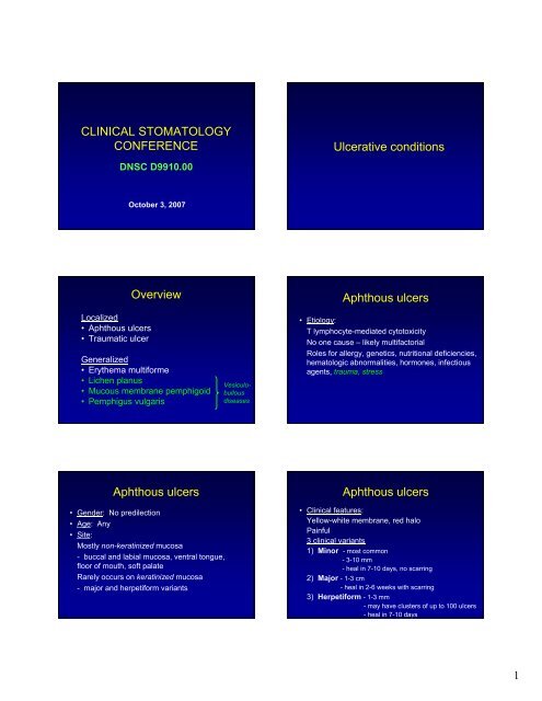
CLINICAL STOMATOLOGY CONFERENCE Ulcerative conditions ...
CLINICAL STOMATOLOGY CONFERENCE Ulcerative conditions ...
CLINICAL STOMATOLOGY CONFERENCE Ulcerative conditions ...

Create successful ePaper yourself
Turn your PDF publications into a flip-book with our unique Google optimized e-Paper software.
<strong>CLINICAL</strong> <strong>STOMATOLOGY</strong><br />
<strong>CONFERENCE</strong><br />
Localized<br />
• Aphthous ulcers<br />
• Traumatic ulcer<br />
DNSC D9910.00<br />
October 3, 2007<br />
Overview<br />
Generalized<br />
• Erythema multiforme<br />
• Lichen planus<br />
• Mucous membrane pemphigoid<br />
• Pemphigus vulgaris<br />
Aphthous ulcers<br />
• Gender: No predilection<br />
• Age: Any<br />
• Site:<br />
Mostly non-keratinized mucosa<br />
- buccal and labial mucosa, ventral tongue,<br />
floor of mouth, soft palate<br />
Rarely occurs on keratinized mucosa<br />
- major and herpetiform variants<br />
Vesiculobullous<br />
diseases<br />
<strong>Ulcerative</strong> <strong>conditions</strong><br />
Aphthous ulcers<br />
• Etiology:<br />
T lymphocyte-mediated cytotoxicity<br />
No one cause – likely multifactorial<br />
Roles for allergy, genetics, nutritional deficiencies,<br />
hematologic abnormalities, hormones, infectious<br />
agents, trauma, stress<br />
Aphthous ulcers<br />
• Clinical features:<br />
Yellow-white membrane, red halo<br />
Painful<br />
3 clinical variants<br />
1) Minor - most common<br />
-3-10 mm<br />
- heal in 7-10 days, no scarring<br />
2) Major - 1-3 cm<br />
- heal in 2-6 weeks with scarring<br />
3) Herpetiform -1-3 mm<br />
- may have clusters of up to 100 ulcers<br />
- heal in 7-10 days<br />
1
Aphthous ulcers<br />
• Association with systemic diseases:<br />
1) Behçet’s syndrome<br />
2) Inflammatory bowel disease<br />
- Crohn’s disease<br />
- ulcerative colitis<br />
3) Celiac disease<br />
4) Cyclic neutropenia<br />
5) Reiter’s syndrome<br />
6) Immunocompromised states<br />
- AIDS, HIV<br />
AU minor AU minor<br />
AU major<br />
Aphthous ulcers<br />
Herpetiform AU<br />
• Differential diagnosis:<br />
1) Recurrent herpetic infection, including herpes<br />
simplex virus (HSV), herpes zoster<br />
– HSV: on keratinized mucosa<br />
2) Other viral infections (e.g. enterovirus, etc.)<br />
3) Ulcers associated with neutropenia<br />
4) Traumatic ulcer<br />
2
Aphthous ulcers<br />
Recurrent<br />
intraoral herpes Herpes zoster<br />
Ulcer assoc. with neutropenia<br />
- Down’s syndrome, s/p heart<br />
transplant<br />
• Differential diagnosis (cont’d):<br />
If major aphthous ulcers, consider:<br />
1) Pemphigus vulgaris<br />
2) Mucous membrane pemphigoid<br />
3) Traumatic ulcer<br />
4) Squamous cell carcinoma<br />
Traumatic ulcer<br />
Pemphigus vulgaris<br />
3
Mucous membrane<br />
pemphigoid<br />
Topical steroids used in oral<br />
pathology<br />
1) Dexamethasone elixir, 0.5mg/5ml<br />
Disp: 8 oz<br />
Label: Swish and spit 1 tsp QID<br />
2) Fluocinonide (Lidex) gel, 0.05%<br />
Disp: 1 tube<br />
Label: Apply to affected area BID<br />
3) Qvar, 40mg<br />
Disp: 1 canister<br />
Label: 2 puffs QID<br />
Traumatic ulcer<br />
- anesthetic<br />
• Histology:<br />
- fibrinopurulent membrane<br />
- lymphocytes, histiocytes,<br />
neutrophils<br />
- epithelial spongiosis<br />
Aphthous ulcers<br />
• Treatment: Minor aphthae – topical steroids<br />
Major aphthae – systemic steroids<br />
Traumatic ulcerations<br />
• Etiology: Mechanical, thermal, electrical<br />
Some factitial in nature<br />
• Gender: No predilection<br />
• Age: Any age<br />
• Site: Tongue, lips, buccal mucosa<br />
• Clinical features:<br />
Erythema surrounding yellow-white membrane<br />
Older lesions – elevated/rolled, white borders<br />
4
Traumatic ulcerations<br />
Eosinophilic ulcerations (traumatic granuloma)<br />
• Unique variant of traumatic ulceration<br />
• Unique histology<br />
• Gender: Male predilection<br />
• Age: Any age<br />
• Site: Tongue<br />
• Clinical features:<br />
Ulceration with surrounding erythema<br />
Exuberant proliferation ~ pyogenic granuloma<br />
Can appear worrisome clinically for SCC<br />
Traumatic ulcerations<br />
• Differential diagnosis:<br />
Simple traumatic ulcers<br />
1) Aphthous ulcers/stomatitis<br />
2) Chemical injury<br />
3) Leukoplakia; erythroplakia<br />
4) Squamous cell carcinoma<br />
Before biopsy After biopsy<br />
Major aphthous ulcer<br />
5
Traumatic ulcerations<br />
• Treatment:<br />
1) Remove source of irritation<br />
2) If symptomatic:<br />
a) Topical corticosteroids<br />
Rx: Lidex gel, 0.05%<br />
Apply to affected area BID<br />
b) Topical analgesics<br />
Rx: Magic mouthwash or KBL<br />
Swish and spit PRN pain<br />
Chemical injury<br />
- aspirin burn<br />
Squamous cell<br />
carcinoma<br />
Traumatic ulceration<br />
• Histology:<br />
- fibrinopurulent membrane<br />
and neutrophils (=ulcer)<br />
- granulation tissue<br />
- epithelial hyperplasia +<br />
hyperkeratosis<br />
Eosinophilic ulcer:<br />
- deep inflammatory<br />
infiltrate; eosinophils and<br />
histiocytes<br />
Traumatic ulcerations<br />
• Treatment (cont’d):<br />
Squamous cell<br />
carcinoma<br />
3) If: - high-risk site (lat./ventral tongue, FOM)<br />
- patient with risk factors<br />
- no identifiable source of irritation<br />
- > 2 weeks in duration<br />
- not responding to tx…<br />
** BIOPSY to rule out malignancy **<br />
6
Erythema multiforme<br />
• Etiology: ? Hypersensitivity reaction<br />
May be induced by:<br />
1. Herpes simplex infection,<br />
2. Exposure to medications (esp. antibiotics, analgesics)<br />
3. Mycoplasma pneumoniae infection<br />
• Types:<br />
1) EM minor<br />
2) EM major (Stevens-Johnson syndrome)<br />
- drug exposure<br />
3) Toxic epidermal necrolysis<br />
- drug exposure<br />
Erythema multiforme<br />
• Clinical features (skin):<br />
Flat, round, dusky-red<br />
May become bullous<br />
May develop “target”/”bulls-eye” lesions<br />
Erythema multiforme<br />
• Gender: M>F<br />
• Age: Young adults (20-30 yo)<br />
• Site: Oral mucosa<br />
Skin<br />
If also ocular or genital SJ syndrome<br />
• Clinical course:<br />
Sudden-onset<br />
1) Fever, malaise, headache, sore throat<br />
2) Skin and/or oral lesions<br />
Self-limiting disease – resolves in 2-6 weeks<br />
Recurrences common<br />
Erythema multiforme<br />
• Clinical features (oral):<br />
Hemorrhagic crusting of lips<br />
Lips, buccal mucosa, tongue, FOM, palate<br />
Erythematous patches erosions, ulcerations<br />
Painful; difficult to examine<br />
7
Erythema multiforme<br />
• Differential diagnosis:<br />
1) Primary herpetic gingivostomatitis (primary<br />
herpes)<br />
2) Pemphigus vulgaris<br />
3) Mucous membrane pemphigoid<br />
4) Erosive lichen planus<br />
** Histology and direct immunofluorescence<br />
studies can help to rule out some of these entities ** Intraoral herpes<br />
8
Pemphigus vulgaris Erosive LP<br />
Erythema multiforme<br />
• Histology:<br />
-vesicles<br />
- epithelial necrosis<br />
- mixed inflammation,<br />
including eosinophils<br />
- perivascular inflammation<br />
• Treatment: Self-limiting; hydration<br />
Systemic steroids<br />
Topical steroids – EM minor<br />
Discontinue suspected drug<br />
If HSV-related, prophylactic Acyclovir<br />
Lichen planus<br />
• Clinical features (skin):<br />
Site: Flexor surfaces of extremities<br />
4 P’s<br />
Purple, pruritic, polygonal papules<br />
White striations<br />
Nails may also be affected<br />
Lichen planus<br />
• Etiology:<br />
Immunologically mediated disease<br />
? Role for stress, anxiety<br />
Association with diseases of altered immunity<br />
and hepatitis C<br />
• Gender: F>M (3:2)<br />
• Age: Middle-aged adults<br />
Can affect children<br />
9
Lichen planus<br />
• Clinical features (oral):<br />
Site: Posterior buccal mucosa, tongue, gingiva,<br />
vermillion of lip<br />
2 forms<br />
1) Reticular LP<br />
- more common form<br />
- usually asymptomatic<br />
- interlacing white striations<br />
- lesions wax and wane<br />
- dorsum of tongue: plaque-like<br />
Lichen planus<br />
- plaque-like, tongue<br />
Lichen planus<br />
Gingival LP, reticular<br />
• Clinical (oral) (cont’d):<br />
2) Erosive LP<br />
- less common than reticular form<br />
- usually symptomatic<br />
- atrophic, erythematous areas, + ulceration<br />
- white striations at periphery<br />
** - if limited to gingiva, may mimic – pemphigoid<br />
– pemphigus<br />
10
Lichen planus<br />
• Differential diagnosis:<br />
4) Oral lesions of lupus erythematosus<br />
- other skin, hematologic, laboratory abnormalities<br />
- oral lesions clinically and histologically ~ to LP<br />
5) Graft-versus-host disease<br />
- h/o transplant<br />
- oral lesions clinically and histologically ~ to LP<br />
6) Mucous membrane pemphigoid<br />
7) Pemphigus vulgaris<br />
** Histology and direct immunofluorescence<br />
studies can help to rule out some of these entities **<br />
• Differential diagnosis:<br />
Lichen planus<br />
Gingival LP, erosive<br />
1) Lichenoid drug reaction<br />
2) Contact reaction to amalgam, cinnamon<br />
3) Erythroplakia; speckled leukoplakia<br />
Contact reaction to amalgam<br />
11
Contact reaction to cinnamon<br />
Lichen planus<br />
• Treatment:<br />
Asymptomatic<br />
- usually reticular form<br />
- no treatment necessary<br />
Symptomatic<br />
- usually erosive form<br />
- topical steroids<br />
Periodic follow-up (6mos to 1 year)<br />
Erosive form – small risk malignant Δ<br />
Graft-versus-host disease<br />
• Histology:<br />
-hyperkeratosis<br />
- “saw-toothed” rete pegs<br />
- hydrophic degeneration of<br />
basal layer<br />
- band-like infiltrate of<br />
lymphocytes<br />
Lichen planus<br />
Systemic lupus<br />
erythematosus<br />
Mucous membrane pemphigoid<br />
• Etiology: Autoimmune<br />
Autoantibodies target component of<br />
basement membrane<br />
• Prevalence: 2x as common as pemphigus<br />
• Gender: F>M<br />
• Age: Older adults (50-60 yo)<br />
• Site:<br />
Oral mucosa – especially gingiva<br />
Conjunctiva, nasal, esophageal, laryngeal<br />
12
Mucous membrane pemphigoid<br />
• Clinical features (oral):<br />
Vesicles or bullae<br />
If not intact, erosions and ulcers<br />
Usually painful<br />
May persist for weeks to months<br />
May be limited to gingiva<br />
- “desquamative gingivitis”<br />
Blisters may be induced by lateral pressure<br />
- “+ Nikolsky sign”<br />
Mucous membrane pemphigoid<br />
• Clinical (ocular):<br />
~25% of patients<br />
Adhesions scarring blindness<br />
13
Pemphigus vulgaris<br />
“Plasma cell” gingivitis<br />
Mucous membrane pemphigoid<br />
• Differential diagnosis:<br />
1) Pemphigus vulgaris<br />
2) Lichen planus<br />
3) Plasma cell gingivitis – related to cinnamon<br />
4) Angina bullosa hemorrhagica – spontaneously<br />
healing, blood-filled blisters<br />
5) Medication-induced pemphigoid-like reaction<br />
** Histology and direct immunofluorescence<br />
studies can help to rule out some of these entities **<br />
Lichen planus<br />
Angina bullosa hemorrhagica<br />
14
Mucous membrane pemphigoid Mucous membrane pemphigoid<br />
• Histology:<br />
- subepithelial split<br />
- chronic inflammation<br />
* Biopsy perilesional tissue *<br />
Pemphigus vulgaris<br />
• Etiology: Autoimmune<br />
Autoantibodies to desmosome<br />
- structures that bind epithelial cells together<br />
• Genetics: HLA-DRw4 (Jewish population)<br />
• Gender: F>M<br />
• Age: Adults (>50 yo)<br />
• Site:<br />
Soft palate, labial mucosa, ventral tongue, gingiva<br />
Skin<br />
Rarely ocular<br />
• Treatment:<br />
* Refer to ophthalmologist *<br />
Topical steroids<br />
Periostat (doxycycline)<br />
Systemic steroids (if topical therapy ineffective)<br />
Maintain good oral hygiene<br />
Pemphigus vulgaris<br />
• Clinical features (oral):<br />
Oral lesions “first to show, last to go”<br />
Erythematous, denuded lesions<br />
Ragged borders<br />
Adjacent epithelium “piled up”<br />
Rarely intact vesicles<br />
Desquamative gingivitis<br />
+ Nikolsky sign<br />
15
Erythema multiforme<br />
Pemphigus vulgaris<br />
• Differential diagnosis:<br />
1) Mucous membrane pemphigoid<br />
2) Erosive lichen planus<br />
3) Erythema multiforme<br />
4) Chemical injury<br />
5) Paraneoplastic pemphigus – associated malignancy<br />
6) Medication-induced pemphigus-like reaction<br />
** Histology and direct immunofluorescence<br />
studies can help to rule out some of these entities **<br />
Erosive LP<br />
16
Pemphigus vulgaris<br />
• Histology:<br />
- acantholysis =<br />
intraepithelial separation<br />
- basal cells remain<br />
attached to basement<br />
membrane<br />
* Biopsy perilesional tissue *<br />
Pemphigus vulgaris<br />
• Treatment:<br />
Systemic steroids + “steroid-sparing” drugs<br />
Goal: as low steroid dose as necessary for<br />
disease control<br />
Topical steroids may be of benefit<br />
Mortality rate of 5-10%<br />
17