SBBD - SBES 2007 - Universidade Federal de Minas Gerais
SBBD - SBES 2007 - Universidade Federal de Minas Gerais
SBBD - SBES 2007 - Universidade Federal de Minas Gerais

Create successful ePaper yourself
Turn your PDF publications into a flip-book with our unique Google optimized e-Paper software.
WAAMD <strong>2007</strong><br />
III Workshop em Algoritmos e Aplicações <strong>de</strong> Mineração <strong>de</strong> Dados<br />
Mining structural signatures of proteins<br />
R.C. Melo 12 , J.S. Gomi<strong>de</strong> 1 , P.S.L. Dias 1 ,W. Meira Jr. 1 , M.M. Santoro 2<br />
1 Departamento <strong>de</strong> Ciência da Computação - <strong>Universida<strong>de</strong></strong> <strong>Fe<strong>de</strong>ral</strong> <strong>de</strong> <strong>Minas</strong> <strong>Gerais</strong><br />
2 Departamento <strong>de</strong> Bioquímica e Imunologia - <strong>Universida<strong>de</strong></strong> <strong>Fe<strong>de</strong>ral</strong> <strong>de</strong> <strong>Minas</strong> <strong>Gerais</strong><br />
{raquelcm,janaina,samer,meira}@dcc.ufmg.br, santoro@icb.ufmg.br<br />
Abstract. Proteins are the most important macromolecules in living systems. It<br />
is well known that their function is totally <strong>de</strong>pen<strong>de</strong>nt on their structure. However,<br />
i<strong>de</strong>ntical structures can be formed by very dissimilar amino acid sequences<br />
and little is known about how so different sequences fold into i<strong>de</strong>ntical structures<br />
and functions. In this work, we propose a clustering approach to obtain<br />
patterns of amino acid chemical interactions in protein families that compose a<br />
structural signature for each family.<br />
1. Introduction<br />
Bioinformatics is an emerging field un<strong>de</strong>rgoing rapid growth. This has been fueled<br />
by advances in DNA sequencing techniques. The Human Genome Project resulted in<br />
an exponentially growth in the database of genetic sequences and Structural Genomics<br />
Initiative is doing the same for Protein Data Bank (PDB) [Berman et al. 2000]. One of<br />
the most active research areas is protein topology analysis. Proteins are the most versatile<br />
macromolecules in living systems serving crucial functions in all biological processes,<br />
such as catalysts, transporters, and mechanical support. They are composed by a sequence<br />
of amino acids which is called primary structure. Different regions of the sequence form<br />
regular secondary structures such as α-helices or beta-sheets. The tertiary structure,<br />
which is the 3D structure of the protein, is formed by packing such structural elements into<br />
compact globular units called domains. The functional properties of proteins <strong>de</strong>pend upon<br />
their 3D structures that arises because a particular chain of amino acids folds to generate<br />
domains with specific 3D structures. It is known that the chain completely <strong>de</strong>termines<br />
the structure of a protein. However, there may be several proteins with the same structure<br />
(or family), and the same function, but with very different sequences and many variations<br />
in secondary structure sizes and positions. Hence, the study of protein topology is very<br />
important because the topology <strong>de</strong>termines protein function.<br />
PDB has on its archives approximately 45,000 proteins and this number has been<br />
increasing year after year. Even thought it is a huge data set, significant knowledge still<br />
needs to be extracted from it. As a mo<strong>de</strong>st step towards this ambitious long-term goal,<br />
in this work we use a data mining approach to analyze similarity of proteins and to extract<br />
conserved information on dissimilar sequences of proteins of the same family. These<br />
patterns are part of what we call structural signature of a protein family. A structural signature<br />
is a set of characteristics that can univocally i<strong>de</strong>ntify a family, and thus structure<br />
and function proteins. It could additionally give clues about the nature of interactions<br />
that a protein can establish with ligands and other proteins. In the present work, we<br />
use a database that contains information about chemical interactions within proteins, represented<br />
by contact maps, exploiting the spatial co-location of interactions as evi<strong>de</strong>nce<br />
towards <strong>de</strong>fining a protein signature.<br />
105
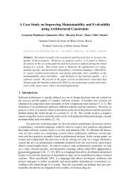
Contact maps enco<strong>de</strong> long-range interactions within proteins in a compact way<br />
and have been used in the literature as two-dimensional representations of proteins’ 3D<br />
topology. We present some examples of contact maps in Figure 1. In fact, a chain folds<br />
into a 3D structure because of chemical interactions between its amino acid residues.<br />
These interactions are indispensable for the action of proteins, being of interest to study<br />
the similarity of proteins based on their chemical interactions patterns. The 3 most important<br />
kinds of interactions are hydrophobic, hydrogen bonds and electrostatic. The<br />
hydrophobic contacts arise because of the molecules water aversion. This effect makes<br />
apolar parts of chain closer in the 3D structure. The hydrogen bonds are short-distance<br />
interactions between residues that share a common hydrogen atom. They are essential in<br />
the stabilization of secondary strutures. The electrostatic interactions are the attraction or<br />
repulsion of charged amino acids, which occur far apart in structure. They are not being<br />
consi<strong>de</strong>red in this work because the occurence of charge clusters are very rare in proteins.<br />
In [Agarwal et al. <strong>2007</strong>], the authors prove that contact map overlap problem is NP-Hard<br />
and <strong>de</strong>scribe an algorithm which solves the problem in polynomial time for some particular<br />
cases. The advantage of our proposal is that we do not overlap contact maps but<br />
signatures which are much more concise representations of them.<br />
140<br />
120<br />
100<br />
80<br />
60<br />
40<br />
20<br />
WAAMD <strong>2007</strong><br />
III Workshop em Algoritmos e Aplicações <strong>de</strong> Mineração <strong>de</strong> Dados<br />
Hydrophobic contacts<br />
Hydrogen bonds<br />
20 40 60 80 100 120 140<br />
160<br />
140<br />
120<br />
100<br />
80<br />
60<br />
40<br />
20<br />
Hydrophobic contacts<br />
Hydrogen bonds<br />
20 40 60 80 100 120 140 160<br />
160<br />
140<br />
120<br />
100<br />
80<br />
60<br />
40<br />
20<br />
Hydrophobic contacts<br />
Hydrogen bonds<br />
20 40 60 80 100 120 140 160<br />
100<br />
80<br />
60<br />
40<br />
20<br />
Hydrophobic contacts<br />
Hydrogen bonds<br />
(a) (b) (c) (d)<br />
20 40 60 80 100<br />
Figure 1. Protein families and their contact maps. In (a), human being Myoglobin,<br />
an oxigen carrier in the muscles; in (b) an Apolipoprotein from human being, a<br />
lipid transporter; in (c) a Retinol Binding Protein (R.B.P.) from pig plasma; in (d)<br />
a human being Thioredoxin, an electron transporter.<br />
2. Mining structural signatures<br />
In this section we present both our mining strategy and its application to samples<br />
of Myoglobins, Apolipoproteins (helical proteins similar to Myoglobins), Plastocyanins,<br />
Retinol Binding Proteins (R.B.P.s) and Thioredoxins from varied animal species selected<br />
from PDB according to SCOP manually curated classification [Murzin et al. 1995]. The<br />
initial step was to compute contacts given a set of atomic coordinates from a PDB file<br />
which is partially based on [Sobolev et al. 1999] and [Mancini et al. 2004]. We analyze<br />
each atom of the 20 most commonly found residues as in [Sobolev et al. 1999] and attribute<br />
for each one a class: acceptor and donnor (for hydrogen bond <strong>de</strong>termination)<br />
and hydrophobic (for hydrophobic interactions) and use the same distance thresholds of<br />
[Mancini et al. 2004].<br />
106
140<br />
120<br />
100<br />
80<br />
60<br />
40<br />
20<br />
WAAMD <strong>2007</strong><br />
III Workshop em Algoritmos e Aplicações <strong>de</strong> Mineração <strong>de</strong> Dados<br />
20 40 60 80 100 120 140<br />
140<br />
120<br />
100<br />
80<br />
60<br />
40<br />
20<br />
20 40 60 80 100 120 140<br />
140<br />
120<br />
100<br />
80<br />
60<br />
40<br />
20<br />
20 40 60 80 100 120 140<br />
1<br />
0.8<br />
0.6<br />
0.4<br />
0.2<br />
0<br />
Hydrophobic contacts 95.74%<br />
Hydrogen bonds 96.04%<br />
0 0.2 0.4 0.6 0.8 1<br />
(a) (b) (c) (d)<br />
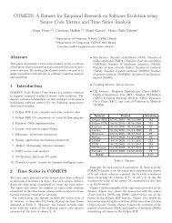
Figure 2. We present the clusters <strong>de</strong>fined by DBSCAN of hydrophobic interactions<br />
and and hydrogen bonds for the Myoglobin of Figure 1 in (a). In (b), we<br />
have the the vectors that represent these clusters, i.e., the structural signature<br />
of the protein. In (c), we have Myoglobin family structural signature and in (d),<br />
precision of the classification of Myoglobins according to protein structural signature.<br />
Contact maps of protein families present conserved clusters, which we <strong>de</strong>tect using<br />
a <strong>de</strong>nsity-based clustering algorithm, DBSCAN [Ester et al. 1996]. This algorithm<br />
is able to handle an important characteristic of the clusters of contacts: they present a<br />
linear shape and are always parallel or anti-parallel to the map diagonal. The parallel<br />
clusters indicate that, numbering the sequence from the beginning to the end, we have<br />
two increasing parts of the chain close w.r.t. the structure establishing chemical interactions,<br />
while the anti-parallel means the reverse. The parameters of the algorithm were<br />
<strong>de</strong>termined usign the heuristic suggested by the authors. In Figure 2.(a), we can see some<br />
examples of clusters i<strong>de</strong>ntified for the Myoglobin presented in Figure 1. Notice that some<br />
of the clusters are not linear, but are <strong>de</strong>tected by DBSCAN. As an example, we can see<br />
a ”L”-shape hydropobic cluster in Figure 2.(a). Such clusters represent that a secondary<br />
structure is in contact with two or more secondary structures.<br />
The next step of our strategy is to <strong>de</strong>termine lines that characterize the clusters. We<br />
use the Hought Transform [Illingworth and Kittler 1988] to <strong>de</strong>tect the single or multiple<br />
lines that characterize a cluster. In Figure 2.(b) we can see the representative lines for<br />
the clusters of Figure 2.(a). The representation of cluster using lines makes easier to<br />
recognize relevant patterns and to <strong>de</strong>tect the lines (and thus clusters) that are conserved<br />
among several proteins. In Figure 2.(c), we plot the representative lines for the clusters<br />
of 9 Myoglobin samples. They are from human being, whale, horse, pig, elephant, turtle,<br />
tuna, seal, and mollusc. The diagonal clusters (hydrogen bond clusters) mark the helix<br />
occurence and are very well conserved. Proteins of Myoglobin family are composed<br />
by, generally, 6 to 8 helices. They are named from A to H. Helices C and D are very<br />
small and in many samples of family they do not occur. There are 5 hydrophobic clusters<br />
conserved in all Myoglobin samples. They are probably an important component of the<br />
structural signature we are looking for. They represent contacts between parts of the<br />
chain in helices AB with E, AB with G, AB with H, G with H and F with H. According<br />
to [Hughson et al. 1997], the Myoglobin folding intermediate contains the A, G, and H,<br />
i.e., these helices are the first to fold and their contacts are important to the stabiization of<br />
the molecule. Particularly, the A and G helices interact to stabilize each other, as shown<br />
by the effect of mutations in the G helix on the unfolding of the A helix. Notice that<br />
helices A, G and H are present in all the conserved hydrophobic clusters of the proposed<br />
107
structural signature.<br />
WAAMD <strong>2007</strong><br />
III Workshop em Algoritmos e Aplicações <strong>de</strong> Mineração <strong>de</strong> Dados<br />
After <strong>de</strong>termining the structural signatures for the various families of proteins,<br />
we used the signature vectors to classify proteins, i.e., <strong>de</strong>termine their families. As we<br />
wanted to <strong>de</strong>termine the matches of vectors which minimize the cost of mutate the map of<br />
a protein into the map of another, we mo<strong>de</strong>led the problem of comparing two sets of vectors<br />
in a 2D space (each set is a protein structure signature) as a Transportation Problem.<br />
The dissimilarity of the maps is measured by the minimum cost of moving all the origin<br />
and <strong>de</strong>stination points of the vectors from a signature to the vectors from another signature.<br />
Comparing the same 9 Myoglobins of different animal species against 62 proteins of<br />
different classes (Apolipoproteins, Plastocyanins, R.B.P.s and Thioredoxins), i.e., classifying<br />
a protein as Myoglobin or non-Myoglobin, we achieved a 95% precision (measured<br />
as the area un<strong>de</strong>r the ROC curves) using hydrophobic contacts and hydrogen bonds, as we<br />
can see in Figure 2.(d). The classification methodology consist of comparing all against<br />
all proteins, ranking them according to the dissimilarity in<strong>de</strong>x given by the cost of transporting<br />
vectors of one protein to the other and selecting the k most similar as Myoglobin<br />
and the others as non-Myoglobin. By cutting these ranks in different ks and computeing<br />
the false positive rate and true positive rate, we obtain the curves of Figure 2.(d).<br />
3. Conclusions<br />
In this paper, we presented a novel methodology for un<strong>de</strong>rstanding contact maps<br />
and through data mining tecnhiques extract patterns that we call protein structural signatures.<br />
This is a new concept in molecular biology and can be helpful in the solution of the<br />
wi<strong>de</strong>ly studied and still open Protein Folding Problem. We also show that the structural<br />
signatures can be used to classify proteins according to their structure and function.<br />
References<br />
Agarwal, P. et al. (<strong>2007</strong>). Fast molecular shape matching using contact maps. J. Comput.<br />
Biol., 14(2):131–143.<br />
Bourne, P. et al. (2000). The protein data bank. Nucleic Acid Research, 28:235–242.<br />
Xu, X. et al. (1996). A <strong>de</strong>nsity-based algorithm for discovering clusters in large spatial<br />
databases with noise. In Proceedings of the Second International Conference on<br />
Knowledge Discovery and Data Mining, pages 226–231.<br />
Baldwin, R. et al. (1997). Cooperativity of folding of the apomyoglobin ph 4 intermediate<br />
studied by glycine and proline mutations. Nature Structural Biology, 4:925–930.<br />
Kittler, J. et al. (1988). A survey of the hough transform. Computer Vision, Graphics,<br />
and Image Processing, pages 87–116.<br />
Neshich, G. et al. (2004). Sting contacts: a web-based application for i<strong>de</strong>ntification and<br />
analysis of amino acid contacts within protein structure and across protein interfaces.<br />
Bioinformatics, 20(13):2145–2147.<br />
Chothia, C. et al. (1995). Scop: a structural classification of proteins database for the<br />
investigation of sequences and structures. Journal of Molecular Biology, 247:536–<br />
540.<br />
Sobolev, V. et al. (1999). Automated analysis of interatomic contacts in proteins. Bioinformatics,<br />
15:327–332.<br />
108