Maxillary Molar Intrusion with Fixed Appliances and Mini-implant ...
Maxillary Molar Intrusion with Fixed Appliances and Mini-implant ...
Maxillary Molar Intrusion with Fixed Appliances and Mini-implant ...

You also want an ePaper? Increase the reach of your titles
YUMPU automatically turns print PDFs into web optimized ePapers that Google loves.
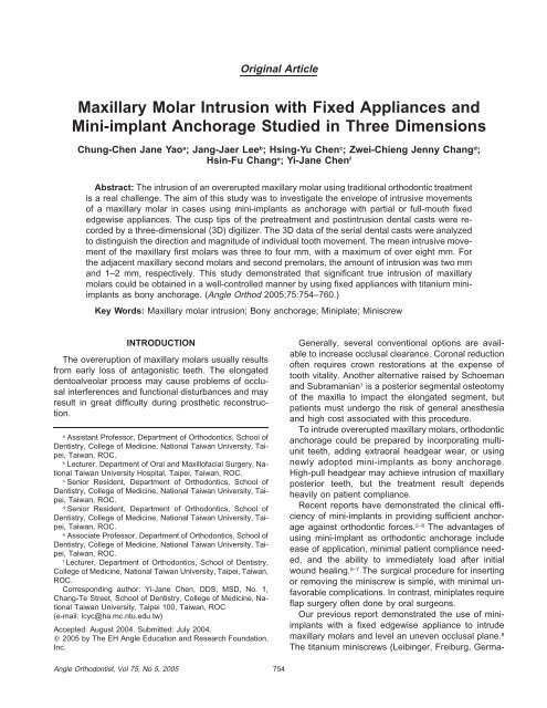
Angle Orthodontist, Vol 75, No 5, 2005<br />
Original Article<br />
<strong>Maxillary</strong> <strong>Molar</strong> <strong>Intrusion</strong> <strong>with</strong> <strong>Fixed</strong> <strong>Appliances</strong> <strong>and</strong><br />
<strong>Mini</strong>-<strong>implant</strong> Anchorage Studied in Three Dimensions<br />
Chung-Chen Jane Yao a ; Jang-Jaer Lee b ; Hsing-Yu Chen c ; Zwei-Chieng Jenny Chang d ;<br />
Hsin-Fu Chang e ; Yi-Jane Chen f<br />
Abstract: The intrusion of an overerupted maxillary molar using traditional orthodontic treatment<br />
is a real challenge. The aim of this study was to investigate the envelope of intrusive movements<br />
of a maxillary molar in cases using mini-<strong>implant</strong>s as anchorage <strong>with</strong> partial or full-mouth fixed<br />
edgewise appliances. The cusp tips of the pretreatment <strong>and</strong> postintrusion dental casts were recorded<br />
by a three-dimensional (3D) digitizer. The 3D data of the serial dental casts were analyzed<br />
to distinguish the direction <strong>and</strong> magnitude of individual tooth movement. The mean intrusive movement<br />
of the maxillary first molars was three to four mm, <strong>with</strong> a maximum of over eight mm. For<br />
the adjacent maxillary second molars <strong>and</strong> second premolars, the amount of intrusion was two mm<br />
<strong>and</strong> 1–2 mm, respectively. This study demonstrated that significant true intrusion of maxillary<br />
molars could be obtained in a well-controlled manner by using fixed appliances <strong>with</strong> titanium mini<strong>implant</strong>s<br />
as bony anchorage. (Angle Orthod 2005;75:754–760.)<br />
Key Words: <strong>Maxillary</strong> molar intrusion; Bony anchorage; <strong>Mini</strong>plate; <strong>Mini</strong>screw<br />
INTRODUCTION<br />
The overeruption of maxillary molars usually results<br />
from early loss of antagonistic teeth. The elongated<br />
dentoalveolar process may cause problems of occlusal<br />
interferences <strong>and</strong> functional disturbances <strong>and</strong> may<br />
result in great difficulty during prosthetic reconstruction.<br />
a Assistant Professor, Department of Orthodontics, School of<br />
Dentistry, College of Medicine, National Taiwan University, Taipei,<br />
Taiwan, ROC.<br />
b Lecturer, Department of Oral <strong>and</strong> Maxillofacial Surgery, National<br />
Taiwan University Hospital, Taipei, Taiwan, ROC.<br />
c Senior Resident, Department of Orthodontics, School of<br />
Dentistry, College of Medicine, National Taiwan University, Taipei,<br />
Taiwan, ROC.<br />
d Senior Resident, Department of Orthodontics, School of<br />
Dentistry, College of Medicine, National Taiwan University, Taipei,<br />
Taiwan, ROC.<br />
e Associate Professor, Department of Orthodontics, School of<br />
Dentistry, College of Medicine, National Taiwan University, Taipei,<br />
Taiwan, ROC.<br />
f Lecturer, Department of Orthodontics, School of Dentistry,<br />
College of Medicine, National Taiwan University, Taipei, Taiwan,<br />
ROC.<br />
Corresponding author: Yi-Jane Chen, DDS, MSD, No. 1,<br />
Chang-Te Street, School of Dentistry, College of Medicine, National<br />
Taiwan University, Taipei 100, Taiwan, ROC<br />
(e-mail: lcyc@ha.mc.ntu.edu.tw)<br />
Accepted: August 2004. Submitted: July 2004.<br />
2005 by The EH Angle Education <strong>and</strong> Research Foundation,<br />
Inc.<br />
754<br />
Generally, several conventional options are available<br />
to increase occlusal clearance. Coronal reduction<br />
often requires crown restorations at the expense of<br />
tooth vitality. Another alternative raised by Schoeman<br />
<strong>and</strong> Subramanian 1 is a posterior segmental osteotomy<br />
of the maxilla to impact the elongated segment, but<br />
patients must undergo the risk of general anesthesia<br />
<strong>and</strong> high cost associated <strong>with</strong> this procedure.<br />
To intrude overerupted maxillary molars, orthodontic<br />
anchorage could be prepared by incorporating multiunit<br />
teeth, adding extraoral headgear wear, or using<br />
newly adopted mini-<strong>implant</strong>s as bony anchorage.<br />
High-pull headgear may achieve intrusion of maxillary<br />
posterior teeth, but the treatment result depends<br />
heavily on patient compliance.<br />
Recent reports have demonstrated the clinical efficiency<br />
of mini-<strong>implant</strong>s in providing sufficient anchorage<br />
against orthodontic forces. 2–8 The advantages of<br />
using mini-<strong>implant</strong> as orthodontic anchorage include<br />
ease of application, minimal patient compliance needed,<br />
<strong>and</strong> the ability to immediately load after initial<br />
wound healing. 5–7 The surgical procedure for inserting<br />
or removing the miniscrew is simple, <strong>with</strong> minimal unfavorable<br />
complications. In contrast, miniplates require<br />
flap surgery often done by oral surgeons.<br />
Our previous report demonstrated the use of mini<strong>implant</strong>s<br />
<strong>with</strong> a fixed edgewise appliance to intrude<br />
maxillary molars <strong>and</strong> level an uneven occlusal plane. 8<br />
The titanium miniscrews (Leibinger, Freiburg, Germa-
MOLAR INTRUSION WITH BONY ANCHORAGE<br />
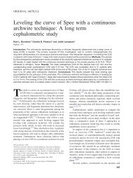
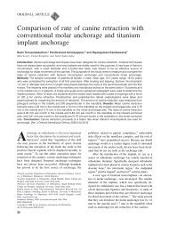
FIGURE 1. <strong>Mini</strong>-<strong>implant</strong> anchorage preparation. (a) Left maxillary<br />
first molar elongation due to loss of antagonist. (b) <strong>Mini</strong>plate was<br />
<strong>implant</strong>ed by a flap surgery. (c) Palatal miniscrew. (d) Buccal miniplate.<br />
ny), two mm in diameter <strong>and</strong> 15 mm long, <strong>and</strong> L-shape<br />
miniplates (Modeal, Tuttlingen, Germany) were adopted<br />
by our department to prepare bony anchorage in<br />
various adult cases, especially when molar intrusion is<br />
required (Figure 1).<br />
The precise quantitative assessment of the envelope<br />
of motion associated <strong>with</strong> molar intrusion <strong>with</strong><br />
mini-<strong>implant</strong>s has not been explored. The aim of this<br />
study was to investigate the envelope of intrusive<br />
movement of maxillary molars in cases receiving mini<strong>implant</strong>s<br />
as bony anchorage during orthodontic treatment<br />
<strong>with</strong> partial or full-mouth fixed edgewise appliances.<br />
MATERIALS AND METHODS<br />
Patient characteristics<br />
Data were obtained from the records of 22 patients<br />
in our department, who had undergone orthodontic<br />
treatment to intrude overerupted maxillary molars (Table<br />
1). Their ages ranged from 15 to 42 years, <strong>with</strong> a<br />
mean of 27.6 years. There were 12 Class I <strong>and</strong> 10<br />
Class II molar relationships, <strong>and</strong> nine cases were hyperdivergent<br />
(SN-MP 37), <strong>with</strong> only two cases being<br />
hypodivergent (SN-MP 28). Six of the 22 patients<br />
had local treatment <strong>with</strong> partial fixed orthodontic<br />
appliances. The other 16 patients initially had partial<br />
appliances to intrude the overerupted teeth, followed<br />
by full-mouth fixed appliances to correct the malocclusion.<br />
Eighteen of the 22 patients received <strong>implant</strong>ation<br />
of both a buccal miniplate <strong>and</strong> palatal miniscrew. The<br />
other four patients had buccal <strong>and</strong> palatal miniscrews<br />
inserted. The average treatment duration of active intrusion<br />
was 7.6 months <strong>with</strong> a range of five to 12<br />
months. A pretreatment <strong>and</strong> postintrusion maxillary<br />
dental cast was collected for each patient.<br />
755<br />
TABLE 1. Demographic Information of 22 Subjects in This Study<br />
Gender (Male/female) 20/2<br />
Age (y) (Mean SD) 27.6 8.11<br />
(Range) 15–42<br />
Malocclusion (Class I/II/III)<br />
Facial divergency<br />
12/10/0<br />
Hyperdivergent (SN-MP 37) 9<br />
Hypodivergent (SN-MP 28) 2<br />
Normal divergent 11<br />
Combination of mini-<strong>implant</strong> for anchorage preparation<br />
Buccal miniplates <strong>and</strong> palatal miniscrews<br />
Bilateral/unilateral<br />
Buccal miniscrews <strong>and</strong> palatal miniscrews<br />
8/10<br />
Bilateral/unilateral 1/3<br />
To prepare bony anchorage for maxillary molar intrusion,<br />
a titanium L-shaped miniplate <strong>and</strong> a miniscrew<br />
were <strong>implant</strong>ed onto the buccal <strong>and</strong> palatal sides of<br />
the overerupted molars. <strong>Mini</strong>screws were <strong>implant</strong>ed<br />
<strong>with</strong>out flap elevation or <strong>with</strong> just a stab incision. To<br />
<strong>implant</strong> a miniplate, a mucoperiosteal flap was elevated<br />
under local anesthesia. The miniplate was adjusted<br />
to fit the contour of the cortical bony surface <strong>and</strong> was<br />
fixed by bone screws <strong>with</strong> the intention to expose the<br />
free fixation hole to the oral cavity from the incised<br />
wound, which was located in the zone of attached gingiva.<br />
Sufficient distance was left between root apex<br />
<strong>and</strong> mini-<strong>implant</strong> to avoid interference <strong>with</strong> the intended<br />
intrusive tooth movement. After initial healing of the<br />
soft tissue around the mini-<strong>implant</strong>s, a medium intrusive<br />
force (150–200 g) was applied <strong>with</strong> the elastic<br />
chains between the buccal miniplate <strong>and</strong> the attachment<br />
on the first molar b<strong>and</strong>, <strong>and</strong> also between the<br />
palatal miniscrew <strong>and</strong> the cleat of the molar attachment<br />
(Figure 1c,d).<br />
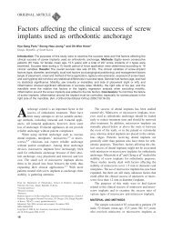
When adjacent teeth need to be intruded, those<br />
teeth were bonded <strong>and</strong> a sectional archwire was inserted<br />
<strong>with</strong> the intrusive force primarily applied only on<br />
the maxillary first molar. After sufficient intrusion was<br />
attained, their vertical position was maintained by ligating<br />
the molars to the miniscrew <strong>and</strong> the miniplate<br />
(Figure 2). In those cases where posterior occlusion<br />
could be restored immediately after the overerupted<br />
tooth was leveled, no retainer was required (Figure<br />
2d). Otherwise, full-coverage retention was used if no<br />
subsequent full-mouth comprehensive therapy was<br />
performed.<br />
Model analysis<br />
The bases of the before- <strong>and</strong> after-treatment dental<br />
casts for each of the 22 patients were trimmed, <strong>with</strong><br />
the occlusal plane <strong>and</strong> the horizontal plane parallel.<br />
The tip of the mesiobuccal cusp, mesiopalatal cusp,<br />
distobuccal cusp, <strong>and</strong> distopalatal cusp of the molars<br />
Angle Orthodontist, Vol 75, No 5, 2005
756 YAO, LEE, CHEN, CHANG, CHANG, CHEN<br />
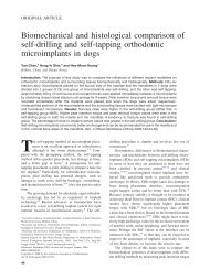
FIGURE 2. <strong>Intrusion</strong> of left maxillary first molar. (a) Elastic chain<br />
between miniscrew <strong>and</strong> palatal cleat of first molar. (b) Elastic chain<br />
between miniplate <strong>and</strong> buccal attachment. (c) Initial radiographic image.<br />
(d) Postintrusion radiographic image.<br />
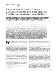
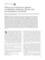
FIGURE 3. Desktop mechanical 3D digitizer.<br />
as well as the buccal <strong>and</strong> palatal cusps of the premolars<br />
were marked <strong>with</strong> a sharp pencil. To compare<br />
the movement of cusp tips on the serial models, a minimum<br />
of four palatal rugae points were registered as<br />
the reference l<strong>and</strong>marks.<br />
The spatial data on the pretreatment <strong>and</strong> postintrusion<br />
maxillary dental casts were recorded by a mechanical<br />
desktop three-dimensional (3D) digitizer (Mi-<br />
Angle Orthodontist, Vol 75, No 5, 2005<br />
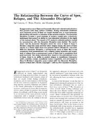
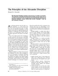
FIGURE 4. Rhinoceros program to analyze 3D point data.<br />
croscribe G2, Immersion Corporation, San Jose, Calif)<br />
(Figure 3) through a stylus tip connected to a mechanical<br />
arm that allowed a full range of movements.<br />
The 3D coordinate data of unique points on the two<br />
casts were analyzed by a Rhinoceros software program<br />
(Robert McNeel & Associates, Seattle, Wash)<br />
(Figure 4). With this information, it was possible to calculate<br />
the positional change of the overerupted maxillary<br />
teeth by superimposing two sets of coordinate<br />
data to assess the relocation of specific cusp tips. The<br />
difference in the z-coordinates of the cusp tips on the<br />
serial dental casts reflected the amount <strong>and</strong> direction<br />
of the movement of the analyzed tooth. The ‘‘positive’’<br />
sign indicated intrusion <strong>and</strong> ‘‘negative’’ sign extrusion.<br />
To examine the reliability of measurements by the<br />
3D digitizer, 20 maxillary dental models were chosen<br />
r<strong>and</strong>omly <strong>and</strong> digitized a second time after a two-week<br />
interval. The l<strong>and</strong>marks digitized included eight points<br />
on the cusp tips of the maxillary teeth <strong>and</strong> another<br />
eight identifiable l<strong>and</strong>marks on the palatal rugae.<br />
The reliability of measurement <strong>with</strong> the 3D digitizer<br />
was 0.41 mm for the cusp tip <strong>and</strong> 0.45 mm for l<strong>and</strong>mark<br />
on the palatal rugae. Their means <strong>and</strong> deviations<br />
on x-, y-, <strong>and</strong> z-coordinates, both individually <strong>and</strong> collectively,<br />
are shown in Table 2. The reliability of linear<br />
measurement established by digitizing the comparable<br />
l<strong>and</strong>marks on serial dental models to assess the magnitude<br />
<strong>and</strong> direction of tooth movement was considered<br />
clinically acceptable.<br />
TABLE 2. Reliability of Measurements (mm): Individual <strong>and</strong> Collective Means <strong>and</strong> St<strong>and</strong>ard Deviations of x, y, <strong>and</strong> z Coordinates<br />
x Coordinate y Coordinate z Coordinate<br />
Point<br />
Measurement<br />
Point on cusp tip 0.15 0.38 0.11 0.49 0.23 0.24 0.41 0.54<br />
Point on palatal rugae 0.16 0.32 0.10 0.29 0.05 0.21 0.45 0.23
MOLAR INTRUSION WITH BONY ANCHORAGE<br />
TABLE 3. Amount of <strong>Intrusion</strong> (mm) at Various Cusp Tips of <strong>Maxillary</strong> Teeth a<br />
Tooth Cusp Mean SD Maximum <strong>Mini</strong>mum<br />
First molar (n 26) Mesiobuccal 3.17 1.70 8.67 0.42<br />
Mesiopalatal 3.25 1.74 8.34 0.64<br />
Distobuccal 3.42 1.94 8.35 0.34<br />
Distopalatal 3.39 1.90 8.12 0.55<br />
Second molar (n 17) Mesiobuccal 1.86 2.73 7.27 3.68<br />
Mesiopalatal 2.09 2.06 6.56 1.39<br />
Distobuccal 2.18 2.26 6.64 1.38<br />
Distopalatal 2.23 2.32 8.34 1.25<br />
First premolar (n 11) Buccal 1.44 2.12 5.86 1.06<br />
Palatal 1.08 1.58 3.98 0.84<br />
Second premolar (n 21) Buccal 1.93 2.03 8.04 1.50<br />
Palatal 1.86 1.69 5.86 0.83<br />
a Positive value: intrusion; negative value: extrusion.<br />
Statistical methods<br />
The SPSS statistical package program for Windows<br />
was used for the evaluation of measurements. The<br />
amount of maxillary molar intrusion was represented<br />
by the changes in the z-coordinate at the mesiobuccal<br />
cusp, distobuccal cusp, mesiopalatal cusp, <strong>and</strong> distopalatal<br />
cusp tips. All the data were expressed as<br />
means <strong>with</strong> st<strong>and</strong>ard deviations <strong>and</strong> the range for<br />
maximal <strong>and</strong> minimal values. The statistical significance<br />
of crown tipping, both in the buccal-palatal direction<br />
<strong>and</strong> the mesial-distal direction, was assessed<br />
by student t-test. P .05 was accepted as significant.<br />
RESULTS<br />
The maxillary molars were successfully intruded<br />
<strong>with</strong> the mini-<strong>implant</strong> system in all the patients. The<br />
intrusion was also noted at the maxillary teeth adjacent<br />
to the first molar, ie, at the second molar <strong>and</strong> at the<br />
first <strong>and</strong> the second premolars. Table 3 shows the<br />
quantitative assessment for intrusion of 26 maxillary<br />
first molars, 17 maxillary second molars, 11 maxillary<br />
first premolars, <strong>and</strong> 21 maxillary second premolars.<br />
The amount of intrusion was expressed as the difference<br />
of the z-coordinates between the pretreatment<br />
<strong>and</strong> postintrusion dental casts, which was equivalent<br />
to the positional change of each cusp tip. The positive<br />
value denoted intrusion of the cusp tip.<br />
The variation of the maxillary molar intrusion was<br />
considerable, ranging from 3.68 to 8.67 mm. The<br />
amount of intrusion at the maxillary second molar was<br />
generally less than that of the first molar. The mean<br />
amount of intrusion at the maxillary first molar was 3<br />
to 4 mm <strong>and</strong> 2 to 2.5 mm at the maxillary second<br />
molar. The larger variation in the second molar intrusion<br />
resulted from extrusion of the buccal cusps usually<br />
noted in cases <strong>with</strong> buccal tipping of second molars<br />
before treatment. The amount of maxillary premolar<br />
intrusion ranged from 1.50 to 8.04 mm <strong>and</strong><br />
757<br />
FIGURE 5. <strong>Intrusion</strong> of maxillary posterior teeth <strong>with</strong> the mini-<strong>implant</strong><br />
anchorage. (a) Significant elongation of maxillary teeth. (b) <strong>Maxillary</strong><br />
posterior teeth were intruded. (c) Initial radiographic image. (d) Postintrusion<br />
radiographic image.<br />
was generally less than that of the maxillary molars.<br />
The mean intrusion of the maxillary premolar was<br />
around 2 mm at the second premolar <strong>and</strong> 1 mm at the<br />
first premolar.<br />
The maximum amount of intrusion (8.67 mm at mesiobuccal<br />
cusp of right maxillary first molar <strong>and</strong> 8.04<br />
mm at second premolar) was noted in a case of a 23year-old<br />
female patient <strong>with</strong> all her right m<strong>and</strong>ibular molars<br />
missing (Figure 5a) <strong>and</strong> the teeth of the opposing<br />
arch (15 to 17) overerupted. The gingival line of the<br />
right maxillary posterior teeth was obviously below that<br />
of the anterior teeth. <strong>Intrusion</strong> was started <strong>with</strong> a sectional-arch<br />
fixed appliance <strong>with</strong> mini-<strong>implant</strong> anchorage<br />
<strong>and</strong> completed in seven months. The intruded molars<br />
were ligated to the miniplate for retention <strong>and</strong> full-mouth<br />
orthodontic treatment followed (Figure 5b–d).<br />
The buccal-lingual tipping of the intruded maxillary<br />
Angle Orthodontist, Vol 75, No 5, 2005
758 YAO, LEE, CHEN, CHANG, CHANG, CHEN<br />
TABLE 4. Assessment of Crown Tipping (mm) of <strong>Maxillary</strong> Posterior Teeth a<br />
Angle Orthodontist, Vol 75, No 5, 2005<br />
Difference<br />
in <strong>Intrusion</strong> b Mean SD Maximum <strong>Mini</strong>mum<br />
First molar (n 26) MB-MP 0.08 0.92 1.40 1.61<br />
DB-DP 0.03 1.05 2.68 1.39<br />
Second molar (n 17) MB-MP 0.22 2.02 1.78 7.65<br />
DB-DP 0.05 0.83 1.39 1.71<br />
First premolar (n 11) B-P 0.36 0.69 1.88 0.41<br />
Second premolar (n 21) B-P 0.07 1.14 2.18 2.51<br />
a Positive value: buccal tipping; negative value: palatal tipping.<br />
b MB indicates mesiobuccal cusp; MP, mesiopalatal cusp; DB, distobuccal cusp; DP, distopalatal cusp; B, buccal cusp; <strong>and</strong> P, palatal cusp.<br />
posterior tooth was quantitatively assessed by subtracting<br />
the difference between the intrusion of the<br />
buccal <strong>and</strong> palatal cusps (Table 4). A positive value<br />
denoted buccal tipping of maxillary posterior teeth.<br />
The difference between the buccal <strong>and</strong> palatal cusps<br />
<strong>and</strong> either the mesial or distal position of the crown<br />
was generally small compared <strong>with</strong> the total intrusion<br />
achieved. Similar to the negligible tipping of the maxillary<br />
molar, the difference between the buccal <strong>and</strong> palatal<br />
cusps of the maxillary premolar was generally little.<br />
None of the differences in intrusion between the<br />
buccal <strong>and</strong> palatal cusps reached the level of significance<br />
(t-test <strong>with</strong> expected value 0, P .05). This<br />
indicated that the intrusion was achieved <strong>with</strong>out significant<br />
buccal-palatal tipping.<br />
The wide range of values resulted from the initial<br />
malposition of some teeth, especially buccoversion of<br />
the maxillary second molars. Correction of a buccally<br />
tipped second molar requires changing the tooth’s axis<br />
by intruding palatal cusps <strong>and</strong> possibly extrusion of<br />
buccal cusps. In one case, the mesiobuccal cusp of<br />
the left maxillary second molar was extruded 3.68 mm.<br />
The initial position of this second molar was in a severe<br />
buccal version, <strong>with</strong> buccal cusps out of occlusal<br />
contact. Indeed in this type of case, the leveling <strong>and</strong><br />
alignment of these teeth is achieved by palatal tipping<br />
<strong>with</strong> buccal cusp extrusion <strong>and</strong> palatal cusp intrusion.<br />
DISCUSSION<br />
Anchorage control plays an important role in orthodontic<br />
mechanics. During conventional orthodontic<br />
treatment for intruding overerupted molars, it is difficult<br />
to avoid the side effect of extrusion of the anchorage<br />
teeth. Some appliances such as high-pull headgears<br />
could be used for molar intrusion, but the patient’s<br />
compliance is essential.<br />
In contrast to traditional orthodontics, the molar intrusion<br />
facilitated <strong>with</strong> the mini-<strong>implant</strong>s causes minimum<br />
extrusion of the adjacent teeth. Incorporation of<br />
mini-<strong>implant</strong>s can achieve a significant amount of<br />
maxillary molar intrusion <strong>and</strong> is an excellent alternative<br />
to traditional method. 3,9–12 The results of this study<br />
demonstrate that the intrusive movement of maxillary<br />
premolars could also be anticipated even when the<br />
bony anchorage was set originally for the maxillary first<br />
molar only. This enables the orthodontist to intrude<br />
multiple teeth at the same time <strong>with</strong> a rigid anchorage.<br />
To quantify the amount of intrusion, we adopted a<br />
3D digitizer to measure the movement of cusp tips of<br />
specific teeth relative to the stable structures on the<br />
dental casts. The superimposition of lateral cephalometric<br />
radiographs may provide us information about<br />
molar intrusion, but the image of posterior teeth is often<br />
blurred <strong>and</strong> buccal-palatal crown tipping could not<br />
be assessed. Panoramic radiographs offer a better alternative<br />
because no image overlapping of contralateral<br />
posterior teeth occurs. Recently, panoramic radiographs<br />
were used to identify pure molar intrusion <strong>with</strong><br />
miniplate anchorage. 3 Contrary to analysis using 2D<br />
images, the 3D approach could quantify the tooth<br />
movement by analyzing spatial data from the serial<br />
models. 13<br />
In this study, palatal rugae were used as references<br />
to evaluate the positional change of maxillary teeth on<br />
serial dental casts. Almedia et al 14 reported that the<br />
medial rugae point is more stable than the lateral point<br />
for longitudinal cast analysis. Bailey et al 15 suggested<br />
that the medial <strong>and</strong> lateral points of the third rugae<br />
could be used as the reference points in longitudinal<br />
cast analysis. In our study, to track the tooth movement<br />
by superimposing two sets of dental casts, a<br />
minimum of four identical <strong>and</strong> reliable reference points<br />
were selected from the medial <strong>and</strong> lateral rugae<br />
points. Despite some controversy regarding the longterm<br />
stability of rugae during growth, the stability of<br />
rugae points was not an issue because the subjects<br />
in our study were mostly adult <strong>and</strong> the observation<br />
period was <strong>with</strong>in a few months.<br />
Various <strong>implant</strong> systems have been used for orthodontic<br />
intrusion. Southard et al 10 reported that molar<br />
intrusion is possible using dental <strong>implant</strong>s. Sherwood<br />
et al 3 reported four cases <strong>with</strong> miniplate anchorage to<br />
close skeletal open bite. They reported that superimposition<br />
of panoramic tracings showed that a mean
MOLAR INTRUSION WITH BONY ANCHORAGE<br />
molar intrusion of 1.99 mm. Kanomi 16 reported an adult<br />
patient <strong>with</strong> a deep bite, which was corrected <strong>with</strong> six<br />
mm of lower incisor intrusion by an intrusive force from<br />
a mini-<strong>implant</strong>. Umemori et al 2 presented a skeletal<br />
anchorage system to correct an anterior open bite.<br />
They <strong>implant</strong>ed the titanium miniplates at buccal aspects<br />
of the m<strong>and</strong>ibular molars <strong>and</strong> intruded the molars<br />
about three to five mm. Daimaruya et al 17 intruded<br />
the m<strong>and</strong>ibular molars 3.4 mm by the intrusive force<br />
from buccal miniplate <strong>and</strong> lingual bone screw in dogs.<br />
Erverdi et al 11 reported that the zygomatic area was a<br />
useful anchorage site for maxillary molar intrusion. A<br />
cephalometric study demonstrated the effectiveness of<br />
skeletal anchorage for intrusion of maxillary posterior<br />
teeth to correct anterior open-bite malocclusion. 12 Our<br />
experience substantiates that successful intrusion of<br />
molars can be consistently achieved <strong>with</strong> mini-<strong>implant</strong>s<br />
as anchorage.<br />
In this study, the severity of the overeruption of individual<br />
maxillary teeth varied among the 22 cases.<br />
The intrusion mechanics were terminated when the<br />
overerupted tooth was leveled. Therefore, the large individual<br />
variation of our data was reasonable because<br />
the treatment goal for each case was different. In addition<br />
to intruding the overerupted maxillary posterior<br />
teeth, we also used the mini-<strong>implant</strong> anchorage system<br />
to correct the Class II malocclusion <strong>with</strong> excessive<br />
dentoalveolar height <strong>and</strong> anterior open bite. The intrusion<br />
of maxillary molars was followed by counterclockwise<br />
rotation of the m<strong>and</strong>ible <strong>with</strong> a decrease in the<br />
sagittal jaw discrepancy. Our clinical observation indicated<br />
successful orthodontic management of these<br />
Class II hyperdivergent patients <strong>with</strong>out orthognathic<br />
surgeries. However, the stability of changing vertical<br />
dimension in these patients requires long-term followup.<br />
<strong>Intrusion</strong> of molars by only applying an apically directed<br />
force to the buccal tooth attachment will tip molars<br />
to the buccal. There are several ways to counteract<br />
the buccal tipping moment produced by the intrusive<br />
force applied from buccal aspect.<br />
Most commonly a transpalatal arch is used to help<br />
minimize the tendency of buccal tipping. This appliance<br />
should be kept well away from the palate to avoid<br />
soft tissue impingement. A second option for minimizing<br />
buccal tipping is the use of a constricted overlay<br />
archwire. Sherwood et al 3 used an overlay round archwire<br />
to provide a counterbalancing moment <strong>and</strong><br />
control the buccal crown tipping. However, the most<br />
efficient control may result from simultaneous application<br />
of intrusive force from buccal <strong>and</strong> palatal aspects.<br />
With the miniplate <strong>and</strong> miniscrew anchorage<br />
system, the intrusive force can be applied by elastic<br />
chains between the buccal miniplate <strong>and</strong> attachment<br />
on first molar b<strong>and</strong>, <strong>and</strong> between the palatal miniscrew<br />
759<br />
<strong>and</strong> the cleat of the molar attachment. The levels of<br />
buccal <strong>and</strong> palatal force need to be closely monitored<br />
to minimize the tendency for crown tipping, which is<br />
usually not desirable. In this study, the palatal cusp<br />
responded faster <strong>and</strong> the intrusive movement occurred<br />
earlier than that of buccal cusp. The interradicular septum<br />
between the two buccal roots may partly explain<br />
the different rate of intrusion in the buccal <strong>and</strong> palatal<br />
cusps.<br />
This retrospective study has certain limitations. First,<br />
its retrospective nature does not allow a detailed analysis<br />
of the sequence of events such as a temporary<br />
halt for better torque control. Second, tracking the<br />
movement of the cusp tip of the posterior tooth simply<br />
indicates the direction of movement but not the type<br />
of movement (tip or torque). 13 Third, we observed noticeable<br />
clinical crown shortening of the intruded teeth<br />
but little was known regarding changes of the surrounding<br />
periodontal tissues. Indeed, deepening of<br />
gingival sulcus <strong>and</strong> bleeding on probing were frequently<br />
noted. The deepened sulcus may prevent proper<br />
cleansing <strong>and</strong> predispose the gingiva to inflammation.<br />
We recommend an intense oral hygiene regimen for<br />
these intruded teeth to resolve the redundant soft tissue<br />
spontaneously. Further study is needed to investigate<br />
the changes of periodontal tissue around the intruded<br />
teeth.<br />
CONCLUSIONS<br />
This study revealed that the average intrusion of<br />
maxillary molars was more than three to four mm. A<br />
combination of mini-<strong>implant</strong>s <strong>and</strong> fixed appliances is a<br />
predictable <strong>and</strong> effective procedure to achieve maxillary<br />
molar intrusion.<br />
ACKNOWLEDGMENT<br />
This work was supported by a research grant from the National<br />
Science Council of Taiwan (NSC-92-2212-E-002-061).<br />
REFERENCES<br />
1. Schoeman R, Subramanian L. The use of orthognathic surgery<br />
to facilitate <strong>implant</strong> placement: a case report. Int J Oral<br />
Maxillofac Implants. 1996;11:682–684.<br />
2. Umemori M, Sugawara J, Mitani H, Nagasaka H, Kawamura<br />
H. Skeletal anchorage system for open-bite correction. Am<br />
J Orthod Dentofacial Orthop. 1999;115:166–174.<br />
3. Sherwood KH, Nurchg JG, Thompson WJ. Closing anterior<br />
open bites by intruding molars <strong>with</strong> titanium miniplate anchorage.<br />
Am J Orthod Dentofacial Orthop. 2002;122:593–<br />
600.<br />
4. Miyawaki S, Koyama I, Inoue M, Mishima K, Sugahara T,<br />
Takano-Yamamoto T. Factors associated <strong>with</strong> the stability<br />
of titanium screws placed in the posterior region for orthodontic<br />
anchorage. Am J Orthod Dentofacial Orthop. 2003;<br />
124:373–378.<br />
5. Park HS, Bae SM, Kyung HM, Sung JH. Micro-<strong>implant</strong> an-<br />
Angle Orthodontist, Vol 75, No 5, 2005
760 YAO, LEE, CHEN, CHANG, CHANG, CHEN<br />
chorage for treatment of skeletal Class I bialveolar protrusion.<br />
J Clin Orthod. 2001;35:417–422.<br />
6. Bae SM, Park HS, Kyung HM, Kwon OW, Sung JH. Clinical<br />
application of micro-<strong>implant</strong> anchorage. J Clin Orthod. 2002;<br />
36:298–302.<br />
7. Costa A, Raffini M, Melsen B. <strong>Mini</strong>screws as orthodontic<br />
anchorage: a preliminary report. Int J Adult Orthod Orthog<br />
Surg. 1998;13:201–209.<br />
8. Yao CC, Wu CB, Wu HY, Kok SH, Chang HF, Chen YJ.<br />
<strong>Intrusion</strong> of the overerupted upper left first <strong>and</strong> second molars<br />
by mini-<strong>implant</strong>s <strong>with</strong> partial-fixed orthodontic appliances:<br />
a case report. Angle Orthod. 2004;74:501–507.<br />
9. Ohmae M, Saito S, Morohashi T, et al. A clinical <strong>and</strong> histological<br />
evaluation of titanium mini-<strong>implant</strong>s as anchors for<br />
orthodontic intrusion in the beagle dog. Am J Orthod Dentofacial<br />
Orthop. 2001;119:489–497.<br />
10. Southard TE, Buckley MJ, Spivey JD, Krizan KE, Casko JS.<br />
<strong>Intrusion</strong> anchorage potential of teeth versus rigid endosseous<br />
<strong>implant</strong>s: a clinical <strong>and</strong> radiographic evaluation. Am<br />
J Orthod Dentofacial Orthop. 1995;107:115–120.<br />
11. Erverdi N, Tosun T, Keles A. A new anchorage site for the<br />
Angle Orthodontist, Vol 75, No 5, 2005<br />
treatment of anterior openbite: zygomatic anchorage case<br />
report. World J Orthod. 2002;3:147–153.<br />
12. Erverdi N, Keles A, N<strong>and</strong>a R. The use of skeletal anchorage<br />
in openbite treatment: a cephalometric evaluation. Angle<br />
Orthod. 2004;74:381–390.<br />
13. Jennifer LA. A 3-dimensional analysis of molar movement<br />
during headgear treatment. Am J Orthod Dentofacial Orthop.<br />
2002;21:18–30.<br />
14. Almedia MA, Philips C, Kula K, Tulloch C. Stability of the<br />
palatal rugae as l<strong>and</strong>marks for analysis of dental casts. Angle<br />
Orthod. 1995;65:43–48.<br />
15. Bailey LT, Esmailnejad A, Almeida MA. Stability of the palatal<br />
rugae as l<strong>and</strong>marks for analysis of dental casts in extraction<br />
<strong>and</strong> nonextraction cases. Angle Orthod. 1996;66:<br />
73–78.<br />
16. Kanomi R. <strong>Mini</strong>-<strong>implant</strong> for orthodontic anchorage. J Clin<br />
Orthod. 1997;31:763–767.<br />
17. Daimaruya T, Nagasaka H, Sugawara J, Mitani H. The influences<br />
of molar intrusion on the inferior alveolar neurovascular<br />
bundle <strong>and</strong> root using the skeletal anchorage system<br />
in dog. Angle Orthod. 2001;71:60–70.