Catalyst - Marine Biological Laboratory
Catalyst - Marine Biological Laboratory
Catalyst - Marine Biological Laboratory

You also want an ePaper? Increase the reach of your titles
YUMPU automatically turns print PDFs into web optimized ePapers that Google loves.
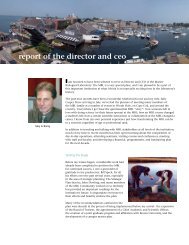
Founded in 1888 as the<br />
<strong>Marine</strong> <strong>Biological</strong> <strong>Laboratory</strong><br />
fall 2012<br />
Volume 7, Number 2<br />
i n t h i S i S S U e<br />
4<br />
From Molecules<br />
to Mental Health<br />
8<br />
Opening<br />
the Senses<br />
10<br />
A Revolution<br />
Born in Nature<br />
<strong>Catalyst</strong><br />
Illuminating<br />
the<br />
Brain<br />
Discovery in Neuroscience<br />
at the MBL<br />
Page 2
<strong>Catalyst</strong><br />
FAll 2012 Volume 7, Number 2<br />
MBL<br />
MBL <strong>Catalyst</strong> is published twice yearly by the Office of<br />
Communications at the <strong>Marine</strong> <strong>Biological</strong> <strong>Laboratory</strong><br />
(MBL) in Woods Hole, Massachusetts. The MBL is<br />
dedicated to scientific discovery and improving the<br />
human condition through research and education<br />
in biology, biomedicine, and environmental science.<br />
Founded in 1888, the MBL is an independent, nonprofit<br />
corporation.<br />
Senior advisors<br />
President and Director: Gary Borisy<br />
Chief Academic and<br />
Scientific Officer: Joshua Hamilton<br />
Director of External Relations: Pamela Clapp Hinkle<br />
Director of Communications: Andrea Early<br />
MBl <strong>Catalyst</strong> Staff<br />
Editor-in-Chief: Diana Kenney<br />
Guest Science Editor: Paul De Weer<br />
Designer: Beth Ready Liles<br />
Contributors: Dorothy Berthel, Paul De Weer, David Gallagher,<br />
Gina Hebert, Lizzie Kripke, Pamela Wilmot<br />
images: Inside cover: Nerve fibers in a human brain revealed using<br />
diffusion tensor imaging (DTI) (Allen Institute for Brain Science); Gary<br />
Borisy (E. Armstrong). P. 1, Nerve fiber tracks in the human brain based on<br />
DTI (Allen Institute for Brain Science); Axon fibers connecting the primary<br />
motor cortex to part of the thalamus in a mouse brain (Allen Institute for<br />
Brain Science); Methods in Computational Neuroscience course co-directors<br />
Adrienne Fairhall and Michael Berry (T. Kleindinst); Chlamydomonas<br />
reinhardtii (L. Hilton, courtesy of L. Quarmby). Pp. 2-3, clockwise from<br />
top: Human neuronal cells (green) and glial cells (red), with the nuclei of<br />
the cells in blue, derived from iPS cells (R. Karmacharya, S.L. Schreiber, and<br />
S.J. Haggarty); Grass Fellow Raquel Vasconcelos (A.R. Martinez); Gross<br />
anatomy of the major outer brain structures: frontal lobe (yellow), temporal<br />
lobe (peach/pink), parietal lobe (red), cerebellum (blue)(Allen Institute for<br />
Brain Science); Leech motor neuron (T. Balmer); neuron illustration by Lizzie<br />
Kripke. Pp. 4-5, Golgi stained pyramidal cell (B. Jacobs); Olivia Mullins<br />
of U of VA in NS&B (D. Kenney). Pp. 6-7 left to right, Choanoflagellates<br />
(Codosiga sp., two conjoined cysts) (D. Stoupin); Trachemys (US Fish &<br />
Wildlife); Bacterial species found in the human gut in a mouse model (Y.<br />
Hasegawa); Squid giant axon (J. Ortega). Pp. 8-9 clockwise from left, Squid<br />
fin skin with nerves in red, chromatophores in pink, iridophores in gray<br />
(T. Wardill et al., Proc. Royal Soc. B, 2012); Roger Hanlon (courtesy of R.<br />
Hanlon); Stephen Highstein (courtesy of S. Highstein); Hair cell (J. Meyers).<br />
Pp. 10-11, C. reinhardtii (L. Hilton, courtesy of L. Quarmby); George<br />
Augustine (T. Kleindinst). Pp. 12-13, Christof Koch (Allen Institute for Brain<br />
Science); Coronal section of an entire mouse brain (Allen Institute for Brain<br />
Science). Pp. 14-15 left to right, deep ocean vents (NOAA and NSF); Swope<br />
Center (MBL); Neuron expressing the ArcLight Q239 probe (L. Jin et al.,<br />
Neuron, 2012). Pp. 16-17, Gerald Fischbach (Simons Foundation); Plate 2<br />
from The Anatomy of the Common Squid, Loligo pealii, Lesueur, by<br />
L.W. Williams.<br />
Back cover: MBL courses through the years (MBL).<br />
About the cover: Nerve fibers in a human brain revealed using diffusion<br />
tensor imaging (DTI). The fiber pathways are derived from the characteristics<br />
of diffusion of water in brain tissue, revealing connections between brain<br />
regions. There is an average of 7,000 connections per neuron in the adult<br />
human brain (Allen Institute for Brain Science).<br />
Online extras: For full image descriptions, supplemental materials,<br />
and other information related to this issue, visit:<br />
www.mbl.edu/catalyst<br />
Send correspondence to:<br />
MBL Communications Office<br />
7 MBL Street, Woods Hole, MA 02543<br />
508-289-7423, comm@mbl.edu<br />
Dear Friends,<br />
Last September, I was thrilled to attend the ceremony at which my colleagues<br />
Ron Vale, Mike Sheetz, and Jim Spudich received the 2012 Lasker Award for Basic<br />
Medical Research (see p. 7). As was noted several times at the ceremony, this was<br />
truly an honor not only for these distinguished biologists, but for the MBL.<br />
When Vale and Sheetz arrived at the MBL in 1983, they were in pursuit of the<br />
squid’s giant axon, the best-studied neuron on the planet. But they were also<br />
drawn by astonishing movies Vale had seen by MBL investigators Bob Allen,<br />
Scott Brady, and Ray Lasek. These movies were among the first to use video<br />
enhancement of microscopic images, a technology independently invented by Bob<br />
and Nina Allen and Shinya Inoué. Video-enhanced microscopy revealed sights that<br />
had never been seen before, such as minuscule organelles zipping along the squid<br />
giant axon. What, they all wondered, was causing this movement? What followed<br />
was an intense effort to answer this question, culminating with Vale and Sheetz’s<br />
middle-of-the-night discovery of a new motor protein, kinesin. As the Lasker<br />
Award recognized, kinesin and other motor proteins are now known to be essential<br />
for critical processes such as muscle contraction, intracellular movement, and cell<br />
locomotion. And the fundamental discoveries by Vale, Sheetz, and Spudich have<br />
spurred research on new treatments for cardiac problems, neurological disorders,<br />
and cancer.<br />
Video-enhanced microscopy, which soon progressed to digital imaging,<br />
represented a sea change not only in the tools cell biologists had, but in the<br />
kinds of questions they could ask and ultimately answer. Today, neuroscience is<br />
invigorated by a new technology, optogenetics, that is having a similar impact<br />
(see p. 10). Neuroscientists can now activate or silence subsets of neurons using<br />
pulses of light, providing a precise tool for testing brain circuitry that hundreds<br />
of laboratories have adopted since 2005. Equally important are optical sensors<br />
that are fast and sensitive enough to report the millisecond-scale ranges of action<br />
potentials in individual neurons, a field pioneered by MBL investigator Larry<br />
Cohen in the 1970s that has matured to where its use in real-time studies of<br />
mammalian brain circuitry can be envisaged (see p. 15). It’s early yet to tell what<br />
biological discoveries will come from these new technologies. But the MBL, with<br />
its strong history of optical imaging in the neurosciences, will be in the middle of<br />
the action.<br />
A great debt is owed to Paul De Weer of University of Pennsylvania Medical<br />
School, a longtime MBL investigator and Neurobiology course faculty member,<br />
who served as guest science editor for this issue of MBL <strong>Catalyst</strong>. With abundant<br />
patience, good humor, and a meticulous editorial eye, Paul guided the <strong>Catalyst</strong><br />
staff through the “impenetrable jungles where many investigators have lost<br />
themselves,” as Ramón Y Cajal once described the brain. I am also pleased to<br />
announce that Diana Kenney, managing editor and senior writer of MBL <strong>Catalyst</strong><br />
for the past five years, has been named editor-in-chief. Diana welcomes ideas for<br />
and contributions to the magazine; please contact her at dkenney@mbl.edu.<br />
Gary Borisy<br />
President and Director<br />
F r o m the Director
i N this issue<br />
D e pA r t m e N t s<br />
6 News & Notes<br />
The latest findings from our laboratories<br />
and field sites.<br />
12 mbl momeNt<br />
<strong>Catalyst</strong><br />
Cracking the Brain’s Code<br />
Christof Koch reflects on co-founding<br />
the Methods in Computational<br />
Neuroscience course in 1988 and the<br />
state of the field today.<br />
14 GiFts & GrANts<br />
14 A c c o l A D e s<br />
15 c o o l tool<br />
F e At u r e s<br />
2<br />
4<br />
8<br />
10<br />
illuminating the Brain<br />
A network of neuroscientists at the MBL is<br />
tackling some of the most profound questions in<br />
biomedical research today.<br />
from Molecules to Mental health<br />
When will a basic understanding of the brain<br />
begin to translate to cures for its disorders? MBL<br />
course faculty members are optimistic that the<br />
time is near.<br />
Opening the Senses<br />
How do we see, hear, taste, smell, and feel, and<br />
how do our senses direct our behavior? Scientists<br />
in the MBL’s new Program in Sensory Physiology<br />
and Behavior are finding out.<br />
a revolution Born in nature<br />
Optogenetics, a powerful technology for<br />
illuminating the functional anatomy of the brain,<br />
has its origins in the microbial world.<br />
the Ultimate Optical Voltage Sensor<br />
Can brain activity be recorded<br />
without using an electrode? Larry<br />
Cohen, a.k.a. “Mr. Light,” was the<br />
first to say, “yes.”<br />
16 s c i e N t i s t’s eye View<br />
What We need to learn<br />
Gerald D. Fischbach lays out<br />
a roadmap toward the goal of<br />
treatments for neuropsychiatric<br />
disorders.<br />
17 m e m o r A b i l i A<br />
Squid Central<br />
Over a century of neuroscientific<br />
discovery at the MBL began with<br />
colored drawings of the squid.<br />
MBL <strong>Catalyst</strong> FALL 2012<br />
1
Cell body<br />
MBL<br />
Target cell<br />
Synapse<br />
the NeuroN<br />
neuron: from the Greek<br />
“bowstring, sinew.” Basic unit<br />
of the nervous system, consisting<br />
of a cell body with two<br />
kinds of specialized extensions:<br />
one axon and (usually many)<br />
dendrites.<br />
Axon<br />
Direction<br />
of action<br />
potential<br />
Ion channels<br />
Dendrites<br />
AnAtomy of CommuniCAtion<br />
2 MBL <strong>Catalyst</strong> FALL 2012<br />
illuminating<br />
Axon<br />
terminals<br />
axon or nerve fiber:<br />
tube-like extension of a neuron.<br />
Axons can be very long<br />
(e.g. from spinal cord to toe).<br />
Dendrite: from the Greek<br />
“tree.” Branched extension<br />
from a neuron body. Its membrane<br />
contains receptors that<br />
receive information from other<br />
neurons via synapses.<br />
How the brain works is one of<br />
the great mysteries of our time.<br />
Over the past century and in part at the<br />
MBL, scientists successfully detailed how<br />
neurons conduct electrical signals and<br />
“communicate.” Through behavioral<br />
studies combined with brain imaging,<br />
they also discovered that each thought,<br />
perception, or feeling coincides with the<br />
frantic firing of neurons in a particular<br />
circuit or brain region. One set of neurons<br />
fires, and you taste an apple; another set<br />
fires and you feel joy, think of your child,<br />
or see the sky.<br />
But despite heroic efforts, tracing the finescale,<br />
functional anatomy of the brain<br />
remains an enormous challenge, due to<br />
the sheer number (100 billion) of neurons<br />
it contains and the even more staggering<br />
number of interconnections. Discerning<br />
which neurons are electrically active, and<br />
in what sequence, is even more difficult,<br />
since they fire at millisecond-range speeds.<br />
Undeterred, neuroscientists are tackling<br />
action potential (or<br />
spike): propagation of electrical<br />
activity along the membrane<br />
of a cell. In most neurons<br />
this involves sudden influx of<br />
sodium ions followed by efflux<br />
of potassium ions, through ion<br />
channels in their membrane.<br />
Some cells fire 100 or more<br />
spikes per second.<br />
ion channel: protein embedded<br />
in the cell membrane<br />
that allows certain ions to cross<br />
the membrane. (Ions are atoms<br />
that have lost or gained one or<br />
more electrons.) The movement<br />
of (mainly) sodium, potassium,<br />
and calcium ions underlies<br />
electrical signaling in animal<br />
biology.
the Brain<br />
Discovery in neuroscience at the MBl<br />
these major unknowns head<br />
on. The secrets of the brain,<br />
they believe, will be revealed<br />
through its connections.<br />
In the MBL’s neuroscience<br />
community, as well,<br />
connections are all important.<br />
Hundreds of the field’s top<br />
research faculty and students<br />
converge at the MBL every<br />
year, creating a “meeting of<br />
minds” that is unmatched,<br />
anywhere. They were joined<br />
in 2012 by more than 60<br />
Whitman Investigators and<br />
Grass Fellows in neuroscience,<br />
along with a growing cadre of<br />
resident researchers in the field.<br />
Jennifer Morgan, who studies<br />
neural regeneration in the sea<br />
lamprey, joined the MBL’s Bell<br />
Center for Regenerative Biology<br />
and Tissue Engineering in the<br />
spring. And with the launching<br />
Synapse: from the Greek<br />
“to join.” Locus where an axon<br />
terminal communicates with<br />
its target cell. If the target is<br />
another nerve, the synapse<br />
can be at the target’s dendrite,<br />
body, or axon. Although<br />
purely electrical synapses exist,<br />
most are chemical. Action<br />
potentials reaching the axon<br />
Why did I choose<br />
to study this organ<br />
that is so awesome in<br />
its complexity,<br />
that it might well<br />
be infinite?<br />
— Sebastian Seung of MIT;<br />
faculty, MBL Methods in<br />
Computational Neuroscience<br />
terminal stimulate the release<br />
of a chemical neurotransmitter<br />
which diffuses across the synaptic<br />
cleft, binds to postsynaptic<br />
receptors, and thereby opens<br />
(or sometimes closes) certain<br />
ion channels. Under appropriate<br />
conditions this succeeds in<br />
initiating an action potential in<br />
the target cell.<br />
neural circuit: ensemble of<br />
interconnected neurons carrying<br />
signals between parts of<br />
the brain, e.g. from retina to<br />
visual cortex; also, arrangement<br />
of neurons underlying a specific<br />
function, e.g. recognizing<br />
a straight line or the timing of<br />
sound arrival in the left vs.<br />
right ear.<br />
of the Program in Sensory<br />
Physiology and Behavior in July,<br />
the MBL established a resident<br />
program in neurobiology for the<br />
first time since the 1970s.<br />
Between all these MBL<br />
courses and labs, the doors<br />
are always open. New ideas<br />
are swapped, equipment<br />
is borrowed, experiments<br />
are hatched, discoveries are<br />
made. MBL students find<br />
mentors, employers, lifelong<br />
collaborators, and even spouses<br />
in Woods Hole. They return as<br />
Grass Fellows, postdocs, and<br />
later as principal investigators<br />
or course directors. The MBL<br />
regularly teems with some of the<br />
most eminent neuroscientists<br />
in the world. It is through their<br />
interconnections that the power<br />
to illuminate the brain resides.<br />
• — DK<br />
Connectome: fanciful<br />
term (pun on “genome”) for<br />
the ensemble of neuronal<br />
connections in a given tissue<br />
or organ (brain; retina).<br />
• — PDW<br />
MBL <strong>Catalyst</strong> FALL 2012<br />
3
Karl Deisseroth opened his MBL neuroimaging<br />
seminar last summer with<br />
an anecdote about one of his patients,<br />
a man with autism spectrum disorder<br />
(ASD). Deisseroth had asked the man<br />
why he always looks down when conversing.<br />
Is it frightening to make eye<br />
contact? No, the patient responded.<br />
Overwhelming? “Yes. Your face changes<br />
when you talk, and I have to think<br />
about what that means. There is too<br />
much information.”<br />
“Here is a patient with autism-spectrum<br />
disease who can communicate just<br />
enough to tell us what is going on in his<br />
brain,” Deisseroth said. “Even the most<br />
eloquent among us may have trouble<br />
articulating such things. This story highlights<br />
how informative, though sometimes<br />
frustrating, psychiatry can be.”<br />
Insight gleaned from his patients informs<br />
Deisseroth’s research at Stanford<br />
University, as he develops a very promising<br />
technology for illuminating brain<br />
activity called optogenetics (see p. 10).<br />
And yet, Deisseroth’s frustration is familiar<br />
to psychiatrists, neuroscientists,<br />
patients and their families everywhere.<br />
Mental disorders are common—1 in every<br />
88 American children is diagnosed<br />
with ASD, for example—and serious<br />
mental illnesses cost more than $300<br />
billion in 2002 (in disability benefits, loss<br />
of earnings, and health care expenses).<br />
Yet cures for those who suffer remain<br />
elusive.<br />
4 MBL <strong>Catalyst</strong> FALL 2012<br />
from Molecules to<br />
In the MBL’s nine<br />
neuroscience courses, the search<br />
for cures looms large<br />
“For many diseases, such as cancer and<br />
heart disease, the scientific enterprise<br />
has really made an impact in extending<br />
life and even finding cures. But in<br />
mental health, we have barely made a<br />
dent,” says Tim Ryan of Cornell Weill<br />
Medical College, current co-director of<br />
the MBL Neurobiology course.<br />
The difficulty lies in the staggering complexity<br />
of the human brain. Over 100<br />
billion neurons—each one communicating<br />
with thousands more—send<br />
and receive precisely timed, electrical<br />
and chemical signals that underlie<br />
all perception, thought, emotions, and<br />
behavior. What’s more, the brain is dynamic—it<br />
is continually reshaped and<br />
rewired by experience. Unlike the welldefined<br />
mechanisms underlying cancer<br />
(uncontrolled growth of abnormal cells)<br />
and cardiovascular disease (narrowed or<br />
blocked arteries), with mental disorders<br />
“we are only beginning to define the<br />
problems,” says Graeme Davis of University<br />
of California, San Francisco, who<br />
also co-directs the Neurobiology course.<br />
The MBL’s advanced neurosciences<br />
courses, with their rigorous orientation<br />
toward discovery at the cutting edge of<br />
research, are widely considered essential<br />
to the quest for a fundamental understanding<br />
of the brain that will eventually<br />
bring treatments and cures.<br />
NeuroscieNce u<br />
Graduate and post-graduate<br />
level MBL courses include:<br />
• Neurobiology<br />
• Neural Systems & Behavior<br />
• Biology of the Inner Ear<br />
• Fundamental Issues in Vision<br />
Research<br />
• Methods in Computational<br />
Neuroscience<br />
• Neuroinformatics<br />
• NeuroStereology<br />
• Summer Program in Neuroscience,<br />
Ethics, and Survival<br />
• Teaching about Neurobiology of<br />
Brain Dysfunction
Mental Health<br />
“The MBL courses are spectacular.<br />
They have top-notch faculty doing topnotch<br />
research. Whenever something<br />
new comes along in neuroscience, the<br />
courses pick up on it super fast,” says<br />
former Neurobiology faculty member<br />
Richard Kramer of University of California,<br />
Berkeley, now an MBL Whitman<br />
investigator.<br />
Case in point: Last summer, Neurobiology<br />
students tried out a new, fast, genetically<br />
engineered sensor of neural activity<br />
from Adam Cohen’s lab at Harvard<br />
(see p. 15). “Those students were about<br />
the fourth and fifth people in the world<br />
to use this technology,” says Ryan, who<br />
had brought the sensor with him from<br />
Cornell. As it turned out, several MBL<br />
scientists became keenly interested in<br />
the sensor. Across campus, and across<br />
the world, the Neurobiology course is<br />
“at the center of the neuroscience community,”<br />
says faculty member Ryohei<br />
Yasuda, scientific director of the Max<br />
Planck Florida Institute.<br />
Every five years, fresh co-directors<br />
take up the reins in each MBL summer<br />
course, updating its content to push the<br />
limits of knowledge and innovation.<br />
Add gifted research scientists serving as<br />
faculty; a select, interdisciplinary group<br />
of very sharp students; and $26 million<br />
in high-end research equipment loaned<br />
to the MBL by 140 vendors, and you get<br />
a transformative experience of scientific<br />
discovery, for students and faculty alike.<br />
The Neurobiology course studies the<br />
brain at the level of individual neurons<br />
and synapses. Both Ryan and Davis believe<br />
the field is at a crucial juncture,<br />
where fundamental understanding will<br />
soon pay off with therapeutic insights.<br />
“Neurosocience is, right now, where<br />
cancer biology was 30 years ago, when<br />
scientists were just beginning to understand<br />
the cell cycle [which controls cell<br />
division]. The next two decades in neuroscience<br />
should yield incredible biology,”<br />
Davis says.<br />
In the Neural Systems & Behavior<br />
(NS&B) course, students investigate<br />
the neural circuits that underlie particular<br />
behaviors in several creatures, from<br />
leech to fruit fly to songbird to mouse.<br />
By studying so-called “simpler” organisms,<br />
students learn to think critically<br />
and innovatively about complex issues<br />
in neuroscience.<br />
“I found NS&B to be life-changing,”<br />
says Ed Boyden of MIT, who was transitioning<br />
from physics to neuroscience<br />
when he took the course in 2000. In a<br />
few short months, Boyden says, NS&B<br />
helped him learn “how to ask deep<br />
questions in neuroscience, and then<br />
how to go about devising novel approaches<br />
to tackle them.” A few years<br />
later, Boyden, Deisseroth, and others<br />
developed optogenetics.<br />
Deisseroth, as well, considers his MBL<br />
experience (he took Methods in Computational<br />
Neuroscience in 1997) crucial<br />
in defining his basic research path to illuminate<br />
mental health and illness. “To<br />
all the students in the room,” he told<br />
his MBL audience last summer, “You are<br />
doing the right thing.” • — DK<br />
MBL <strong>Catalyst</strong> FALL 2012<br />
5
N e w s & Notes<br />
evolution of Voltage-Gated Sodium Channels: the first 800 Million Years<br />
Voltage-gated sodium channels are proteins in<br />
the membranes of neurons that rapidly open<br />
and close to allow a brief influx of sodium,<br />
initiating a nerve impulse. These channels<br />
underlie electrical excitability in all animals.<br />
Humans have 10 sodium channel genes and<br />
mutations in some of them cause epilepsy<br />
or other neurological diseases. Did sodium<br />
channels first evolve along with the nervous<br />
system? When did sodium channel genes<br />
increase in number, and what did this mean<br />
for humans? Answers to these questions are<br />
being found through the research of harold<br />
Zakon of University of Texas, Austin, an<br />
adjunct scientist in the MBL’s Bay Paul Center.<br />
turtle Middle ear is Specialized<br />
for Underwater hearing<br />
As reviewed in a recent article, Zakon’s group<br />
discovered a gene for voltage-gated sodium<br />
channels in single-celled marine organisms<br />
called choanoflagellates, which are distant<br />
relatives of all animals. Since the common<br />
ancestor of animals and choanoflagellates<br />
lived 800 million years ago, sodium channels<br />
predated neurons, and were “recruited” by<br />
neurons as they evolved 600 million years ago<br />
in a jellyfish-like animal. Previously, the Zakon<br />
lab found that the number of sodium channel<br />
genes increased in tetrapods (amphibians,<br />
reptiles, birds, mammals) about 300-400<br />
million years ago. This occurred during a<br />
period when many marine organisms became<br />
extinct. Our fish-like ancestors survived this<br />
extinction and prospered by leaving the water<br />
and colonizing the land. The brain increased its<br />
anatomical complexity at this time and Zakon<br />
believes that this, along with the increase in<br />
the number of sodium channel types, allowed<br />
the brains of our ancestors to perform more<br />
complex computations. (PNAS 109: 10619-<br />
10625, 2012) •<br />
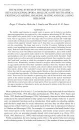
The turtle Trachemys has been developed as a model<br />
organism for studying hair cells, specialized neurons<br />
of the vertebrate inner ear that sense sound, as well<br />
as balance and acceleration. Yet very little is known<br />
about turtle hearing sensitivity. In a recent study,<br />
Whitman Investigators Jakob Christensen-Dalsgaard,<br />
Peggy edds-Walton, richard fay, and Catherine Carr and collaborators report that Trachemys<br />
has a large, air-filled, tympanic middle ear that is specialized for underwater hearing. The<br />
auditory threshold for the turtle, they found, is approximately 20-30 dB lower in water than in<br />
air. This study, performed in collaboration with Darlene Ketten of Woods Hole Oceanographic<br />
Institution, adds to the understanding both of hair cell function and of the evolution of<br />
vertebrate auditory systems. (Proc. Royal Soc. B: 279: 2816-24, 2012) •<br />
6 MBL <strong>Catalyst</strong> FALL 2012<br />
first Map of the Bacterial Makeup<br />
of healthy humans is Published<br />
Five years ago, the National Institutes of<br />
Health launched the ambitious Human<br />
Microbiome Project (HMP) to define the<br />
boundaries of bacterial variation found<br />
in 242 healthy human beings. More than<br />
200 scientists nationwide recently reported<br />
the results, including Susan huse, hilary<br />
Morrison, and Mitchell Sogin of the MBL’s<br />
Bay Paul Center. To analyze more than 5,000<br />
samples of human and bacterial DNA, the<br />
HMP adopted state-of-the-art sequencing<br />
and analysis methods, some of which were<br />
developed at the MBL for the decade-long<br />
International Census of <strong>Marine</strong> Microbes.<br />
Perhaps not surprisingly, the HMP discovered<br />
that microbial distributions in the human<br />
body are not so different from those in ocean<br />
ecosystems. In both, microbial communities<br />
contain a few highly abundant bacterial<br />
types plus many, many more low-abundance<br />
types (the “rare biosphere”). The HMP also<br />
confirmed that in people, like in the ocean,<br />
which bacteria are abundant and which are<br />
rare varies from site to site. The common<br />
bacterium Bacteroides, for instance, can<br />
constitute nearly 100 percent of the microbes<br />
in one person’s gut, yet be barely present<br />
in another’s. “What this means is, there is<br />
not just one way to be healthy,” says Huse.<br />
“There doesn’t have to be one or two ‘just<br />
right’ gut communities, but rather a range of<br />
‘just fine’ communities.” Another key finding<br />
of the HMP is that nearly everyone, including<br />
healthy individuals, carries pathogens—<br />
microbes known to cause illness. Researchers<br />
now must figure out why some pathogens<br />
turn deadly and under what conditions,<br />
likely revising current concepts of how<br />
microorganisms cause disease. (Nature 486:<br />
207-214; Nature 486: 215-221; PLoS ONE<br />
7(6): e34242. doi:10.1371, 2012) •
Vale, Sheetz, and Spudich Receive<br />
Lasker Award for Basic Medical Research<br />
MBL and Stanford scientists who invented<br />
ways to study how “cargo” is moved<br />
within cells and, as a result, discovered<br />
a new biological motor protein, kinesin,<br />
at the MBL in 1984 have received the<br />
prestigious 2012 Lasker Award for Basic<br />
Medical Research. They are Michael<br />
Sheetz of Columbia University; James<br />
Spudich of Stanford University School of<br />
Medicine; and ron Vale of University of<br />
California, San Francisco/Howard Hughes<br />
Medical Institute. The Lasker Awards are<br />
among the most respected science prizes<br />
in the world, and often anticipate the<br />
Nobel Prize for their recipients.<br />
“Sheetz, Spudich, and Vale opened up<br />
the study of cytoskeletal motor proteins,<br />
machines that move cargoes within<br />
cells, contract muscles, and enable cell<br />
movements …. The minuscule motors<br />
underlie numerous vital processes, and<br />
[the awardees’] landmark achievements<br />
are driving drug-discovery efforts aimed at<br />
cardiac problems as well as cancer,” stated<br />
the Lasker Foundation announcement.<br />
The discovery of kinesin emerged from<br />
an intense period of investigation<br />
(1983 to 1985) at the MBL, where<br />
several key elements were essential to<br />
the work: an abundance of squid; the<br />
landmark introduction of video-enhanced<br />
microscopy, independently made by<br />
Robert and Nina Allen at Dartmouth<br />
College and at the MBL and Shinya Inoué<br />
at the MBL; and an environment dedicated<br />
to internationally collaborative, risk-taking<br />
science.<br />
As a neuroscience graduate student at<br />
Stanford in the early 1980s, Vale became<br />
interested in how material is transported<br />
inside the axon of nerve cells, which in<br />
humans can be more than three feet<br />
long. Vale was inspired by 1982 papers<br />
by MBL investigators Robert Allen, Scott<br />
Brady, and Ray Lasek, in which they used<br />
Allen’s video-enhanced microscopy to<br />
make movies of axonal transport in the<br />
squid, whose giant axon is over 100 times<br />
larger in diameter than the human axon.<br />
Allen’s technology dramatically increased<br />
the contrast of the images, allowing one to<br />
see details that could never be seen before.<br />
“What they saw was absolutely fantastic,”<br />
Vale said. “All these little dots [organelles]<br />
zipping along the axon, moving between<br />
the cell body and the nerve terminal. It was<br />
really thrilling to watch.”<br />
Meanwhile, at Stanford, Spudich and<br />
Sheetz (who was on sabbatical from<br />
University of Connecticut) were successfully<br />
developing a way to study the motion<br />
of myosin—a motor protein known to<br />
cause muscle movement—in a test tube.<br />
Vale began to wonder if axonal transport<br />
might be explained by a similar myosin<br />
mechanism. He and Sheetz decided to<br />
test the idea in the squid giant axon, and<br />
came to the MBL in 1983. Originally they<br />
collaborated with Allen’s and Lasek’s labs,<br />
but later teamed up with Tom Reese and<br />
Bruce Schnapp of the National Institutes of<br />
Health. All of these scientists were working<br />
intently on axonal transport, and all made<br />
important contributions to the final picture.<br />
Through what Vale calls a “tour de force”<br />
experiment, Schnapp and Reese showed<br />
that axonal transport in the squid took<br />
place on tracks called microtubules. This<br />
meant the mystery motor protein that<br />
carries out axonal transport couldn’t be<br />
myosin, which is associated with a different<br />
type of track, called actin. The culminating<br />
observation came at 2 AM one night in<br />
1984 when Vale and Sheetz looked at the<br />
video screen and saw “something totally<br />
shocking and surprising,” Vale said. It<br />
was purified microtubules that Vale had<br />
isolated and deposited on the glass surface<br />
of a microscope slide, together with<br />
some cytoplasm; the microtubules were<br />
“crawling along like little snakes,” he said.<br />
“It was fully in the spirit<br />
of MBL, in the sense of<br />
collaborations without<br />
boundaries. The MBL gave<br />
us the opportunity<br />
to just get down and<br />
do the science.”<br />
— Michael Sheetz<br />
To their surprise, the unknown motor in<br />
the cytoplasm was a soluble protein that<br />
serendipitously bound to glass. The motor<br />
proteins then caused the microtubules to<br />
“crowd surf” over them! It was now a<br />
matter of tracking that motor protein down.<br />
Vale, Sheetz, Reese, and Schnapp continued<br />
studying the system all winter and spring<br />
in Reese’s MBL lab. Within a few months,<br />
they had purified the protein, and called<br />
it kinesin (from the Greek root “kine” for<br />
motion).<br />
Today we know that humans have dozens<br />
of myosins and kinesins,” stated the award<br />
announcement. “Through their vision,<br />
ingenuity, and persistence, Sheetz, Spudich<br />
and Vale…illuminated crucial features of a<br />
fundamental biological process.”<br />
Vale is an MBL Whitman Investigator and<br />
former co-director of the Physiology course.<br />
Sheetz (Physiology and Neurobiology) and<br />
Spudich (Physiology) have served as course<br />
faculty members several times since the<br />
mid-1980s. •<br />
MBL <strong>Catalyst</strong> FALL 2012<br />
7
Opening<br />
The MBL Launches a new PrograM in sensory PhysioLogy and Behavior<br />
A lively open house last July for the new<br />
Program in Sensory Physiology and Behavior<br />
drew scientists from all over the campus, which<br />
is just what it aimed to do. The PSPB provides a<br />
nexus for existing strengths in sensory biology<br />
at the MBL—especially for research on vision,<br />
balance, and hearing.<br />
“We are forming a strong link between the<br />
MBL’s resident laboratories in sensory biology<br />
and summer activities in this field, including<br />
at the Whitman Center, the Grass <strong>Laboratory</strong>,<br />
and in many of the neuroscience courses,” says<br />
MBL Senior Scientist Roger Hanlon, who directs<br />
the PSPB. “The integration and interpretation<br />
of sensory information guides the lives of all<br />
animals and humans. There are many key questions<br />
in basic and applied sensory science to be<br />
addressed,” he says, including treatments for<br />
human sensory disorders.<br />
Judging from the mix of people at the open<br />
house, the program’s goal of stimulating intellectual<br />
exchange and research collaborations<br />
through many channels (seminars, student<br />
and faculty residencies, outreach) had already<br />
begun. In the hallway, MBL Senior Research<br />
Scientist Richard Chappell, who studies the role<br />
8 MBL <strong>Catalyst</strong> FALL 2012<br />
“The integration<br />
and interpretation<br />
of sensory<br />
information<br />
guides the lives of<br />
all animals<br />
and humans.”<br />
—Roger Hanlon,<br />
MBL Senior Scientist<br />
of zinc in vision and disease, was chatting with<br />
Hanlon lab affiliate Robyn Crook, an evolutionary<br />
neuroethologist and squid expert. Encircling<br />
MBL Adjunct Scientists Enrico Nasi and Maria<br />
Gomez, experts on the electrical response of<br />
visual cells to light, were several students from<br />
the Universidad Nacional de Columbia whom<br />
they had brought to their MBL lab. Neurophysiologists<br />
from many corners of the MBL —Alan<br />
Kuzirian, Richard Rabbitt, Robert Baker—were<br />
rubbing elbows.<br />
“We especially encourage collaborations with<br />
people who take advantage of the unique resources<br />
at the MBL, and the marine organisms<br />
we can obtain,” says Stephen Highstein, the<br />
program’s associate director. Highstein is one of<br />
the world’s foremost researchers on the innerear<br />
organs that govern balance and acceleration,<br />
and he has developed the toadfish and the<br />
turtle as model organisms for this purpose.<br />
“There are so many organisms at the MBL,” says<br />
Felix Schweizer of UCLA’s David Geffen School<br />
of Medicine. “And there is a lot to be learned<br />
from them. Many have functional and anatomical<br />
specializations that make it much easier to<br />
study certain research questions.”<br />
Above: A single hair cell from a frog ear. Hair cells detect sound and acceleration.
the Senses<br />
The chance to work with squid is one reason<br />
Paloma Gonzalez-Bellido and her husband,<br />
Trevor Wardill, left Howard Hughes Medical<br />
Institute’s Janelia Farm campus last year to join<br />
the Hanlon lab. “Most neuroscience nowadays<br />
is in [the traditional genetic] model animals—<br />
the mouse, the fruit fly, the nematode worm,”<br />
she says. “It’s rare that you can do neuroscience<br />
in any other model, and it’s extremely rare that<br />
you can do it in a marine system. The MBL<br />
presented us with a unique opportunity. We had<br />
to run with it!”<br />
Until recently, research on models like the<br />
toadfish or midshipman was limited to electrophysiology,<br />
because the molecular tools weren’t<br />
there to plumb their genomes. “But that is<br />
changing,” Schweizer says. “High-throughput<br />
sequencing, and novel molecular methods, are<br />
making it much easier to work in marine and<br />
other models.”<br />
Schweizer is president of the Grass Foundation,<br />
which grants fellowships to early-career neuroscientists<br />
to conduct summer research at the<br />
MBL. He sees great opportunities for continued<br />
exchange between the MBL Grass Lab and the<br />
PSPB. “Our senses inform us about the world<br />
around us. And, in some cases, we still know<br />
very little about how sensory information gets<br />
into the nervous system,” he says.<br />
The mystery of the “neural code”—how stimuli<br />
such as light or sound are converted into electrical<br />
spikes in the nervous system—is at the core<br />
of much PSPB research, including Highstein’s<br />
lab. It is indeed a code to be cracked, since sensory<br />
organs detect stimuli in a graded manner<br />
(a fire can feel warm, hot, or burning, for example)<br />
yet most of the neurons that drive the<br />
appropriate responses, such as moving the hand<br />
away from the fire, spike in an on/off, digital<br />
manner. What stimulus produces a certain spike<br />
pattern to get a certain behavioral outcome?<br />
“This is the Holy Grail of neuroscience. What<br />
is the code?” Gonzalez-Bellido says. “If you understand<br />
that coding, and you know the wiring<br />
of the neurons, then you may be able to predict<br />
what type of signals create a specific spike pattern/response.<br />
Then, you will understand that<br />
aspect of the nervous system very well.” • — DK<br />
The mystery of the “neural code”<br />
—how stimuli such as light or sound<br />
are converted into electrical spikes<br />
in the nervous system—is at the<br />
core of much PsPB research.<br />
That Squid Can Dance<br />
In terms of outreach,<br />
no one could have planned a<br />
more sensational kick-off for<br />
the PSPB than a video that<br />
went viral on the Internet. It<br />
came from one of those serendipitous<br />
encounters at the<br />
MBL that are often chalked<br />
up to the “MBL magic.” In<br />
July, the program’s first publication<br />
appeared, a Hanlon<br />
lab study that showed, for<br />
the first time, that squid<br />
have precise neural control<br />
of the iridescence cells in<br />
their skin. The authors<br />
(Wardill, Gonzalez-Bellido,<br />
Crook and Hanlon) had<br />
developed a novel squid<br />
preparation that allowed<br />
them to dramatically shift<br />
the skin’s iridescence from<br />
red to green by electrically<br />
stimulating certain nerves.<br />
That prep caught the attention<br />
of Greg Gage, an MBL<br />
course lecturer whose Backyard<br />
Brains company sells<br />
affordable brain-recording<br />
kits to the public. What<br />
might happen, Gage wondered,<br />
if he pumped the<br />
digitized signal of hip-hop<br />
music from his iPod into<br />
those squid skin nerves? He<br />
tried it, and the fascinating<br />
result was a colorful dance<br />
of the pigment organs<br />
(chromatophores) pulsating<br />
with the song’s bass beat.<br />
Gage made a music video<br />
of the dancing squid skin,<br />
called “Insane in the Chromatophores,”<br />
and posted it<br />
on YouTube. Within a week<br />
it had racked up more than<br />
1.3 million views! To see<br />
the video, go to<br />
news.backyardbrains.com.<br />
MBL <strong>Catalyst</strong> FALL 2012<br />
9
A Revolution<br />
frOM the MiCrOBial WOrlD COMe POWerfUl neW tOOlS tO illUMinate the hUMan Brain<br />
A<br />
familiar furnishing in the MBLWHOI Library is this<br />
elegantly penned motto of 19th-century zoologist Louis<br />
Agassiz: “Study Nature, Not Books.” The wisdom of<br />
this phrase is often reproven, as in the serendipitous<br />
discovery of a green fluorescent protein (GFP) in jellyfish by<br />
Osamu Shimomura, now an MBL Distinguished Scientist, in<br />
1961. Decades later, others turned GFP into a revolutionary<br />
tool for illuminating the workings of cells, one that brought<br />
Shimomura the Nobel Prize. Agassiz’s motto still reverberates<br />
today, most recently in the form of a sensational technique that<br />
is taking neuroscience by storm: Optogenetics.<br />
“Optogenetics is a revolution, and the MBL is in the midst of<br />
it,” says George Augustine, a researcher and frequent course<br />
faculty member at the MBL since 1979. Barely ten years old,<br />
optogenetics is so promising that its discoverers are widely<br />
considered candidates for the Nobel Prize.<br />
With amazing precision, optogenetics allows scientists to<br />
control the electrical activity of neurons simply by targeting<br />
them with beams of light. Besides being much less invasive<br />
than using electrodes—which has been the standard way to<br />
study neural activity since the 1930s—optogenetics allows<br />
scientists to turn on or off specific types of neurons in the<br />
brain (say, only those that produce dopamine), which is a huge<br />
advance. It throws open the door to pinpointing exactly which<br />
neurons and circuits cause specific human sensations, thoughts,<br />
and behaviors—and perhaps one day, to light-based therapies<br />
for mental and sensory disorders.<br />
10 MBL <strong>Catalyst</strong> FALL 2012<br />
Yet this powerful tool for illuminating the animal brain emerged<br />
out of a quite distant realm of curiosity. Since the early 1970s,<br />
a small group of scientists had been studying the membrane<br />
proteins that some microbes (which, of course, lack eyes) use<br />
to respond to light. Called opsins, they help the microbe do a<br />
variety of things, such as move toward a light source or hold<br />
steady at an ocean depth. Animals, too, have opsins, which<br />
they use for vision.<br />
The first microbe with an opsin was discovered in a high-salt<br />
environment; others were later found in variety of ecosystems.<br />
Although they fascinated those who discovered them,<br />
hardly anyone else took notice. When scientists reported<br />
in 2002 that an opsin from a green alga, now aptly called<br />
Channelrhodopsin, seemed to “open” just like an ion channel<br />
when stimulated by light, neuroscientists barely noticed. “It’s<br />
not something I, or pretty much anyone outside of the aquatic<br />
algae field, was paying attention to,” says Augustine.<br />
Fortunately, at least two brilliant neuroscientists (both alumni<br />
of MBL courses) were paying attention: Ed Boyden, a graduate<br />
student and engineering whiz, and Karl Deisseroth, an MD-<br />
PhD who was devoted to finding cures for the devastating<br />
mental disorders he saw during his psychiatric training. The two<br />
met at Stanford and began dreaming about ways to control<br />
subsets of neurons in the brain and, ultimately, repair neural<br />
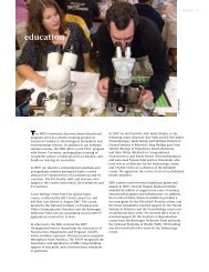
circuits that were malfunctioning. When the Channelrhodopsin<br />
paper appeared, they and others checked out the far-fetched<br />
possibility that this green alga (Chlamydomonas reinhardtii)<br />
might contain the tool they needed.<br />
Above: The green alga C. reinhardtii, in which the first Channelrhodopsins were discovered.
Born in Nature<br />
They were not prepared for how spectacularly well it would<br />
work. Using standard genetic engineering techniques, they<br />
inserted the Channelrhodopsin gene into mammalian neurons<br />
in culture, and then pulsed them with blue light. Amazingly,<br />
the neurons fired. “It worked right out of the gate,” Augustine<br />
says, which happens almost never in science. The startling<br />
findings were published in 2005. Later, a different opsin (a<br />
Halorhodopsin) was shown to inhibit or silence neurons when<br />
hit with yellow light.<br />
Augustine, who had been one of Deisseroth’s mentors on<br />
optics in the 1990s, right away recognized the discovery’s<br />
power. He and his colleagues at Duke University tried<br />
“Optogenetics is a revolution,<br />
and the MBl is in the midst of it.”<br />
Augustine is excited about the more immediate prospect<br />
of using optogenetics to construct a “wiring diagram” of<br />
the neural circuits in the brain, a field of research known<br />
as connectomics. “In the past, figuring out how a whole<br />
brain works would have been just lunacy. But I think with<br />
optogenetic approaches, it’s within the realm of feasibility,”<br />
he says.<br />
“The historical way to define a neural circuit is anatomically.<br />
You simply look at this neuron and see if it touches the next<br />
neuron. If it does, they are probably talking to each other,”<br />
Augustine says. “But the problem with this method is, it<br />
doesn’t tell you anything about the functioning of that circuit.<br />
Do all of its synapses actually work, or are<br />
some of them silent? Before optogenetics, it<br />
was well nigh impossible to tell.”<br />
Buoyed by the possibilities, Augustine founded<br />
— George Augustine, MBL Whitman Investigator the Center for Functional Connectomics in<br />
Seoul, South Korea, in 2010. So far, he has<br />
used optogenetics to selectively activate about<br />
introducing the Channelrhodopsin gene into a live mouse, to 100 different kinds of neurons in the mouse brain. “The goal<br />
see if they could activate subsets of neurons from the millions is to get them all, by hook or by crook,” he says. The big<br />
in the animal’s brain. “We turned on the light and, boom, the remaining challenge is getting a reliable readout of which<br />
mouse’s neurons fired. It was magical,” Augustine recalls. neurons are talking, which are listening, and when (see Cool<br />
Tool, page 15). Then, Augustine says, all the tools will be in<br />
A distinct buzz about the new “optogenetics” soon spread place to map every circuit in the brain.<br />
through the neuroscience community. “It has absolutely<br />
enabled experiments that before were impossible to do,” As for the origin of this revolutionary tool? Deisseroth, for one,<br />
says MBL Neurobiology course co-director Graeme Davis of hasn’t forgotten that it sprang from the study of microbes that<br />
University of California, San Francisco. And it allows one to may seem just weird. To him, the discovery of optogenetics<br />
dream that cures for long-intractable brain disorders might carries two big lessons: The importance of undirected, basic<br />
someday be at hand.<br />
inquiry into nature and of protecting biodiversity, even in<br />
“the harsh, barren and otherwise useless Saharan salt lakes<br />
from which some of the most useful opsins originated.”<br />
Undoubtedly, Agassiz would have agreed. • — DK<br />
MBL <strong>Catalyst</strong> FALL 2012<br />
11
mBL momeNt Cracking<br />
with …<br />
Christof Koch<br />
Co-founder, Methods in<br />
Computational Neuroscience course<br />
Christof Koch is the Lois and Victor<br />
Troendle Professor of Cognitive<br />
and Behavioral Biology at the<br />
California Institute of Technology.<br />
Since April 2011, he has been on<br />
partial leave of absence to serve<br />
as chief scientific officer of the<br />
Allen Institute for Brain Science,<br />
a nonprofit medical research<br />
institution founded and funded<br />
by Paul G. Allen, co-founder of<br />
Microsoft. Koch’s research lab at<br />
Caltech studies how individual<br />
nerve cells process information;<br />
the primate visual system; and the<br />
relationship between consciousness<br />
and brain activity. The latter<br />
interest developed from a nearly<br />
two-decade collaboration with the<br />
late Francis Crick, co-discoverer<br />
of the structure of DNA, during<br />
which they pioneered a framework<br />
for the neurobiological study of<br />
consciousness. Koch received his<br />
PhD in 1982 from the Max Planck<br />
Institute for <strong>Biological</strong> Cybernetics<br />
in Tübingen, Germany, and was<br />
a postdoctoral fellow at MIT’s<br />
Artificial Intelligence <strong>Laboratory</strong>. He<br />
holds eight patents, has authored<br />
several hundred peer-reviewed<br />
scientific publications and three<br />
books, including Consciousness:<br />
Confessions of a Romantic<br />
Reductionist (MIT Press, 2012).<br />
12 MBL <strong>Catalyst</strong> FALL 2012<br />
chrisTof Koch spent a<br />
“glorious” month at the MBL each<br />
summer from 1988 to 1992<br />
while co-directing the Methods<br />
in Computational Neuroscience<br />
course, which he founded with<br />
James M. Bower. Today, Koch<br />
is launching neuroscience’s<br />
first “big science” project: a<br />
massive, multidisciplinary quest<br />
to understand the structure<br />
and the function of the mouse<br />
cortex, focusing on vision and<br />
visual behaviors. The mission<br />
of this open-source effort at<br />
the Allen Institute for Brain<br />
Science in Seattle—funded by<br />
a $300 million donation for its<br />
first 3.5 years by Paul Allen—is<br />
a quantitative understanding of<br />
how the mouse sees, thinks, and<br />
decides in terms of the underlying<br />
neurons and their synaptic<br />
interconnections. Meanwhile, the<br />
MBL “Methods” course, currently<br />
co-directed by Adrienne Fairhall<br />
of University of Washington and<br />
Michael Berry of Princeton,<br />
is going strong.
The Brain’s Code<br />
MBL: what motivated you to launch an MBL course on<br />
computer modeling of the brain?<br />
CK: The course was inspired by Caltech’s new Computation &<br />
Neural Systems graduate program, which I had joined in 1986<br />
as an assistant professor. This was the first PhD program in the<br />
world that took in undergraduates from physics, engineering,<br />
and mathematics and exposed them to neurobiology and<br />
molecular biology. Today, every university has faculty doing<br />
modeling of the brain, but 30 years ago, you were kind of looked<br />
at askance, like, “This is not real biology! Real biology is in the<br />
lab, poking electrodes in things.”<br />
MBL: The MBL course today studies neural computation<br />
at many levels, from individual ion channels to the<br />
whole brain. did you organize the course that way?<br />
CK: Yes. Jim Bower and I were really trying to go from the [cell]<br />
biophysical level all the way to the final output, which is<br />
behavior and consciousness. Another unusual thing we did is<br />
bring computers and insist each student do some kind of<br />
simulation project. Laptops didn’t exist then; the MBL had a few<br />
central computers in the library. So Jim persuaded Sun<br />
Equipment to ship us computer workstations, which they did at<br />
great expense to them. The course was really intense,<br />
particularly in the last week, when people were working day and<br />
night to finish their projects!<br />
MBL: you first started thinking about the scientific study<br />
of consciousness while in woods hole. Please share that<br />
story.<br />
CK: One night after teaching I had a toothache, and I was lying<br />
in bed trying to distract myself from the pain. And then it hit<br />
me. Where the hell does this feeling of pain come from? Why<br />
does some physical event—activity of neurons —give rise to a<br />
feeling in the head? This dilemma goes back hundreds of years;<br />
it’s the heart of the traditionally conceived mind-body problem.<br />
So, the pulp of your tooth is inflamed, electrical activity travels<br />
down the trigeminus nerve to your spinal cord, up through<br />
relays in the thalamus and elsewhere, and finally activates<br />
neurons in your neocortex. The neurons fire, and you experience<br />
this firing as pain. Other neurons fire when you make love and<br />
experience pleasure, and some other neurons fire when you<br />
hear Beethoven. When you think about it, this is a complete<br />
non sequitur. It’s like rubbing a brass lamp and suddenly a genie<br />
appears. I mean, if I touch some buttons on my iPhone, charge<br />
flows onto a transistor, the transistor switches and opens other<br />
transistors, and so on. But nobody in their right mind thinks<br />
the iPhone has any feelings, or can hear Beethoven! Where do<br />
conscious experiences come from? Science has to explain this!<br />
This is the realization I had in Woods Hole. As Francis Crick<br />
had already occupied himself with the same problem—how<br />
can the excitable matter in the brain generate any conscious<br />
feelings—we started to meet and discuss these questions. We<br />
then wrote our first paper on consciousness together, in 1990.<br />
MBL: you have described neuroscience as being a very<br />
splintered field today. given that, would you design a<br />
new course at the MBL differently?<br />
CK: It’s a difficult question. The field has grown vastly, in<br />
terms of practitioners and publications. And yet, unlike in<br />
physics and chemistry, we still don’t have a single, unifying<br />
theory. Indeed, it is not at all clear what principles allow one<br />
to go from a roundworm with 302 neurons, to a fruit fly with<br />
100,000 neurons, to a mouse with 70 million neurons. The<br />
neurons are obviously the same, the ionic channels are very<br />
similar, but how they are put together is not clear. Have you<br />
ever been to a Society for Neuroscience meeting? It’s utterly<br />
bewildering. There are 30,000+ people with 20,000 posters<br />
and presentations. Each one involves a different species, at<br />
a different developmental time stage, a different question<br />
using a different technique. It’s like attending a conference of<br />
Dadaist painters, all of whom insist on being utterly unique!<br />
We have not had a clearing out (and maybe this will never<br />
come) when people realize, oh, we have these two or three<br />
model, exemplary paradigms, and everything coalesces<br />
around them.<br />
So, in terms of teaching, I’m not sure I would do things<br />
differently today. I still feel it is important to go from the ion<br />
channel and synapse levels all the way to the behavioral level.<br />
In physics, you can study forces in isolation because they<br />
differ by many orders of magnitude, but that is not the case<br />
in biology. If you want to understand perception, memory<br />
and consciousness, you need to understand the details of the<br />
“and then it hit me. where the hell does this feeling of pain come from?”<br />
hardware, the neurons and their interconnections. They really<br />
matter.<br />
In 1988, our “Methods” course looked at neurobiological<br />
lessons from many animals, from invertebrates to humans.<br />
I probably wouldn’t do that today. Instead, I am thinking<br />
of starting a course somewhere focused on just the mouse,<br />
but from many different vantage points: brain development,<br />
different cell types, behavior, computational theory.<br />
I have also thought about teaching a course on the neural<br />
basis of consciousness, where we invite neurobiologists,<br />
psychologists, some clinicians, and one or two philosophers.<br />
Right now, I’m just a little overwhelmed, having to hire a few<br />
hundred scientists and engineers for our Allen mouse project!<br />
But I definitely want to do it someday. • —DK<br />
MBL <strong>Catalyst</strong> FALL 2012<br />
13
G i F t s & GrANts<br />
A c c o L A D e s The American Society for Cell Biology presented an award to the Physiology course “for its tremendous impact<br />
on developing the classical and modern eras of cell biology.” The award was given in honor of the course’s<br />
120th anniversary.<br />
14 MBL <strong>Catalyst</strong> FALL 2012<br />
The Gordon and<br />
Betty Moore<br />
foundation awarded<br />
a $2,258,548<br />
grant to investigate<br />
microbial ecosystems<br />
at Axial Seamount,<br />
a deep-sea volcano<br />
in the North Pacific.<br />
Julie Huber is the<br />
principal investigator.<br />
The national<br />
institutes of health<br />
awarded $1,593,277<br />
for a project titled<br />
“Orientation<br />
Independent DIC<br />
and Polarization<br />
Microscopy.”<br />
Michael Shribak<br />
is the principal<br />
investigator.<br />
The national<br />
institutes of health<br />
awarded $1,340,167<br />
for a project titled<br />
“The Tunicate<br />
Ciona: A New Model<br />
for the Effects of<br />
Aging on Tissue<br />
Regeneration.”<br />
William Jeffery is the<br />
principal investigator.<br />
The national<br />
institute of Diabetes<br />
and Digestive and<br />
Kidney Diseases<br />
awarded $1,309,040<br />
for a project titled<br />
“<strong>Biological</strong> Roles of<br />
Copper in Human<br />
Nutrition.” Jonathan<br />
Gitlin is the principal<br />
investigator.<br />
The Department<br />
of energy awarded<br />
$1,048,327 for<br />
a project titled<br />
“Hydraulic<br />
redistribution of<br />
water through plant<br />
roots – implications<br />
for carbon cycling<br />
and energy flux at<br />
multiple scales.”<br />
Zoe Cardon is the<br />
principal investigator.<br />
The following members of the MBL community were elected to the National Academy of Sciences: William<br />
Bialek, faculty, Methods in Computational Neuroscience; Karl Deisseroth, alumnus, Methods in Computational<br />
Neuroscience, faculty, Neurobiology; Gideon Dreyfuss, faculty, Physiology; Paul t. englund, emeritus Corporation<br />
member, alumnus, Invertebrate Zoology and Physiology, faculty and former director, Biology of Parasitism;<br />
roy Parker, faculty, Physiology; Mary Power, alumna, Neural Systems & Behavior.<br />
Senior Scientist Jonathan Gitlin was elected to the Institute of Medicine.<br />
An anonymous<br />
donor contributed<br />
$600,000 to<br />
help support the<br />
construction of a<br />
public elevator in the<br />
Swope Conference<br />
Center.<br />
The Charles evans<br />
foundation awarded<br />
$500,000 to<br />
establish The Charles<br />
Evans Research<br />
Development Fund.<br />
The William<br />
randolph hearst<br />
foundation awarded<br />
$100,000 to support<br />
the William Randolph<br />
Hearst Scholarship<br />
Program.<br />
Dr. and Mrs. John<br />
W. rowe contributed<br />
$100,000 to the<br />
MBL Annual Fund. •<br />
The following members of the MBL community were elected to the American Academy of Arts and Sciences:<br />
Ben a. Barres, faculty, Fundamental Issues in Vision Research; emery n. Brown, alumnus, Computational<br />
Neuroscience, faculty, Neuroinformatics; Sarah elgin, faculty, Physiology; Margaret J. Mcfall-ngai, faculty,<br />
Microbial Diversity, Embryology; Steven Siegelbaum, faculty, Neurobiology; larry Simpson, faculty, Biology<br />
of Parasitism.<br />
Winfried Denk, faculty, Neurobiology, received the Kavli Prize in Neuroscience for elucidating the basic neuronal<br />
mechanisms underlying perception and decision. Denk shared the prize with Cornelia Isabella Bargmann and Ann<br />
M. Graybiel.<br />
Whitman Investigator and Neural Systems & Behavior course alumnus richard fay received the Silver Medal in<br />
Animal Bioacoustics for pioneering studies of fish hearing by the Acoustical Society of America, the premier<br />
international scientific society in acoustics.<br />
Gary ruvkun, former director of the Biology of Aging course, and Victor Ambros were awarded the Dr. Paul<br />
Janssen Award for Biomedical Research. Ruvkun and Ambros identified and characterized the first microRNA,<br />
pivotal regulators of both normal and disease physiology. •
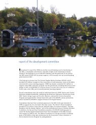
THE ULTIMATE OPTICAL<br />
A neuron expressing the<br />
ArcLight Q239 probe<br />
visualized with confocal<br />
microscopy. Image<br />
courtesy of Larry Cohen<br />
Voltage Sensor<br />
Off to the Races . . .<br />
Alongstanding dream in neuroscience<br />
has been finding a way to monitor<br />
neural activity across the entire brain. In the<br />
1960s, the only way to record a neuron’s<br />
voltage was to stick an electrode in it, an<br />
approach limited to a few cells at a time.<br />
The person who changed all that is Larry<br />
Cohen of Yale University, who has been<br />
a near-constant summer presence at the<br />
MBL since 1962. “Larry is the original ‘Mr.<br />
Light,’” says David Gadsby of Rockefeller<br />
University. “He pioneered the study of brain<br />
activity using electro-optical measures.”<br />
Cohen started his journey after codiscovering<br />
that when neurons fire, they<br />
undergo changes in light scattering and<br />
other optical properties. Perhaps a camera<br />
could record such changes, and use them<br />
as a proxy for electrical activity? The idea<br />
was “pretty far out,” says Cohen’s MBL<br />
colleague George Augustine. But it was<br />
evidently fertile, as functional neuroimaging<br />
is now a huge endeavor, at the MBL and<br />
around the world.<br />
c o o L tooL<br />
In 1973, Cohen, Vicencio Davila, and<br />
Brian Salzberg at the MBL successfully<br />
made the first optical recording of a leech<br />
neuron firing. They did this by bathing the<br />
neuron in a fluorescent dye. When the<br />
neuron fired, the fluorescence changed,<br />
and a detector captured the change.<br />
Since then, with many contributions<br />
from MBL researchers, various optical<br />
sensors have been introduced, the latest<br />
being genetically engineered ones that,<br />
in principle, can target specific neurons.<br />
Today, the race is on to design better<br />
sensors, ones that give fast, big signals<br />
and—crucially —show which individual<br />
neurons in a brain region fire, and when.<br />
Two promising sensors are Cohen’s latest,<br />
called ArcLight, and one from Adam<br />
Cohen of Harvard (MBL Physiology<br />
alumnus, 2007), called Arch.<br />
“Once we get that ultimate sensor, it’s off<br />
to the races, in terms of mapping the connectome,”<br />
says Augustine, who recruited<br />
Larry Cohen to the Center for Functional<br />
Connectomics (see p. 10). “When that<br />
happens, everybody is going to jump into<br />
the game. Things are poised right now,<br />
ready to blow.” • — DK<br />
MBL <strong>Catalyst</strong> FALL 2012<br />
15
s c i e N t i s t’s eye View<br />
Gerald D. Fischbach, M.D., is<br />
director of the Simons Foundation<br />
Autism Research Initiative and<br />
Health Sciences Dean Emeritus<br />
at Columbia University. An<br />
MBL Corporation member<br />
since 1976 and now emeritus<br />
Trustee, Fischbach taught for<br />
many years in the Neurobiology<br />
course and served on numerous<br />
MBL committees, including the<br />
Discovery Campaign Committee.<br />
For more than a decade, he and<br />
his wife, Ruth, have sponsored<br />
the annual MBL Bioethics Lecture.<br />
In 2012, they established the<br />
Fischbach Scholarship at MBL, and<br />
the Simons Foundation endowed<br />
the MBL Fischbach Fellowship<br />
in their honor. A distinguished<br />
researcher who served as president<br />
of the Society for Neuroscience,<br />
Fischbach chaired the<br />
Departments of Neurobiology at<br />
Washington University (St. Louis)<br />
and Harvard University before<br />
being appointed director of the<br />
National Institute of Neurological<br />
Diseases and Stroke (NIH) and<br />
then dean at Columbia University.<br />
His memberships include the<br />
National Academy of Sciences,<br />
the American Academy of Arts<br />
and Sciences, and the Institute of<br />
Medicine.<br />
16 MBL <strong>Catalyst</strong> FALL 2012<br />
What We Need to Learn<br />
By Gerald D. Fischbach<br />
Despite remarkable technical and conceptual advances in neuroscience, at all levels from<br />
molecules to cognition and behavior, we still lack deep enough understanding at any level<br />
to pursue rational therapies for neuropsychiatric disorders. A brief discussion of autism, with<br />
which I have become familiar over the past six years as director of the Simons Foundation<br />
Autism Research Initiative, illustrates these points.<br />
High-resolution genome analyses have revealed genetic variants that raise the risk of autism.<br />
Ordinarily we carry two copies of each gene, one from each parent. Many children on the<br />
autism spectrum have only one of the pair; some have more than two. Such deletions or<br />
duplications may lead to symptoms. Gene copy number matters in the brain. Some of these<br />
variants are inherited, but many others arise de novo in the germ line of a parent.<br />
Note that I avoid the claim that any single variant “causes” autism, a multifaceted condition<br />
marked by deficits in social communication, restricted interests, repetitive movements and,<br />
sometimes, intellectual impairment and language delay. But copy number variants are highly<br />
penetrant (meaning, they often have measurable effects): they do increase risk. Such variants,<br />
though individually rare, together may account for over 25 percent of autism cases.<br />
Many risk genes encode proteins engaged in synaptic transmission or plasticity, the ability to<br />
change with experience. This important insight has suggested novel therapies. For example,<br />
blockers of a postsynaptic receptor are now under investigation for treatment of Fragile X<br />
syndrome and autism. But more is needed to meaningfully relate genes to cognition and<br />
behavior. We must move beyond individual molecules, synapses, and cells to understand<br />
entire molecular networks and large neuron populations.<br />
Breakthroughs are needed that will allow manipulation of molecules within cells while<br />
recording from many hundreds of individual neurons in real time. Analysis of the massive data<br />
generated in such efforts is the subject of the MBL’s extraordinary Methods in Computational<br />
Neuroscience course. These approaches aim to reduce complex, multidimensional data to a<br />
manageable set of parameters.<br />
Highly time-resolved recordings may capture the neural correlates of fleeting thoughts<br />
and decisions essential for daily living and social cognition. Perhaps these correlates will<br />
provide “biomarkers” (biological indicators) more relevant to autism and related disorders<br />
than particular molecules operating at certain synapses. Novel biomarkers are essential for<br />
mechanism-based therapeutics.<br />
Another challenge involves human experimentation. We must move beyond mice to address<br />
the complexities of human cognition and behavior directly. It is accepted that effective<br />
therapies must begin before critical periods have elapsed and compensatory changes set in.<br />
Changes in Alzheimer’s and Parkinson’s diseases begin 10 to 15 years ahead of clinical signs!<br />
When do the first changes of autism and related developmental disorders occur?<br />
Large samples and quantitative, non-invasive measurements are needed to characterize<br />
normal brain states and subtle changes during early development. Relevant protocols must<br />
be devised, measurements standardized, and terabytes of imaging data collected and shared.<br />
Profound ethical and educational issues regarding living persons and collection of postmortem<br />
brains must be addressed.<br />
Resolution of these issues is within reach. Now is an exciting time in neuroscience as<br />
fundamental research is closer than ever to improving the human condition. •
T r e a s u r e s f r o m T h e m B L ’ s a r c h i v e s<br />
Plate 2 from The Anatomy of the<br />
Common Squid, Loligo pealii,<br />
Lesueur, by Leonard Worcester<br />
Williams (Leiden, Holland: E.J.<br />
Brill, 1910). Fig. 15: Nervous<br />
system, right aspect. (The arrow,<br />
added here to the original,<br />
indicates the squid giant axon,<br />
which is 0.5 to 1 mm in diameter<br />
or nearly 100 times larger than a<br />
typical human axon.)<br />
FiGure 15<br />
<br />
Squid Central<br />
When L.W. Williams first described the squid’s nervous system at<br />
m e m o r A B i L i A<br />
the MBL in 1910, he had no inkling he was launching a century of<br />
neuroscientific discovery. Little was known about how nerves work when<br />
J.Z. Young a quarter-century later suggested studying the squid, whose<br />
axons are “giant” compared to those in humans. By the late 1930s, MBL<br />
scientists like Kenneth Cole were putting electrodes inside the squid<br />
giant axon to measure electrical events associated with nerve impulses.<br />
Andrew Huxley (who died last May) and Alan Hodgkin pursued similar<br />
squid research in England. Using an improvement on Cole’s electronics<br />
(the voltage clamp), they formulated their model of voltage-controlled<br />
nerve conductance to explain the nerve impulse. Hodgkin and Huxley<br />
won the Nobel Prize in 1963, along with John Eccles, who described<br />
nerve cell communication at the synapse. Each of these pioneers at some<br />
point worked at the MBL, and their intellectual descendants are still here<br />
today. As for the squid, it is now so familiar that it is valued for many<br />
kinds of studies, from genomics to the pathology of Alzheimer’s disease.<br />
In the pantheon of model organisms in neuroscience, the squid has been<br />
honored by time. • — DK<br />
MBL <strong>Catalyst</strong> FALL 2012<br />
17
MBL<br />
7 MBL Street<br />
Woods Hole, MA 02543 USA<br />
www.mbl.edu<br />
Printed with recycled materials.<br />
<strong>Catalyst</strong><br />
i N the Next MBL C AtA Ly s t<br />
The MBL Turns 125<br />
Smartphone users:<br />
Scan QR code for<br />
online content.<br />
In celebration of the MBL’s 125th anniversary in 2013, we will reflect on the laboratory’s significant impact on the<br />
biological sciences and envision the next generation of inquiry and discovery.<br />
NON-PROFIT ORG.<br />
U.S. POSTAGE<br />
PAID<br />
Marshfield, MA<br />
PERMIT NO. 51