

Identification of important interactions between subchondral bone ...
Identification of important interactions between subchondral bone ...
Identification of important interactions between subchondral bone ...

Create successful ePaper yourself
Turn your PDF publications into a flip-book with our unique Google optimized e-Paper software.
CHAPTER 2: Introduction<br />
follows normal development or healing. Lamellar <strong>bone</strong> is characterized by organized fibres,<br />
which are forming arches for optimal <strong>bone</strong> strength. The lamellae can be parallel to each other if<br />
deposited along a flat surface (trabecular <strong>bone</strong>) or concentric if deposited on a surface<br />
surrounding a channel centred on a blood vessel 17,22,23 .<br />
2.2.3 The cells <strong>of</strong> <strong>bone</strong> and their communications<br />
19<br />
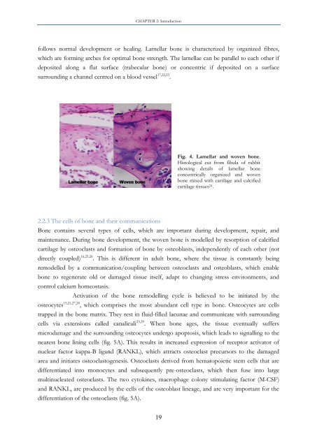
Fig. 4. Lamellar and woven <strong>bone</strong>.<br />
Histological cut from fibula <strong>of</strong> rabbit<br />
showing details <strong>of</strong> lamellar <strong>bone</strong><br />
concentrically organized and woven<br />
<strong>bone</strong> mixed with cartilage and calcified<br />
cartilage tissues 24 .<br />
Bone contains several types <strong>of</strong> cells, which are <strong>important</strong> during development, repair, and<br />
maintenance. During <strong>bone</strong> development, the woven <strong>bone</strong> is modelled by resorption <strong>of</strong> calcified<br />
cartilage by osteoclasts and formation <strong>of</strong> <strong>bone</strong> by osteoblasts, independently <strong>of</strong> each other (not<br />
directly coupled) 16,25,26 . This is different in adult <strong>bone</strong>, where the tissue is constantly being<br />
remodelled by a communication/coupling <strong>between</strong> osteoclasts and osteoblasts, which enable<br />
<strong>bone</strong> to regenerate old or damaged tissue itself, adapt to changing stress environments, and<br />
control calcium homeostasis.<br />
Activation <strong>of</strong> the <strong>bone</strong> remodelling cycle is believed to be initiated by the<br />
osteocytes 15,21,27,28 , which comprises the most abundant cell type in <strong>bone</strong>. Osteocytes are cells<br />
trapped in the <strong>bone</strong> matrix. They rest in fluid-filled lacunae and communicate with surrounding<br />
cells via extensions called canaliculi 23,29 . When <strong>bone</strong> ages, the tissue eventually suffers<br />
microdamage and the surrounding osteocytes undergo apoptosis, which leads to signalling to the<br />
nearest <strong>bone</strong> lining cells (fig. 5A). This results in increased expression <strong>of</strong> receptor activator <strong>of</strong><br />
nuclear factor kappa-B ligand (RANKL), which attracts osteoclast precursors to the damaged<br />
area and initiates osteoclastogenesis. Osteoclasts derived from hematopoietic stem cells that are<br />
differentiated into monocytes and subsequently pre-osteoclasts, which then fuse into large<br />
multinucleated osteoclasts. The two cytokines, macrophage colony stimulating factor (M-CSF)<br />
and RANKL, are produced by the cells <strong>of</strong> the osteoblast lineage, and are very <strong>important</strong> for the<br />
differentiation <strong>of</strong> the osteoclasts (fig. 5A).


