

PhD thesis Accessory Proteins at ERES-
PhD thesis Accessory Proteins at ERES-
PhD thesis Accessory Proteins at ERES-

You also want an ePaper? Increase the reach of your titles
YUMPU automatically turns print PDFs into web optimized ePapers that Google loves.
FACULTY OF SCIENCE<br />
UNIVERSITY OF COPENHAGEN<br />
<strong>PhD</strong> <strong>thesis</strong><br />
David Klinkenberg<br />
<strong>Accessory</strong> <strong>Proteins</strong> <strong>at</strong> <strong>ERES</strong>‐<br />
Assembly of ER exit sites is regul<strong>at</strong>ed by interactions of p125A with lipid signals.<br />
Academic advisor: Lektor Lars Ellgaard, <strong>PhD</strong><br />
Co‐Supervisor: Assoc. Prof. Meir Aridor, <strong>PhD</strong> (University of Pittsburgh)<br />
This <strong>thesis</strong> has been submitted to the <strong>PhD</strong> School of The Faculty of Science, University<br />
of Copenhagen: 26/02/2013
Name of department: Department of Biology<br />
Author: David Klinkenberg<br />
Title / Subtitle: <strong>Accessory</strong> <strong>Proteins</strong> <strong>at</strong> <strong>ERES</strong>‐<br />
p125A Couples Lipid Signals with Functional ER Exit Site Assembly<br />
Subject description: This <strong>thesis</strong> provides a characteriz<strong>at</strong>ion of the accessory protein p125A and its<br />
functions <strong>at</strong> Endoplasmic Reticulum Exit Sites (<strong>ERES</strong>) in response to membrane<br />
lipid composition by dissecting two functional domains within p125A. The<br />
results provide evidence for a mechanism where p125A response to lipid<br />
signals, i.e. PI(4)P, promotes both COPII displacement from the scaffolding<br />
protein mSec16A, as well as stabilizing linkage between the two layers<br />
forming the COPII cage <strong>at</strong> <strong>ERES</strong>.<br />
Academic advisor: Lektor Lars Ellgaard, <strong>PhD</strong><br />
Co‐Supervisor: Assoc. Prof. Meir Aridor, <strong>PhD</strong> (University of Pittsburgh)<br />
Submitted: 26 February 2013<br />
Grade: <strong>PhD</strong><br />
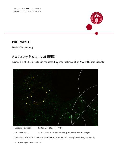
Front Page Image<br />
EGFPp125A expression co-localized <strong>at</strong> <strong>ERES</strong><br />
(yellow) with Sec31A (green) <strong>at</strong> 10°C.<br />
Recorded on a Olympus Fluoview 1000 PLAPON<br />
60 x objective, NA = 1.42
Abstract<br />
Traffic medi<strong>at</strong>ed by vesicles budding from the membranes of the Endoplasmic Reticulum (ER) <strong>at</strong><br />
specific sites termed ER Exit Sites (<strong>ERES</strong>) is medi<strong>at</strong>ed by the COPII machinery. The molecular<br />
1<br />
interactions th<strong>at</strong> COPII utilize to form the basic bud have been examined and mapped. However, not<br />
much is known about how these interactions are regul<strong>at</strong>ed, in particular with respect to COPII<br />
interactions in rel<strong>at</strong>ion to specific ER membrane lipid signals.<br />
This <strong>thesis</strong> explores the mechanisms by which the accessory protein p125A (aka Sec23IP) regul<strong>at</strong>es<br />
COPII <strong>at</strong> <strong>ERES</strong>. The work shows th<strong>at</strong> p125A recognizes lipids, and in particular phosph<strong>at</strong>idylinositol‐4‐<br />
phosph<strong>at</strong>e (PI(4)P), through concerted actions between two internal domains – a sterile α‐motif<br />
(SAM) and a put<strong>at</strong>ive lipid recognizing domain termed a DDHD domain. We demonstr<strong>at</strong>e th<strong>at</strong> p125A<br />
binding <strong>at</strong> <strong>ERES</strong>, in response to local presence of PI(4)P, medi<strong>at</strong>es displacement of the two COPII<br />
layers from the Sec16A <strong>ERES</strong> nucle<strong>at</strong>ion scaffold. We furthermore show evidence th<strong>at</strong> p125A<br />
provides a linkage between the two COPII layers during vesicle budding. Additional observ<strong>at</strong>ions<br />
indic<strong>at</strong>e th<strong>at</strong> p125A lipid recognition and binding supports the steady‐st<strong>at</strong>e transport levels between<br />
ER and Golgi. We also provide evidence th<strong>at</strong> Sec16A functions <strong>at</strong> an early stage of <strong>ERES</strong> assembly, as<br />
we can show clear segreg<strong>at</strong>ion of Sec16A from <strong>ERES</strong> during temper<strong>at</strong>ure imposed inhibition of the<br />
cellular transport.<br />
We finally explore the membrane binding mechanism of mammalian Sec16 (mSec16) A and B, and<br />
identify domains within each mSec16 subtype th<strong>at</strong> show membrane binding, but do not support<br />
selective <strong>ERES</strong> targeting.<br />
A major part of the experimental work presented in this <strong>thesis</strong> is included in the following<br />
manuscript th<strong>at</strong> has been submitted for review to the Journal of Cell Biology:<br />
Assembly of ER exit sites is regul<strong>at</strong>ed by interactions of p125A with lipid signals.<br />
David Klinkenberg, Kimberly R. Long, Kuntala Shome, Simon C. W<strong>at</strong>kins and Meir Aridor<br />
26/2‐2013<br />
David Klinkenberg
Acknowledgments<br />
The experiments of this <strong>thesis</strong> were all performed in the labor<strong>at</strong>ory of Ph.D. Meir Aridor <strong>at</strong> the<br />
Department of Cell Biology, University of Pittsburgh, Pittsburgh PA, USA, while employed as<br />
Research Technician.<br />
I would first like to thank Meir for all his encouragement and long scientific discussions th<strong>at</strong> have<br />
2<br />
fueled my fascin<strong>at</strong>ion for this particular field of Cell Biology, and in particular for allowing me to turn<br />
a position as Research Technician into a Ph.D. project.<br />
I would also like to give a very special thanks to Ph.D. Kimberley Long for helping verify and finalize<br />
the results of the appended manuscript, Ph.D. Kuntala Shome for providing technical assistance, and<br />
the rest of the members of the Aridor lab, past and present, th<strong>at</strong> I had the gre<strong>at</strong> pleasure to work<br />
with while in Pittsburgh and whom have made me grow as a scientist.<br />
I would finally like to thank my mother Marta and my step‐dad Carsten for all their support and aid<br />
th<strong>at</strong> made it possible for me to move for an extended period to the beautiful city of Pittsburgh,<br />
thereby giving me the opportunity to conduct the research presented in this <strong>thesis</strong>.<br />
In memoriam Teresa.
Summary<br />
The components of the COPII machinery, which are essential in establishing an effective<br />
Endoplasmic Reticulum (ER) to Golgi transport from ER exit sites (<strong>ERES</strong>), have been identified and<br />
characterized within the last 25 years. These consist of the essential Sec12, Sec23, Sec24, Sec13,<br />
Sec31 and Sar1 proteins. Together these components co‐oper<strong>at</strong>e in cargo‐selection as well as<br />
forming, loading and releasing budding vesicles from specific regions on the membrane surface of<br />
the ER. Co<strong>at</strong> components furthermore convey vesicle targeting towards the Golgi. However, not<br />
much is known about the mechanisms th<strong>at</strong> regul<strong>at</strong>e the COPII assembly <strong>at</strong> the vesicle bud site.<br />
This <strong>thesis</strong> provides the first regul<strong>at</strong>ory mechanism of COPII assembly in rel<strong>at</strong>ion to ER‐membrane<br />
lipid‐signal recognition by the accessory protein p125A (Sec23IP).<br />
The aim of the project was to characterize p125A function by dissecting two main domains in the<br />
protein; a put<strong>at</strong>ive lipid‐associ<strong>at</strong>ing domain termed the DDHD domain th<strong>at</strong> is defined by the four<br />
3<br />
amino acid motif th<strong>at</strong> gives the domain its name; and a ubiquitously found domain termed Sterile α‐<br />
motif (SAM), which is mostly associ<strong>at</strong>ed with oligomeriz<strong>at</strong>ion and polymeriz<strong>at</strong>ion.<br />
We first show, th<strong>at</strong> the DDHD domain of p125A utilizes a stretch of positively charged residues<br />
(KGRKR) to bind lipid membranes th<strong>at</strong> are enriched in Phosph<strong>at</strong>idylinositol‐4‐phosph<strong>at</strong>es (PI(4)P).<br />
The specificity of the DDHD domain lipid recognition is demonstr<strong>at</strong>ed to be enhanced through p125A<br />
oligomeriz<strong>at</strong>ion medi<strong>at</strong>ed by the upstream SAM domain.<br />
We then show th<strong>at</strong> p125A is targeted specifically to ER exit sites (<strong>ERES</strong>) through a series of<br />
experiments where p125A expressing cells are incub<strong>at</strong>ed <strong>at</strong> lower temper<strong>at</strong>ures. Incub<strong>at</strong>ion <strong>at</strong> either<br />
15°C or 10°C inhibits cargo transport out of specific compartments th<strong>at</strong> represent defined stages<br />
during the biosynthetic transport between the ER and the Golgi. We find th<strong>at</strong> p125A associ<strong>at</strong>es<br />
predominantly with COPII‐marked <strong>ERES</strong> and dissoci<strong>at</strong>es from both the ER‐to Golgi‐intermedi<strong>at</strong>e‐<br />
compartment (ERGIC) and from the cis‐Golgi compartment.<br />
The same set of experiments also provides evidence th<strong>at</strong> p125A functions <strong>at</strong> a l<strong>at</strong>er stage of the ER<br />
export. The temper<strong>at</strong>ure‐dependent block of ER export is shown to cause a clear segreg<strong>at</strong>ion of <strong>ERES</strong><br />
composed of Sec31A, Sec23 and p125A from the known COPII‐associ<strong>at</strong>ing <strong>ERES</strong> nucle<strong>at</strong>ion scaffold<br />
protein mSec16A. The temper<strong>at</strong>ure block furthermore causes mSec16A to collect on the ER<br />
membrane in structures th<strong>at</strong> neither co‐localize with ERGIC nor Golgi.
Using p125A double mutants th<strong>at</strong> are impaired in lipid recognition, we show th<strong>at</strong> the lipid<br />
recognizing activity of p125A regul<strong>at</strong>es COPII organiz<strong>at</strong>ion. These double mutants are produced by<br />
4<br />
introducing a point mut<strong>at</strong>ion (L690E) in the SAM domain th<strong>at</strong> causes inhibition of its oligomeriz<strong>at</strong>ion,<br />
combined with either a charge reversal of the KGRKR lipid recognition motif within the DDHD<br />
domain (850(KGRKR/EGEEE)854 – DDHD‐PI‐X) or by deleting the entire DDHD domain (ΔDDHD). We<br />
demonstr<strong>at</strong>e th<strong>at</strong> p125A double mutants with defective lipid recognition strongly disperse <strong>ERES</strong>. This<br />
dispersal of the <strong>ERES</strong> can be rescued by replacing the DDHD with the PI(4)P recognizing Fapp1‐PH<br />
domain even if SAM(L690E) is still present in p125A. We additionally show th<strong>at</strong> a stretch of c<strong>at</strong>ionic<br />
residues (KGRKR) in the DDHD abrog<strong>at</strong>ed p125A lipid recognition influences the proteins residency<br />
time <strong>at</strong> <strong>ERES</strong>.<br />
Comparison of overexpressed of p125A wt, p125A(L690E)(PI‐X) and p125A(L690E)(ΔDDHD) with the<br />
expression of a GFP‐tagged mSec16A provides evidence th<strong>at</strong> p125A lipid recognition furthermore<br />
promotes the displacement of COPII from the mSec16A scaffold during <strong>ERES</strong> assembly. The<br />
overexpression of p125A wt and p125A(L690E)(ΔDDHD), but not p125A(L690E)(PI‐X), causes p125A<br />
to aggreg<strong>at</strong>e in enlarged structures. The enlarged p125A wt structures show clear segreg<strong>at</strong>ion from<br />
mSec16A, whereas the enlarged p125A(L690E)(ΔDDHD) structures become engulfed by the<br />
mSec16A. Surprisingly, no inhibition in the overall export of the temper<strong>at</strong>ure sensitive VSV‐G<br />
transport marker can be measured during these conditions.<br />
Depletion of p125A by RNAi is additionally shown to cause perturb<strong>at</strong>ion of steady st<strong>at</strong>e level<br />
transport in HeLa cells. The transport perturb<strong>at</strong>ion manifests itself by the dispersion/sh<strong>at</strong>tering of<br />
the Golgi ribbon, where the Golgi instead appears to be broken into multiple mini‐stacks adjacent to<br />
<strong>ERES</strong>. The steady st<strong>at</strong>e transport level can be rescued by the introduction of an RNAi resistant p125A<br />
wt clone, but not by an RNAi resistant p125A double mutant.<br />
These findings taken together point towards a model of p125A regul<strong>at</strong>ion <strong>at</strong> <strong>ERES</strong>, where p125A<br />
associ<strong>at</strong>ion with Sec31A, Sec23 and to specific ER membrane lipid signals provides linkage between<br />
the two COPII layers, and furthermore promotes displacement of the COPII cage from the mSec16A<br />
scaffold.<br />
We additionally identify a structural fold termed WWE in the unstructured region of the p125A N‐<br />
terminus th<strong>at</strong> may potentially promote p125A binding to Sec31A.<br />
We then further expand the temper<strong>at</strong>ure dependent ER export analysis of mSec16A to its smaller<br />
homolog mSec16B. Here, we examine mSec16B and mSec16A with regards to both proteins<br />
membrane targeting and associ<strong>at</strong>ion with <strong>ERES</strong>. We determine the localiz<strong>at</strong>ion of Sec16B by
5<br />
transient expression in HeLa cells, and find th<strong>at</strong> the protein is evenly distributed throughout the cell<br />
except the nucleus <strong>at</strong> 37°C, as is also observed with mSec16A. When the temper<strong>at</strong>ure is lowered to<br />
15°C, mSec16B mimics mSec16A further by associ<strong>at</strong>ing and forming larger defined structures <strong>at</strong> the<br />
ER membrane th<strong>at</strong> do not co‐localize with COPII, ERGIC53 or cis‐Golgi. Lowering the temper<strong>at</strong>ure<br />
further to 10°C, which arrests cargo <strong>at</strong> the <strong>ERES</strong>, maintains the formed structures substantially and<br />
decreases the even cellular distribution of mSec16B.<br />
We further dissect both mSec16A and mSec16B, and show th<strong>at</strong> the region in human mSec16B<br />
encompassing residues 35‐194 and the region in human mSec16A comprising residues 1096‐1190<br />
maintain membrane binding irrespective of the removal of membrane associ<strong>at</strong>ing proteins by salt<br />
wash or proteolytic digestion. However, neither mSec16B (35‐194) nor mSec16A (1096‐1190)<br />
maintain <strong>ERES</strong> targeting.<br />
These findings support previous observ<strong>at</strong>ions of the need for the membrane binding regions to be<br />
expressed in cis with a Central Conserved Domain (CCD) in both proteins to convey <strong>ERES</strong> targeting.
Dansk Resumé (Summary in Danish)<br />
6<br />
De komponenter, der er essentielle for etableringen af en effektiv Endoplasm<strong>at</strong>isk Reticulum (ER)‐til‐<br />
Golgi transport, er blevet identificeret og karakteriseret indenfor de sidste 25 år. De udgøres af<br />
proteinerne Sec12, Sec23, Sec24, Sec13, Sec31 og Sar1, der samarbejder ved sorteringen af cargo,<br />
samt former, laster og afsnører vesikler fra særlige regioner på membranoverfladen af ER, hvor de<br />
endvidere sørger for, <strong>at</strong> vesiklerne målrettes henimod Golgi. Desværre ved man meget lidt om de<br />
mekanismer, som regulerer COPII ved ”bud sitet” for vesikler.<br />
I denne afhandling giver vi for første gang en beskrivelse af en reguleringsmekanisme for COPII<br />
samling, der er varetaget af "accessory" proteinet p125A (Sec23IP) ved hjælp af dets evne til <strong>at</strong><br />
genkende særlige lipid‐signaler i ER‐membranen.<br />
Formålet med dette projekt har været <strong>at</strong> karakterisere p125A’s funktion ved <strong>at</strong> dissekere to<br />
hoveddomæner i proteinet: Et formodet lipidbindende domæne, der defineres af et 4‐aminosyre‐<br />
motiv (DDHD domænet), samt et oligomeriserings‐domæne kaldet Sterile α‐Motif (SAM), som findes<br />
i en række multidomæneproteiner, og som endvidere oftest er tilknyttet oligomerisering og<br />
polymerisering.<br />
Vi viser først, <strong>at</strong> p125As DDHD‐domæne igennem et positivt ladet motiv (KGRKR) interagerer med<br />
lipidmembraner, der er beriget med phosph<strong>at</strong>idylinositol‐4‐phosph<strong>at</strong>er (PI(4)P). Specificiteten for<br />
DDHD‐domænets lipidgenkendelse forstærkes gennem p125A's oligomeriseringen medieret af det<br />
opstrøms SAM‐domæne.<br />
Dernæst viser vi, <strong>at</strong> p125A hovedsagligt forefindes ved ER exit sites (<strong>ERES</strong>). Ved <strong>at</strong> inkubere celler,<br />
der udtrykker p125A, ved forskellige temper<strong>at</strong>urer lavere end 37C, hæmmes cargo‐transporten ud<br />
af specifikke compartments. Disse compartments repræsenterer hver især forskellige stadier af den<br />
biosyntetiske transport. Disse eksperimenter viser, <strong>at</strong> p125A især lokaliserer til <strong>ERES</strong>. Desuden viser<br />
vi, <strong>at</strong> p125A hovedsagligt associerer med COPII‐markerede <strong>ERES</strong> og dissocierer fra både "ER‐to‐Golgi‐<br />
intermediary compartments" (ERGIC) og fra cis‐Golgi.<br />
Den samme eksperimentrække antyder også, <strong>at</strong> p125A fungerer under et senere stadium af ER<br />
eksporten. Den temper<strong>at</strong>urafhængige blokering af ER eksport medfører en klar adskillelse af <strong>ERES</strong><br />
bestående af Sec31A, Sec23 og p125A fra mSec16A, der er et kendt <strong>ERES</strong> dannende "scaffold"
protein. Endvidere medfører den temper<strong>at</strong>urafhængige blokering til, <strong>at</strong> mSec16A samles på ER<br />
membranen i strukturer, der ikke co‐lokaliserer med hverken ERGIC eller Golgi.<br />
Gennem brugen af p125A dobbeltmutanter, der er hæmmede i deres evne til <strong>at</strong> genkende lipider,<br />
påviser vi, <strong>at</strong> p125A's lipidgenkendelse er med til <strong>at</strong> regulere COPII organis<strong>at</strong>ionen. De pågældende<br />
dobbeltmutanter er skabt ved <strong>at</strong> introducere en punktmut<strong>at</strong>ion (L690E) i SAM domænet, der<br />
inhiberer domænets evne til oligomerisere, kombineret med enten en positiv til neg<strong>at</strong>iv<br />
7<br />
ladningsændring i en strækning af aminosyrer i DDHD domænet (850(KGRKR/EGEEE)854 – DDHD‐PI‐<br />
X), eller ved helt <strong>at</strong> fjerne DDHD domænet igennem en deletion (ΔDDHD). <strong>ERES</strong> spredes som<br />
konsekvens af den introducerede hæmning af p125A's lipidgenkendelse. Spredningen af <strong>ERES</strong> kan<br />
reddes ved <strong>at</strong> udskifte DDHD domænet i p125A med det PI(4)P genkendende Fapp1‐PH domæne,<br />
også under indflydelse af SAM(L690E). Vi påviser ydermere, <strong>at</strong> den hæmmede lipidgenkendelse har<br />
indflydelse på p125A's opholdstid ved <strong>ERES</strong>.<br />
Sammenligning af overudtrykt p125A wt, p125A(L690E)(PI‐X) og p125A(L690E)(ΔDDHD) i forhold til<br />
GFP‐mærket mSec16A antyder, <strong>at</strong> p125A's lipidgenkendelse også fremmer COPII's afkobling fra<br />
mSec16A's "scaffolding" ved <strong>ERES</strong> dannelsen. Overudtrykket af p125A wt og p125A(L690E)(ΔDDHD),<br />
men ikke p125A(L690E)(PI‐X), fører til, <strong>at</strong> p125A aggregerer i større strukturer. Der ses en tydelig<br />
adskillelse imellem de forstørrede p125A wt strukturer og mSec16A, hvorimod de forstørrede<br />
p125A(L690E)(DDHD) strukturer til gengæld lader til <strong>at</strong> være fuldstændigt opslugt af mSec16A. Til<br />
vores overraskelse lader den tilstedeværende ER eksport til ikke <strong>at</strong> være hæmmet nævneværdigt,<br />
når den måles ved hjælp af transportmarkøren VSV‐G.<br />
Vi viser også, <strong>at</strong> nedregulering af p125A ved RNAi forårsager en kraftig forstyrrelse af steady‐st<strong>at</strong>e<br />
niveauet for transporten i HeLa celler, hvilket manifesterer sig i spredning ("sh<strong>at</strong>tering") af Golgi<br />
"ribbon", der i stedet bliver nedbrudt til små mini‐stacks overfor <strong>ERES</strong>. Steady‐st<strong>at</strong>e transporten kan<br />
reddes ved introduktionen af et RNAi‐modstandsdygtigt p125A wt‐konstrukt, men ikke af en RNAi‐<br />
modstandsdygtig dobbeltmutant ‐ p125A (L690E)(PI‐X).<br />
Samlet peger disse observ<strong>at</strong>ioner på en model af p125A's regulering ved <strong>ERES</strong>, hvor p125A<br />
associering med Sec31A, Sec23 og til særlige lipidsignaler i ER‐membranen yder en form for kobling<br />
imellem det indre og det ydre lag af COPII, og samtidig også sørger for <strong>at</strong> COPII "cagen" afkobles fra<br />
mSec16A "scaffoldingen".<br />
Derudover, identificerer vi et strukturelt fold kaldet et WWE domæne, der befinder sig i en N‐<br />
terminal ustruktureret region af p125A, og som har potentiale for <strong>at</strong> formidle p125A’s binding til<br />
Sec31A.
Vi udvider endvidere analyser af den temper<strong>at</strong>ur‐afhængige blokering af ER eksporten til også <strong>at</strong><br />
omhandle mSec16A's mindre homolog mSec16B. Vi undersøger først lokaliseringen af Sec16B ved<br />
transient udryk i HeLa celler og finder, <strong>at</strong> ved 37°C udtrykkes proteinet spredt udover det meste af<br />
8<br />
cellen bortset fra cellekernen, hvilket også er tilfældet med mSec16A. Sænkes temper<strong>at</strong>uren til 15°C,<br />
arter mSec16B sig videre som Sec16A og associerer kraftigt med membraner, hvor mSec16B samler<br />
sig til større definerbare strukturer ved især ER‐membranen. De observerede strukturer co‐<br />
lokaliserer ikke med COPII, ERGIC53 eller cis‐Golgi. Yderligere sænkning af temper<strong>at</strong>uren til 10°C,<br />
hvilket forårsager en blokering for transport af cargo ud af <strong>ERES</strong>, bibeholdes de pågældende<br />
strukturer med en drastisk reduktion i mængden af mSec16B, der før var jævnt fordelt ud over<br />
cellen.<br />
Dernæst dissekerer vi både Sec16A og Sec16B i mere detalje. Vi påviser herved <strong>at</strong> regionen i Sec16B<br />
omf<strong>at</strong>tende aminosyrerne 35‐194, samt regionen i Sec16A omf<strong>at</strong>tende aminosyrerne 1096‐1190,<br />
bibeholder membranbinding uanset om man fjerner membranassocierede proteiner ved enten<br />
saltvask eller proteolytisk fordøjelse. Derimod bibeholder hverken Sec16B (35‐194) eller Sec16A<br />
(1096‐1190) målretningen imod <strong>ERES</strong>.<br />
Disse result<strong>at</strong>er bekræfter forudgående observ<strong>at</strong>ioner med hensyn til behovet af, <strong>at</strong> de<br />
membranbindende regioner i Sec16A og Sec16B skal udtrykkes i cis med et centralt konserveret<br />
domæne (CCD) i begge proteiner for <strong>at</strong> formidle målretning til <strong>ERES</strong>.
Table of Contents<br />
Abstract 1<br />
Acknowledgements 2<br />
Summary 3<br />
Dansk Resumé (Summary in Danish) 6<br />
Table of Contents 9<br />
9<br />
Abbrevi<strong>at</strong>ions 13<br />
Introduction: 16<br />
The Discovery of two Organelles and the Link Between Them 16<br />
The Biosynthetic transport p<strong>at</strong>hway: a brief overview 17<br />
The Endoplasmic Reticulum (ER) and the ER‐to‐Golgi intermedi<strong>at</strong>e compartment – ERGIC 20<br />
ER dynamics, morphology and general function 20<br />
ER exit sites and the ERGIC 21<br />
The ERGIC53 protein 23<br />
The Golgi appar<strong>at</strong>us and COPI 23<br />
General mammalian Golgi morphology 24<br />
Golgi and cisternal m<strong>at</strong>ur<strong>at</strong>ion 25<br />
COPI 25<br />
COPI cargo loading 27<br />
COPII 27<br />
Sec12 28<br />
Sar1 29<br />
Sec23 and Sec24 31<br />
Sec13 and Sec31 32
10<br />
Cargo loading end ER export motifs 35<br />
COPII mut<strong>at</strong>ions and physiological effects 36<br />
Membranes and lipid biogenesis 38<br />
Lipid transport 39<br />
Cholesterol and membrane fluidity 40<br />
Lipids and membrane curv<strong>at</strong>ure 41<br />
PI and phosphoryl<strong>at</strong>ed PI (PIP): their role in signaling 43<br />
Golgi and PI(4)P 45<br />
PI(4)P and <strong>ERES</strong> form<strong>at</strong>ion 45<br />
Sec16 48<br />
Sec16 structure 49<br />
Sec16 functions 50<br />
Sec16B 53<br />
p125A (Sec23IP) 54<br />
p125A architecture 54<br />
p125B 55<br />
Cellular localiz<strong>at</strong>ion of p125A 57<br />
Consequences of modul<strong>at</strong>ing p125A expression levels 57<br />
P125A <strong>ERES</strong> targeting and interactions 58<br />
p125A and disease 60<br />
References 61<br />
Aim of the Project 80<br />
Public<strong>at</strong>ion with Supl.: Assembly of ER exit sites is regul<strong>at</strong>ed by interactions of p125A with lipid signals 81<br />
Abstract 82<br />
Introduction 83
11<br />
Results 85<br />
p125 is recruited with COPII to PI4P enriched liposomes 85<br />
The DDHD and SAM domains cooper<strong>at</strong>e to support lipid recognition in vitro and<br />
binding of PI4P‐rich membranes in cells 86<br />
Segreg<strong>at</strong>ion of <strong>ERES</strong> from ERGIC and Golgi <strong>at</strong> low temper<strong>at</strong>ures reveals and exclusive<br />
localiz<strong>at</strong>ion of p125A <strong>at</strong> <strong>ERES</strong> 89<br />
COPII‐p125A containing <strong>ERES</strong> segreg<strong>at</strong>e from mSec16A <strong>at</strong> low temper<strong>at</strong>ures 90<br />
Charge and hydrophobic interactions are used by the SAM and DDHD domains to<br />
support lipid recognition and assembly 91<br />
Assembly controlled lipid‐recognition is required to regul<strong>at</strong>e COPII organiz<strong>at</strong>ion <strong>at</strong> <strong>ERES</strong> 92<br />
Lipid recognition controls p125A residency <strong>at</strong> <strong>ERES</strong> 94<br />
p125A functions <strong>at</strong> a l<strong>at</strong>e stage in <strong>ERES</strong> nucle<strong>at</strong>ion 95<br />
Functional contribution of the SAM‐DDHD membrane‐binding module 97<br />
Discussion 98<br />
The SAM‐DDHD lipid‐binding module 98<br />
Role of p125A in <strong>ERES</strong> regul<strong>at</strong>ion 100<br />
M<strong>at</strong>erials and Methods 103<br />
Acknowledgment 109<br />
Abbrevi<strong>at</strong>ions 109<br />
References 110<br />
Figure legends 114<br />
Supplement (Legends) 120<br />
Figures (with Supplemental figures) 122<br />
Investig<strong>at</strong>ions of p125A‐Sec31A associ<strong>at</strong>ions and mammalian Sec16A and B membrane binding 135<br />
Additional explor<strong>at</strong>ion of p125A 135<br />
A study of Sec16A and B membrane binding 141
12<br />
M<strong>at</strong>erials and Methods 152<br />
References 155<br />
Conclusions, Discussion and Perspectives 157<br />
Summary of findings 157<br />
SAM – a domain for oligomeriz<strong>at</strong>ion 159<br />
DDHD Domains and the influence of lipid recognition 161<br />
WWE domain of p125A – a possible Sec31A binding motif 163<br />
Sec16A and B collect into structures <strong>at</strong> low temper<strong>at</strong>ure incub<strong>at</strong>ion 167<br />
p125A medi<strong>at</strong>ed displacement of Sec16A from <strong>ERES</strong> 169<br />
Sec16A and Sec16B membrane binding and <strong>ERES</strong> targeting 173<br />
Physiological Relevance of p125A Regul<strong>at</strong>ion 174<br />
Concluding remarks 176<br />
References 178<br />
Co‐authorship St<strong>at</strong>ement 182
Abbrevi<strong>at</strong>ions<br />
13<br />
ACE 1 – Ancestral Co<strong>at</strong>omer Element 1<br />
ADP – Adenosine di‐phosph<strong>at</strong>e<br />
Alg‐2 – Alix linked gene ‐2<br />
AP – adaptor protein<br />
Arf – ADP ribosyl<strong>at</strong>ion factor<br />
ArfGAP – Arf GTPase Activ<strong>at</strong>ing Protein<br />
ATP – Adenosine tris‐phosph<strong>at</strong>e<br />
BFA – Brefeldin A<br />
CCD – Conserved Central Domain<br />
CDP – Cytosine di‐phosph<strong>at</strong>e<br />
Cer – Ceramide<br />
CERT ‐ Ceramide Transfer protein<br />
CMP – Cytosine mono‐phosph<strong>at</strong>e<br />
co‐IP – co‐immuno‐precipit<strong>at</strong>ion<br />
COP – Co<strong>at</strong> Protein<br />
DAG – diacylglycerol<br />
DAGKδ – diacylglycerol kinase δ<br />
DNA – deoxy‐ribonucleic acid<br />
DRM – detergent resistant membrane<br />
DsRNAi – Dicer specific ribonucleic acid inhibition<br />
ECFP – enhanced cyan fluorescent protein<br />
EGFP – enhanced green fluorescent protein<br />
EH – end‐helix<br />
EM – electron microscopy<br />
ER – Endoplasmic Reticulum<br />
ERAD – ER associ<strong>at</strong>ed degrad<strong>at</strong>ion machinery<br />
<strong>ERES</strong> – ER Exit Sites<br />
ERGIC – ER‐Golgi intermediary compartment<br />
ERK7 – Extracellularly Regul<strong>at</strong>ed Kinase 7<br />
ER‐RLM – endoplasmic reticulum derived r<strong>at</strong> liver microsomes<br />
ERv – ER‐Vesicle protein<br />
EYFP – enhanced yellow fluorescent protein<br />
FAPP – Four‐Phosph<strong>at</strong>e‐Adaptor Protein<br />
FRAP – fluorescence recovery after photobleaching<br />
G3P – glycerol‐3‐phosph<strong>at</strong>e<br />
GalNAc – N‐acetylgalactosaminyltransferase<br />
GAP – Guanosine activ<strong>at</strong>ing protein<br />
GAT1 – GABA transporter 1<br />
GDP – Guanosine di‐phosph<strong>at</strong>e<br />
GEF – Guanosine Exchange Factor<br />
GFP – green fluorescent protein
14<br />
GGA's – Golgi‐Localized γ‐ear containing, Arf‐binding proteins<br />
GPI – glycosylphosph<strong>at</strong>idylinositol<br />
GST – glut<strong>at</strong>hione transferase<br />
GTP – Guanosine tris‐phosph<strong>at</strong>e<br />
HeLa – Henrietta Lacks<br />
kDa – kilo Dalton<br />
KDELR – KDEL recognizing receptor<br />
LPA – lysophosph<strong>at</strong>idic acid<br />
LPAT – lysophosph<strong>at</strong>idic acid transferase<br />
mAB – monoclonal antibody<br />
ML – mid‐loop<br />
mRFP – monomeric red fluorescent protein<br />
mRNA – messenger ribonucleic acid<br />
MT – microtubule<br />
MTOC – microtubule organizing center<br />
MVB – Multivesicular Body<br />
MW – molecular weight<br />
Nir – N‐Terminal domain interacting receptor<br />
NM – Nodular Melanoma<br />
NRK – newborn r<strong>at</strong> kidney<br />
PA – phosph<strong>at</strong>idic acid<br />
PA‐PLA1 – phosph<strong>at</strong>idic acid preferring‐Phospholipase A1<br />
PAR – poly(ADP)‐ribosyl<strong>at</strong>ion<br />
PARP – poly(ADP)‐ribosyl<strong>at</strong>ion protein<br />
PC – phosph<strong>at</strong>idylcholine<br />
PCR – polymerase chain reaction<br />
PE – phosph<strong>at</strong>idylethanolamine<br />
Pex – Peroxisome specific transport receptor<br />
PG – phosph<strong>at</strong>idylglycerol<br />
PH – pleckstrin homology<br />
PI – phosph<strong>at</strong>idylinositol<br />
PI(3)P – phosph<strong>at</strong>idylinositol‐3‐phosph<strong>at</strong>e<br />
PI(3,5)P2 – phosph<strong>at</strong>idylinositol‐3,5‐bis‐phosph<strong>at</strong>e<br />
PI(4)P – phosph<strong>at</strong>idylinositol‐4‐phosph<strong>at</strong>e<br />
PI(4,5)P2 – phosph<strong>at</strong>idylinositol‐4,5‐bis‐phosph<strong>at</strong>e<br />
PI4KinIIIα – phosph<strong>at</strong>idylinositol‐4 Kinase type III α<br />
PI4KinIIIβ – phosph<strong>at</strong>idylinositol‐4 Kinase type III β<br />
PI4KinIIα – phosph<strong>at</strong>idylinositol‐4 Kinase type II α<br />
PIK – Phopsph<strong>at</strong>idylinositol Kinase<br />
PIP – phosph<strong>at</strong>idylinositol‐phosph<strong>at</strong>es<br />
PITP – PI transfer domain<br />
PM – plasma membrane<br />
PMA – phorbol 12‐myrist<strong>at</strong>e 13‐acet<strong>at</strong>e<br />
P‐Q – proline‐glutamine rich<br />
PS – phosph<strong>at</strong>idylserine<br />
qPCR – quantit<strong>at</strong>ive polymerase chain reaction
ER – rough<br />
RLC ‐ r<strong>at</strong> liver cytosol<br />
RNA – ribonucleic acid<br />
RNAi – ribonucleic acid inhibition<br />
SAM – Sterile α‐Motif<br />
SEC (Sec) – secretory deficient<br />
SFV – Simliki Forest Virus<br />
siRNA – small inhibitory ribonucleic acid<br />
SNARE – soluble NSF (N‐ethylmaleimide‐sensitive factor) <strong>at</strong>tachment protein (SNAP) receptors<br />
SSM – Superficial Spreading Melanoma<br />
STAM – signal‐transducing adaptor molecule<br />
TAC – Tip Attachment Complex<br />
TEL – transloc<strong>at</strong>ion ETS leukemia<br />
tER – transitional ER<br />
TFG‐1 – Tyrosine Receptor Kinase Fused Gene‐ 1<br />
TGN – trans Golgi network<br />
TRAPP – Transport/Trafficking Protein Particle<br />
VAP‐A & VAP‐B – Vesicle Associ<strong>at</strong>ed membrane Protein A & B<br />
VSV‐G – Vesicular Stom<strong>at</strong>itis Virus Glycoprotein<br />
VSV‐G‐tsO45 – temper<strong>at</strong>ure sensitive l<strong>at</strong>e phase G‐protein from Vesicular Stom<strong>at</strong>itis Virus<br />
capsid<br />
VTC – Vesicular Tubular Clusters<br />
WB – Western Blot<br />
15
Introduction<br />
16<br />
This introduction will briefly describe the function and the different steps of the secretory<br />
transport p<strong>at</strong>hway. An overview of some of the st<strong>at</strong>ions, organelles, and important<br />
components involved in the different stages of transport will also be given in the first part.<br />
The second part provides a comprehensive review of the actual initi<strong>at</strong>ion of the transport <strong>at</strong><br />
its origin, the Endoplasmic Reticulum (ER), with particular emphasis on two important<br />
proteins necessary for the transport initi<strong>at</strong>ion process, namely Sec16 and p125A.<br />
The discovery of two organelles and the link between them<br />
The discovery of the major components involved in the biosynthetic transport p<strong>at</strong>hway<br />
begins in 1898 when the Italian physician Camillo Golgi was able to visualize a "cellular<br />
body" in Purkinje cells by staining it with silver nitr<strong>at</strong>e [1]. He describes this cellular structure<br />
as: "..ora ha struttura reticolare, ora appare in forma di str<strong>at</strong>o continuo omogeneo, ora si<br />
direbbe costituito da fine squammette applic<strong>at</strong>e in continuità l'una dall'altra..". Wh<strong>at</strong> he saw<br />
was a net‐like/reticular structure surrounding the nucleus of the Purkinje cell in<br />
homogenous continuous str<strong>at</strong>as. He calls them "appar<strong>at</strong>o interno reticolare". L<strong>at</strong>er this<br />
organelle gets named "the Golgi structure" in honor of its discoverer [1]. The function of this<br />
cellular body does not become clear until 70 years l<strong>at</strong>er when James D. Jamieson and<br />
George E. Palade are able to define the Golgi as a regular way st<strong>at</strong>ion in protein transport<br />
between the ER and vacuoles [2]. Just prior to this, Marian Neutra and C.P. Leblond were<br />
able to recognize th<strong>at</strong> the Golgi played a role in the syn<strong>thesis</strong> of complex carbohydr<strong>at</strong>es and<br />
glycoproteins in secretory mucosal cell of r<strong>at</strong>s [3, 4].<br />
In 1945 Keith R. Porter, Albert Claude and Ernest F. Fullam discover a second major<br />
component of the transport p<strong>at</strong>hway while examining different types of electron<br />
microscopy (EM) staining procedures on tissue cultures derived from chicken embryos. In<br />
samples stained with osmium they cannot help noticing a "lace‐like reticulum" th<strong>at</strong> extends
throughout the cytoplasm [5]. K.R. Porter l<strong>at</strong>er names the network the Endoplasmic<br />
Reticulum [6].<br />
13 years l<strong>at</strong>er Philip Siekevitz and George E. Palade noticed th<strong>at</strong> they were able to extract<br />
protein precursors, zymogens, from microsomes th<strong>at</strong> they could identify as mainly being<br />
derived from fractions associ<strong>at</strong>ed to the rough ER (rER) [7‐9]. In 1960, these researchers<br />
17<br />
finally demonstr<strong>at</strong>ed th<strong>at</strong> the ER played an important role in the production of proteins th<strong>at</strong><br />
were bound for export out of the cell. They injected DL‐leucine‐1‐C 14 into guinea pigs 1 h<br />
after feeding. Through pulse‐chase analysis they were then able to show th<strong>at</strong> the majority of<br />
the digestive protease pre‐cursor chymotrypsinogen was found in rER microsome fractions<br />
from pancreas extracted 1‐3 min post‐injection [10]. In 1964, Lucien G. Caro and George E.<br />
Palade made it possible to map the directional transport in the same pancre<strong>at</strong>ic cells from<br />
guinea pigs by injecting them with DL‐leucine‐ 4,5‐H 3 . They then followed the isotopically<br />
labeled secretory proteins from their transl<strong>at</strong>ion in the ER, across the Golgi ending up in<br />
discernible vacuoles [11].<br />
Today, we have gained an in‐depth understanding of a variety of functions of both the ER<br />
and the Golgi appar<strong>at</strong>us, including knowledge about central processes such as protein and<br />
lipid syn<strong>thesis</strong>, protein folding and misfolding, co‐ and posttransl<strong>at</strong>ional modific<strong>at</strong>ion, Ca 2+ ‐<br />
storage, membrane transport and much, much more.<br />
The biosynthetic transport p<strong>at</strong>hway: a brief overview.<br />
The main purpose of the biosynthetic transport p<strong>at</strong>hway is to shuttle newly formed proteins<br />
and lipids to various destin<strong>at</strong>ions within and outside of the cell (see fig. 1). The process<br />
begins with the syn<strong>thesis</strong> of protein or lipid <strong>at</strong> the ER. Export of the newly formed<br />
components out of the ER is initi<strong>at</strong>ed by assembly and packaging of the components into<br />
transport vesicles <strong>at</strong> sites dedic<strong>at</strong>ed to vesicle budding. N<strong>at</strong>urally, these sites have been<br />
named ER exit sites (<strong>ERES</strong>). The budding of the actual transport vesicles is controlled by an<br />
intric<strong>at</strong>e machinery named the CO<strong>at</strong> Protein (COP) II complex. Vesicle form<strong>at</strong>ion starts with<br />
the ER membrane resident protein Sec12 recruiting and initi<strong>at</strong>ing the small GTPase Sar1.<br />
Sar1 initi<strong>at</strong>ion causes the protein to tether and deform the membrane, and then recruits the
additional components of the COPII machinery Sec23, Sec24, Sec13 and Sec31. Together<br />
18<br />
they form a cage structure th<strong>at</strong> acts as a protein scaffold during the vesicle form<strong>at</strong>ion. The<br />
actual COPII cage consist of two protein layers built up of two different heteromeric<br />
complexes; an inner layer consisting of the proteins Sec23 and Sec24, and an outer layer<br />
consisting of the proteins Sec13 and Sec31. The COPII complex is also responsible for<br />
maintaining the transport targeting from the ER towards the Golgi. The transport proceeds<br />
through different way st<strong>at</strong>ions where additional sorting and processing occurs.<br />
Figure 1 ‐ ER to Golgi transport p<strong>at</strong>hway ‐ The biosynthetic transport p<strong>at</strong>hway between ER and Golgi. COPII cargo<br />
vesicles are formed from the ER <strong>at</strong> Vesicular Tubular Structures (VTC) by multiple ER Exit sites. Vesicles are budded in<br />
response to Sec12 recruiting and activ<strong>at</strong>ing Sar1. Activ<strong>at</strong>ion of Sar1 in turn recruits the inner layer of COPII,<br />
Sec23/Sec24, where Sec24 aids in loading cargo into the forming vesicle. Next, the outer layer of the cage ‐<br />
Sec13/Sec31 ‐ is recruited followed by membrane fission th<strong>at</strong> releases the budding vesicle. Released COPII vesicles<br />
move anterograde and fuse with themselves and with retrograde COPI vesicles into the ER‐to‐Golgi intermedi<strong>at</strong>e<br />
compartment (ERGIC), marked by the lectin ERGIC53. Transport continues from ERGIC to the cis‐Golgi where cargo<br />
enters the Golgi stack for further post‐transl<strong>at</strong>ional processing. Recycling of ER components is maintained by<br />
retrograde COPI vesicles, th<strong>at</strong> bud off the cis‐Golgi and return to the ER via the ERGIC.<br />
The first stop occurs <strong>at</strong> a dynamically maintained organelle loc<strong>at</strong>ed between the ER and the<br />
Golgi, the ER‐to‐Golgi intermedi<strong>at</strong>e compartment (ERGIC). The main function of the ERGIC<br />
has not yet been fully determined, but it is believed to act as a primary sorting st<strong>at</strong>ion used<br />
for the retrieval of ER resident factors not destined for the Golgi. From the ERGIC, the<br />
transport proceeds towards and into the Golgi. Within the Golgi, the newly formed proteins<br />
are further processed and m<strong>at</strong>ured for their final function. The proteins are conveyed<br />
through the Golgi either within one of the cisternae th<strong>at</strong> form the Golgi, or the proteins are<br />
transported between the individual Golgi cisternae by a Golgi‐dedic<strong>at</strong>ed vesicle transport
19<br />
system. The lipids on the other hand become parts of the Golgi membranes where further<br />
modific<strong>at</strong>ion may occur to prime them for specific tasks, either as signaling molecules or for<br />
altering membrane properties such as fluidity, rigidity or bending.<br />
Transport out of the Golgi finally shuttles the processed proteins or modified lipids to their<br />
site of function through transport in distinct popul<strong>at</strong>ions of vesicles. These are destined<br />
either for exocytosis by fusion with the plasma membrane (PM), or targeted to intra‐cellular<br />
organelles or compartments by an endocytic vesicle transport system. The initi<strong>at</strong>ion of the<br />
transport out of the Golgi is medi<strong>at</strong>ed by mechanisms very similar to mechanisms employed<br />
by the COPII machinery. A main difference is th<strong>at</strong> transport out of the Golgi utilizes a<br />
different subset of co<strong>at</strong> components named cl<strong>at</strong>hrin. Cl<strong>at</strong>hrin is also used on the PM to<br />
initi<strong>at</strong>e and stabilize vesicle transport processes targeting components in contact with or<br />
from the extracellular environment to compartments within the cell. These vesicles are part<br />
of the endocytic vesicle system mentioned above. The endocytic vesicles target a wide<br />
variety of intracellular organelles, such as endosomes, multivesicular bodies (MVB),<br />
lysosomes, and even return components to the Golgi.<br />
The transport direction away from the ER is generally termed anterograde transport. A<br />
transport system also exists th<strong>at</strong> has directionality towards the ER. Transport in this<br />
direction is termed retrograde transport. An important complex involved in the retrograde<br />
transport is the COPI machinery. COPI functions are quite homologous to the functions of<br />
COPII, the main difference being th<strong>at</strong> COPI vesicle form<strong>at</strong>ion mainly occurs on the<br />
membranes of the Golgi. The mechanisms involved in COPI‐medi<strong>at</strong>ed vesicle form<strong>at</strong>ion are<br />
also very similar to the mechanisms used by COPII. The previously mentioned Golgi‐<br />
dedic<strong>at</strong>ed vesicle transport system is believed to mainly consist of COPI co<strong>at</strong>ed vesicles.<br />
These are furthermore responsible for the retrieval of ER‐resident factors as well as ER<br />
export‐associ<strong>at</strong>ed cargo receptors from the Golgi and the ERGIC back to the ER.
20<br />
The Endoplasmic Reticulum (ER) and the ER‐to‐Golgi intermedi<strong>at</strong>e compartment – ERGIC<br />
ER dynamics, morphology and general function<br />
The ER is by far the most extensive organelle within the cell. Rough estim<strong>at</strong>es makes it out<br />
to be a bit more than 10 % of the cell volume [12]. Microscopical analysis of the organelle<br />
reveals th<strong>at</strong> it has a multitude of morphological traits and differences organized in<br />
noticeable regions. It appears to entail fe<strong>at</strong>ures such as sheets, and tubules th<strong>at</strong> form vast<br />
polygonal shapes all interconnected through three‐way junctions.<br />
The extensive membrane network of the ER associ<strong>at</strong>es with both the microtubule (MT)<br />
network and actin skeleton to stretch out the organelle into the lace‐like structure th<strong>at</strong><br />
defines it. Stable <strong>at</strong>tachment and tethering between the ER and MT's are for example<br />
medi<strong>at</strong>ed by the ER resident CLIMP63 via its binding to the MT‐bound MAP‐2 protein [13].<br />
Movement of the ER can happen in unison with MT polymeriz<strong>at</strong>ion by the Tip Attachment<br />
Complex (TAC), which connects the ER to the plus‐end of the MT's [14]. The ER can also use<br />
kinesin‐1 connections to slide along acetyl<strong>at</strong>ed MT's, which also explains the weak effects<br />
MT depolymeriz<strong>at</strong>ion by nocadazole tre<strong>at</strong>ment has on ER dynamics [15, 16].<br />
The tubular structure of the ER is maintained by <strong>at</strong> least two families of membrane proteins,<br />
the eukaryotic membrane‐bound reticulons and the DP1/YOP‐1 protein family [17]. Both<br />
families use a hair‐pin wedging mechanism to distort the membranes. Subsequent<br />
oligomeriz<strong>at</strong>ion of the proteins into arc‐like scaffolds molds the ER layers into tubules [17‐<br />
20]. ER sheets are believed to be gener<strong>at</strong>ed by the high transl<strong>at</strong>ional activity on the surface<br />
of the ER. This activity connects multiple translocon‐ribosome complexes across a wide span<br />
of the ER membrane, and as a consequence inhibits reticulon and/or DP1/Yop1 binding [21‐<br />
23].<br />
Finally, the branching of ER tubules into a network is medi<strong>at</strong>ed by a class of membrane‐<br />
bound dynamin‐like GTPase proteins named <strong>at</strong>lastins, in the mammalian system. The<br />
<strong>at</strong>lastins interact with both the reticulons and DP1/Yop1, and medi<strong>at</strong>e fusion between<br />
different ER tubules [24, 25].
21<br />
EM studies have defined two overall types of ER, the rER and the smooth ER (sER). In these<br />
studies, the bi‐layer of the rER appeared to have a "studded" fe<strong>at</strong>ure along the outer<br />
surface of extensively stacked sheet‐like cisternae [26, 27]. The "studs" were quickly<br />
identified as membrane‐bound ribosomes. Today we have gained a fundamental knowledge<br />
on how this region plays a vital role in the biogenesis of proteins [28‐30]. The <strong>at</strong>tachment of<br />
ribosomes to the membrane surface occurs through interactions with the Sec61<br />
transloc<strong>at</strong>ion complex, an ER membrane channel responsible for the transport of nascent<br />
polypeptides into the lumen of the ER [31‐36]. The translocon also controls the insertion of<br />
membrane‐spanning regions into the lipid bi‐layer [37‐39]. Within the ER lumen, newly<br />
formed polypeptides get properly folded with the aid of a variety of chaperones such as BIP,<br />
calnexin, calreticulin and protein disulfide isomerase [40, 41]. If proteins fail to achieve a<br />
n<strong>at</strong>ive conform<strong>at</strong>ion they are targeted for degrad<strong>at</strong>ion by the ER‐associ<strong>at</strong>ed degrad<strong>at</strong>ion<br />
(ERAD) machinery. By this system, misfolded proteins are transported back to the cytosol<br />
where they get tagged with ubiquitin, which destines them for proteolytic degrad<strong>at</strong>ion by<br />
the 26S proteasome [40, 41].<br />
The major type of ER observed in early EM studies showed a highly convoluted tubular<br />
"unstudded"/smooth structure, implying a different role than protein syn<strong>thesis</strong> [27]. These<br />
tubules have today been recognized as a major site of sterol and steroid syn<strong>thesis</strong> (See<br />
section "Lipids, cholesterol and membrane bi‐layer organiz<strong>at</strong>ion in the cell") [42‐44].<br />
ER exit sites and the ERGIC<br />
As proteins fold and pass ER quality control they are quickly transported towards the Golgi<br />
for further processing, passing through the ERGIC on their way. Specific transitional areas<br />
within the sER termed transitional ER (tER) th<strong>at</strong> are enriched in COPII co<strong>at</strong>ed budding<br />
structures in associ<strong>at</strong>ion with vesicular structures, have been recognized as the major hubs<br />
for initi<strong>at</strong>ing biosynthetic transport. These structures are wh<strong>at</strong> have been defined as <strong>ERES</strong>.<br />
<strong>ERES</strong> assemble around an organized center, which is formed by juxtaposition of one or more<br />
tER‐enriched ER cisternae. High levels of budding takes place to fill up the enclosed region<br />
with vesicles th<strong>at</strong> can undergo homotypic fusion. As a consequence, the budded structures<br />
merge to form Vesicular Tubular Clusters (VTC's) (see fig. 1 and 2) [45].
Incub<strong>at</strong>ing cells <strong>at</strong> 10°C has been known to prevent cargo exit and to cause gre<strong>at</strong>er<br />
22<br />
abundance of tER structures [46, 47]. Anna Mezzacasa and Ari Helenius have shown th<strong>at</strong> the<br />
cargo in this case is arrested in the <strong>ERES</strong>. They utilized a useful temper<strong>at</strong>ure‐sensitive folding<br />
mutant of the l<strong>at</strong>e phase G‐protein from the Vesicular Stom<strong>at</strong>itis Virus capsid (VSV‐G‐<br />
tsO45), which accumul<strong>at</strong>es in an unfolded st<strong>at</strong>e in the ER <strong>at</strong> 39.5°C. The protein refolds and<br />
exports when switched to 32°C, and can be easily followed throughout the secretory<br />
p<strong>at</strong>hway. They observed th<strong>at</strong> incub<strong>at</strong>ion of VSV‐G‐tsO45‐expressing Vero cells <strong>at</strong> 10°C<br />
caused folded VSV‐G‐tsO45 to accumul<strong>at</strong>e in COPII‐marked <strong>ERES</strong>, not being able to move to<br />
VTC's or the Golgi [48].<br />
The dynamic VTC's were originally observed as "pre‐Golgi compartments" by Jaakko Saraste<br />
and Esa Kuismanen when they studied the transport kinetics of Semliki Forest Virus (SFV)<br />
membrane glycoproteins. Incub<strong>at</strong>ing cells infected with SFV <strong>at</strong> 15°C they found th<strong>at</strong> SFV<br />
glycoproteins accumul<strong>at</strong>ed in defined structures distal to the ER, and identified these<br />
structures as probable ER‐to‐Golgi intermediary st<strong>at</strong>ions [49]. These clusters were<br />
subsequently mapped and termed the ER‐Golgi Intermedi<strong>at</strong>e Compartment – ERGIC. As<br />
Saraste and Kuismanen presumed, the ERGIC represents a collection of intermediary<br />
st<strong>at</strong>ions in the transport p<strong>at</strong>hway between the ER and Golgi where initial post‐ER sorting is<br />
carried out. An interesting fe<strong>at</strong>ure is th<strong>at</strong> within the ERGIC, COPII‐co<strong>at</strong>ed vesicles start to<br />
recruit COPI components, which indic<strong>at</strong>es th<strong>at</strong> ER retrieval is initi<strong>at</strong>ed immedi<strong>at</strong>ely following<br />
budding [50, 51]. Fusion of COPI vesicles th<strong>at</strong> return ER‐specific factors retrieved from the<br />
Golgi, also helps establish and maintain the ERGIC [52].<br />
Figure 2 ‐ ER Exit Complex ‐ Reconstruction of an ER Exit<br />
complex from EM recording. Multiple cisternae (green)<br />
assembled around an organized center. High level of<br />
budding from COPII enriched <strong>ERES</strong> (see red circle) on the<br />
cisternae fill up the center, where they undergo high level<br />
of homotypic fusion forming vesicul<strong>at</strong>ed tubular structures<br />
(VTC's) aka ER‐to‐Golgi intermedi<strong>at</strong>e compartments<br />
(ERGIC). (Adapted from Bannykh, S. I. et al (1996)) [45].
23<br />
The ERGIC exists as an organelle th<strong>at</strong> is stabilized through tethering promoted by a specific<br />
hexameric complex named Transport/Trafficking Protein Particle I (TRAPPI). TRAPPI binds to<br />
the COPII component Sec23 and thereby medi<strong>at</strong>es vesicle tethering between individual<br />
COPII vesicles, and between COPII vesicles and the Golgi. It also medi<strong>at</strong>es the homotypic<br />
fusion between the vesicles, and COPII fusion to the ERGIC and the cis‐Golgi. This fusion<br />
event is controlled by the interactions of a set of COPII associ<strong>at</strong>ed SNARE's (soluble NSF (N‐<br />
ethylmaleimide‐sensitive factor) <strong>at</strong>tachment protein (SNAP) receptors) and their tethers<br />
such as p115 or Rab1 th<strong>at</strong> bridge individual membranes and promote their mixing and<br />
fusion [53‐56].<br />
The ERGIC53 protein<br />
One protein has become synonymous with the ERGIC as a marker of this dynamic organelle,<br />
a mannose‐binding Ca 2+ ‐dependent L‐type lectin of 510 residues named ERGIC53. ERGIC53<br />
is a cargo receptor th<strong>at</strong> in a 1:1 complex with another cargo receptor – the multiple<br />
coagul<strong>at</strong>ion factor deficiency protein 2 (MCFD2) – is essential in secretion of two soluble<br />
glycoproteins important in blood clotting, Factor V and VII [57‐59]. Mut<strong>at</strong>ions in ERGIC53<br />
have been identified as the cause of a rare bleeding disorder named combined Factor V and<br />
VII deficiency (F5F8D) [58, 60‐62]. When bound to cargo, ERGIC53 associ<strong>at</strong>es with the COPII<br />
complex through an FF motif in its cytoplasmic domain [63, 64]. The cargo is released in<br />
response to reduced Ca 2+ as well as acidific<strong>at</strong>ion <strong>at</strong> post‐ER compartments prior to arrival <strong>at</strong><br />
the cis‐Golgi. The lectin gets retrieved to the ER through a di‐lysine ER‐retrieval signal<br />
recognized by the COPI machinery [65, 66]. Therefore, ERGIC53 appears to be cycling within<br />
the boundaries th<strong>at</strong> make up the ER‐ERGIC interface.<br />
The Golgi appar<strong>at</strong>us and COPI<br />
A majority of the newly formed polypeptides made in the ER need extensive processing to<br />
m<strong>at</strong>ure regardless of their final target destin<strong>at</strong>ion. A major hub for these post‐transl<strong>at</strong>ional<br />
processes is the Golgi appar<strong>at</strong>us. The Golgi is by n<strong>at</strong>ure dependent upon a functional COPII<br />
machinery for the delivery of the cargo th<strong>at</strong> needs to be processed. However, the Golgi is<br />
also dependent on COPI for its maintenance as will be evident in this section.
General mammalian Golgi morphology<br />
The mammalian Golgi can be seen by light microscopical methods as a set of stacked<br />
continuous ribbons with perinuclear localiz<strong>at</strong>ion close to, or on top of, the microtubule<br />
24<br />
organizing center (MTOC). The reason for the ribbon morphology is thought to be rel<strong>at</strong>ed to<br />
the polariz<strong>at</strong>ion of the cell. Trafficking directionality is essential in many polarized cell<br />
functions, such as migr<strong>at</strong>ion or polarized secretion. Positioning of intact Golgi ribbons helps<br />
the cell to keep an internal orient<strong>at</strong>ion, for instance by sensing the apical and basal axis of<br />
the cell or ensuring both directionality and optimal delivery of membrane factors towards<br />
the leading edge of a migr<strong>at</strong>ing cell [67‐69]. Inhibiting or perturbing Golgi ribbon form<strong>at</strong>ion<br />
does not interrupt global trafficking through the Golgi, but causes major defects in targeting<br />
of polarized secretion and disturbs directional migr<strong>at</strong>ion during in vitro scr<strong>at</strong>ch wounding<br />
assays [70‐72].<br />
The Golgi of the mammalian system is sub‐divided into four sets of compartments; the cis‐,<br />
medial‐, trans‐Golgi cisternae and the Trans Golgi Network (TGN), named according to their<br />
positions in rel<strong>at</strong>ion to the nucleus. Each compartment contains site‐specific enzymes<br />
involved in the sequential processing of passing cargo. Cargo will generally traverse between<br />
3‐8 cisternae on the way to its final destin<strong>at</strong>ion, all depending on the type of processing<br />
demands [73].<br />
At the final stage of transport, the cargo passes through the trans‐Golgi and the eman<strong>at</strong>ing<br />
reticular membrane network, the TGN [74‐77]. Cargo exits the TGN towards the PM mainly<br />
by vesicular transport, initi<strong>at</strong>ed by the activ<strong>at</strong>ion of the small GTPase from the ADP<br />
ribosyl<strong>at</strong>ion factor (Arf) family proteins, Arf1. Arf1 binds to cl<strong>at</strong>hrin and to a family of<br />
heteromeric Adaptor <strong>Proteins</strong> (AP‐1, AP‐3 or AP‐4), and γ‐ear containing, Arf‐binding<br />
proteins (GGA's) [78‐86]. The adaptor proteins each recognize a specific collection of signal<br />
motifs in the polypeptide destined for transport, as well as monoubiquitin in the case of the<br />
GGA's [78‐86]. Exit out of the very last TGN cisternae seems to only be medi<strong>at</strong>ed through<br />
cl<strong>at</strong>hrin co<strong>at</strong>ed vesicles, whereas the preceding cisternae are capable of initi<strong>at</strong>ing non‐<br />
co<strong>at</strong>ed budding and transport [75, 87].
Golgi and cisternal m<strong>at</strong>ur<strong>at</strong>ion<br />
A long standing model of the Golgi assumed th<strong>at</strong> each individual cisternae was a stable<br />
25<br />
predefined compartment through which secrectory proteins were shuttled for processing.<br />
The shuttling was believed to be maintained by anterograde COPI vesicle trafficking. The<br />
COPI vesicles would specifically sort out and leave Golgi‐resident proteins behind while<br />
trafficking cargo proteins in need of processing. This model provides a good explan<strong>at</strong>ion for<br />
the polarity of the organelle and the high concentr<strong>at</strong>ions of COPI vesicles observed around<br />
the appar<strong>at</strong>us [88‐91].<br />
More recent studies have returned to an earlier model, where the Golgi is more likely<br />
maintained by a highly dynamic cisternal m<strong>at</strong>ur<strong>at</strong>ion process analogous to a conveyor belt<br />
[92, 93]. According to this model, each individual cisternae undergoes a m<strong>at</strong>ur<strong>at</strong>ion process.<br />
Here, a cis‐Golgi cisternae is assembled by the fusion of COPII vesicles and ERGIC<br />
compartments. The cisternae is then moved through the system from cis‐ through medial‐<br />
and trans‐Golgi, all the way to the TGN, where the cisternae disperses as targeted tubules<br />
and vesicles containing the processed cargo destined for storage or the intended site of<br />
function. In this model, COPI vesicles are assumed to shuttle Golgi‐resident proteins<br />
retrograde from older to younger cisternae [93, 94].<br />
Of the two models, the l<strong>at</strong>ter accounts better for the transport of larger molecules, in<br />
particular pro‐collagen, th<strong>at</strong> has been shown not to fit into a conventional COPI and COPII<br />
vesicle. Additionally, no observ<strong>at</strong>ions to d<strong>at</strong>e have been made of pro‐collagen leaving<br />
individual cisternae in specific carriers. Measurements of transport r<strong>at</strong>es show th<strong>at</strong> a<br />
majority of larger cargo, such as pro‐collagen I, moves <strong>at</strong> the same r<strong>at</strong>e as small cargo<br />
markers such as VSV‐G glycoprotein [95].<br />
COPI<br />
COPI vesicles were initially identified as essential components in maintaining the transport<br />
flow through the Golgi, which explains their primary localiz<strong>at</strong>ion <strong>at</strong> the cis‐face of the Golgi<br />
[50, 96, 97]. Several similarities have been identified between COPI and COPII. COPI vesicle<br />
form<strong>at</strong>ion initi<strong>at</strong>es through Arf1 th<strong>at</strong> gets recruited by a family of oligomerizing cargo<br />
receptor proteins named p24 [98‐102]. The COPI co<strong>at</strong> assembles in response to Arf1<br />
activ<strong>at</strong>ion by an Arf Guanosine Exchange Factor (GEF), exchanging a bound GDP to GTP in
the Arf1 [103‐106]. This exchange facilit<strong>at</strong>es the insertion of a myristoyl<strong>at</strong>ed N‐terminal<br />
26<br />
helix into the lipid bi‐layer of the Golgi surface, thereby tethering Arf1 to the membrane as<br />
well as initi<strong>at</strong>ing membrane deform<strong>at</strong>ion which starts forming the vesicle bud [107‐112].<br />
The activ<strong>at</strong>ed Arf1 recruits the preassembled COPI complex consisting of the following 7<br />
subunits: α‐, β‐, β'‐, γ‐, δ‐, ε‐ and ζ‐COP [113‐119].<br />
Recent advances in the crystalliz<strong>at</strong>ion of the COPI complex have indic<strong>at</strong>ed th<strong>at</strong> the co<strong>at</strong><br />
most likely assembles into a quarternary structure similar to the well‐known triskelion of<br />
cl<strong>at</strong>hrin, but with a tertiary structure subunit th<strong>at</strong> resembles the basic COPII Sec13/31<br />
associ<strong>at</strong>ions (see fig. 3) [120]. A very recent study has shown th<strong>at</strong> Arf1 binds both to the γζ‐<br />
COP and βδ‐COP, meaning th<strong>at</strong> each COPI co<strong>at</strong>omer associ<strong>at</strong>es with the lipid membrane<br />
through two Arf1 molecules [121].<br />
Figure 3 ‐ Comparison of COPI, COPII cage and the cl<strong>at</strong>hrin<br />
triskelion – Top: The crystal structure of assembled COPI complex,<br />
with solenoid arms curving outwards from an assembled hinge<br />
made out of β‐COP β‐propeller domains. Bottom: Graphic<br />
comparison of COPII, COPI and cl<strong>at</strong>hrin cage structures. Notice the<br />
similarities between the sub‐unit architecture of COPII and COPI<br />
with β‐propeller domains forming the hinge of each cage vertice.<br />
Also notice the curv<strong>at</strong>ure of the assembled COPI vertices th<strong>at</strong><br />
have apparent similarities to the known structure of the cl<strong>at</strong>hrin<br />
triskellion represented to the far right (Adapted from Lee, C. and<br />
Goldberg, J. (2010)) [120].
COPI cargo loading<br />
27<br />
The loading of cargo into the COPI vesicles is controlled either by direct interaction of cargo<br />
transport motifs with the co<strong>at</strong>omers ‐ as observed for membrane‐bound cargo ‐ or through<br />
a loading machinery th<strong>at</strong> helps retrieving soluble cargo to the ER.<br />
Classic examples of transport motifs for membrane‐bound cargo are the dilysine motifs<br />
KKXX and KXKXX, found in a wide variety of ER proteins. Retrieval is medi<strong>at</strong>ed by direct<br />
interactions with the α‐ and β'‐COP subunits [97, 122‐128]. Lumenal cargo, on the other<br />
hand, carry a KDEL or KDEL‐like sequence th<strong>at</strong> directs their binding to KDEL‐recognizing<br />
receptors (KDELR) loc<strong>at</strong>ed within the cis‐Golgi. This re‐directs the cargo back to the ER by<br />
associ<strong>at</strong>ion of KDELR's with COPI vesicles [129‐131]. The KDELR's dissoci<strong>at</strong>e from their cargo<br />
in response to the pH change from acidic to neutral observed between the cis‐Golgi and the<br />
ER, and recycles to the early Golgi for additional rounds of transport [132].<br />
Unco<strong>at</strong>ing of COPI vesicles, a necessary step prior to fusion with the target membrane, is<br />
triggered and controlled by Arf GTPase Activ<strong>at</strong>ing <strong>Proteins</strong> (ArfGAPs), which c<strong>at</strong>alyze the<br />
hydrolysis of the Arf‐bound GTP [133‐135].<br />
It is important to note th<strong>at</strong> COPI and COPII activities are coupled. Blocking COPI dependent<br />
retrograde transport causes inhibition of COPII‐medi<strong>at</strong>ed anterograde transport. Because<br />
COPI is largely implic<strong>at</strong>ed in retrograde trafficking, transport of novel proteins is apparently<br />
highly dependent upon the efficient return of the Golgi targeting factors th<strong>at</strong> escorted the<br />
previous b<strong>at</strong>ch of the COPII‐associ<strong>at</strong>ed cargo [50].<br />
COPII<br />
COPII was initially discovered using yeast genetics. Temper<strong>at</strong>ure‐sensitive mut<strong>at</strong>ions within<br />
a specific set of genes were shown by Peter Novick, Charles Field and Randy Schekman to<br />
inhibit the transport of marker enzymes [136]. Protein production was observed to be still<br />
ongoing, while vesicular clusters or expanded ER membranes accumul<strong>at</strong>ed. Subsequently,<br />
the identified genes were termed SEC (secretory deficient) [136‐139].
The screen revealed a vast and intric<strong>at</strong>e network of particip<strong>at</strong>ing proteins. Among these,<br />
seven particip<strong>at</strong>e in the budding on the ER: Sec12, Sar1, Sec23, Sec24, Sec13, Sec 31 and<br />
28<br />
Sec16. These are the essential components of the COPII complex. These proteins have been<br />
shown to co‐oper<strong>at</strong>e in an ordered fashion to both ensure vesicle form<strong>at</strong>ion as well as<br />
selection and packaging of cargo into budding vesicles (see fig. 4) [140].<br />
Figure 4 ‐ COPII recruitment and budding‐ Sequence in the form<strong>at</strong>ion of COPII vesicles. The Sec12 GEF recruits and tethers<br />
Sar1 to the ER membrane by exchanging a bound GDP to GTP. In turn, Sar1 recruits the inner layer of the COPII co<strong>at</strong><br />
consisting of the GAP Sec23 and the cargo receptor Sec24. Next the Sec13/Sec31 gets recruited, stabilizing the cage<br />
structure and c<strong>at</strong>alyzing the membrane constriction th<strong>at</strong> leads to release of the vesicle (Adapted from S<strong>at</strong>o, K. (2004)<br />
[143]).<br />
Sec12<br />
Sec12 resides predominantly on the cytoplasmic surface of the ER [141‐143], tethered to<br />
the membrane by a C‐terminal domain [144]. Sec12 recruits and activ<strong>at</strong>es a small GTPase<br />
from the Ras superfamily, Sar1, which belongs to the Arf family [145‐148]. The recruitment<br />
of Sar1 by Sec12 causes a conform<strong>at</strong>ional change in Sar1 th<strong>at</strong> medi<strong>at</strong>es its tethering to the<br />
ER membrane [149‐152]. This recruitment initi<strong>at</strong>es the budding process and form<strong>at</strong>ion of a<br />
COPII cargo vesicle (see fig. 4).<br />
The cytosolic domain of S. cerevisiae Sec12 has recently been crystallized. The protein folds<br />
into a seven blade β‐propeller. An extended loop dubbed the "K‐loop" projects upward from<br />
the first propeller blade. This loop has been shown to bind K + and has been identified as
29<br />
important in the Sec12‐Sar1 interaction as it enhances Sec12 GEF activity [153]. Sec12 and<br />
Sar1 work together to initi<strong>at</strong>e COPII vesicle form<strong>at</strong>ion. Currently, Sec12 is the only GEF<br />
known to activ<strong>at</strong>e Sar1 [154].<br />
Sar1<br />
Sar1 was initially identified as a suppressor of the temper<strong>at</strong>ure‐sensitive Sec12 mutant<br />
[145]. Sar1 exists in two isoforms in mammalian cells, Sar1A and Sar1B. The major functional<br />
difference between these isoforms is th<strong>at</strong> Sar1B appears to be essential in the transport of<br />
chylomicrons from the ER [155, 156].<br />
Similar to most small GTP'ases, Sar1 contains a structural core consisting mainly of a<br />
nucleotide binding pocket where a Mg 2+ ion aids in holding the GDP molecule. Moreover, a<br />
threonine <strong>at</strong> position 39 (T39) within the pocket is essential for GTP binding (see fig. 5)<br />
[152]. A mut<strong>at</strong>ion substituting the threonine with an asparagine (T39N) interferes with<br />
interactions necessary for Sec12 to induce the essential nucleotide exchange, and renders<br />
the protein locked in a dominant neg<strong>at</strong>ive GDP‐bound st<strong>at</strong>e [152].<br />
Sar1 contains two switch regions (I and II) positioned on either side of the nucleotide<br />
binding pocket [152]. These regions change their conform<strong>at</strong>ions in response to the bound<br />
nucleotide. The switch II region contains a histidine <strong>at</strong> position 79 th<strong>at</strong> is essential for the<br />
GTP hydrolysis reaction. Changing this residue to a glycine (H79G) locks the protein in a<br />
constitutively active GTP‐bound st<strong>at</strong>e, inhibiting the disassembly of the formed complex and<br />
vesicle fission (see fig. 5) [146, 147, 152, 157, 158].<br />
Figure 5 ‐ Sar1A – The Sar1A structure seen from three angles (from left to right), 1) back with the nucleotide pocket<br />
turned away from view, 2) front with the nucleotide binding pocket turned towards the viewer and 3) sideways. The white<br />
arrows shows a bound GDP in the nucleotide pocket of the protein, and the red arrows show the switch regions (modeled<br />
from PDB accession # 1F6B ‐ Sar1A bound with GDP‐ using 3D‐molecule viewer (Invitrogen)).
A distinct fe<strong>at</strong>ure of Sar1 is th<strong>at</strong> it does not contain any prenyl‐lipid modific<strong>at</strong>ions for<br />
membrane binding and tethering <strong>at</strong> the N‐terminus, a fe<strong>at</strong>ure found in the close rel<strong>at</strong>ives<br />
30<br />
Arf1 and Rab. Instead, the protein utilizes an extended amphip<strong>at</strong>hic N‐terminus th<strong>at</strong> inserts<br />
into the lipid bi‐layer, when the protein is in a GTP bound st<strong>at</strong>e, and thereby tethers the<br />
protein to the membrane [149‐152]. The insertion of the N‐terminus furthermore extends<br />
the surface of the outer leaflet. This starts a nucle<strong>at</strong>ion process, where additional Sar1 gets<br />
recruited and organizes into a helical protofilament‐like scaffold leading to membrane<br />
tubul<strong>at</strong>ion [150, 159]. Accommod<strong>at</strong>ion of the area expansion of the outer leaflet induces<br />
elastic stress on the inner leaflet, which in turn accommod<strong>at</strong>es by causing a local change in<br />
the membrane shape such as forming a tubule [160]. Additional organized local clustering of<br />
the Sar1 N‐terminus results in the tubule further constricting to a 'beads‐on‐a‐string" like<br />
morphology. This is believed to cause sufficient perturb<strong>at</strong>ion of the inner leaflet<br />
organiz<strong>at</strong>ion for the hydrophobic interior of the membrane to become exposed. This causes<br />
the interior to collapse and thereby drive fission [149, 150, 152, 159, 160]. GTP hydrolysis of<br />
Sar1‐GTP promotes the final steps of fission [150, 159].<br />
The activ<strong>at</strong>ion of Sar1 by GTP loading is essential for the recruitment of the inner layer COPII<br />
components, Sec23/Sec24 (see below). Hydrolysis of Sar1‐bound GTP results in rapid<br />
disassembly of the recruited COPII co<strong>at</strong> [45, 157]. The recruited co<strong>at</strong> itself induces the<br />
hydrolysis of the Sar1‐bound GTP [161]. All this implies th<strong>at</strong> a very fine balance is<br />
maintained between COPII recruitment/co<strong>at</strong>ing, the un‐co<strong>at</strong>ing of the bud and<br />
constriction/fission. Here Sar1 is intrinsically involved in controlling the retention time of the<br />
co<strong>at</strong> on the bud, but is also itself controlled by interactions with the co<strong>at</strong> to induce a<br />
productive vesicul<strong>at</strong>ion.<br />
Several lines of evidence suggest th<strong>at</strong> factors other than Sar1 may influence co<strong>at</strong> residency<br />
on the membrane. For instance, changes in the local lipid environment can promote<br />
budding and fission (see the section on Membranes and lipids) [159, 162]. These changes<br />
can be promoted or responded to by accessory proteins, such as Tango1 in concert with<br />
Sedlin [163, 164]. On the bud, Tango1 and Sedlin control and extend the co<strong>at</strong> life‐time and<br />
the Sar1 cycle, respectively, to ensure loading of large cargo, i.e. pro‐collagen [163, 164].<br />
Another example is Sed4 in yeast th<strong>at</strong> promotes GTP hydrolysis in Sar1 when the co<strong>at</strong><br />
associ<strong>at</strong>es to the bud site without cargo [165, 166].
Sec23 and Sec24<br />
The Sec23/Sec24 complex has three major roles during COPII vesicle form<strong>at</strong>ion; 1) Cargo<br />
31<br />
binding (which particularly involves Sec24) to ensure proper packaging of cargo protein into<br />
budding COPII vesicles; 2) membrane lipid binding, which provides the interactions between<br />
the budding lipid membrane and the forming COPII complex, and 3) GAP activity, whereby it<br />
provides a mechanism to control the life‐time of the complex <strong>at</strong> the bud site and also<br />
control Sar1 membrane interaction.<br />
Activ<strong>at</strong>ion of Sar1 leads to recruitment of the inner layer of the COPII complex through<br />
direct interaction with a conserved region on the surface of Sec23 (see fig. 4 and 6) [151,<br />
167, 168]. X‐ray structures of both the Sar1/Sec23 and Sec23/Sec24 complexes have<br />
revealed th<strong>at</strong> they are highly refined for the interaction with a curved surface of a vesicle.<br />
The Sec23/Sec24 complex forms an intric<strong>at</strong>e bow‐like structure in cooper<strong>at</strong>ion with Sar1,<br />
with an inside curv<strong>at</strong>ure th<strong>at</strong> fits a standard 60 nm vesicle (see fig. 6) [151, 169, 170]. A<br />
striking fe<strong>at</strong>ure of Sec23 and Sec24 is th<strong>at</strong> they fold into virtually homologous structures,<br />
even though they only share about 14 % sequence similarity. Both proteins fold into a<br />
twisted triangular shape. Sec23, in contrast to Sec24, furthermore encompasses contact<br />
sites to interact with Sar1 (see fig. 6) [151, 171].<br />
Figure 6 ‐ The GAP Sec23 (yellow) bound to Sar1 (red) in complex with the cargo receptor Sec24 (green) seen from three<br />
angles ‐ Angle b shows the complex when bound to the vesicle surface, with the Sar1 N‐terminus inserted into the<br />
membrane leaflet (red arrow), and the concave surface of Sec23/Sec24 associ<strong>at</strong>ing with the curved membrane surface.<br />
(Adapted from Bi, X. et al (2002) [151]).
32<br />
Two specific sites in Sec24 have been identified th<strong>at</strong> are involved in cargo recognition, the A‐<br />
site and the B‐site. The A‐site forms a pocket loc<strong>at</strong>ed on the periphery of the membrane‐<br />
proximal surface of Sec24 [172]. The B‐site constitutes a shallow groove on the periphery of<br />
the membrane‐interaction surface of Sec24. The B‐site is known to associ<strong>at</strong>e with the DxE<br />
export motif of VSV‐G [172‐174]. A third cargo interaction site, named the C‐site, was<br />
identified through a point mut<strong>at</strong>ion th<strong>at</strong> influenced COPII interactions with the SNARE<br />
protein Sec22 [175]. Co‐crystaliz<strong>at</strong>ion of Sec23/Sec24 with Sec22 revealed th<strong>at</strong> the<br />
interaction site was loc<strong>at</strong>ed in the Sec23/Sec24 interaction groove, and th<strong>at</strong> Sec22 is<br />
recognized by Sec23 and Sec24 not by a conserved motif, but r<strong>at</strong>her a conform<strong>at</strong>ional<br />
epitope [173, 175].<br />
A c<strong>at</strong>alytic arginine <strong>at</strong> position 722 (R722) from Sec23 interacts with Sar1 by inserting into<br />
the nucleotide binding pocket of Sar1. The arginine guanidinium group interacts with the<br />
phosph<strong>at</strong>es of the nucleotide, and helps neutralize neg<strong>at</strong>ive charges formed by the<br />
transition st<strong>at</strong>e during the hydrolysis (see fig. 7B) [151].<br />
A recent study has discovered a novel mutant of yeast Sec24, called m11, containing two<br />
point mut<strong>at</strong>ions (E504A and D505A) on a surface loop flanking the A‐site [176]. These<br />
mut<strong>at</strong>ions perturb Sec24p associ<strong>at</strong>ion with the scaffolding protein Sec16p. As a<br />
consequence, Sec16p medi<strong>at</strong>ed inhibition of the COPII promoted Sar1‐GTP hydrolysis is<br />
inhibited, and a decrease in packaging efficiency as well as an increase in small vesicle<br />
form<strong>at</strong>ion can be detected [176]. These observ<strong>at</strong>ions add to the function of Sec24 beyond<br />
just cargo binding and loading.<br />
Sec13 and Sec31<br />
The associ<strong>at</strong>ion of the inner Sec23/24 layer with Sar1 signals for the recruitment of the<br />
outer layer of the COPII complex th<strong>at</strong> consists of a heterotetramer formed by the two<br />
proteins Sec13 and Sec31. The Sec13/31 heterotetramer forms a "ball‐capped" rod<br />
consisting of a central α‐solenoid region comprised from the two C‐termini of Sec31, which<br />
shape the rod [177]. The "ball‐caps", <strong>at</strong> each end of the rod, are assembled through a<br />
characteristic evolutionarily conserved crown, trunk and tail motif named Ancestral<br />
Co<strong>at</strong>omer Element 1 (ACE1) within Sec31 (see fig. 7A) [177, 178].
The ball cap constitutes the crown in ACE1 and the α‐solenoid forms the trunk. The tail<br />
33<br />
associ<strong>at</strong>es with Sec13 forming a second ball like structure just bene<strong>at</strong>h the crown (see fig.<br />
7A). The crowns of each individual heterotetramer associ<strong>at</strong>e with each other in an off‐edge<br />
assembly, forming the vertices of the COPII cage (see fig. 7A and fig. 8) [179, 180].<br />
Sec31 contacts Sec23, but not Sec24, through a 50‐residue fragment from an unstructured<br />
region. This Sec31 region binds across the surface of Sec23 and extends a short N‐terminal<br />
element into the nucleotide‐binding pocket of Sar1 (see fig. 7B) [181]. A tryptophan in<br />
position 922 (W922) and an asparagine in position 923 (N923) in the Sec31 fragment<br />
interact with the c<strong>at</strong>alytic H79 of Sar1, forming a lid to the pocket cavity of Sar1. This orients<br />
H79 for c<strong>at</strong>alysis, and also acceler<strong>at</strong>es the Sec23‐promoted c<strong>at</strong>alysis up to ten‐fold [151,<br />
161, 181].<br />
Figure 7 ‐ A) Sec13 and Sec31, B) Sec23 and Sar1A<br />
associ<strong>at</strong>ed with a Sec31A fragment ‐ A)<br />
Heterotetramer complex of Sec13 (red and orange)<br />
and Sec31 (dark green and light green). The C‐termini<br />
of Sec31 associ<strong>at</strong>e to form the α‐solenoid rod (trunk)<br />
with a crown as a ball cap. Sec13 (red and orange)<br />
binds to a tail, protruding from the trunk of Sec31,<br />
forming a second ball bene<strong>at</strong>h the crown (Adapted<br />
from F<strong>at</strong>h, S. et al (2007) [177]). B) Sec23 (yellow)<br />
associ<strong>at</strong>ing with Sar1A (red) promoting the hydrolysis<br />
of the bound nucleotide. Also seen in turquoise/blue<br />
is the active Sec31A fragment binding across the<br />
surface of Sec23 don<strong>at</strong>ing a tryptophan (W922) and<br />
an asparagine (N923) th<strong>at</strong> c<strong>at</strong>alyze nucleotide<br />
hydrolysis in Sar1A (Adapted from Bi, X et al (2007)<br />
[181]).<br />
There is some deb<strong>at</strong>e regarding the flexibility of the assembled cage. Early cryo‐EM d<strong>at</strong>a of<br />
the self‐assembled Sec13/31 outer cage components revealed th<strong>at</strong> they have a propensity<br />
for associ<strong>at</strong>ing into cuboctahedral cage‐like particles with an average diameter of app. 600 Å<br />
(60 nm) (See Fig. 8) [182]. In this configur<strong>at</strong>ion, large cargo such as chylomicrons and pro‐
collagen cannot be accommod<strong>at</strong>ed. Specul<strong>at</strong>ions were made over the possibility th<strong>at</strong> the<br />
cage could expand by addition of more vertices, cre<strong>at</strong>ing larger shapes, e.g. octahedrons,<br />
34<br />
icosidodecahedrons or small rhombicosidodecahedrons [182]. L<strong>at</strong>er cryo‐EM experiments<br />
were able to identify icosidodecahedral structures when Sec13/31 was assembled with<br />
Sec23/24. In these studies, it was plain to see th<strong>at</strong> the majority of the accommod<strong>at</strong>ions<br />
occur <strong>at</strong> the vertex interfaces extending the angles between the assembled heterotetramers<br />
[177, 180, 183].<br />
Budding from artificial liposomes has been achieved by mixing just Sar1 with the inner and<br />
outer co<strong>at</strong> components and GMP‐PNP (a non‐hydrolysable analog of GTP), although the<br />
findings did show th<strong>at</strong> liposomes needed to be composed of acidic lipids to induce<br />
recruitment of the co<strong>at</strong>omers [184]. The electrost<strong>at</strong>ic interactions between the neg<strong>at</strong>ively<br />
charged lipids of the liposome membrane and positive charges within the membrane facing<br />
curv<strong>at</strong>ure of the Sec23/24 heterodimer are believed to help stabilize the Sar1‐Sec23<br />
associ<strong>at</strong>ion and overall COPII assembly [151, 169, 170, 184, 185].<br />
Figure 8 – Cuboctahedral COPII cage on vesicle ‐ A)<br />
Assembly of the COPII cage on a vesicle, with the Sec31<br />
crowns forming the hinged vertices of the cuboctahedral<br />
cage and the α‐solenoid stretching across the vesicle<br />
surface. Sec23 (purple)/Sec24 (blue) bound with Sar1<br />
(red) is seen below a vertice on the very left of the cage<br />
(Adapted from F<strong>at</strong>h, S. et al (2007) [177]).<br />
The tight associ<strong>at</strong>ion of the two co<strong>at</strong> layers with lipids, cargo and accessory proteins causes<br />
the l<strong>at</strong>tice to linger upon the bud/vesicle membrane after the hydrolysis of GTP in Sar1<br />
[186]. <strong>Accessory</strong> proteins are non‐COPII factors th<strong>at</strong> have recently been identified to<br />
associ<strong>at</strong>e with Sec13/31. One such accessory protein is the apoptosis linked gene 2 (Alg‐2)<br />
th<strong>at</strong> has been shown to modul<strong>at</strong>e the COPII disassembly kinetics <strong>at</strong> the <strong>ERES</strong> in response<br />
increases in Ca 2+ flux by binding to a proline‐rich region within Sec31 [187‐189]. Also signal‐
35<br />
transducing adaptor molecules (STAM's), which are involved in growth factor and cytokine<br />
signaling as well as receptor degrad<strong>at</strong>ion, have been identified to associ<strong>at</strong>e with Sec31A in<br />
co‐immunoprecipit<strong>at</strong>ion experiments [190]. Overexpression of STAM2 caused a decreased<br />
intensity of the Sec31 signal <strong>at</strong> <strong>ERES</strong> and cytocolic re‐distribution of Sec24. The recruitment<br />
of STAM2 to <strong>ERES</strong> was further shown to be Sar1 dependent and not influenced by the<br />
Sec31A expression levels. Both depletion and over‐expression of STAM2 caused inhibition of<br />
VSV‐G transport [190].<br />
Cargo loading and ER export motifs<br />
Cargo loading has been shown to be medi<strong>at</strong>ed by interactions with Sar1 as well as the inner<br />
co<strong>at</strong>omer layer, in particular with the Sec24 subunit, during assembly of the <strong>ERES</strong> [168, 191,<br />
192]. Sec24 specifically binds to the ER export motifs in the cargo molecule C‐terminus [157,<br />
175, 191, 193‐197]. It is interesting to notice th<strong>at</strong> Sec24 has several orthologoues in both<br />
the yeast and the mammalian system th<strong>at</strong> each respond to different cargo motifs [198‐200].<br />
On the other hand, orthologoues of Sec12, Sar1, Sec23, Sec13 and Sec31 have not been<br />
found in the yeast system but are present in the mammalian system [198, 201, 202].<br />
Different ER export motifs for transmembrane cargo have been recognized to bind to e.g.<br />
Sec23/24. A well‐characterized export motif comprise of a di‐acidic cluster, such as the<br />
YxExD/DxE seen in the carboxy tail of VSV‐G [174, 203, 204]. Other known export motifs<br />
involve hydrophobic or arom<strong>at</strong>ic amino acids <strong>at</strong> the C‐terminus, e.g. two phenylalanine (FF)<br />
residues of the cargo export receptor ERGIC53 or p23/24 family proteins [124, 197, 199], or<br />
a single valine in as seen with CD8 [205].<br />
A different export mechanism has been identified for Sec22, where protein folding cre<strong>at</strong>es a<br />
specific conform<strong>at</strong>ional epitope th<strong>at</strong> is recognized as a transport signal by both Sec23 and<br />
Sec24 [173]. This also suggests th<strong>at</strong> the COPII machinery may act as a component of the<br />
quality control system in the ER, since only proper folding of a protein presents a useful ER<br />
exit signal [173].<br />
Soluble cargo has been proposed to package into vesicles indiscrimin<strong>at</strong>ely by a "bulk flow"<br />
mechanism. Unintentionally transported proteins, such as ER‐resident proteins, would then<br />
be retrieved by an intrinsic sorting signal, e.g. KDEL [206]. The bulk flow mechanism was<br />
adapted from observ<strong>at</strong>ions in plants, where cargo was observed to export via a default
36<br />
p<strong>at</strong>hway [207]. Studies with pro‐α‐factor in yeast seemed to imply th<strong>at</strong> a receptor‐medi<strong>at</strong>ed<br />
transport was also present in ER export [168]. The responsible cargo receptor was l<strong>at</strong>er<br />
identified as ERv29p, a membrane bound protein known to interact with COPII [208‐210].<br />
Other cargo receptors such as the previously mentioned ERGIC53 and MCDF2 have l<strong>at</strong>er<br />
also been identified [57‐59]. Whether a bulk‐flow mechanism is present needs still to be<br />
determined [211, 212].<br />
COPII mut<strong>at</strong>ions and physiological effects<br />
The important cellular function of COPII‐medi<strong>at</strong>ed transport is reflected by a number of<br />
serious diseases caused by COPII dysfunction. At the same time, research into the<br />
underlying molecular mechanisms of these diseases illustr<strong>at</strong>es important principles of COPII<br />
assembly and trafficking.<br />
Of the genetic diseases th<strong>at</strong> have been identified as correl<strong>at</strong>ed with defects in the COPII<br />
machinery, many are rel<strong>at</strong>ed to the deposition of connective tissue and in particular with<br />
the secretion of collagen from cells [213, 214]. A particularly interesting mut<strong>at</strong>ion in a sub‐<br />
type of Sec23, Sec23A, causing a substitution of a phenylalanine to a leucine <strong>at</strong> position 382<br />
(F382L), manifests in humans as bone diseases and problems with closure of the fontanel<br />
causing cranio‐lenticulo‐sutural dysplasia. The mutant Sec23A is still capable of associ<strong>at</strong>ing<br />
with Sec24 and Sar1 and initi<strong>at</strong>ing the budding process, but Sec13/31 does not get recruited<br />
to the budding site. Since the mut<strong>at</strong>ion lies within a part of Sec23 th<strong>at</strong> has been identified to<br />
associ<strong>at</strong>e with the c<strong>at</strong>alytic region of Sec31, this mut<strong>at</strong>ion is believed to inhibit productive<br />
binding of Sec31 to Sec23, and subsequent Sec31 associ<strong>at</strong>ion with the c<strong>at</strong>alytic site within<br />
the Sar1 sub‐type, Sar1B [181, 215, 216].<br />
Further evidence for the necessity of Sec23 and Sec31 associ<strong>at</strong>ion for pro‐collagen transport<br />
has been found in a novel Sec23 point mut<strong>at</strong>ion, changing a methionine <strong>at</strong> position 702 to a<br />
valine (M702V) right next to the aforementioned F328L in the fully folded protein [217]. This<br />
mut<strong>at</strong>ion still retains Sec31 recruitment ability, but the associ<strong>at</strong>ion causes acceler<strong>at</strong>ed<br />
activ<strong>at</strong>ion of Sar1B GTP hydrolysis, and manifests itself by defects in pro‐collagen transport.<br />
The mut<strong>at</strong>ion is still able to maintain transport of smaller molecules e.g. ERGIC53, Sec22 and<br />
amyloid precursor protein. This suggests th<strong>at</strong> a prolonged associ<strong>at</strong>ion of the four COPII
37<br />
co<strong>at</strong>omer subunits is essential in promoting a productive packaging of large cargo molecules<br />
such as pro‐collagen [217].<br />
Sar1B mut<strong>at</strong>ions have been identified in people with lipid processing defects, such as<br />
Anderson's disease or chylomicron disease th<strong>at</strong> presents itself in infants as a failure to thrive<br />
as well as chronic diarrhea. The reason for these symptoms has been identified as a failure<br />
by enterocytes to secrete chylomicrons into the lymph. These lipids particles are instead<br />
retained within the ER causing low lipid levels in the plasma, and this in turn causes a<br />
detrimental decrease in available f<strong>at</strong>‐soluble vitamins th<strong>at</strong> usually manifests in neurological<br />
impairments [155, 218]. Similar lipid disorders are also associ<strong>at</strong>ed with mut<strong>at</strong>ions in Sec24C<br />
th<strong>at</strong> prevent pre‐chylomicron vesicles to dock with the Golgi [156, 219]. Mut<strong>at</strong>ions in<br />
Sec23B have been identified in defects associ<strong>at</strong>ed with erythrocyte m<strong>at</strong>ur<strong>at</strong>ion, causing<br />
congenital dyserythropoietic anemia [220]. Mut<strong>at</strong>ions in Sec24B have been shown to affect<br />
planar cell polarity, causing major developmental defects in mice such as craniorachischisis,<br />
neural tube closure defects, disturbed cochlea development, to name a few [221].<br />
Recent knock‐outs in mice of both Sec23A and B as well as Sec24D have turned out to be<br />
embryonic lethal. Knock out Sec23A animals die mid‐embryogenesis and Sec23B knock‐outs<br />
showed massive pancre<strong>at</strong>ic degener<strong>at</strong>ions and perin<strong>at</strong>al lethality. Sec24D knock‐outs were<br />
aborted prior either to the blastocyte stage or between E10.5 and E18.5 with none surviving<br />
to the l<strong>at</strong>ter. The varying effects where ascribed to differences in intron insertions of the<br />
gene trap [222, 223].<br />
The assembly of both COPI vesicles and of COPII vesicles has been shown to be very<br />
dependent upon the local membrane environment. Several factors governing recruitment of<br />
the components as well as shaping the membrane are controlled by reactions th<strong>at</strong> target<br />
and modify the individual lipids, which make up the membranes. These modul<strong>at</strong>ions will be<br />
described in the following section.
Membranes and lipid biogenesis<br />
38<br />
In 1972 S.J. Singer and Garth L. Nicolson proposed a view of the biological membrane as "a<br />
fluid mosaic" where proteins flo<strong>at</strong>ed and interacted in a two‐dimensional sea of lipids, and<br />
the lipids acted more or less as "solvent" where "(…) a small fraction of the lipids may<br />
interact specifically with the membrane proteins." [224]. Today we know th<strong>at</strong> lipids and<br />
membranes are far more dynamic, and exert far more influence on proteins and the<br />
functions of the cell than Singer and Nicolson envisioned in their model.<br />
Membranes are an integral part of the cell, where they compartmentalize several vital<br />
processing centers such as the nucleus, Golgi, the mitochondria and the ER. Cellular<br />
membranes are composed by a large variety of lipids. Most membrane lipids consist of two<br />
hydrophobic acyl tails and a hydrophilic head group. The predominant head groups found in<br />
phospholipids are usually derived from either serine, choline, ethanolamine or the sugar<br />
inositol. The lipids are furthermore classified by said head group deriv<strong>at</strong>e (see fig. 9) [225].<br />
The acyl tails vary in length, usually 14 to 24 carbons, with one of the chains being poly‐<br />
uns<strong>at</strong>ur<strong>at</strong>ed and the other either mono‐uns<strong>at</strong>ur<strong>at</strong>ed or s<strong>at</strong>ur<strong>at</strong>ed [225, 226]. This high<br />
degree of variability means th<strong>at</strong> the cell uses more than 1000 different types of lipids in the<br />
assembly of membranes [227].<br />
The majority of phospholipid biogenesis occurs upon the cytosolic leaflet of the ER<br />
membrane [228, 229]. The ER furthermore synthesizes Ceramide (Cer), the precursor for<br />
most of the glycolipids and sphingomyelin (SM) th<strong>at</strong> are mainly produced on the Golgi [230,<br />
231]. The Golgi also has the capability of producing phosph<strong>at</strong>idylcholine (PC) and<br />
phosph<strong>at</strong>idylethanolamine (PE) by head group substitution (see fig. 9) [232, 233].<br />
Phosph<strong>at</strong>idic Acid (PA) is synthesized on the cytosolic leaflet of the ER by acetyl<strong>at</strong>ion of<br />
glycerol‐3‐phosph<strong>at</strong>e (G3P) [225, 234‐237]. The formed PA is subsequently<br />
dephosphoryl<strong>at</strong>ed into diacylglycerol, and serves as a precursor for the form<strong>at</strong>ion of PC, PE,<br />
PS and triacylglycerols [225]. Altern<strong>at</strong>ively, the PA is converted into CDP‐diacylglycerol (CDP‐<br />
DAG) [225]. CDP‐DAG gets converted to either cardiolipins, phosph<strong>at</strong>idylglycerol (PG) or to<br />
phosph<strong>at</strong>idylinositol (PI) [225, 234‐237]. The l<strong>at</strong>ter, PI, is produced through the ER and PM<br />
resident PI synthase, which substitutes the CDP molecule of the CDP‐DAG with a myo‐
inositol molecule releasing a CMP in the process [237‐239]. PI and its phosphoryl<strong>at</strong>ed<br />
deriv<strong>at</strong>ives are important for signal transduction and in initi<strong>at</strong>ing vesicle trafficking in the<br />
cell [240‐242].<br />
39<br />
The most abundant phospholipid within the cell is PC, which makes up for app. 50 % of the<br />
total phospholipid mass of a eukaryotic cell. Second most abundant is PE, which makes up<br />
for app. 25 % of the total lipid mass in a eukaryotic cell [232].<br />
Lipid transport<br />
The lipid composition varies gre<strong>at</strong>ly between the various organelle membranes of the cell<br />
[226, 227]. How the cell distributes the various lipids from their site of syn<strong>thesis</strong> has still not<br />
been mapped properly [243]. Within membranes, lipid composition is managed by<br />
scramblases, flippases and floppases th<strong>at</strong> are capable of transloc<strong>at</strong>ing lipids between the<br />
two leaflets [244‐248]. Inter‐organellar lipid exchange is presumed to occur either via vesicle<br />
trafficking, or phospholipid exchange proteins th<strong>at</strong> are capable of extracting lipids out of<br />
one membrane, shielding it from the surrounding aqueous environment while delivering the<br />
lipids to target membranes [225, 249‐252].<br />
Whether specific lipid sorting happens during the form<strong>at</strong>ion of transport vesicles on the ER<br />
is still not fully known. Studies with SM and glycerophospholipids imply th<strong>at</strong> their<br />
movement out of the ER is maintained by bulk flow into COPII vesicles [253, 254], whereas<br />
inhibiting Golgi traffic or disrupting the cytoskeletal railing system appear to have no<br />
influence on the distribution of PE or PI to the PM [255‐257].<br />
Figure 9 – Phospholipids and their<br />
head groups – The four main<br />
phospholipids and their associ<strong>at</strong>ed<br />
head groups. Note th<strong>at</strong> carbon<br />
position 1 in the inositol sugar is<br />
<strong>at</strong>tached to phosph<strong>at</strong>e of the<br />
diacyl‐phosphoglycerol (Adapted<br />
from<br />
http://resources.jorum.ac.uk/xmlui<br />
/bitstream/handle/123456789/138<br />
08/page29.htm).
Lipid exchange may occur via contact sites between the ER and the recipient organelle<br />
40<br />
membrane. The lipid exchange occurs either by diffusion across the short cytosolic span or<br />
the lipids may be carried across via specific lipid transfer proteins. One example is the<br />
recruitment of Cer transfer protein (CERT) and Vesicle Associ<strong>at</strong>ed membrane Protein (VAP‐A<br />
and VAP‐B), which promote shuttling of Cer from the ER to the Golgi by connecting a<br />
juxtaposed ER region to a trans‐Golgi cisternae [258‐261]. Similar examples are found<br />
connecting the ER to the mitochondria or the PM, where fraction<strong>at</strong>ion of these regions have<br />
shown them enriched in PS synthase [262‐268].<br />
Cholesterol and membrane fluidity<br />
The fluidity and the shape of the lipid membrane can be varied in response to the<br />
heterogeneous fe<strong>at</strong>ures of the composing lipids. Varying lengths and levels of s<strong>at</strong>ur<strong>at</strong>ion in<br />
the acyl chains can cause varied alignment within in the bi‐layer [269‐272]. The easier the<br />
acyl chains are able to align, the higher degree of order is achieved, to a point where the bi‐<br />
layer becomes rigid and gel like. This is termed the membrane's solid st<strong>at</strong>e (so) [273].<br />
Disordered acyl chain alignment tend to give the membranes a morphology resembling<br />
crystalline liquid, and is termed liquid disordered phase (Ld) [273]. By incorpor<strong>at</strong>ion of<br />
sterols, and in particular cholesterol, into the bi‐layer, the membranes can achieve a<br />
transitional st<strong>at</strong>e between gel and crystalline st<strong>at</strong>e, termed liquid ordered phase (Lo)[273].<br />
The cholesterol thereby aids in organizing and aligning the acyl chains, mixing so‐ with Ld ‐<br />
preferring lipids and maintaining them in an Lo phase [274, 275]. It is believed th<strong>at</strong> the<br />
decrease in line tension in the boundary between the Lo and Ld favors the Ld phase to bulge<br />
which thereby supports vesicul<strong>at</strong>ion [276, 277].<br />
Cells acquire cholesterol through two p<strong>at</strong>hways: either by a complic<strong>at</strong>ed syn<strong>thesis</strong> p<strong>at</strong>hway,<br />
involving more than 30 different enzymes, th<strong>at</strong> condens<strong>at</strong>e acetyl‐Coenzyme A over several<br />
turns to yield cholesterol [278, 279]; or the cell acquires the cholesterol by receptor‐<br />
medi<strong>at</strong>ed endocytic uptake of low‐density lipoprotein following sorting out through the<br />
lysosomes [280].<br />
Cholesterol can be fraction<strong>at</strong>ed from a variety of eukaryotic cells in detergent resistant<br />
membrane (DRM) fractions th<strong>at</strong> are also enriched in sphingolipids [274, 281]. Suggestions<br />
have been made th<strong>at</strong> the DRM's exist as insoluble rafts on the membrane used to tether,
sort and transport associ<strong>at</strong>ed proteins, i.e. glycosylphosph<strong>at</strong>idylinositol (GPI)‐anchored<br />
proteins from the Golgi to the PM [282, 283].<br />
41<br />
Although sterols and cholesterol are synthesized on the ER membrane, they only constitute<br />
a few percent of the ER membrane's total lipids [284, 285]. Still, they play an important role<br />
in cargo packaging <strong>at</strong> the <strong>ERES</strong>. Cells cultiv<strong>at</strong>ed in lipoprotein depleted serum and<br />
subsequently exposed to 2‐hydroxypropyl‐β‐cyclodextrin, which causes extraction of<br />
cholesterol from the cell, showed a significant delay in VSV‐G‐ts‐O45‐YFP transport from the<br />
ER to the Golgi [286]. FRAP of the <strong>ERES</strong>, and in particular the COPII component Sec23,<br />
revealed th<strong>at</strong> the turn‐over of Sec23 had increased in the cholesterol‐depleted cells,<br />
suggesting an inhibition in COPII function as a consequence of the tre<strong>at</strong>ment [286].<br />
However, the direct mechanistic role of cholesterol in packaging has yet to be deduced. It<br />
has been suggested th<strong>at</strong> initial raft form<strong>at</strong>ion and raft‐induced protein sorting may occur<br />
already <strong>at</strong> the <strong>ERES</strong>, and depletion of cholesterol would therefore cause a sorting delay<br />
[286‐289]. It has also been implied th<strong>at</strong> dynamic cholesterol micro‐domains directs Sar1<br />
activity <strong>at</strong> <strong>ERES</strong>, and furthermore decreases the membrane elasticity <strong>at</strong> the bud site causing<br />
lipid packaging defects th<strong>at</strong> promote fission and subsequent release of the vesicle [159,<br />
289].<br />
Recruitment of yeast Sar1p to synthetic liposomes has shown an increased nucleotide‐<br />
independent binding of Sar1p when lysophospholipids and oleic acid were added. The same<br />
study also showed a difference in Sar1p binding ability on synthetic liposomes with<br />
vari<strong>at</strong>ions in the phospholipid s<strong>at</strong>ur<strong>at</strong>ion levels. Both these experiments showed the need<br />
for a certain level of membrane fluidity to exist to support efficient Sar1p binding [184].<br />
Lipids and membrane curv<strong>at</strong>ure<br />
The shape of the actual lipid molecule is used by the cell to promote or discourage the<br />
form<strong>at</strong>ion of domains and vesicles [290]. The influence of the area occupied by the head<br />
group in comparison to the volume occupied by the acyl chains of the lipid affects the<br />
internal organiz<strong>at</strong>ion of the membrane, and can cause the membrane to curve<br />
spontaneously [290]. Lipids can be divided into three different shapes: cylindrical, conical<br />
and inverted cones, with each shape promoting a different type of organiz<strong>at</strong>ion, i.e.<br />
membrane bi‐layer, neg<strong>at</strong>ive curv<strong>at</strong>ure and positive curv<strong>at</strong>ure (see fig. 10) [269, 290].
The cell is able to control membrane curv<strong>at</strong>ure by changing the lipid composition globally<br />
42<br />
and locally [291]. This can be done through; 1) adding or removing acyl chains of lipids, e.g.<br />
PA conversion to LPA by phospholipase A2 removing an acyl chain, or LPA conversion to PA<br />
by lysophosph<strong>at</strong>idic acyl transferase (LPAT) [291, 292]; 2) substituting or modifying the lipid<br />
head group thereby changing the area occupied by the head group [293]; 3) flippase‐<br />
medi<strong>at</strong>ed re‐distribution of the lipids between the bi‐layer leaflets [294‐296].<br />
Membrane curv<strong>at</strong>ure can also be induced and controlled by protein interactions and<br />
insertions [291]. Microtubules and cytoskeletal elements can use bundles to protrude and<br />
push local areas of a membrane, or pull out membrane tubules using kinesin motors running<br />
along microtubule tracks [291, 297, 298]. Scaffolding can be medi<strong>at</strong>ed by binding of a rigid<br />
protein structure with intrinsic curv<strong>at</strong>ure to a membrane surface, which causes the<br />
membrane to bend, as seen with the BAR domain of dynamin [299‐301]. Scaffolding also<br />
occurs as stabiliz<strong>at</strong>ion of induced curv<strong>at</strong>ure by co<strong>at</strong> protein polymeriz<strong>at</strong>ion, as seen with<br />
cl<strong>at</strong>hrin, COPI and COPII co<strong>at</strong>s [180, 299, 302]. Local spontaneous curv<strong>at</strong>ure can be induced<br />
by insertions of amphip<strong>at</strong>hic helices between the polar head groups of a leaflet in a bi‐layer.<br />
The induced elastic stress on the inner leaflet by the local expansion of the outer leaflet<br />
promotes a change in the membrane shape. This can be seen in protein‐membrane<br />
interactions of epsin and, as previously mentioned, the small GTPases such as Arf1 and Sar1<br />
[111, 149, 159, 303].<br />
Figure 10 – Lipid geometry and spontaneous<br />
curv<strong>at</strong>ure. Graphical description of lipid geometry.<br />
Vari<strong>at</strong>ions in head group size compared to the volume<br />
occupied by the acyl chains. PC and PI, have a<br />
cylindrical shape and spontaneously form bi‐layers.<br />
Conically shaped lipids such as PE, with smaller head<br />
groups compared to the volume occupied by the acyl<br />
chains, have a tendency to spontaneously form<br />
membrane layers with neg<strong>at</strong>ive curv<strong>at</strong>ure. Inverted<br />
cones, where the head group is substantially larger<br />
than the volume occupied by the acyl chain(s) e.g.<br />
LPA, are more prone to associ<strong>at</strong>e in membrane layers<br />
with positive curv<strong>at</strong>ure, and easily form micelles.<br />
Membrane curv<strong>at</strong>ure <strong>at</strong> <strong>ERES</strong> is primarily influenced by the actions of Sar1 and the insertion<br />
of its N‐terminal amphip<strong>at</strong>ic helix [149, 150, 159]. In addition, several indic<strong>at</strong>ions of lipid
organiz<strong>at</strong>ion and modific<strong>at</strong>ion have been found to influence the stability of <strong>ERES</strong>. The<br />
43<br />
phorbol ester analogs calphostin C and phorbol 12‐myrist<strong>at</strong>e 13‐acet<strong>at</strong>e have been shown<br />
to influence the export of VSV‐G from the ER [304]. Phorbol esters are known to mimic DAG<br />
[305]. As DAG has a rel<strong>at</strong>ively small head group it can potentially induce a neg<strong>at</strong>ive<br />
membrane curv<strong>at</strong>ure [290, 306]. It was shown th<strong>at</strong> calphostin C inhibited the export of VSV‐<br />
G from the ER, and th<strong>at</strong> PMA had an opposite effect, stimul<strong>at</strong>ing VSV‐G ER export [304]. It<br />
should be noted though, th<strong>at</strong> DAG also serves as signaling molecule, and recruitment of<br />
additional membrane modul<strong>at</strong>ing factors could not be ruled out [304].<br />
PA is known to induce neg<strong>at</strong>ive curv<strong>at</strong>ure in membrane bi‐layers [290, 307], and plays an<br />
important role in the assembly and stabiliz<strong>at</strong>ion of COPII <strong>at</strong> the <strong>ERES</strong> [308‐310].<br />
Overexpression of diacylglycerol kinase δ (DAGKδ), which phosphoryl<strong>at</strong>es DAG into PA, has<br />
also been shown to re‐distribute Golgi markers and to inhibit ER export of VSV‐G [309].<br />
Ethanol‐induced inhibition of phospholipase D (PLD), a protein th<strong>at</strong> c<strong>at</strong>alyzes the form<strong>at</strong>ion<br />
of PA, has been observed to inhibit VSV‐G exit from the ER [308]. PLD activity is stimul<strong>at</strong>ed<br />
on the ER in response to Sar1A activ<strong>at</strong>ion, and PLD activity influences Sar1‐promoted<br />
tubul<strong>at</strong>ion <strong>at</strong> <strong>ERES</strong> [310]. The study was able to show th<strong>at</strong> PA enhanced Sar1A‐dependent<br />
recruitment of the Sec23/24. These studies suggest th<strong>at</strong> introduction of PA during <strong>ERES</strong><br />
assembly may promote neg<strong>at</strong>ive membrane curv<strong>at</strong>ure and thus support tubule and vesicle<br />
form<strong>at</strong>ion [308‐310].<br />
The presence of the small LPAT antagonist called CI‐976 during temper<strong>at</strong>ure‐induced<br />
transport of ts‐O45‐VSV‐G caused a general inhibition of ER export which also blocked the<br />
viral protein from budding <strong>at</strong> <strong>ERES</strong>, concentr<strong>at</strong>ing VSV‐G <strong>at</strong> <strong>ERES</strong> foci. Interestingly, Sar1‐<br />
induced tubule form<strong>at</strong>ion during VSV‐G transport in semi‐intact cells was enhanced in the<br />
presence of CI‐976 [311]. These observ<strong>at</strong>ions imply th<strong>at</strong> a remodeling of the lipid bi‐layer,<br />
from positive to neg<strong>at</strong>ive curv<strong>at</strong>ure, during a l<strong>at</strong>e stage of the vesicle form<strong>at</strong>ion <strong>at</strong> the ER is<br />
necessary [290, 307, 311].<br />
PI and phosphoryl<strong>at</strong>ed PI (PIP): their role in signaling<br />
More and more evidence now support th<strong>at</strong> lipids play a vital role in cell signaling. These lipid<br />
signaling events has proven to be essential for the productive form<strong>at</strong>ion of ER export<br />
vesicles. It has also been found th<strong>at</strong> most of the lipids are utilized in relaying signals within
44<br />
the cell. Examples include PA's particip<strong>at</strong>ion in Ras signaling activ<strong>at</strong>ion [312], and PS cellular<br />
externaliz<strong>at</strong>ion during apoptosis, which signals for macrophage engulfment and clearing of<br />
the apoptotic cell without causing inflamm<strong>at</strong>ion in the surrounding cells [313].<br />
A majority of transport initi<strong>at</strong>ion in the cell is tightly connected to a particular modific<strong>at</strong>ion<br />
of PI, namely phosphoryl<strong>at</strong>ion. Phosphoryl<strong>at</strong>ions of PI change the head group area and<br />
thereby the geometry of the PI lipid towards an inverted cone shape. Thereby, they may<br />
induce positive curv<strong>at</strong>ure [314, 315]. However, due to their ability to become rapidly<br />
phosphoryl<strong>at</strong>ed <strong>at</strong> the 3', 4' and/or 5' position of the inositol ring, PI and its phosphoryl<strong>at</strong>ed<br />
deriv<strong>at</strong>ives have been mainly identified as involved in lipid medi<strong>at</strong>ed signaling [316].<br />
For instance, PM PI(4,5)P2 is used to medi<strong>at</strong>e targeting, docking and priming of exocytic<br />
vesicles to the PM, thereby influencing vesicle fusion and cargo release [317, 318].<br />
PIP(4,5)P2 also recruits several cytosolic cl<strong>at</strong>hrin adaptors, e.g. AP‐2, AP180 and epsin, as<br />
well as the cl<strong>at</strong>hrin triskelion during endocytosis [319‐326]. Early endosomes are enriched in<br />
PI(3)P, and this lipid is also essential in the biogenesis of MVB's and in the form<strong>at</strong>ion as well<br />
as the m<strong>at</strong>ur<strong>at</strong>ion of autophagosomes [318, 321, 327‐329]. PI(5)P has been identified in<br />
controlling p53 medi<strong>at</strong>ed DNA damage repair, and in membrane trafficking from endosomes<br />
to the PM [330]. And finally, PI(3,4,5)P3 has been found as a transient signal connected to<br />
cell prolifer<strong>at</strong>ion, metabolism and apoptosis [331].<br />
PI typically represents less than 15 % of the total amount of phospholipids found in the<br />
eukaryotic cell, and as low as 1 % in total lipid by weight in erythrocytes [324, 332]. PI serves<br />
mainly as substr<strong>at</strong>e for the PIP's during signaling. PIP's are less abundant than PI, between a<br />
10‐ to a 100‐fold, with the majority comprising of PI(4)P and PI(4,5)P2 [324, 332].<br />
Organelles have been found to be enriched in specific species of PIP's – e.g. the plasma<br />
membrane is highly enriched in PI(4,5)P2, whereas MVB's and early endosomes are enriched<br />
in PI(3)P, and the Golgi is highly enriched in PI(4)P [331, 333].<br />
PIP levels <strong>at</strong> organelles are maintained by a variety of phopsph<strong>at</strong>idylinositol kinases (PIK's)<br />
and phosph<strong>at</strong>ases. Each medi<strong>at</strong>es the phosphoryl<strong>at</strong>ion/dephosphoryl<strong>at</strong>ion of a defined<br />
position on the inositol head group [236, 316, 334‐336]. PIK's and phosph<strong>at</strong>ases maintain<br />
PIP's th<strong>at</strong> are not always loc<strong>at</strong>ed on the same membrane as the enzyme, e.g. a substantial<br />
part of the PI(4)P PM pool is supplied by type III PI‐4 Kinase (PI4KinIIIα) th<strong>at</strong> resides in the
ER, where exchange may occur <strong>at</strong> various contact sites between the ER and the PM [337‐<br />
339]. A recent report has also identified a PI4KinIIIα popul<strong>at</strong>ion th<strong>at</strong> is present <strong>at</strong> the PM<br />
which also influences the PM localized amount of PI(4)P [340]. Similarly, PI(4)P<br />
dephosphoryl<strong>at</strong>ion <strong>at</strong> the PM is medi<strong>at</strong>ed by the ER resident Sac1 phosph<strong>at</strong>ase <strong>at</strong> ER‐PM<br />
contact sites [339, 341‐344].<br />
PI modifiers are regularly targeted to specific subdomains by small GTPases, e.g. Rac1<br />
45<br />
recruits the lipid phosph<strong>at</strong>ase synaptojanin 2 to the PM [345]. Small GTPases also function<br />
as activ<strong>at</strong>ors of the PI modifiers. For instance, Rab5 activ<strong>at</strong>es the type III phosph<strong>at</strong>idyl‐3‐<br />
kinase <strong>at</strong> endosomes [346].<br />
Several protein motifs have been identified th<strong>at</strong> recognize and bind PIP's. These include<br />
FYVE, PX, PH, ENTH, ANTH, Tubby, FERM and DDHD domains [347‐357]. These motifs are<br />
found in numerous proteins with varied functions such as cytoskeletal remodeling, protein<br />
sorting, vesicular trafficking, and lipid metabolism [347‐357].<br />
Golgi and PI(4)P<br />
PIP signaling within the early secretory p<strong>at</strong>hway and Golgi is mainly associ<strong>at</strong>ed with PI(4)P<br />
[162, 358, 359]. PI(4)P is highly enriched in the Golgi, where the lipid is integral in transport<br />
signaling and transport initi<strong>at</strong>ion from all compartments of the organelle [331].<br />
The Golgi pool of PI(4)P is supplied either by lipid transfer protein shuttling the from ER to<br />
Golgi, e.g. by PITPβ [360], or by kinase activity through the two kinases PI4KinIIα, and Arf 1<br />
associ<strong>at</strong>ed PI4KinIIIβ [338, 361].<br />
PI(4)P also recruits a family of Four‐Phosph<strong>at</strong>e‐Adaptor <strong>Proteins</strong> (FAPP)‐Rel<strong>at</strong>ed proteins<br />
FAPP1 and FAPP2 through their PH domains [362, 363]. The FAPP proteins promote<br />
transport from Golgi to the PM by supporting Golgi vesicle form<strong>at</strong>ion [364].<br />
The presence of PI(4)P <strong>at</strong> the cis‐Golgi is furthermore necessary for the assembly of the<br />
trans‐SNARE complex and the subsequent fusion of COPII vesicles with Golgi acceptor<br />
membranes [359].<br />
PI(4)P and <strong>ERES</strong> form<strong>at</strong>ion<br />
The form<strong>at</strong>ion of <strong>ERES</strong> has proven to be dependent upon local increases of PI(4)P<br />
concentr<strong>at</strong>ions [162, 184, 358]. The majority of PI(4)P synthesized in the ER is formed by
46<br />
phosphoryl<strong>at</strong>ion of PI through two PI‐4 Kinases, the ER membrane abundant PI4KinIIIα and<br />
PI‐4 Kinase II α (PI4KinIIα) th<strong>at</strong> associ<strong>at</strong>es with membranes throughout the cell [236, 338,<br />
361, 365, 366].<br />
Initial experiments using yeast COPII components showed a dependence of lipid‐<br />
composition for the nucleotide‐induced recruitment of the COPII proteins to proteolipsome<br />
[184]. It was found th<strong>at</strong> Sar1p could bind to liposomes composed of 53 mol% PC, 23 mol%<br />
PE, 8 mol% PS, 5 mol% PA and 11 mol% PI <strong>at</strong> nearly normal levels, but recruitment of<br />
Sec23/24p and Sec13/31p was weak. Replacing a portion of the PI with PI(4)P dram<strong>at</strong>ically<br />
increased the Sar1p‐dependent recruitment of Sec23/24 and Sec13/31. Inclusion of<br />
PI(4,5)P2 and CDP‐DAG further enhanced the binding of the co<strong>at</strong> proteins. Recruited Sar1p<br />
levels did not increase significantly in response to the changes in lipid composition,<br />
suggesting th<strong>at</strong> the PIP's are involved in medi<strong>at</strong>ing and stabilizing the recruitment of the<br />
co<strong>at</strong> layers [184].<br />
These observ<strong>at</strong>ions have been supported by l<strong>at</strong>er experiments, where a decrease in Sar1‐<br />
dependent recruitment of Sec23 was observed in budding assays performed in conditions<br />
with low lipid kinase activ<strong>at</strong>ion due to a reduction in the available pool of ATP [162]. The<br />
Sar1 activ<strong>at</strong>ed recruitment of Sec23 was shown to be ATP dependent, but could be<br />
rendered ATP independent by supplying the reaction with PI(4)P micelles. Similar effects<br />
were also observed when localizing COPII component in morphological transport assays<br />
using semi‐intact cells. Addition of GST‐Fapp1‐PH to the reaction markedly reduced Sar1‐<br />
induced nucle<strong>at</strong>ion of both the Sec23/24 layer as well as the Sec13/31 layer. These results<br />
show th<strong>at</strong> COPII nucle<strong>at</strong>ion and assembly is dependent on the presence of PI(4)P [162].<br />
Depletion of the PI(4)P pool <strong>at</strong> the ER, by knock‐down experiments targeting PI4KinIIIα, has<br />
been shown to decrease the number of visible <strong>ERES</strong> in HeLa cells [358]. A reduction in the<br />
transport efficiency of VSV‐G could also be measured. When PI4KinIIIα was knocked down in<br />
Brefeldin A (BFA) tre<strong>at</strong>ed cells, a reduction in spot intensity and size of GFP‐tsO45‐VSV‐G‐<br />
marked <strong>ERES</strong> was observed [358, 367]. The authors went on to mimic chronic increase in<br />
cargo load, by overexpression of the anterograde GABA transporter 1 (GAT1). This<br />
tre<strong>at</strong>ment caused an app. 30 % increase in the number of visible <strong>ERES</strong>. siRNA‐medi<strong>at</strong>ed<br />
reduction of PI4KinIIIα did not influence the rel<strong>at</strong>ive increase in <strong>ERES</strong> numbers, ruling out an<br />
influence of PI4KinIIIα on de novo <strong>ERES</strong> form<strong>at</strong>ion during chronic cargo load. The factors
promoting PI4KinIIIα‐medi<strong>at</strong>ed elev<strong>at</strong>ion of PI(4)P <strong>at</strong> <strong>ERES</strong> have yet not been identified<br />
[358].<br />
47<br />
Local PI(4)P increases have been observed <strong>at</strong> <strong>ERES</strong> during loading of VSV‐G‐tsO45 [162]. In<br />
vitro budding assays from microsomes isol<strong>at</strong>ed from cells overexpressing ts‐O45‐VSV‐G<br />
showed a clear inhibition of ER export when GST‐Fapp1‐PH was added to the reactions [162,<br />
368]. Adding PH domains targeting PI(3,4,5)P3 did not modul<strong>at</strong>e the ts‐O45‐VSV‐G export,<br />
whereas only high concentr<strong>at</strong>ions of PI(4,5)P2‐binding PH‐domains showed an inhibitory<br />
effect. Furthermore, addition of liposomes composed with PI(4)P (but not PI‐free liposomes)<br />
together with the GST‐Fapp1‐PH fragment to the budding reaction maintained the normal<br />
levels of ER‐export, implying th<strong>at</strong> GST‐Fapp1‐PH inhibited the ER export due to its PI(4)P<br />
binding capabilities [162]. The inhibitory effect of GST‐Fapp1‐PH was repe<strong>at</strong>ed when added<br />
to semi‐intact cells during induced morphological GFP‐tsO45‐VSV‐G transport assays [162,<br />
319]. The specificity of PI(4)P <strong>at</strong> <strong>ERES</strong> was verified by addition of the cytosolic domain of the<br />
PI(4)P preferring yeast phosph<strong>at</strong>ase Sac1 to both the budding reaction and the<br />
morphological transport assay. In both instances, ER export was inhibited [162, 369].<br />
The levels of PI(4)P could be shown to increase in a cyclic manner <strong>at</strong> 3 min and 9 min in<br />
response to Sar1‐GTP activ<strong>at</strong>ion of <strong>ERES</strong> form<strong>at</strong>ion on purified r<strong>at</strong> liver microsomes. Similar<br />
transient activity had been observed previously with Arf1 on Golgi membranes, and was<br />
presumed to reflect limiting PI levels th<strong>at</strong> are dynamically regener<strong>at</strong>ed <strong>at</strong> the membranes<br />
[162, 370]. Sar1‐GTP activ<strong>at</strong>ion in the presence of the phosph<strong>at</strong>ase inhibitor orthovanad<strong>at</strong>e<br />
led to a 4‐fold increase of PI(4)P and PI(4,5)P2 levels. This implies th<strong>at</strong> Sar1 does not control<br />
PIP levels <strong>at</strong> <strong>ERES</strong> by inhibition of phosph<strong>at</strong>ase activity. R<strong>at</strong>her, Sar1 stimul<strong>at</strong>es kinase<br />
activity <strong>at</strong> the <strong>ERES</strong>. This furthermore implies th<strong>at</strong> Sar1 provides a coupling between COPII<br />
assembly and PI(4)P form<strong>at</strong>ion [162].<br />
Sar1‐GTP induces <strong>ERES</strong> tubul<strong>at</strong>ion in semi‐intact cells under cytosol free reaction conditions,<br />
in which cargo such as ts‐O45‐VSV‐G is selectively concentr<strong>at</strong>ed [157]. PI(4)P could be<br />
visualized as present in these structures. The Sar1‐GTP induced tubul<strong>at</strong>ion could<br />
furthermore be inhibited by addition of GST‐Fapp1‐PH. This implied th<strong>at</strong> PI(4)P may also<br />
support Sar1‐induced tubule constriction <strong>at</strong> <strong>ERES</strong> [162].<br />
Why ER budding needs locally elev<strong>at</strong>ed PI(4)P levels still has to be fully investig<strong>at</strong>ed. The
48<br />
recruitment of yeast COPII components to synthetic liposomes suggests a role in assembling<br />
and stabilizing the form<strong>at</strong>ion of the COPII co<strong>at</strong> [184]. These interactions are though<br />
redundant in the presence of Sec16p. Studies in yeast furthermore show th<strong>at</strong> depleting the<br />
major PI4‐kinases does not impede <strong>ERES</strong> budding, but r<strong>at</strong>her inhibits the fusion of COPII<br />
vesicles with Golgi acceptor membranes [359, 371]. In the mammalian system COPII vesicles<br />
fuse homotypically close to the bud site and prior to merging with the Golgi [50, 51]. Taken<br />
together, these results imply th<strong>at</strong> PI(4)P play a more important role for maintaining the<br />
COPII linkage and possibly the subsequent recruitment of SNARE proteins to the vesicle.<br />
Indeed, in vitro experiments where COPII assembly <strong>at</strong> <strong>ERES</strong> was induced by introduction of<br />
Sar1 seem to imply th<strong>at</strong> the lipid may act more as a signal for recruitment of accessory<br />
proteins th<strong>at</strong> can maintain the scaffolding organiz<strong>at</strong>ion during budding [159, 162, 358].<br />
The formed lipids and in particular PI(4)P need to be decoded <strong>at</strong> the <strong>ERES</strong>. Here, two<br />
proteins have been identified with a potential role in responding to the lipid signals, Sec16<br />
and p125A.<br />
Sec16<br />
Maintenance of the transitional domain <strong>at</strong> <strong>ERES</strong> has been associ<strong>at</strong>ed with the Sec16<br />
proteins. Sec16 was early on found to bind to Sec23, 24 and 31, implying a role in the<br />
assembly of the COPII vesicle [136‐138, 372‐375]. A point mut<strong>at</strong>ion in the yeast Sec16<br />
(Sec16p), called dot1, was observed to cause breakdown and dispersion of the otherwise<br />
unique and defined tER in Pichia pastoris (P. pastoris). This organism stands out compared<br />
to Saccharomyces cerevisiae (S. cerevisiae) by actually having stacked Golgi cisternae ‐<br />
almost resembling a mammalian Golgi ‐ in juxtaposition to specified tER/<strong>ERES</strong>. The<br />
breakdown furthermore caused a dispersion of the coalesced Golgi, indic<strong>at</strong>ing th<strong>at</strong> the P.<br />
pastoris Sec16 plays a role in the maintenance of the cohesion between Golgi stacks. This<br />
could be effected either by tethering individual compartments to each other, or through the<br />
inn<strong>at</strong>e role of Sec16 in controlling the integrity of the tER and influence the transport<br />
dynamics needed to maintain a collected Golgi [376].
Sec16 structure<br />
49<br />
Two mammalian homologs of Sec16 (Sec 16 A and B) have been identified. Sec16A is a 250<br />
kDa protein th<strong>at</strong> most closely resembles the S. cerevisiae ortholog functionally. Sec16A<br />
contains a central conserved domain (CCD) and a C‐terminal region of app. 250 residues th<strong>at</strong><br />
is conserved amongst orthologs [376‐378]. Sec16B is a shorter and less characterized 117<br />
kDa protein, which contains a CCD but, compared to Sec16A, has a substantial trunc<strong>at</strong>ion <strong>at</strong><br />
the N‐terminus and lacks the conserved C‐terminal region [378‐381] (see fig. 11).<br />
Figure 11 – Graphical overview of Sec16A and B protein structure – Top: Sec16A, a protein of 2110 residues. Sec16A<br />
contains a Central Conserved Domain (CCD), a highly charged arginine rich sequence (RRS), and finally a conserved C‐<br />
terminal domain of app. 250 residues. Bottom: Sec16B, a protein of 1061 residues, which is a shorter homolog of Sec16A<br />
th<strong>at</strong> contains a CCD. Whether Sec16B utilizes an RRS similarly to Sec16A needs to be further investig<strong>at</strong>ed.<br />
Depletion of either Sec16A or Sec16B has been shown to inhibit ER‐to‐Golgi transport [377,<br />
378], but only Sec16A has been identified to directly associ<strong>at</strong>e with COPII components ‐<br />
Sec23 and Sec13 ‐ through the conserved C‐terminal domain [378, 382, 383].<br />
Both human Sec16 proteins have been observed to localize with tER/<strong>ERES</strong>. Work using the<br />
Drosophila melanogaster (D. melanogaster) ortholog ‐ dSec16 ‐ identified an arginine‐rich<br />
region <strong>at</strong> the N‐terminus th<strong>at</strong>, in conjunction to the Conserved Central Domain, was needed<br />
to localize the domain to the tER [384]. In agreement, experiments using N‐terminal<br />
deletions in mammalian Sec16A caused an apparent loss of tER targeting [378]. L<strong>at</strong>er<br />
observ<strong>at</strong>ions with a different, longer, version of the mammalian Sec16A, mapped the tER<br />
targeting to the CCD and a highly charged region enriched in arginine (RRS) as observed for<br />
dSec16 [383].<br />
The mammalian Sec16A and Sec16B have both been reported to oligomerize [378]. Recent<br />
reports identified sequence similarities within the CCD to the ACE1 in Sec31, and, in<br />
agreement, Sec16A has been crystallized in complex with Sec13 in a similar β‐propeller‐<br />
ACE1 interaction to th<strong>at</strong> observed between Sec13‐Sec31A (see fig. 6) and Sec13‐NUP145C
50<br />
[385]. The crystal structure furthermore revealed th<strong>at</strong> Sec16A and Sec13 form a tetrameric<br />
complex (see fig. 12). The inner core of the complex consists of the trunks of the two Sec16s<br />
forming a curved solenoid backbone. Sec13 associ<strong>at</strong>es <strong>at</strong> each end of the Sec16A dimer core<br />
[385]. It should also be noted th<strong>at</strong> the initially mentioned dot 1 mut<strong>at</strong>ion was mapped to the<br />
ACE1/CCD.<br />
The implic<strong>at</strong>ions of these findings are still not fully understood, but the authors<br />
hypothesized th<strong>at</strong> a Sec13/Sec16A complex could act as a membrane tethered scaffold th<strong>at</strong><br />
initi<strong>at</strong>es the recruitment and stabiliz<strong>at</strong>ion of the inner layer of the COPII co<strong>at</strong> (Sec23/24).<br />
The Sec13/16A scaffold would subsequently get displaced by the Sec13/31A cage [376, 385].<br />
Sec16 functions<br />
A number of the observ<strong>at</strong>ions done on Sec16 provide evidence for the protein's<br />
involvement in both the nucle<strong>at</strong>ion of novel <strong>ERES</strong> and maintaining existing <strong>ERES</strong>. This<br />
conclusion is based on examining Sec16 from several eukaryote systems ranging from yeast<br />
to humans.<br />
Figure 12 – Sec16A/Sec13 Tetramer<br />
Complex ‐ Sec16A (green and light blue)<br />
in complex with Sec13 (yellow and blue)<br />
seen from two angles. Folding of the<br />
Sec16A C‐terminus ACE1 associ<strong>at</strong>ed<br />
"trunks", forms a curved solenoid<br />
backbone (green and light blue). Sec13<br />
(yellow and dark blue) forms separ<strong>at</strong>e<br />
compact ball structures in extension to<br />
the Sec16A solenoid backbone. Modeled<br />
from PDB accession # 3MZK‐ Sec13‐Sec16<br />
Tetramer – using 3D‐molecule viewer<br />
(Invitrogen).
51<br />
Evidence for Sec16 acting as an initial scaffold for the COPII nucle<strong>at</strong>ion has been observed<br />
both in vitro and in vivo. S. cerevisiae Sec16p has been shown to bind to synthetic liposomes<br />
in response to Sar1 activ<strong>at</strong>ion, and aid in the recruitment of the outer and inner layers of<br />
the COPII co<strong>at</strong>. Sec16p has furthermore been observed to deceler<strong>at</strong>e the r<strong>at</strong>e of Sec31<br />
increased GTP hydrolysis in Sar1 when associ<strong>at</strong>ing with Sec23 on microsomes [375]. Sec16p<br />
has also been recognized to stabilize the cage complex post‐GTP hydrolysis on synthetic<br />
liposomes, and thereby inhibit prem<strong>at</strong>ure disassembly. These observ<strong>at</strong>ions imply th<strong>at</strong><br />
Sec16p acts as a scaffold during vesicle co<strong>at</strong> assembly [185]. The same study furthermore<br />
showed th<strong>at</strong> Sec16p targeted and bound liposomes with acidic lipid composition<br />
independent of Sar1p activ<strong>at</strong>ion. Substituting the acidic lipid composed liposomes with non‐<br />
acidic lipid composed liposomes inhibited the Sar1p‐independent binding of Sec16p.<br />
Instead, Sec16p was recruited in response to Sar1p activ<strong>at</strong>ion, and this in turn led to<br />
recruitment and stabiliz<strong>at</strong>ion of the inner and outer layer. Sar1p activ<strong>at</strong>ion on non‐acidic<br />
liposomes in the absence of Sec16p did not lead to the recruitment of Sec23/24 and<br />
Sec13/31. This suggests th<strong>at</strong> Sec16p can overcome the necessity of acidic lipids for <strong>ERES</strong><br />
nucle<strong>at</strong>ion, and act as a scaffold for the stabiliz<strong>at</strong>ion of the COPII co<strong>at</strong> [185].<br />
In vivo results in mammalian systems have observed th<strong>at</strong> Sec16A remains associ<strong>at</strong>ed with<br />
tER during mitosis in contrast to the COPII co<strong>at</strong>omers th<strong>at</strong> become largely cytosolic [386].<br />
Sec16A wt was observed to nucle<strong>at</strong>e COPII <strong>at</strong> tER during exit from metaphase, whereas<br />
deletion mut<strong>at</strong>ions inhibiting Sec16A associ<strong>at</strong>ion with Sec23A caused dispersed recruitment<br />
of Sec23A. These findings indic<strong>at</strong>e th<strong>at</strong> the mammalian Sec16A , equivalent to Sec16p, also<br />
plays a role in defining and initi<strong>at</strong>ing the assembly of <strong>ERES</strong> [386].<br />
Investig<strong>at</strong>ions of the dynamics of Sec16A <strong>at</strong> <strong>ERES</strong> show the protein to have role in<br />
maintaining and controlling the <strong>ERES</strong> during ER export. This function has been observed<br />
both <strong>at</strong> steady st<strong>at</strong>e transport levels as well as during conditions of cellular stress due to<br />
elev<strong>at</strong>ed levels of protein expression and imposed export pressure.<br />
FRAP studies of the protein showed th<strong>at</strong> Sec16A has a generally slower recycling time<br />
compared to Sec23 [383]. The same study used EM tomography to localize the binding of<br />
Sec16A to the outer edge of distinct cup‐like structures on the ER membrane. Interestingly,<br />
the tomography showed clear sp<strong>at</strong>ial separ<strong>at</strong>ion between Sec16A and Sec31A. It was further
shown th<strong>at</strong> blocked recycling of Sec23/24 still maintained Sec16A localizing to distinct<br />
52<br />
puncta, and did not alter the r<strong>at</strong>e of Sec16A recycling [383].<br />
The function of Sec16A has also been shown to play a pivotal role in responding to both<br />
acute and chronic increases in cargo load [358]. In the acute situ<strong>at</strong>ion, cells normally<br />
respond by coalescing <strong>ERES</strong> into fewer and larger puncta, probably due to elev<strong>at</strong>ed local<br />
COPII assembly in response to the increased protein production. Knock‐down of Sec16A<br />
decreased the number of <strong>ERES</strong>, and when acute cargo load was induced, using BFA and<br />
followed by washout, no increase in <strong>ERES</strong> numbers nor size was observed. This indic<strong>at</strong>ed<br />
th<strong>at</strong> Sec16A is a necessary component in response to acute increases in cargo load [358,<br />
367]. During chronic elev<strong>at</strong>ed cargo load, the number of observed <strong>ERES</strong> was increased and a<br />
marginal increase in Sec16A expression was also recorded. In an effort to get a better<br />
Sec16A response, levels of the protein were lowered with siRNA before a chronic cargo load<br />
was introduced by overexpression of a cargo protein. These results showed a clearly<br />
induced elev<strong>at</strong>ion of Sec16A in response to the chronic load. The higher levels of Sec16A<br />
could be linked to the induction of the Unfolded Protein Response. This implies th<strong>at</strong> a<br />
Sec16A threshold has to be reached, and th<strong>at</strong> this threshold usually is present during regular<br />
steady st<strong>at</strong>e levels of the protein for the handling of chronic cargo loading [358].<br />
Various roles for Sec16, which might not necessarily transl<strong>at</strong>e to the mammalian system,<br />
have been found in orthologous systems. As described in the following, these tasks indic<strong>at</strong>e<br />
th<strong>at</strong> Sec16 functions are controlled and regul<strong>at</strong>ed in response to changes in the local cellular<br />
environment.<br />
A kinase‐depletion screen in the D. melanogaster identified dSec16 to be regul<strong>at</strong>ed by the<br />
Extracellularly Regul<strong>at</strong>ed Kinase 7 (ERK7) in response to serum or amino acid starv<strong>at</strong>ion<br />
[387]. The amino acid deprav<strong>at</strong>ion caused a stabiliz<strong>at</strong>ion of ERK7 as well as disassembly of<br />
the tER. Overexpression of ERK7 was furthermore shown to cause dispersal of dSec16.<br />
These results imply a tight regul<strong>at</strong>ion between dSec16 and the establishment of highly<br />
productive tERs [387].<br />
Sec16 in Caenorhabditis elegans (C. elegans) (Sec16(Ce)) was recently shown to interact<br />
with Tyrosine Receptor Kinase Fused Gene‐ 1 (TFG‐1), a factor required for protein secretion<br />
in the nem<strong>at</strong>ode [388]. TFG‐1 was shown to aid Sec16(Ce) accumul<strong>at</strong>ion <strong>at</strong> <strong>ERES</strong>. The TFG‐1
hexamer was further shown to influence Sec16(Ce) complex assembly. It should be noted<br />
th<strong>at</strong> Sec16(Ce) did still form <strong>ERES</strong>‐like structures in TFG‐1‐depleted cells, whereas TFG‐1<br />
seemed to aggreg<strong>at</strong>e in Sec16‐depleted cells. A following co‐immunoprecipit<strong>at</strong>ion<br />
experiment from transient expressions in HeLa cells with the N‐terminus of the human<br />
53<br />
homolog of TFG‐1 and an mCherry‐tagged human Sec16B, showed th<strong>at</strong> these two proteins<br />
do interact, adding another possible regul<strong>at</strong>or in the <strong>ERES</strong> maintenance [388].<br />
A C‐terminal fragment of Sec16p (565‐1235) is capable of delaying the Sec31‐induced GTP<br />
hydrolysis in Sar1 [176]. The authors hypothesized th<strong>at</strong> coupling of Sec16p with the<br />
co<strong>at</strong>omer, likely with Sec24, would ensure th<strong>at</strong> only m<strong>at</strong>ure vesicles with loaded cargo<br />
would be released from the bud site [176]. It should also be noted th<strong>at</strong> the C‐terminus of<br />
Sec16p has been identified to bind the Sec12p homolog Sed4p, which is known to stimul<strong>at</strong>e<br />
Sar1p‐GTP hydrolysis in response to absence of cargo [165, 166, 389]. The Sec16p influence<br />
on GAP activity was further substanti<strong>at</strong>ed by a recent report showing th<strong>at</strong> a fragment of<br />
Sec16p (1639‐2195) can compete with full lengthSec16p during budding. The fragment was<br />
shown to cause a substantial delay in the Sec23 medi<strong>at</strong>ed GAP activity [375]. In vitro<br />
budding assays on artificial liposomes demonstr<strong>at</strong>ed th<strong>at</strong> the Sec16p (1639‐2195) inhibited<br />
the recruitment of Sec31 to the <strong>ERES</strong> and thus inhibiting the Sec31 medi<strong>at</strong>ed c<strong>at</strong>alysis of the<br />
Sar1‐GTP hydrolysis. These add to the function of Sec16p a role as a monitor of vesicle<br />
m<strong>at</strong>ur<strong>at</strong>ion.<br />
Sec16B<br />
The functions of Sec16B have still not been fully investig<strong>at</strong>ed. Sec16B seems to exist in a<br />
larger complex together with Sec16A, and targets towards <strong>ERES</strong> through an N‐terminal<br />
region [378].The protein was originally identified as potentially binding to the regucalcin<br />
promoter [379]. It was l<strong>at</strong>er suggested th<strong>at</strong> Sec16B enhances regucalcin expression in<br />
response to cytokines and various Ca 2+ signaling factors [379, 390, 391]. How exactly Sec16B<br />
regul<strong>at</strong>es regucalcin has still not been fully determined [379, 390, 391]. Depletion of Sec16B<br />
has a disruptive effect on tER morphology and inhibits export of a GFP‐tagged Golgi protein,<br />
GalNac‐T2‐GFP, from the ER. However, as mentioned above, Sec16B does not comprise the<br />
C‐terminal region conserved among Sec16A orthologs involved in binding to Sec23 and<br />
Sec13, and has not been identified to associ<strong>at</strong>e with any COPII components [378].
Still, Sec16B has been identified as important in the budding of specialized vesicle<br />
compartments from the ER in the mammalian system. Sec16B was recently shown to<br />
54<br />
particip<strong>at</strong>e in the peroxisome biogenesis [392]. Depletion of Sec16B, but not Sec16A, caused<br />
changes in peroxisome morphology and changed the distribution of peroxisomal membrane<br />
proteins. These changes in distribution could be <strong>at</strong>tributed to a transport inhibition of the<br />
peroxisome‐specific transport receptor Pex16, implying a specific role for Sec16B in a<br />
peroxisome‐dedic<strong>at</strong>ed ER export machinery [392].<br />
Overall, the mechanistic roles of the Sec16 proteins are presently not well understood.<br />
Apparent functional differences between the protein in yeast and higher eukaryotes seem<br />
to indic<strong>at</strong>e th<strong>at</strong> roles maintained by a single protein in yeast and lower order organisms may<br />
have been split up in l<strong>at</strong>er evolutionary stages to support more elabor<strong>at</strong>e physiological<br />
demands for cargo regul<strong>at</strong>ion, unique structural <strong>ERES</strong> morphology and overall <strong>ERES</strong><br />
organiz<strong>at</strong>ion in higher eukaryotes.<br />
p125A (Sec23IP)<br />
The mechanisms th<strong>at</strong> regul<strong>at</strong>e <strong>ERES</strong> assembly especially in response to signals in the local<br />
lipid environment are still unknown. In this <strong>thesis</strong> we hypothesize th<strong>at</strong> p125A – also known<br />
as Sec23IP – might be one plausible regul<strong>at</strong>or of <strong>ERES</strong> th<strong>at</strong> utilizes selective lipid recognition.<br />
p125A architecture<br />
p125A – a 125 kDa (1000 aa) protein (see fig. 13) – was identified in GST pull‐down assays<br />
using GST‐Sec23 [354]. p125A consists of an N‐terminal WWE motif, a central non‐functional<br />
lipase motif, followed by a domain with a sterile α‐motif (SAM domain) and a DDHD (Asp‐<br />
Asp‐His‐Asp) domain <strong>at</strong> the C‐terminus [393]. Additionally, p125A comprises a proline‐<br />
glutamine (P‐Q) rich N‐terminal domain, to which Sec23 binding has been localized. More<br />
specifically, Sec23 binding was mapped to residues 135‐259 [393, 394].
WWE motifs are named after the characteristic two conserved tryptophans and a single<br />
glutamic acid residue [395]. They are also found in several E3 ubiquitin ligases, poly‐ADP‐<br />
55<br />
Figure 13 – Graphical overview of p125A – The 1000 residues long p125A consists of an N‐terminal proline – glutamine<br />
(P‐Q) rich region (yellow) with a WWE motif <strong>at</strong> the end (WWE). The P‐Q region has been identified to associ<strong>at</strong>e with<br />
both Sec23 and Sec31. It contains a non‐functional lipase motif (black) in front of a put<strong>at</strong>ive oligomerizing SAM domain<br />
(blue) in the central part of the protein. Finally, p125A has a DDHD domain (Bordeaux red) <strong>at</strong> the C‐terminus th<strong>at</strong> is<br />
presumed to provide the proteins lipid recognition and binding activity.<br />
ribose polymerases and in p125B [395, 396]. There is a low degree of sequence homology<br />
within this domain family, which <strong>at</strong> the structural level superficially resembles ubiquitin. The<br />
WWE motifs are presumed to medi<strong>at</strong>e protein‐protein interactions th<strong>at</strong> promote poly‐ADP‐<br />
ribosyl<strong>at</strong>ion or ubiqutin<strong>at</strong>ion [395, 396].<br />
SAM domains are among the most abundant protein‐protein interaction motifs known [397,<br />
398]. They are mostly found in the context of larger multidomain proteins loc<strong>at</strong>ed in all<br />
cellular compartments mirroring the particip<strong>at</strong>ion in a wide variety of processes [398]. A<br />
general commonality is their capability to modul<strong>at</strong>e function by homo‐ or hetero‐<br />
oligomeriz<strong>at</strong>ion [399‐402].<br />
p125A is a homolog of DDHD1 (previously known as phosph<strong>at</strong>idic acid preferring‐<br />
Phospholipase A1 (PA‐PLA1)) [354, 355], and belongs to a family of proteins defined by a<br />
conserved DDHD motif. The family consists of p125A, DDHD1 and a smaller homolog of<br />
p125A called p125B.<br />
p125B<br />
p125B consists of a C‐terminal DDHD domain, an N‐terminal WWE motif, and a centrally<br />
loc<strong>at</strong>ed lipase motif followed by a SAM domain (see fig. 14).<br />
Figure 14 – Graphical overview of p125B ‐ The 711 residues long p125B contains an N‐terminal WWE motif (red) whose<br />
binding target has not been determined. p125B furthermore comprises of a central lipase domain (black) with activity<br />
th<strong>at</strong> targets several phospholipids, followed by a SAM domain (blue) th<strong>at</strong> has been identified to cooper<strong>at</strong>e with the C‐<br />
terminal DDHD domain (Bordeaux red) in targeting and binding to lipids. The DDHD domain functionality has been<br />
identified as essential for p125B's lipase activity.
56<br />
In contrast to p125A, p125B does not bind to Sec23A [393, 403, 404]. Although p125B has<br />
several domains in common with p125A, and appears to target membranes enriched in<br />
specific lipids, the protein does not seem to have the same functions as p125A.<br />
p125B has been shown to target and bind <strong>at</strong> the cis‐Golgi [405]. The protein has also been<br />
shown to have PLA1 activity with a preference towards hydrolyzing PA. Minor activity is<br />
observed against PS and PC in the presence of Triton X‐100, and towards PE in detergent<br />
free conditions [403]. The lipase activity has been shown influential for the targeting<br />
towards the Golgi [405]. The localiz<strong>at</strong>ion has been further shown to be influenced by Sac1<br />
phosph<strong>at</strong>ase activity [393].<br />
The mechanism for p125B targeting appears to also influence the protein's lipase activity, so<br />
how does p125B recognize specific lipids? A recent study has identified the DDHD domain of<br />
p125B as part of a monophosphoryl<strong>at</strong>ed PI binding element together with the SAM domain<br />
[393]. The study showed th<strong>at</strong> purified full length GST‐tagged p125B recognized PI(3)P, PI(4)P<br />
and PI(5)P in a lipid blot over‐lay assay. Furthermore, a marked increase in targeting<br />
towards PI(4)P and PI(4,5)P2‐containing liposomes was detected using a lipase‐inactive<br />
mutant of the purified protein. As potential lipid binding capabilities by SAM domains have<br />
also been reported [406, 407], and since a SAM domain is present in both p125A and p125B,<br />
the authors reasoned th<strong>at</strong> the present SAM domain might either confer PIP binding, or<br />
collabor<strong>at</strong>e with the downstream DDHD domain for targeting and lipid binding [393].<br />
Purified GST‐tagged p125B (SAM) domain, p125B (DDHD) domain and a C‐terminal fragment<br />
comprising both the p125B (SAM) and the p125B (DDHD) domain – p125B (SAM‐DDHD) –<br />
were examined by lipid blot over‐lay [393]. It was found th<strong>at</strong> neither SAM alone nor DDHD<br />
alone conferred any lipid targeting. However, the SAM‐DDHD containing fragment showed<br />
targeting towards PI(4)P, implying th<strong>at</strong> this module was responsible for the p125B targeting<br />
towards the Golgi. Alanine substitutions of a positively charged arginine, lysine and alanine<br />
(RKA) group in the SAM domain caused the full‐length protein to lose Golgi targeting,<br />
supporting a role for the SAM domain in membrane targeting of p125B. In addition, the<br />
DDHD domain was shown to be essential for the p125B lipase activity, as alanine<br />
substitutions of the actual DDHD motif abolished the PLA1 activity markedly [393]. It should<br />
be noted th<strong>at</strong> this group did not analyze the structural effects of the RKA mut<strong>at</strong>ion and as<br />
consequence the group did not define a specific role for the SAM domain [393].
A more recent genetic study has recognized several mut<strong>at</strong>ions in p125B th<strong>at</strong> causes a<br />
recessive form of complex spastic paraplegia as well as intellectual disabilities [408]. The<br />
majority of the defects cause frame shifts trunc<strong>at</strong>ing or abolishing the DDHD region. One<br />
57<br />
interesting point mut<strong>at</strong>ion influences a conserved RIDYXL motif found throughout the DDHD<br />
family of proteins causing the arginine to be substituted with a histidine. Cerebral proton<br />
MRS of affected individuals showed an abnormal lipid peak similar to a characteristic lipid<br />
peak found in individuals afflicted with Sjögren‐Larssen syndrome, indic<strong>at</strong>ive of abnormal<br />
brain lipid accumul<strong>at</strong>ion [408, 409]. These observ<strong>at</strong>ions imply th<strong>at</strong> p125B plays an important<br />
role in the normal development of the central nervous system (CNS) [408].<br />
Cellular Localiz<strong>at</strong>ion of p125A<br />
p125A is expressed ubiquitously in an expression p<strong>at</strong>tern similar to Sec23 [354]. p125A's<br />
homology to p125B, and the presence of both a lipase motif and of a DDHD domain th<strong>at</strong> is<br />
known to bind lipids, implies th<strong>at</strong> p125A may also possess similar specific lipid recognition.<br />
But, as will become apparent, p125A and p125B do not seem to target the same type of<br />
membranes.<br />
Transient expression of p125A showed th<strong>at</strong> it co‐localized with ERGIC53 and β‐COP to VTC's<br />
[354]. Further dissection, using a mAb raised against the first 134 residues of the protein,<br />
has specified a strong perinuclear co‐localiz<strong>at</strong>ion with the COPII components Sec31 and<br />
Sec23, and a lesser overlap with β‐COP, clearly indic<strong>at</strong>ing a role <strong>at</strong> <strong>ERES</strong>. EM analysis showed<br />
th<strong>at</strong> p125A does not localize within Golgi stacks, yet resides in regions between the ER and<br />
Golgi in a p<strong>at</strong>tern similar to Sec31 [354].<br />
Inhibition of retrograde transport by BFA tre<strong>at</strong>ment does not disperse p125A in a similar<br />
manner as the rapidly recycling ERGIC53. Instead, the protein retains its perinuclear co‐<br />
localiz<strong>at</strong>ion with Sec31. Expression of Sar1 (H79G) furthermore clusters Sec23, Sec31 and<br />
p125A <strong>at</strong> perinuclear <strong>ERES</strong>. Additionally, recruitment of p125A to purified ER microsomes is<br />
Sar1A dependent, indic<strong>at</strong>ing th<strong>at</strong> p125A is recruited as part of the COPII complex [410].<br />
Consequences of modul<strong>at</strong>ing p125A expression levels<br />
So wh<strong>at</strong> type of influence does p125A confer <strong>at</strong> the <strong>ERES</strong>? During overexpression of p125A,<br />
<strong>ERES</strong> perturb and the overexpression induces disorganiz<strong>at</strong>ion of both VTC's and the cis‐<br />
Golgi, which causes the VTC's and subsequently the Golgi to disperse [354, 394, 410]. Under
these conditions, p125A collects into larger perinuclear structures th<strong>at</strong> contain <strong>ERES</strong><br />
components such as Sec23, Sec31, p115 and GM130, whereas β‐COP and ERGIC53 are<br />
58<br />
dispersed. This has been interpreted as p125A being capable of inducing organized cellular<br />
localiz<strong>at</strong>ion of the <strong>ERES</strong> with the ERGIC and the cis‐Golgi as a result of ectopical expression.<br />
p125A also influences the l<strong>at</strong>er compartments of the Golgi, e.g. medial‐, trans‐Golgi and the<br />
TGN. siRNA‐induced p125A depletion disperses the Golgi, yet it maintains its cis‐trans<br />
organiz<strong>at</strong>ion. This implies th<strong>at</strong> p125A knock‐down compromises the stacking and fusion<br />
necessary for Golgi ribbon form<strong>at</strong>ion. Knock‐down also causes dispersion of the usually<br />
perinuclear‐concentr<strong>at</strong>ed and <strong>ERES</strong>‐associ<strong>at</strong>ed Sec23 and Sec31 within 48 hours. After an<br />
additional 24 hours, β‐COP becomes dispersed, indic<strong>at</strong>ing th<strong>at</strong> p125A plays a role in<br />
maintaining the cellular distribution of <strong>ERES</strong> <strong>at</strong> an early stage of <strong>ERES</strong> form<strong>at</strong>ion [410, 411].<br />
The p125A associ<strong>at</strong>ion with the COPII components and its influence on the organiz<strong>at</strong>ion of<br />
the compartments within the biosynthetic transport p<strong>at</strong>hway means th<strong>at</strong> ER export is also<br />
affected. Traffic delays are observed in p125A‐depleted cells when monitoring VSV‐G‐tsO45‐<br />
GFP transport. Transport inhibition was also observed for secretion of alkaline phosph<strong>at</strong>ase.<br />
Both findings indic<strong>at</strong>e an accessory role for p125A in influencing COPII‐medi<strong>at</strong>ed ER export.<br />
Since transport is still ongoing albeit delayed in knock‐down cells, implic<strong>at</strong>ions are th<strong>at</strong><br />
p125A is an essential <strong>ERES</strong> regul<strong>at</strong>or involved in improving the efficiency of COPII‐medi<strong>at</strong>ed<br />
cargo export from the ER [411]. These effects are almost identical to the effects observed<br />
during knock‐down of Sec16A, where steady st<strong>at</strong>e trafficking is also perturbed [358].<br />
These observ<strong>at</strong>ions have been transl<strong>at</strong>ed into biological systems. p125A knock‐out mice are<br />
viable, but show impaired male fertility due to defective spermiogenesis. Specifically,<br />
sperm<strong>at</strong>ids lacked acrosomes, an organelle covering part of the head of the sperm<br />
containing the enzymes responsible for dissolving the zona pelucida of the ovum. The<br />
form<strong>at</strong>ion of the acrosome is medi<strong>at</strong>ed by fusion of pro‐acrosome vesicles derived from the<br />
trans‐Golgi. The specific role of p125A in spermiogenesis has yet to be determined [412].<br />
p125A <strong>ERES</strong> targeting and interactions<br />
Targeting of p125A to <strong>ERES</strong> is dependent on both the protein's associ<strong>at</strong>ion with <strong>ERES</strong><br />
protein components as well as its targeting to specific lipids. Targeting of p125A to lipids has<br />
prior to the paper of this <strong>thesis</strong> not been fully resolved.
59<br />
Preceding observ<strong>at</strong>ions with a fragment comprising the N‐terminal 259 residues of p125A,<br />
containing the P‐Q rich region and the Sec23 binding domain, showed targeting to <strong>ERES</strong>.<br />
This is also the case when the phospholipase motif is expressed without the P‐Q‐domain<br />
[394]. Chimeric substitution of the DDHD domain with the DDHD of p125B still retained<br />
targeting towards <strong>ERES</strong>, whereas substituting the DDHD domain of p125A with the DDHD<br />
domain from DDHD1 did not retain targeting, but r<strong>at</strong>her dispersed the Sec23 expression<br />
p<strong>at</strong>tern. These results imply th<strong>at</strong> specific phospholipid recognition is necessary for <strong>ERES</strong><br />
localiz<strong>at</strong>ion of p125A [403, 405, 410]. Importantly, p125A appears to lack lipase activity<br />
[382, 393, 403]. However, a recent study has shown th<strong>at</strong> a purified GST‐tagged full‐length<br />
p125A targets and binds PI(3)P, PI(4)P and PI(5)P in lipid blot over‐lays [393].<br />
Additional parts of p125A are also involved in associ<strong>at</strong>ing with COPII components. Pull‐<br />
downs of p125A with a GST‐tagged fragment of Sec31A (1041‐1220) show th<strong>at</strong> p125A<br />
interacts directly with Sec31A [411]. In gel filtr<strong>at</strong>ion assays, p125A co‐elutes with Sec31A<br />
and Sec13 but not with Sec23, implying th<strong>at</strong> Sec13/Sec31/p125A co‐exist as a complex in<br />
the cytosol. This finding may also explain the unexpectedly high molecular weight th<strong>at</strong> has<br />
been seen in previous reports of the Sec13/31 complex in gel‐filtr<strong>at</strong>ion assays. The<br />
theoretical MW of the complex is app. 370 kDa, but the components elute as a 600‐700 kDa<br />
complex [169, 411, 413‐415]. Immunodepletion of p125A caused a proportional depletion<br />
of Sec31A, indic<strong>at</strong>ing th<strong>at</strong> the two proteins are likely bound together in the cytosol. The<br />
region responsible for binding to Sec31A has been mapped to residues 260‐600 of p125A<br />
and comprises the end of the P‐Q domain containing the WWE motif (259‐342). The Sec31A‐<br />
p125A associ<strong>at</strong>ion was also inferred by live‐cell imaging experiments, where the two<br />
proteins co‐localized in dynamic vesicular structures capable of undergoing homotypic<br />
fusion for time periods of more than 30 min [411].<br />
Taken together, these studies indic<strong>at</strong>e th<strong>at</strong> p125A is recruited to the <strong>ERES</strong> together with the<br />
outer COPII co<strong>at</strong> layer, likely as an integral part of the Sec13/31 complex. At the <strong>ERES</strong>,<br />
p125A binds Sec23 and thus promotes the stabiliz<strong>at</strong>ion and scaffolding of the COPII co<strong>at</strong>.<br />
COPII co<strong>at</strong> stabiliz<strong>at</strong>ion is further promoted by the binding of p125A to a specific sub‐set of<br />
phospholipids.
p125A and disease<br />
Since p125A appears to be an important part of the COPII complex, various p<strong>at</strong>hological<br />
diseases may be associ<strong>at</strong>ed with p125A dysfunction. Indeed, a few diseases have been<br />
linked to vari<strong>at</strong>ions in p125A expression levels.<br />
p125A has been identified as a potential candid<strong>at</strong>e involved in Waardenburg Disease th<strong>at</strong><br />
60<br />
causes craniofacial dysmorphy due to defects in the developing neural crest [416]. The study<br />
identified p125A through screening and comparing orthologous disease phentoypes<br />
between neg<strong>at</strong>ive gravitropism (growth direction) defects of Arabidopsis Thaliana<br />
transl<strong>at</strong>ed to vertebr<strong>at</strong>e systems. Indeed, Xenopus p125A is prominently expressed in<br />
migr<strong>at</strong>ing neural crest cells in embryos. Morpholino experiments targeting p125A<br />
unil<strong>at</strong>erally caused marked defects in the neural crest migr<strong>at</strong>ion p<strong>at</strong>tern <strong>at</strong> the injection<br />
side, corrobor<strong>at</strong>ing the results of the screen [416].<br />
The p125A gene has a chromosomal position <strong>at</strong> a region th<strong>at</strong> has quantit<strong>at</strong>ive trait loci (QTL)<br />
for femur strength in a specific cross of inbred r<strong>at</strong>s. Thus, low femur strength showed a<br />
strong correl<strong>at</strong>ion with lower levels of p125A expression in these breeds [417].<br />
Both the observed morpholino p125A migr<strong>at</strong>ion defect and the influence of p125A on r<strong>at</strong><br />
femur strength imply th<strong>at</strong> p125A influences collagen secretion. These observ<strong>at</strong>ions are in<br />
agreement with studies showing th<strong>at</strong> defects in COPII affect collagen secretion [213, 214,<br />
418].<br />
Knock‐down of the p125A and p125B ortholog CG8552 in D. melanogaster did not cause<br />
visual phenotypical abnormalities in the flies [408]. This study did though observe a mild but<br />
highly significant decrease in chemical synapses, a.k.a. active zones, in the observed<br />
individuals. These observ<strong>at</strong>ions indic<strong>at</strong>e the p125A and p125B might play an important role<br />
in the development of the CNS.<br />
Recently, p125A gene expression was identified as a possible effector in melanoma<br />
progression [419]. Superficial Spreading Melanoma (SSM) showed a lower degree of p125A<br />
mRNA expression compared to Nodular Melanoma (NM), which correl<strong>at</strong>ed with frequent<br />
deletions of the p125A gene. NM has a higher r<strong>at</strong>e of re‐occurrence and does not show a<br />
high degree of initial downward‐stage migr<strong>at</strong>ion compared to SSM, but the overall role of<br />
p125A in carcinogenesis needs to be defined [419].
61<br />
References<br />
1. Golgi, C., Intorno Alla Struttura Delle Cellule Nervose. Bolletino Della Società Medico‐<br />
Chirurgica Di Pavia, 1898(13): p. 3‐16.<br />
2. Jamieson, J.D. and G.E. Palade, Intracellular transport of secretory proteins in the pancre<strong>at</strong>ic<br />
exocrine cell. I. Role of the peripheral elements of the Golgi complex. J Cell Biol, 1967. 34(2):<br />
p. 577‐96.<br />
3. Neutra, M. and C.P. Leblond, Syn<strong>thesis</strong> of the carbohydr<strong>at</strong>e of mucus in the golgi complex as<br />
shown by electron microscope radioautography of goblet cells from r<strong>at</strong>s injected with<br />
glucose‐H3. J Cell Biol, 1966. 30(1): p. 119‐36.<br />
4. Neutra, M. and C.P. Leblond, Radioautographic comparison of the uptake of galactose‐H and<br />
glucose‐H3 in the golgi region of various cells secreting glycoproteins or<br />
mucopolysaccharides. J Cell Biol, 1966. 30(1): p. 137‐50.<br />
5. Porter, K.R., A. Claude, and E.F. Fullam, A Study of Tissue Culture Cells by Electron Microscopy<br />
: Methods and Preliminary Observ<strong>at</strong>ions. The Journal of experimental medicine, 1945. 81(3):<br />
p. 233‐46.<br />
6. Porter, K.R., Observ<strong>at</strong>ions on a submicroscopic basophilic component of cytoplasm. J Exp<br />
Med, 1953. 97(5): p. 727‐50.<br />
7. Siekevitz, P. and G.E. Palade, A cytochemical study on the pancreas of the guinea pig. I.<br />
Isol<strong>at</strong>ion and enzym<strong>at</strong>ic activities of cell fractions. J Biophys Biochem Cytol, 1958. 4(2): p.<br />
203‐18.<br />
8. Siekevitz, P. and G.E. Palade, A cytochemical study on the pancreas of the guinea pig. II.<br />
Functional vari<strong>at</strong>ions in the enzym<strong>at</strong>ic activity of microsomes. J Biophys Biochem Cytol, 1958.<br />
4(3): p. 309‐18.<br />
9. Siekevitz, P. and G.E. Palade, A cyto‐chemical study on the pancreas of the guinea pig. III. In<br />
vivo incorpor<strong>at</strong>ion of leucine‐1‐C14 into the proteins of cell fractions. J Biophys Biochem<br />
Cytol, 1958. 4(5): p. 557‐66.<br />
10. Siekevitz, P. and G.E. Palade, A cytochemical study on the pancreas of the guinea pig. 5. In<br />
vivo incorpor<strong>at</strong>ion of leucine‐1‐C14 into the chymotrypsinogen of various cell fractions. J<br />
Biophys Biochem Cytol, 1960. 7: p. 619‐30.<br />
11. Caro, L.G. and G.E. Palade, Protein Syn<strong>thesis</strong>, Storage, and Discharge in the Pancre<strong>at</strong>ic<br />
Exocrine Cell. An Autoradiographic Study. J Cell Biol, 1964. 20: p. 473‐95.<br />
12. Voeltz, G.K., M.M. Rolls, and T.A. Rapoport, Structural organiz<strong>at</strong>ion of the endoplasmic<br />
reticulum. EMBO reports, 2002. 3(10): p. 944‐50.<br />
13. Vedrenne, C., D.R. Klopfenstein, and H.P. Hauri, Phosphoryl<strong>at</strong>ion controls CLIMP‐63‐<br />
medi<strong>at</strong>ed anchoring of the endoplasmic reticulum to microtubules. Mol Biol Cell, 2005. 16(4):<br />
p. 1928‐37.<br />
14. Grigoriev, I., et al., STIM1 is a MT‐plus‐end‐tracking protein involved in remodeling of the ER.<br />
Curr Biol, 2008. 18(3): p. 177‐82.<br />
15. Friedman, J.R., et al., ER sliding dynamics and ER‐mitochondrial contacts occur on acetyl<strong>at</strong>ed<br />
microtubules. The Journal of cell biology, 2010. 190(3): p. 363‐75.<br />
16. Terasaki, M., L.B. Chen, and K. Fujiwara, Microtubules and the endoplasmic reticulum are<br />
highly interdependent structures. J Cell Biol, 1986. 103(4): p. 1557‐68.<br />
17. Voeltz, G.K., et al., A class of membrane proteins shaping the tubular endoplasmic reticulum.<br />
Cell, 2006. 124(3): p. 573‐86.<br />
18. Hu, J., et al., Membrane proteins of the endoplasmic reticulum induce high‐curv<strong>at</strong>ure tubules.<br />
Science, 2008. 319(5867): p. 1247‐50.<br />
19. Shib<strong>at</strong>a, Y., et al., The reticulon and DP1/Yop1p proteins form immobile oligomers in the<br />
tubular endoplasmic reticulum. The Journal of biological chemistry, 2008. 283(27): p. 18892‐<br />
904.
62<br />
20. Shib<strong>at</strong>a, Y., et al., Mechanisms determining the morphology of the peripheral ER. Cell, 2010.<br />
143(5): p. 774‐88.<br />
21. Becker, F., et al., Expression of the 180‐kD ribosome receptor induces membrane<br />
prolifer<strong>at</strong>ion and increased secretory activity in yeast. The Journal of cell biology, 1999.<br />
146(2): p. 273‐84.<br />
22. Puhka, M., et al., Endoplasmic reticulum remains continuous and undergoes sheet‐to‐tubule<br />
transform<strong>at</strong>ion during cell division in mammalian cells. The Journal of cell biology, 2007.<br />
179(5): p. 895‐909.<br />
23. Benyamini, P., P. Webster, and D.I. Meyer, Knockdown of p180 elimin<strong>at</strong>es the terminal<br />
differenti<strong>at</strong>ion of a secretory cell line. Molecular biology of the cell, 2009. 20(2): p. 732‐44.<br />
24. Orso, G., et al., Homotypic fusion of ER membranes requires the dynamin‐like GTPase<br />
<strong>at</strong>lastin. N<strong>at</strong>ure, 2009. 460(7258): p. 978‐83.<br />
25. Hu, J., et al., A class of dynamin‐like GTPases involved in the gener<strong>at</strong>ion of the tubular ER<br />
network. Cell, 2009. 138(3): p. 549‐61.<br />
26. Palade, G.E., A small particul<strong>at</strong>e component of the cytoplasm. The Journal of biophysical and<br />
biochemical cytology, 1955. 1(1): p. 59‐68.<br />
27. Palade, G.E., The endoplasmic reticulum. The Journal of biophysical and biochemical<br />
cytology, 1956. 2(4 Suppl): p. 85‐98.<br />
28. Palade, G.E., Intracisternal granules in the exocrine cells of the pancreas. The Journal of<br />
biophysical and biochemical cytology, 1956. 2(4): p. 417‐22.<br />
29. Palade, G.E. and P. Siekevitz, Pancre<strong>at</strong>ic microsomes; an integr<strong>at</strong>ed morphological and<br />
biochemical study. The Journal of biophysical and biochemical cytology, 1956. 2(6): p. 671‐<br />
90.<br />
30. Palade, G.E. and P. Siekevitz, Liver microsomes; an integr<strong>at</strong>ed morphological and biochemical<br />
study. The Journal of biophysical and biochemical cytology, 1956. 2(2): p. 171‐200.<br />
31. Deshaies, R.J. and R. Schekman, A yeast mutant defective <strong>at</strong> an early stage in import of<br />
secretory protein precursors into the endoplasmic reticulum. The Journal of cell biology,<br />
1987. 105(2): p. 633‐45.<br />
32. Krieg, U.C., A.E. Johnson, and P. Walter, Protein transloc<strong>at</strong>ion across the endoplasmic<br />
reticulum membrane: identific<strong>at</strong>ion by photocross‐linking of a 39‐kD integral membrane<br />
glycoprotein as part of a put<strong>at</strong>ive transloc<strong>at</strong>ion tunnel. The Journal of cell biology, 1989.<br />
109(5): p. 2033‐43.<br />
33. Wiedmann, M., et al., Photocrosslinking demonstr<strong>at</strong>es proximity of a 34 kDa membrane<br />
protein to different portions of preprolactin during transloc<strong>at</strong>ion through the endoplasmic<br />
reticulum. FEBS letters, 1989. 257(2): p. 263‐8.<br />
34. Gorlich, D., et al., A protein of the endoplasmic reticulum involved early in polypeptide<br />
transloc<strong>at</strong>ion. N<strong>at</strong>ure, 1992. 357(6373): p. 47‐52.<br />
35. Gorlich, D., et al., A mammalian homolog of SEC61p and SECYp is associ<strong>at</strong>ed with ribosomes<br />
and nascent polypeptides during transloc<strong>at</strong>ion. Cell, 1992. 71(3): p. 489‐503.<br />
36. Gorlich, D. and T.A. Rapoport, Protein transloc<strong>at</strong>ion into proteoliposomes reconstituted from<br />
purified components of the endoplasmic reticulum membrane. Cell, 1993. 75(4): p. 615‐30.<br />
37. Do, H., et al., The cotransl<strong>at</strong>ional integr<strong>at</strong>ion of membrane proteins into the phospholipid<br />
bilayer is a multistep process. Cell, 1996. 85(3): p. 369‐78.<br />
38. Martoglio, B., et al., The protein‐conducting channel in the membrane of the endoplasmic<br />
reticulum is open l<strong>at</strong>erally toward the lipid bilayer. Cell, 1995. 81(2): p. 207‐14.<br />
39. Mothes, W., et al., Molecular mechanism of membrane protein integr<strong>at</strong>ion into the<br />
endoplasmic reticulum. Cell, 1997. 89(4): p. 523‐33.<br />
40. Ellgaard, L. and A. Helenius, Quality control in the endoplasmic reticulum. N<strong>at</strong>ure reviews.<br />
Molecular cell biology, 2003. 4(3): p. 181‐91.
63<br />
41. Brodsky, J.L. and W.R. Skach, Protein folding and quality control in the endoplasmic<br />
reticulum: Recent lessons from yeast and mammalian cell systems. Current opinion in cell<br />
biology, 2011. 23(4): p. 464‐75.<br />
42. Brown, D.A. and R.D. Simoni, Biogenesis of 3‐hydroxy‐3‐methylglutaryl‐coenzyme A<br />
reductase, an integral glycoprotein of the endoplasmic reticulum. Proceedings of the<br />
N<strong>at</strong>ional Academy of Sciences of the United St<strong>at</strong>es of America, 1984. 81(6): p. 1674‐8.<br />
43. Skalnik, D.G., et al., The membrane domain of 3‐hydroxy‐3‐methylglutaryl‐coenzyme A<br />
reductase confers endoplasmic reticulum localiz<strong>at</strong>ion and sterol‐regul<strong>at</strong>ed degrad<strong>at</strong>ion onto<br />
beta‐galactosidase. The Journal of biological chemistry, 1988. 263(14): p. 6836‐41.<br />
44. Gil, G., et al., Membrane‐bound domain of HMG CoA reductase is required for sterol‐<br />
enhanced degrad<strong>at</strong>ion of the enzyme. Cell, 1985. 41(1): p. 249‐58.<br />
45. Bannykh, S.I., T. Rowe, and W.E. Balch, The organiz<strong>at</strong>ion of endoplasmic reticulum export<br />
complexes. J Cell Biol, 1996. 135(1): p. 19‐35.<br />
46. Tartakoff, A.M., Temper<strong>at</strong>ure and energy dependence of secretory protein transport in the<br />
exocrine pancreas. The EMBO journal, 1986. 5(7): p. 1477‐82.<br />
47. Lotti, L.V., et al., Morphological analysis of the transfer of VSV ts‐045 G glycoprotein from the<br />
endoplasmic reticulum to the intermedi<strong>at</strong>e compartment in vero cells. Experimental cell<br />
research, 1996. 227(2): p. 323‐31.<br />
48. Mezzacasa, A. and A. Helenius, The transitional ER defines a boundary for quality control in<br />
the secretion of tsO45 VSV glycoprotein. Traffic, 2002. 3(11): p. 833‐49.<br />
49. Saraste, J. and E. Kuismanen, Pre‐ and post‐Golgi vacuoles oper<strong>at</strong>e in the transport of Semliki<br />
Forest virus membrane glycoproteins to the cell surface. Cell, 1984. 38(2): p. 535‐49.<br />
50. Aridor, M., et al., Sequential coupling between COPII and COPI vesicle co<strong>at</strong>s in endoplasmic<br />
reticulum to Golgi transport. J Cell Biol, 1995. 131(4): p. 875‐93.<br />
51. Peter, F., et al., Beta‐COP is essential for transport of protein from the endoplasmic reticulum<br />
to the Golgi in vitro. J Cell Biol, 1993. 122(6): p. 1155‐67.<br />
52. Scales, S.J., R. Pepperkok, and T.E. Kreis, Visualiz<strong>at</strong>ion of ER‐to‐Golgi transport in living cells<br />
reveals a sequential mode of action for COPII and COPI. Cell, 1997. 90(6): p. 1137‐48.<br />
53. Yu, S., et al., mBet3p is required for homotypic COPII vesicle tethering in mammalian cells.<br />
The Journal of cell biology, 2006. 174(3): p. 359‐68.<br />
54. Cai, H., et al., TRAPPI tethers COPII vesicles by binding the co<strong>at</strong> subunit Sec23. N<strong>at</strong>ure, 2007.<br />
445(7130): p. 941‐4.<br />
55. Lord, C., et al., Sequential interactions with Sec23 control the direction of vesicle traffic.<br />
N<strong>at</strong>ure, 2011. 473(7346): p. 181‐6.<br />
56. Jahn, R. and R.H. Scheller, SNAREs‐‐engines for membrane fusion. N<strong>at</strong>ure reviews. Molecular<br />
cell biology, 2006. 7(9): p. 631‐43.<br />
57. Zhang, B., et al., Bleeding due to disruption of a cargo‐specific ER‐to‐Golgi transport complex.<br />
N<strong>at</strong> Genet, 2003. 34(2): p. 220‐5.<br />
58. Nichols, W.C., et al., Mut<strong>at</strong>ions in the ER‐Golgi intermedi<strong>at</strong>e compartment protein ERGIC‐53<br />
cause combined deficiency of coagul<strong>at</strong>ion factors V and VIII. Cell, 1998. 93(1): p. 61‐70.<br />
59. Zheng, C., et al., EF‐hand domains of MCFD2 medi<strong>at</strong>e interactions with both LMAN1 and<br />
coagul<strong>at</strong>ion factor V or VIII. Blood, 2010. 115(5): p. 1081‐7.<br />
60. Itin, C., et al., Recycling of the endoplasmic reticulum/Golgi intermedi<strong>at</strong>e compartment<br />
protein ERGIC‐53 in the secretory p<strong>at</strong>hway. Biochem Soc Trans, 1995. 23(3): p. 541‐4.<br />
61. Schweizer, A., et al., Identific<strong>at</strong>ion, by a monoclonal antibody, of a 53‐kD protein associ<strong>at</strong>ed<br />
with a tubulo‐vesicular compartment <strong>at</strong> the cis‐side of the Golgi appar<strong>at</strong>us. J Cell Biol, 1988.<br />
107(5): p. 1643‐53.<br />
62. Appenzeller, C., et al., The lectin ERGIC‐53 is a cargo transport receptor for glycoproteins. N<strong>at</strong><br />
Cell Biol, 1999. 1(6): p. 330‐4.
64<br />
63. Kappeler, F., et al., The recycling of ERGIC‐53 in the early secretory p<strong>at</strong>hway. ERGIC‐53<br />
carries a cytosolic endoplasmic reticulum‐exit determinant interacting with COPII. J Biol<br />
Chem, 1997. 272(50): p. 31801‐8.<br />
64. Nufer, O., et al., ER export of ERGIC‐53 is controlled by cooper<strong>at</strong>ion of targeting determinants<br />
in all three of its domains. Journal of cell science, 2003. 116(Pt 21): p. 4429‐40.<br />
65. Itin, C., R. Schindler, and H.P. Hauri, Targeting of protein ERGIC‐53 to the ER/ERGIC/cis‐Golgi<br />
recycling p<strong>at</strong>hway. J Cell Biol, 1995. 131(1): p. 57‐67.<br />
66. Appenzeller‐Herzog, C., et al., pH‐induced conversion of the transport lectin ERGIC‐53<br />
triggers glycoprotein release. J Biol Chem, 2004. 279(13): p. 12943‐50.<br />
67. Bisel, B., et al., ERK regul<strong>at</strong>es Golgi and centrosome orient<strong>at</strong>ion towards the leading edge<br />
through GRASP65. J Cell Biol, 2008. 182(5): p. 837‐43.<br />
68. Stinchcombe, J.C., et al., Centrosome polariz<strong>at</strong>ion delivers secretory granules to the<br />
immunological synapse. N<strong>at</strong>ure, 2006. 443(7110): p. 462‐5.<br />
69. Horton, A.C., et al., Polarized secretory trafficking directs cargo for asymmetric dendrite<br />
growth and morphogenesis. Neuron, 2005. 48(5): p. 757‐71.<br />
70. Vasiliev, J.M., et al., Effect of colcemid on the locomotory behaviour of fibroblasts. Journal of<br />
embryology and experimental morphology, 1970. 24(3): p. 625‐40.<br />
71. Yadav, S., S. Puri, and A.D. Linstedt, A primary role for Golgi positioning in directed secretion,<br />
cell polarity, and wound healing. Molecular biology of the cell, 2009. 20(6): p. 1728‐36.<br />
72. Hurtado, L., et al., Disconnecting the Golgi ribbon from the centrosome prevents directional<br />
cell migr<strong>at</strong>ion and ciliogenesis. The Journal of cell biology, 2011. 193(5): p. 917‐33.<br />
73. Wilson, C., et al., The Golgi appar<strong>at</strong>us: an organelle with multiple complex functions.<br />
Biochem J, 2011. 433(1): p. 1‐9.<br />
74. Clermont, Y., A. Rambourg, and L. Hermo, Trans‐Golgi network (TGN) of different cell types:<br />
three‐dimensional structural characteristics and variability. An<strong>at</strong> Rec, 1995. 242(3): p. 289‐<br />
301.<br />
75. Ladinsky, M.S., et al., Golgi structure in three dimensions: functional insights from the normal<br />
r<strong>at</strong> kidney cell. J Cell Biol, 1999. 144(6): p. 1135‐49.<br />
76. Marsh, B.J., et al., Structural evidence for multiple transport mechanisms through the Golgi in<br />
the pancre<strong>at</strong>ic beta‐cell line, HIT‐T15. Biochemical Society transactions, 2001. 29(Pt 4): p.<br />
461‐7.<br />
77. Mogelsvang, S., et al., Predicting function from structure: 3D structure studies of the<br />
mammalian Golgi complex. Traffic, 2004. 5(5): p. 338‐45.<br />
78. Bonifacino, J.S. and L.M. Traub, Signals for sorting of transmembrane proteins to endosomes<br />
and lysosomes. Annual review of biochemistry, 2003. 72: p. 395‐447.<br />
79. Stamnes, M.A. and J.E. Rothman, The binding of AP‐1 cl<strong>at</strong>hrin adaptor particles to Golgi<br />
membranes requires ADP‐ribosyl<strong>at</strong>ion factor, a small GTP‐binding protein. Cell, 1993. 73(5):<br />
p. 999‐1005.<br />
80. Traub, L.M., J.A. Ostrom, and S. Kornfeld, Biochemical dissection of AP‐1 recruitment onto<br />
Golgi membranes. The Journal of cell biology, 1993. 123(3): p. 561‐73.<br />
81. Boman, A.L., et al., A family of ADP‐ribosyl<strong>at</strong>ion factor effectors th<strong>at</strong> can alter membrane<br />
transport through the trans‐Golgi. Molecular biology of the cell, 2000. 11(4): p. 1241‐55.<br />
82. Dell'Angelica, E.C., et al., GGAs: a family of ADP ribosyl<strong>at</strong>ion factor‐binding proteins rel<strong>at</strong>ed<br />
to adaptors and associ<strong>at</strong>ed with the Golgi complex. The Journal of cell biology, 2000. 149(1):<br />
p. 81‐94.<br />
83. Hirst, J., et al., A family of proteins with gamma‐adaptin and VHS domains th<strong>at</strong> facilit<strong>at</strong>e<br />
trafficking between the trans‐Golgi network and the vacuole/lysosome. The Journal of cell<br />
biology, 2000. 149(1): p. 67‐80.<br />
84. Bilodeau, P.S., et al., The GAT domains of cl<strong>at</strong>hrin‐associ<strong>at</strong>ed GGA proteins have two<br />
ubiquitin binding motifs. The Journal of biological chemistry, 2004. 279(52): p. 54808‐16.
65<br />
85. Scott, P.M., et al., GGA proteins bind ubiquitin to facilit<strong>at</strong>e sorting <strong>at</strong> the trans‐Golgi network.<br />
N<strong>at</strong>ure cell biology, 2004. 6(3): p. 252‐9.<br />
86. Puertollano, R. and J.S. Bonifacino, Interactions of GGA3 with the ubiquitin sorting<br />
machinery. N<strong>at</strong>ure cell biology, 2004. 6(3): p. 244‐51.<br />
87. Ladinsky, M.S., et al., Structure of the Golgi and distribution of reporter molecules <strong>at</strong> 20<br />
degrees C reveals the complexity of the exit compartments. Molecular biology of the cell,<br />
2002. 13(8): p. 2810‐25.<br />
88. Glick, B.S. and A. Nakano, Membrane traffic within the Golgi appar<strong>at</strong>us. Annu Rev Cell Dev<br />
Biol, 2009. 25: p. 113‐32.<br />
89. Orci, L., B.S. Glick, and J.E. Rothman, A new type of co<strong>at</strong>ed vesicular carrier th<strong>at</strong> appears not<br />
to contain cl<strong>at</strong>hrin: its possible role in protein transport within the Golgi stack. Cell, 1986.<br />
46(2): p. 171‐84.<br />
90. Balch, W.E., B.S. Glick, and J.E. Rothman, Sequential intermedi<strong>at</strong>es in the p<strong>at</strong>hway of<br />
intercompartmental transport in a cell‐free system. Cell, 1984. 39(3 Pt 2): p. 525‐36.<br />
91. Pelham, H.R. and J.E. Rothman, The deb<strong>at</strong>e about transport in the Golgi‐‐two sides of the<br />
same coin? Cell, 2000. 102(6): p. 713‐9.<br />
92. Franke, W.W., et al., Syn<strong>thesis</strong> and turnover of membrane proteins in r<strong>at</strong> liver: an<br />
examin<strong>at</strong>ion of the membrane flow hypo<strong>thesis</strong>. Z N<strong>at</strong>urforsch B, 1971. 26(10): p. 1031‐9.<br />
93. Mironov, A.A., P. Weidman, and A. Luini, Vari<strong>at</strong>ions on the intracellular transport theme:<br />
m<strong>at</strong>uring cisternae and trafficking tubules. J Cell Biol, 1997. 138(3): p. 481‐4.<br />
94. Glick, B.S. and V. Malhotra, The curious st<strong>at</strong>us of the Golgi appar<strong>at</strong>us. Cell, 1998. 95(7): p.<br />
883‐9.<br />
95. Mironov, A.A., et al., Small cargo proteins and large aggreg<strong>at</strong>es can traverse the Golgi by a<br />
common mechanism without leaving the lumen of cisternae. J Cell Biol, 2001. 155(7): p.<br />
1225‐38.<br />
96. Presley, J.F., et al., Dissection of COPI and Arf1 dynamics in vivo and role in Golgi membrane<br />
transport. N<strong>at</strong>ure, 2002. 417(6885): p. 187‐93.<br />
97. Letourneur, F., et al., Co<strong>at</strong>omer is essential for retrieval of dilysine‐tagged proteins to the<br />
endoplasmic reticulum. Cell, 1994. 79(7): p. 1199‐207.<br />
98. Gommel, D.U., et al., Recruitment to Golgi membranes of ADP‐ribosyl<strong>at</strong>ion factor 1 is<br />
medi<strong>at</strong>ed by the cytoplasmic domain of p23. The EMBO journal, 2001. 20(23): p. 6751‐60.<br />
99. Majoul, I., et al., KDEL‐cargo regul<strong>at</strong>es interactions between proteins involved in COPI vesicle<br />
traffic: measurements in living cells using FRET. Developmental cell, 2001. 1(1): p. 139‐53.<br />
100. Contreras, I., E. Ortiz‐Zap<strong>at</strong>er, and F. Aniento, Sorting signals in the cytosolic tail of<br />
membrane proteins involved in the interaction with plant ARF1 and co<strong>at</strong>omer. The Plant<br />
journal : for cell and molecular biology, 2004. 38(4): p. 685‐98.<br />
101. Reinhard, C., et al., Receptor‐induced polymeriz<strong>at</strong>ion of co<strong>at</strong>omer. Proceedings of the<br />
N<strong>at</strong>ional Academy of Sciences of the United St<strong>at</strong>es of America, 1999. 96(4): p. 1224‐8.<br />
102. Langer, J.D., et al., A conform<strong>at</strong>ional change in the alpha‐subunit of co<strong>at</strong>omer induced by<br />
ligand binding to gamma‐COP revealed by single‐pair FRET. Traffic, 2008. 9(4): p. 597‐607.<br />
103. Kahn, R.A. and A.G. Gilman, ADP‐ribosyl<strong>at</strong>ion of Gs promotes the dissoci<strong>at</strong>ion of its alpha and<br />
beta subunits. The Journal of biological chemistry, 1984. 259(10): p. 6235‐40.<br />
104. Chardin, P., et al., A human exchange factor for ARF contains Sec7‐ and pleckstrin‐homology<br />
domains. N<strong>at</strong>ure, 1996. 384(6608): p. 481‐4.<br />
105. Helms, J.B. and J.E. Rothman, Inhibition by brefeldin A of a Golgi membrane enzyme th<strong>at</strong><br />
c<strong>at</strong>alyses exchange of guanine nucleotide bound to ARF. N<strong>at</strong>ure, 1992. 360(6402): p. 352‐4.<br />
106. Donaldson, J.G., D. Finazzi, and R.D. Klausner, Brefeldin A inhibits Golgi membrane‐c<strong>at</strong>alysed<br />
exchange of guanine nucleotide onto ARF protein. N<strong>at</strong>ure, 1992. 360(6402): p. 350‐2.<br />
107. Antonny, B., et al., N‐terminal hydrophobic residues of the G‐protein ADP‐ribosyl<strong>at</strong>ion factor‐<br />
1 insert into membrane phospholipids upon GDP to GTP exchange. Biochemistry, 1997.<br />
36(15): p. 4675‐84.
66<br />
108. Franco, M., et al., Myristoyl<strong>at</strong>ion‐facilit<strong>at</strong>ed binding of the G protein ARF1GDP to membrane<br />
phospholipids is required for its activ<strong>at</strong>ion by a soluble nucleotide exchange factor. The<br />
Journal of biological chemistry, 1996. 271(3): p. 1573‐8.<br />
109. Liu, Y., R.A. Kahn, and J.H. Prestegard, Structure and membrane interaction of myristoyl<strong>at</strong>ed<br />
ARF1. Structure, 2009. 17(1): p. 79‐87.<br />
110. Antonny, B., et al., Membrane curv<strong>at</strong>ure and the control of GTP hydrolysis in Arf1 during<br />
COPI vesicle form<strong>at</strong>ion. Biochemical Society transactions, 2005. 33(Pt 4): p. 619‐22.<br />
111. Bigay, J., et al., ArfGAP1 responds to membrane curv<strong>at</strong>ure through the folding of a lipid<br />
packing sensor motif. The EMBO journal, 2005. 24(13): p. 2244‐53.<br />
112. Bigay, J., et al., Lipid packing sensed by ArfGAP1 couples COPI co<strong>at</strong> disassembly to membrane<br />
bilayer curv<strong>at</strong>ure. N<strong>at</strong>ure, 2003. 426(6966): p. 563‐6.<br />
113. Serafini, T., et al., A co<strong>at</strong> subunit of Golgi‐derived non‐cl<strong>at</strong>hrin‐co<strong>at</strong>ed vesicles with homology<br />
to the cl<strong>at</strong>hrin‐co<strong>at</strong>ed vesicle co<strong>at</strong> protein beta‐adaptin. N<strong>at</strong>ure, 1991. 349(6306): p. 215‐20.<br />
114. W<strong>at</strong>ers, M.G., T. Serafini, and J.E. Rothman, 'Co<strong>at</strong>omer': a cytosolic protein complex<br />
containing subunits of non‐cl<strong>at</strong>hrin‐co<strong>at</strong>ed Golgi transport vesicles. N<strong>at</strong>ure, 1991. 349(6306):<br />
p. 248‐51.<br />
115. Duden, R., et al., Beta‐COP, a 110 kd protein associ<strong>at</strong>ed with non‐cl<strong>at</strong>hrin‐co<strong>at</strong>ed vesicles and<br />
the Golgi complex, shows homology to beta‐adaptin. Cell, 1991. 64(3): p. 649‐65.<br />
116. Stenbeck, G., et al., beta'‐COP, a novel subunit of co<strong>at</strong>omer. The EMBO journal, 1993. 12(7):<br />
p. 2841‐5.<br />
117. Harrison‐Lavoie, K.J., et al., A 102 kDa subunit of a Golgi‐associ<strong>at</strong>ed particle has homology to<br />
beta subunits of trimeric G proteins. The EMBO journal, 1993. 12(7): p. 2847‐53.<br />
118. Hara‐Kuge, S., et al., En bloc incorpor<strong>at</strong>ion of co<strong>at</strong>omer subunits during the assembly of COP‐<br />
co<strong>at</strong>ed vesicles. The Journal of cell biology, 1994. 124(6): p. 883‐92.<br />
119. Kuge, O., et al., zeta‐COP, a subunit of co<strong>at</strong>omer, is required for COP‐co<strong>at</strong>ed vesicle assembly.<br />
The Journal of cell biology, 1993. 123(6 Pt 2): p. 1727‐34.<br />
120. Lee, C. and J. Goldberg, Structure of co<strong>at</strong>omer cage proteins and the rel<strong>at</strong>ionship among<br />
COPI, COPII, and cl<strong>at</strong>hrin vesicle co<strong>at</strong>s. Cell, 2010. 142(1): p. 123‐32.<br />
121. Yu, X., M. Breitman, and J. Goldberg, A structure‐based mechanism for arf1‐dependent<br />
recruitment of co<strong>at</strong>omer to membranes. Cell, 2012. 148(3): p. 530‐42.<br />
122. Cosson, P. and F. Letourneur, Co<strong>at</strong>omer interaction with di‐lysine endoplasmic reticulum<br />
retention motifs. Science, 1994. 263(5153): p. 1629‐31.<br />
123. Jackson, M.R., T. Nilsson, and P.A. Peterson, Identific<strong>at</strong>ion of a consensus motif for retention<br />
of transmembrane proteins in the endoplasmic reticulum. The EMBO journal, 1990. 9(10): p.<br />
3153‐62.<br />
124. Dominguez, M., et al., gp25L/emp24/p24 protein family members of the cis‐Golgi network<br />
bind both COP I and II co<strong>at</strong>omer. The Journal of cell biology, 1998. 140(4): p. 751‐65.<br />
125. Gommel, D., et al., p24 and p23, the major transmembrane proteins of COPI‐co<strong>at</strong>ed<br />
transport vesicles, form hetero‐oligomeric complexes and cycle between the organelles of the<br />
early secretory p<strong>at</strong>hway. FEBS letters, 1999. 447(2‐3): p. 179‐85.<br />
126. Vincent, M.J., A.S. Martin, and R.W. Compans, Function of the KKXX motif in endoplasmic<br />
reticulum retrieval of a transmembrane protein depends on the length and structure of the<br />
cytoplasmic domain. The Journal of biological chemistry, 1998. 273(2): p. 950‐6.<br />
127. Eugster, A., et al., The alpha‐ and beta'‐COP WD40 domains medi<strong>at</strong>e cargo‐selective<br />
interactions with distinct di‐lysine motifs. Molecular biology of the cell, 2004. 15(3): p. 1011‐<br />
23.<br />
128. Jackson, L.P., et al., Molecular basis for recognition of dilysine trafficking motifs by COPI. Dev<br />
Cell, 2012. 23(6): p. 1255‐62.<br />
129. Dean, N. and H.R. Pelham, Recycling of proteins from the Golgi compartment to the ER in<br />
yeast. The Journal of cell biology, 1990. 111(2): p. 369‐77.
67<br />
130. Pelham, H.R., Evidence th<strong>at</strong> luminal ER proteins are sorted from secreted proteins in a post‐<br />
ER compartment. The EMBO journal, 1988. 7(4): p. 913‐8.<br />
131. Pelham, H.R., K.G. Hardwick, and M.J. Lewis, Sorting of soluble ER proteins in yeast. The<br />
EMBO journal, 1988. 7(6): p. 1757‐62.<br />
132. Wilson, D.W., M.J. Lewis, and H.R. Pelham, pH‐dependent binding of KDEL to its receptor in<br />
vitro. The Journal of biological chemistry, 1993. 268(10): p. 7465‐8.<br />
133. Lee, S.Y., et al., ARFGAP1 plays a central role in coupling COPI cargo sorting with vesicle<br />
form<strong>at</strong>ion. The Journal of cell biology, 2005. 168(2): p. 281‐90.<br />
134. Yang, J.S., et al., ARFGAP1 promotes the form<strong>at</strong>ion of COPI vesicles, suggesting function as a<br />
component of the co<strong>at</strong>. The Journal of cell biology, 2002. 159(1): p. 69‐78.<br />
135. Cukierman, E., et al., The ARF1 GTPase‐activ<strong>at</strong>ing protein: zinc finger motif and Golgi<br />
complex localiz<strong>at</strong>ion. Science, 1995. 270(5244): p. 1999‐2002.<br />
136. Novick, P., C. Field, and R. Schekman, Identific<strong>at</strong>ion of 23 complement<strong>at</strong>ion groups required<br />
for post‐transl<strong>at</strong>ional events in the yeast secretory p<strong>at</strong>hway. Cell, 1980. 21(1): p. 205‐15.<br />
137. Schekman, R., et al., Yeast secretory mutants: isol<strong>at</strong>ion and characteriz<strong>at</strong>ion. Methods<br />
Enzymol, 1983. 96: p. 802‐15.<br />
138. Ferro‐Novick, S., et al., Yeast secretory mutants th<strong>at</strong> block the form<strong>at</strong>ion of active cell surface<br />
enzymes. J Cell Biol, 1984. 98(1): p. 35‐43.<br />
139. Kaiser, C.A. and R. Schekman, Distinct sets of SEC genes govern transport vesicle form<strong>at</strong>ion<br />
and fusion early in the secretory p<strong>at</strong>hway. Cell, 1990. 61(4): p. 723‐33.<br />
140. S<strong>at</strong>o, K., COPII co<strong>at</strong> assembly and selective export from the endoplasmic reticulum. Journal of<br />
biochemistry, 2004. 136(6): p. 755‐60.<br />
141. Nakano, A., D. Brada, and R. Schekman, A membrane glycoprotein, Sec12p, required for<br />
protein transport from the endoplasmic reticulum to the Golgi appar<strong>at</strong>us in yeast. J Cell Biol,<br />
1988. 107(3): p. 851‐63.<br />
142. Nishikawa, S. and A. Nakano, Identific<strong>at</strong>ion of a gene required for membrane protein<br />
retention in the early secretory p<strong>at</strong>hway. Proc N<strong>at</strong>l Acad Sci U S A, 1993. 90(17): p. 8179‐83.<br />
143. S<strong>at</strong>o, M., K. S<strong>at</strong>o, and A. Nakano, Endoplasmic reticulum localiz<strong>at</strong>ion of Sec12p is achieved by<br />
two mechanisms: Rer1p‐dependent retrieval th<strong>at</strong> requires the transmembrane domain and<br />
Rer1p‐independent retention th<strong>at</strong> involves the cytoplasmic domain. J Cell Biol, 1996. 134(2):<br />
p. 279‐93.<br />
144. d'Enfert, C., et al., Structural and functional dissection of a membrane glycoprotein required<br />
for vesicle budding from the endoplasmic reticulum. Mol Cell Biol, 1991. 11(11): p. 5727‐34.<br />
145. Nakano, A. and M. Muram<strong>at</strong>su, A novel GTP‐binding protein, Sar1p, is involved in transport<br />
from the endoplasmic reticulum to the Golgi appar<strong>at</strong>us. The Journal of cell biology, 1989.<br />
109(6 Pt 1): p. 2677‐91.<br />
146. d'Enfert, C., et al., Sec12p‐dependent membrane binding of the small GTP‐binding protein<br />
Sar1p promotes form<strong>at</strong>ion of transport vesicles from the ER. J Cell Biol, 1991. 114(4): p. 663‐<br />
70.<br />
147. Barlowe, C. and R. Schekman, SEC12 encodes a guanine‐nucleotide‐exchange factor essential<br />
for transport vesicle budding from the ER. N<strong>at</strong>ure, 1993. 365(6444): p. 347‐9.<br />
148. Weissman, J.T., H. Plutner, and W.E. Balch, The mammalian guanine nucleotide exchange<br />
factor mSec12 is essential for activ<strong>at</strong>ion of the Sar1 GTPase directing endoplasmic reticulum<br />
export. Traffic, 2001. 2(7): p. 465‐75.<br />
149. Lee, M.C., et al., Sar1p N‐terminal helix initi<strong>at</strong>es membrane curv<strong>at</strong>ure and completes the<br />
fission of a COPII vesicle. Cell, 2005. 122(4): p. 605‐17.<br />
150. Bielli, A., et al., Regul<strong>at</strong>ion of Sar1 NH2 terminus by GTP binding and hydrolysis promotes<br />
membrane deform<strong>at</strong>ion to control COPII vesicle fission. J Cell Biol, 2005. 171(6): p. 919‐24.<br />
151. Bi, X., R.A. Corpina, and J. Goldberg, Structure of the Sec23/24‐Sar1 pre‐budding complex of<br />
the COPII vesicle co<strong>at</strong>. N<strong>at</strong>ure, 2002. 419(6904): p. 271‐7.
68<br />
152. Huang, M., et al., Crystal structure of Sar1‐GDP <strong>at</strong> 1.7 A resolution and the role of the NH2<br />
terminus in ER export. J Cell Biol, 2001. 155(6): p. 937‐48.<br />
153. McMahon, C., et al., The Structure of Sec12 Implic<strong>at</strong>es Potassium Ion Coordin<strong>at</strong>ion in Sar1<br />
Activ<strong>at</strong>ion. J Biol Chem, 2012.<br />
154. Spang, A., On vesicle form<strong>at</strong>ion and tethering in the ER‐Golgi shuttle. Curr Opin Cell Biol,<br />
2009. 21(4): p. 531‐6.<br />
155. Jones, B., et al., Mut<strong>at</strong>ions in a Sar1 GTPase of COPII vesicles are associ<strong>at</strong>ed with lipid<br />
absorption disorders. N<strong>at</strong> Genet, 2003. 34(1): p. 29‐31.<br />
156. Siddiqi, S.A., et al., COPII proteins are required for Golgi fusion but not for endoplasmic<br />
reticulum budding of the pre‐chylomicron transport vesicle. J Cell Sci, 2003. 116(Pt 2): p. 415‐<br />
27.<br />
157. Aridor, M., et al., The Sar1 GTPase coordin<strong>at</strong>es biosynthetic cargo selection with endoplasmic<br />
reticulum export site assembly. J Cell Biol, 2001. 152(1): p. 213‐29.<br />
158. Plutner, H., et al., Morphological analysis of protein transport from the ER to Golgi<br />
membranes in digitonin‐permeabilized cells: role of the P58 containing compartment. The<br />
Journal of cell biology, 1992. 119(5): p. 1097‐116.<br />
159. Long, K.R., et al., Sar1 assembly regul<strong>at</strong>es membrane constriction and ER export. J Cell Biol,<br />
2010. 190(1): p. 115‐28.<br />
160. Heinrich, V.V., S. Svetina, and B. Zeks, Nonaxisymmetric vesicle shapes in a generalized<br />
bilayer‐couple model and the transition between obl<strong>at</strong>e and prol<strong>at</strong>e axisymmetric shapes.<br />
Physical review. E, St<strong>at</strong>istical physics, plasmas, fluids, and rel<strong>at</strong>ed interdisciplinary topics,<br />
1993. 48(4): p. 3112‐3123.<br />
161. Antonny, B., et al., Dynamics of the COPII co<strong>at</strong> with GTP and stable analogues. N<strong>at</strong> Cell Biol,<br />
2001. 3(6): p. 531‐7.<br />
162. Blumental‐Perry, A., et al., Phosph<strong>at</strong>idylinositol 4‐phosph<strong>at</strong>e form<strong>at</strong>ion <strong>at</strong> ER exit sites<br />
regul<strong>at</strong>es ER export. Developmental cell, 2006. 11(5): p. 671‐82.<br />
163. Saito, K., et al., TANGO1 facilit<strong>at</strong>es cargo loading <strong>at</strong> endoplasmic reticulum exit sites. Cell,<br />
2009. 136(5): p. 891‐902.<br />
164. Venditti, R., et al., Sedlin controls the ER export of procollagen by regul<strong>at</strong>ing the Sar1 cycle.<br />
Science, 2012. 337(6102): p. 1668‐72.<br />
165. Saito‐Nakano, Y. and A. Nakano, Sed4p functions as a positive regul<strong>at</strong>or of Sar1p probably<br />
through inhibition of the GTPase activ<strong>at</strong>ion by Sec23p. Genes to cells : devoted to molecular<br />
& cellular mechanisms, 2000. 5(12): p. 1039‐48.<br />
166. Kodera, C., et al., Sed4p stimul<strong>at</strong>es Sar1p GTP hydrolysis and promotes limited co<strong>at</strong><br />
disassembly. Traffic, 2011. 12(5): p. 591‐9.<br />
167. Barlowe, C., et al., COPII: a membrane co<strong>at</strong> formed by Sec proteins th<strong>at</strong> drive vesicle budding<br />
from the endoplasmic reticulum. Cell, 1994. 77(6): p. 895‐907.<br />
168. Kuehn, M.J., J.M. Herrmann, and R. Schekman, COPII‐cargo interactions direct protein<br />
sorting into ER‐derived transport vesicles. N<strong>at</strong>ure, 1998. 391(6663): p. 187‐90.<br />
169. Lederkremer, G.Z., et al., Structure of the Sec23p/24p and Sec13p/31p complexes of COPII.<br />
Proc N<strong>at</strong>l Acad Sci U S A, 2001. 98(19): p. 10704‐9.<br />
170. Bickford, L.C., E. Mossessova, and J. Goldberg, A structural view of the COPII vesicle co<strong>at</strong>.<br />
Current opinion in structural biology, 2004. 14(2): p. 147‐53.<br />
171. Peng, R., et al., Specific interaction of the yeast cis‐Golgi syntaxin Sed5p and the co<strong>at</strong> protein<br />
complex II component Sec24p of endoplasmic reticulum‐derived transport vesicles.<br />
Proceedings of the N<strong>at</strong>ional Academy of Sciences of the United St<strong>at</strong>es of America, 1999.<br />
96(7): p. 3751‐6.<br />
172. Mossessova, E., L.C. Bickford, and J. Goldberg, SNARE selectivity of the COPII co<strong>at</strong>. Cell, 2003.<br />
114(4): p. 483‐95.<br />
173. Mancias, J.D. and J. Goldberg, The transport signal on Sec22 for packaging into COPII‐co<strong>at</strong>ed<br />
vesicles is a conform<strong>at</strong>ional epitope. Mol Cell, 2007. 26(3): p. 403‐14.
69<br />
174. Nishimura, N. and W.E. Balch, A di‐acidic signal required for selective export from the<br />
endoplasmic reticulum. Science, 1997. 277(5325): p. 556‐8.<br />
175. Miller, E.A., et al., Multiple cargo binding sites on the COPII subunit Sec24p ensure capture of<br />
diverse membrane proteins into transport vesicles. Cell, 2003. 114(4): p. 497‐509.<br />
176. Kung, L.F., et al., Sec24p and Sec16p cooper<strong>at</strong>e to regul<strong>at</strong>e the GTP cycle of the COPII co<strong>at</strong>.<br />
The EMBO journal, 2011. 31(4): p. 1014‐27.<br />
177. F<strong>at</strong>h, S., et al., Structure and organiz<strong>at</strong>ion of co<strong>at</strong> proteins in the COPII cage. Cell, 2007.<br />
129(7): p. 1325‐36.<br />
178. Brohawn, S.G., et al., Structural evidence for common ancestry of the nuclear pore complex<br />
and vesicle co<strong>at</strong>s. Science, 2008. 322(5906): p. 1369‐73.<br />
179. Stagg, S.M., P. LaPointe, and W.E. Balch, Structural design of cage and co<strong>at</strong> scaffolds th<strong>at</strong><br />
direct membrane traffic. Current opinion in structural biology, 2007. 17(2): p. 221‐8.<br />
180. Stagg, S.M., et al., Structural basis for cargo regul<strong>at</strong>ion of COPII co<strong>at</strong> assembly. Cell, 2008.<br />
134(3): p. 474‐84.<br />
181. Bi, X., J.D. Mancias, and J. Goldberg, Insights into COPII co<strong>at</strong> nucle<strong>at</strong>ion from the structure of<br />
Sec23.Sar1 complexed with the active fragment of Sec31. Developmental cell, 2007. 13(5): p.<br />
635‐45.<br />
182. Stagg, S.M., et al., Structure of the Sec13/31 COPII co<strong>at</strong> cage. N<strong>at</strong>ure, 2006. 439(7073): p.<br />
234‐8.<br />
183. Gurkan, C., et al., The COPII cage: unifying principles of vesicle co<strong>at</strong> assembly. N<strong>at</strong> Rev Mol<br />
Cell Biol, 2006. 7(10): p. 727‐38.<br />
184. M<strong>at</strong>suoka, K., et al., COPII‐co<strong>at</strong>ed vesicle form<strong>at</strong>ion reconstituted with purified co<strong>at</strong> proteins<br />
and chemically defined liposomes. Cell, 1998. 93(2): p. 263‐75.<br />
185. Supek, F., et al., Sec16p potenti<strong>at</strong>es the action of COPII proteins to bud transport vesicles. J<br />
Cell Biol, 2002. 158(6): p. 1029‐38.<br />
186. Forster, R., et al., Secretory cargo regul<strong>at</strong>es the turnover of COPII subunits <strong>at</strong> single ER exit<br />
sites. Curr Biol, 2006. 16(2): p. 173‐9.<br />
187. Yamasaki, A., et al., The Ca2+‐binding protein ALG‐2 is recruited to endoplasmic reticulum<br />
exit sites by Sec31A and stabilizes the localiz<strong>at</strong>ion of Sec31A. Mol Biol Cell, 2006. 17(11): p.<br />
4876‐87.<br />
188. Bentley, M., et al., Vesicular calcium regul<strong>at</strong>es co<strong>at</strong> retention, fusogenicity, and size of pre‐<br />
Golgi intermedi<strong>at</strong>es. Mol Biol Cell, 2010. 21(6): p. 1033‐46.<br />
189. Shib<strong>at</strong>a, H., et al., The ALG‐2 binding site in Sec31A influences the retention kinetics of<br />
Sec31A <strong>at</strong> the endoplasmic reticulum exit sites as revealed by live‐cell time‐lapse imaging.<br />
Biosci Biotechnol Biochem, 2010. 74(9): p. 1819‐26.<br />
190. Rismanchi, N., R. Puertollano, and C. Blackstone, STAM adaptor proteins interact with COPII<br />
complexes and function in ER‐to‐Golgi trafficking. Traffic, 2009. 10(2): p. 201‐17.<br />
191. Miller, E., et al., Cargo selection into COPII vesicles is driven by the Sec24p subunit. EMBO J,<br />
2002. 21(22): p. 6105‐13.<br />
192. Giraudo, C.G. and H.J. Maccioni, Endoplasmic reticulum export of glycosyltransferases<br />
depends on interaction of a cytoplasmic dibasic motif with Sar1. Molecular biology of the<br />
cell, 2003. 14(9): p. 3753‐66.<br />
193. Bannykh, S., et al., Regul<strong>at</strong>ed export of cargo from the endoplasmic reticulum of mammalian<br />
cells. Cold Spring Harb Symp Quant Biol, 1995. 60: p. 127‐37.<br />
194. Aridor, M., et al., Cargo selection by the COPII budding machinery during export from the ER.<br />
J Cell Biol, 1998. 141(1): p. 61‐70.<br />
195. Aridor, M., et al., Cargo can modul<strong>at</strong>e COPII vesicle form<strong>at</strong>ion from the endoplasmic<br />
reticulum. The Journal of biological chemistry, 1999. 274(7): p. 4389‐99.<br />
196. Votsmeier, C. and D. Gallwitz, An acidic sequence of a put<strong>at</strong>ive yeast Golgi membrane<br />
protein binds COPII and facilit<strong>at</strong>es ER export. EMBO J, 2001. 20(23): p. 6742‐50.
70<br />
197. Nufer, O., et al., Role of cytoplasmic C‐terminal amino acids of membrane proteins in ER<br />
export. J Cell Sci, 2002. 115(Pt 3): p. 619‐28.<br />
198. Pagano, A., et al., Sec24 proteins and sorting <strong>at</strong> the endoplasmic reticulum. J Biol Chem,<br />
1999. 274(12): p. 7833‐40.<br />
199. Wendeler, M.W., J.P. Paccaud, and H.P. Hauri, Role of Sec24 isoforms in selective export of<br />
membrane proteins from the endoplasmic reticulum. EMBO Rep, 2007. 8(3): p. 258‐64.<br />
200. Tang, B.L., et al., A family of mammalian proteins homologous to yeast Sec24p. Biochem<br />
Biophys Res Commun, 1999. 258(3): p. 679‐84.<br />
201. Stankewich, M.C., P.R. Stabach, and J.S. Morrow, Human Sec31B: a family of new<br />
mammalian orthologues of yeast Sec31p th<strong>at</strong> associ<strong>at</strong>e with the COPII co<strong>at</strong>. J Cell Sci, 2006.<br />
119(Pt 5): p. 958‐69.<br />
202. Tang, B.L., et al., Mammalian homologues of yeast sec31p. An ubiquitously expressed form is<br />
localized to endoplasmic reticulum (ER) exit sites and is essential for ER‐Golgi transport. J Biol<br />
Chem, 2000. 275(18): p. 13597‐604.<br />
203. Sevier, C.S., et al., Efficient export of the vesicular stom<strong>at</strong>itis virus G protein from the<br />
endoplasmic reticulum requires a signal in the cytoplasmic tail th<strong>at</strong> includes both tyrosine‐<br />
based and di‐acidic motifs. Mol Biol Cell, 2000. 11(1): p. 13‐22.<br />
204. Barlowe, C., Molecular recognition of cargo by the COPII complex: a most accommod<strong>at</strong>ing<br />
co<strong>at</strong>. Cell, 2003. 114(4): p. 395‐7.<br />
205. Iodice, L., S. Sarn<strong>at</strong>aro, and S. Bon<strong>at</strong>ti, The carboxyl‐terminal valine is required for transport<br />
of glycoprotein CD8 alpha from the endoplasmic reticulum to the intermedi<strong>at</strong>e compartment.<br />
The Journal of biological chemistry, 2001. 276(31): p. 28920‐6.<br />
206. Rothman, J.E. and F.T. Wieland, Protein sorting by transport vesicles. Science, 1996.<br />
272(5259): p. 227‐34.<br />
207. Denecke, J., J. Botterman, and R. Deblaere, Protein secretion in plant cells can occur via a<br />
default p<strong>at</strong>hway. Plant Cell, 1990. 2(1): p. 51‐9.<br />
208. Belden, W.J. and C. Barlowe, Role of Erv29p in collecting soluble secretory proteins into ER‐<br />
derived transport vesicles. Science, 2001. 294(5546): p. 1528‐31.<br />
209. Otte, S., et al., Erv41p and Erv46p: new components of COPII vesicles involved in transport<br />
between the ER and Golgi complex. J Cell Biol, 2001. 152(3): p. 503‐18.<br />
210. Caldwell, S.R., K.J. Hill, and A.A. Cooper, Degrad<strong>at</strong>ion of endoplasmic reticulum (ER) quality<br />
control substr<strong>at</strong>es requires transport between the ER and Golgi. J Biol Chem, 2001. 276(26):<br />
p. 23296‐303.<br />
211. Dancourt, J. and C. Barlowe, Protein sorting receptors in the early secretory p<strong>at</strong>hway. Annu<br />
Rev Biochem, 2010. 79: p. 777‐802.<br />
212. Wiseman, R.L., et al., Protein energetics in m<strong>at</strong>ur<strong>at</strong>ion of the early secretory p<strong>at</strong>hway. Curr<br />
Opin Cell Biol, 2007. 19(4): p. 359‐67.<br />
213. Lang, M.R., et al., Secretory COPII co<strong>at</strong> component Sec23a is essential for craniofacial<br />
chondrocyte m<strong>at</strong>ur<strong>at</strong>ion. N<strong>at</strong> Genet, 2006. 38(10): p. 1198‐203.<br />
214. Townley, A.K., et al., Efficient coupling of Sec23‐Sec24 to Sec13‐Sec31 drives COPII‐<br />
dependent collagen secretion and is essential for normal craniofacial development. J Cell Sci,<br />
2008. 121(Pt 18): p. 3025‐34.<br />
215. Boyadjiev, S.A., et al., Cranio‐lenticulo‐sutural dysplasia associ<strong>at</strong>ed with defects in collagen<br />
secretion. Clin Genet, 2011. 80(2): p. 169‐76.<br />
216. Fromme, J.C., et al., The genetic basis of a craniofacial disease provides insight into COPII<br />
co<strong>at</strong> assembly. Developmental cell, 2007. 13(5): p. 623‐34.<br />
217. Kim, S.D., et al., The SEC23‐SEC31 interface plays a critical role for export of procollagen from<br />
the endoplasmic reticulum. The Journal of biological chemistry, 2012.<br />
218. Charcosset, M., et al., Anderson or chylomicron retention disease: molecular impact of five<br />
mut<strong>at</strong>ions in the SAR1B gene on the structure and the functionality of Sar1b protein.<br />
Molecular genetics and metabolism, 2008. 93(1): p. 74‐84.
71<br />
219. Siddiqi, S., S.A. Siddiqi, and C.M. Mansbach, 2nd, Sec24C is required for docking the<br />
prechylomicron transport vesicle with the Golgi. J Lipid Res, 2010. 51(5): p. 1093‐100.<br />
220. Schwarz, K., et al., Mut<strong>at</strong>ions affecting the secretory COPII co<strong>at</strong> component SEC23B cause<br />
congenital dyserythropoietic anemia type II. N<strong>at</strong>ure genetics, 2009. 41(8): p. 936‐40.<br />
221. Wansleeben, C., et al., Planar cell polarity defects and defective Vangl2 trafficking in mutants<br />
for the COPII gene Sec24b. Development, 2010. 137(7): p. 1067‐73.<br />
222. Everett, B., Zhang, B., Vasievich, M., Adams, E., Khori<strong>at</strong>y, R., Chen, X‐W. & Ginsburg, D. ,<br />
COPII components SEC23A and SEC23B are required for normal murine development<br />
(Poster), 2011, ASCB Annual Meeting 2011: Denver, CO.<br />
223. Adams, E., Baines, A. & Ginsburg, D. , The Role of Mammalian COPII Component SEC24D<br />
(Poster), 2011, ASCB Annual Meeting 2011: Denver, CO.<br />
224. Singer, S.J. and G.L. Nicolson, The fluid mosaic model of the structure of cell membranes.<br />
Science, 1972. 175(4023): p. 720‐31.<br />
225. Hermansson, M., K. Hokynar, and P. Somerharju, Mechanisms of glycerophospholipid<br />
homeostasis in mammalian cells. Progress in lipid research, 2011. 50(3): p. 240‐57.<br />
226. van Meer, G., D.R. Voelker, and G.W. Feigenson, Membrane lipids: where they are and how<br />
they behave. N<strong>at</strong> Rev Mol Cell Biol, 2008. 9(2): p. 112‐24.<br />
227. van Meer, G. and A.I. de Kroon, Lipid map of the mammalian cell. J Cell Sci, 2011. 124(Pt 1):<br />
p. 5‐8.<br />
228. Jelsema, C.L. and D.J. Morre, Distribution of phospholipid biosynthetic enzymes among cell<br />
components of r<strong>at</strong> liver. J Biol Chem, 1978. 253(21): p. 7960‐71.<br />
229. Bell, R.M., L.M. Ballas, and R.A. Coleman, Lipid topogenesis. J Lipid Res, 1981. 22(3): p. 391‐<br />
403.<br />
230. Sprong, H., et al., UDP‐galactose:ceramide galactosyltransferase is a class I integral<br />
membrane protein of the endoplasmic reticulum. J Biol Chem, 1998. 273(40): p. 25880‐8.<br />
231. Futerman, A.H. and H. Riezman, The ins and outs of sphingolipid syn<strong>thesis</strong>. Trends Cell Biol,<br />
2005. 15(6): p. 312‐8.<br />
232. Henneberry, A.L., M.M. Wright, and C.R. McMaster, The major sites of cellular phospholipid<br />
syn<strong>thesis</strong> and molecular determinants of F<strong>at</strong>ty Acid and lipid head group specificity. Mol Biol<br />
Cell, 2002. 13(9): p. 3148‐61.<br />
233. Voelker, D.R., Bridging gaps in phospholipid transport. Trends Biochem Sci, 2005. 30(7): p.<br />
396‐404.<br />
234. Kennedy, E.P., Metabolism of lipides. Annual review of biochemistry, 1957. 26: p. 119‐48.<br />
235. Bell, R.M. and R.A. Coleman, Enzymes of glycerolipid syn<strong>thesis</strong> in eukaryotes. Annual review<br />
of biochemistry, 1980. 49: p. 459‐87.<br />
236. Bishop, W.R. and R.M. Bell, Assembly of phospholipids into cellular membranes: biosyn<strong>thesis</strong>,<br />
transmembrane movement and intracellular transloc<strong>at</strong>ion. Annual review of cell biology,<br />
1988. 4: p. 579‐610.<br />
237. Kent, C., Eukaryotic phospholipid biosyn<strong>thesis</strong>. Annual review of biochemistry, 1995. 64: p.<br />
315‐43.<br />
238. Imai, A. and M.C. Gershengorn, Independent phosph<strong>at</strong>idylinositol syn<strong>thesis</strong> in pituitary<br />
plasma membrane and endoplasmic reticulum. N<strong>at</strong>ure, 1987. 325(6106): p. 726‐8.<br />
239. Galvao, C. and J.A. Shayman, The phosph<strong>at</strong>idylinositol synthase of proximal tubule cells.<br />
Biochim Biophys Acta, 1990. 1044(1): p. 34‐42.<br />
240. Anderson, R.A. and V.T. Marchesi, Regul<strong>at</strong>ion of the associ<strong>at</strong>ion of membrane skeletal<br />
protein 4.1 with glycophorin by a polyphosphoinositide. N<strong>at</strong>ure, 1985. 318(6043): p. 295‐8.<br />
241. Burn, P., Phosph<strong>at</strong>idylinositol cycle and its possible involvement in the regul<strong>at</strong>ion of<br />
cytoskeleton‐membrane interactions. Journal of cellular biochemistry, 1988. 36(1): p. 15‐24.<br />
242. Martin, T.F., Phosphoinositide lipids as signaling molecules: common themes for signal<br />
transduction, cytoskeletal regul<strong>at</strong>ion, and membrane trafficking. Annual review of cell and<br />
developmental biology, 1998. 14: p. 231‐64.
72<br />
243. Lev, S., Non‐vesicular lipid transport by lipid‐transfer proteins and beyond. N<strong>at</strong>ure reviews.<br />
Molecular cell biology, 2010. 11(10): p. 739‐50.<br />
244. Bishop, W.R. and R.M. Bell, Assembly of the endoplasmic reticulum phospholipid bilayer: the<br />
phosph<strong>at</strong>idylcholine transporter. Cell, 1985. 42(1): p. 51‐60.<br />
245. Kawashima, Y. and R.M. Bell, Assembly of the endoplasmic reticulum phospholipid bilayer.<br />
Transporters for phosph<strong>at</strong>idylcholine and metabolites. The Journal of biological chemistry,<br />
1987. 262(34): p. 16495‐502.<br />
246. Bevers, E.M., et al., Gener<strong>at</strong>ion of prothrombin‐converting activity and the exposure of<br />
phosph<strong>at</strong>idylserine <strong>at</strong> the outer surface of pl<strong>at</strong>elets. European journal of biochemistry / FEBS,<br />
1982. 122(2): p. 429‐36.<br />
247. Bitbol, M. and P.F. Devaux, Measurement of outward transloc<strong>at</strong>ion of phospholipids across<br />
human erythrocyte membrane. Proceedings of the N<strong>at</strong>ional Academy of Sciences of the<br />
United St<strong>at</strong>es of America, 1988. 85(18): p. 6783‐7.<br />
248. Connor, J., et al., Bidirectional transbilayer movement of phospholipid analogs in human red<br />
blood cells. Evidence for an ATP‐dependent and protein‐medi<strong>at</strong>ed process. The Journal of<br />
biological chemistry, 1992. 267(27): p. 19412‐7.<br />
249. Helmkamp, G.M., Jr., et al., Phospholipid exchange between membranes. Purific<strong>at</strong>ion of<br />
bovine brain proteins th<strong>at</strong> preferentially c<strong>at</strong>alyze the transfer of phosph<strong>at</strong>idylinositol. The<br />
Journal of biological chemistry, 1974. 249(20): p. 6382‐9.<br />
250. Crain, R.C. and D.B. Zilversmit, Net transfer of phospholipid by the nonspecific phospholipid<br />
transfer proteins from bovine liver. Biochimica et biophysica acta, 1980. 620(1): p. 37‐48.<br />
251. Crain, R.C. and D.B. Zilversmit, Two nonspecific phospholipid exchange proteins from beef<br />
liver. I. Purific<strong>at</strong>ion and characteriz<strong>at</strong>ion. Biochemistry, 1980. 19(7): p. 1433‐9.<br />
252. Morton, R.E. and D.B. Zilversmit, Purific<strong>at</strong>ion and characteriz<strong>at</strong>ion of lipid transfer protein(s)<br />
from human lipoprotein‐deficient plasma. Journal of lipid research, 1982. 23(7): p. 1058‐67.<br />
253. Young, W.W., Jr., M.S. Lutz, and W.A. Blackburn, Endogenous glycosphingolipids move to the<br />
cell surface <strong>at</strong> a r<strong>at</strong>e consistent with bulk flow estim<strong>at</strong>es. J Biol Chem, 1992. 267(17): p.<br />
12011‐5.<br />
254. Gillon, A.D., C.F. L<strong>at</strong>ham, and E.A. Miller, Vesicle‐medi<strong>at</strong>ed ER export of proteins and lipids.<br />
Biochim Biophys Acta, 2012. 1821(8): p. 1040‐9.<br />
255. Vance, J.E., E.J. Aasman, and R. Szarka, Brefeldin A does not inhibit the movement of<br />
phosph<strong>at</strong>idylethanolamine from its sites for syn<strong>thesis</strong> to the cell surface. J Biol Chem, 1991.<br />
266(13): p. 8241‐7.<br />
256. Kaplan, M.R. and R.D. Simoni, Intracellular transport of phosph<strong>at</strong>idylcholine to the plasma<br />
membrane. J Cell Biol, 1985. 101(2): p. 441‐5.<br />
257. Gnamusch, E., et al., Transport of phospholipids between subcellular membranes of wild‐type<br />
yeast cells and of the phosph<strong>at</strong>idylinositol transfer protein‐deficient strain Saccharomyces<br />
cerevisiae sec 14. Biochim Biophys Acta, 1992. 1111(1): p. 120‐6.<br />
258. Kawano, M., et al., Efficient trafficking of ceramide from the endoplasmic reticulum to the<br />
Golgi appar<strong>at</strong>us requires a VAMP‐associ<strong>at</strong>ed protein‐interacting FFAT motif of CERT. The<br />
Journal of biological chemistry, 2006. 281(40): p. 30279‐88.<br />
259. Chandran, S. and C.E. Machamer, Acute perturb<strong>at</strong>ions in Golgi organiz<strong>at</strong>ion impact de novo<br />
sphingomyelin syn<strong>thesis</strong>. Traffic, 2008. 9(11): p. 1894‐904.<br />
260. Perry, R.J. and N.D. Ridgway, Oxysterol‐binding protein and vesicle‐associ<strong>at</strong>ed membrane<br />
protein‐associ<strong>at</strong>ed protein are required for sterol‐dependent activ<strong>at</strong>ion of the ceramide<br />
transport protein. Molecular biology of the cell, 2006. 17(6): p. 2604‐16.<br />
261. Wh<strong>at</strong>more, J., et al., Resyn<strong>thesis</strong> of phosph<strong>at</strong>idylinositol in permeabilized neutrophils<br />
following phospholipase Cbeta activ<strong>at</strong>ion: transport of the intermedi<strong>at</strong>e, phosph<strong>at</strong>idic acid,<br />
from the plasma membrane to the endoplasmic reticulum for phosph<strong>at</strong>idylinositol<br />
resyn<strong>thesis</strong> is not dependent on soluble lipid carriers or vesicular transport. The Biochemical<br />
journal, 1999. 341 ( Pt 2): p. 435‐44.
73<br />
262. Franke, W.W. and J. Kartenbeck, Outer mitochondrial membrane continuous with<br />
endoplasmic reticulum. Protoplasma, 1971. 73(1): p. 35‐41.<br />
263. Ardail, D., F. Lerme, and P. Louisot, Involvement of contact sites in phosph<strong>at</strong>idylserine import<br />
into liver mitochondria. J Biol Chem, 1991. 266(13): p. 7978‐81.<br />
264. Vance, J.E., Phospholipid syn<strong>thesis</strong> in a membrane fraction associ<strong>at</strong>ed with mitochondria. J<br />
Biol Chem, 1990. 265(13): p. 7248‐56.<br />
265. Shiao, Y.J., G. Lupo, and J.E. Vance, Evidence th<strong>at</strong> phosph<strong>at</strong>idylserine is imported into<br />
mitochondria via a mitochondria‐associ<strong>at</strong>ed membrane and th<strong>at</strong> the majority of<br />
mitochondrial phosph<strong>at</strong>idylethanolamine is derived from decarboxyl<strong>at</strong>ion of<br />
phosph<strong>at</strong>idylserine. J Biol Chem, 1995. 270(19): p. 11190‐8.<br />
266. Achleitner, G., et al., Associ<strong>at</strong>ion between the endoplasmic reticulum and mitochondria of<br />
yeast facilit<strong>at</strong>es interorganelle transport of phospholipids through membrane contact. Eur J<br />
Biochem, 1999. 264(2): p. 545‐53.<br />
267. Gaigg, B., et al., Characteriz<strong>at</strong>ion of a microsomal subfraction associ<strong>at</strong>ed with mitochondria<br />
of the yeast, Saccharomyces cerevisiae. Involvement in syn<strong>thesis</strong> and import of phospholipids<br />
into mitochondria. Biochim Biophys Acta, 1995. 1234(2): p. 214‐20.<br />
268. Pichler, H., et al., A subfraction of the yeast endoplasmic reticulum associ<strong>at</strong>es with the<br />
plasma membrane and has a high capacity to synthesize lipids. Eur J Biochem, 2001. 268(8):<br />
p. 2351‐61.<br />
269. Frolov, V.A., A.V. Shnyrova, and J. Zimmerberg, Lipid polymorphisms and membrane shape.<br />
Cold Spring Harb Perspect Biol, 2011. 3(11): p. a004747.<br />
270. Lindblom, G. and G. Oradd, Lipid l<strong>at</strong>eral diffusion and membrane heterogeneity. Biochim<br />
Biophys Acta, 2009. 1788(1): p. 234‐44.<br />
271. Aikawa, Y., et al., Involvement of PITPnm, a mammalian homologue of Drosophila rdgB, in<br />
phosphoinositide syn<strong>thesis</strong> on Golgi membranes. The Journal of biological chemistry, 1999.<br />
274(29): p. 20569‐77.<br />
272. Los, D.A. and N. Mur<strong>at</strong>a, Membrane fluidity and its roles in the perception of environmental<br />
signals. Biochim Biophys Acta, 2004. 1666(1‐2): p. 142‐57.<br />
273. Ipsen, J.H., et al., Phase equilibria in the phosph<strong>at</strong>idylcholine‐cholesterol system. Biochim<br />
Biophys Acta, 1987. 905(1): p. 162‐72.<br />
274. Brown, D.A. and E. London, Structure of detergent‐resistant membrane domains: does phase<br />
separ<strong>at</strong>ion occur in biological membranes? Biochem Biophys Res Commun, 1997. 240(1): p.<br />
1‐7.<br />
275. Mannock, D.A., et al., The effect of vari<strong>at</strong>ions in phospholipid and sterol structure on the<br />
n<strong>at</strong>ure of lipid‐sterol interactions in lipid bilayer model membranes. Chem Phys Lipids, 2010.<br />
163(6): p. 403‐48.<br />
276. Baumgart, T., S.T. Hess, and W.W. Webb, Imaging coexisting fluid domains in biomembrane<br />
models coupling curv<strong>at</strong>ure and line tension. N<strong>at</strong>ure, 2003. 425(6960): p. 821‐4.<br />
277. Vind‐Kezunovic, D., et al., Line tension <strong>at</strong> lipid phase boundaries regul<strong>at</strong>es form<strong>at</strong>ion of<br />
membrane vesicles in living cells. Biochim Biophys Acta, 2008. 1778(11): p. 2480‐6.<br />
278. Vance, D.E. and H. Van den Bosch, Cholesterol in the year 2000. Biochim Biophys Acta, 2000.<br />
1529(1‐3): p. 1‐8.<br />
279. Liscum, L., Cholesterol biosyn<strong>thesis</strong>, in Biochemistry of Lipids, Lipoproteins and Membranes,<br />
D.E.V. Vance, J.E., Editor. 2002, Elsevier Science B.V. p. 409‐431.<br />
280. Brown, M.S. and J.L. Goldstein, Receptor‐medi<strong>at</strong>ed control of cholesterol metabolism.<br />
Science, 1976. 191(4223): p. 150‐4.<br />
281. Yu, J., D.A. Fischman, and T.L. Steck, Selective solubiliz<strong>at</strong>ion of proteins and phospholipids<br />
from red blood cell membranes by nonionic detergents. J Supramol Struct, 1973. 1(3): p. 233‐<br />
48.<br />
282. Simons, K. and G. van Meer, Lipid sorting in epithelial cells. Biochemistry, 1988. 27(17): p.<br />
6197‐202.
74<br />
283. Brown, D.A. and J.K. Rose, Sorting of GPI‐anchored proteins to glycolipid‐enriched membrane<br />
subdomains during transport to the apical cell surface. Cell, 1992. 68(3): p. 533‐44.<br />
284. Colbeau, A., J. Nachbaur, and P.M. Vignais, Enzymic characteriz<strong>at</strong>ion and lipid composition of<br />
r<strong>at</strong> liver subcellular membranes. Biochim Biophys Acta, 1971. 249(2): p. 462‐92.<br />
285. Lange, Y., et al., Regul<strong>at</strong>ion of endoplasmic reticulum cholesterol by plasma membrane<br />
cholesterol. J Lipid Res, 1999. 40(12): p. 2264‐70.<br />
286. Runz, H., et al., Sterols regul<strong>at</strong>e ER‐export dynamics of secretory cargo protein ts‐O45‐G.<br />
EMBO J, 2006. 25(13): p. 2953‐65.<br />
287. Muniz, M., P. Morsomme, and H. Riezman, Protein sorting upon exit from the endoplasmic<br />
reticulum. Cell, 2001. 104(2): p. 313‐20.<br />
288. Heino, S., et al., Dissecting the role of the golgi complex and lipid rafts in biosynthetic<br />
transport of cholesterol to the cell surface. Proc N<strong>at</strong>l Acad Sci U S A, 2000. 97(15): p. 8375‐80.<br />
289. Ridsdale, A., et al., Cholesterol is required for efficient endoplasmic reticulum‐to‐Golgi<br />
transport of secretory membrane proteins. Mol Biol Cell, 2006. 17(4): p. 1593‐605.<br />
290. Israelachvili, J.N., D.J. Mitchell, and B.W. Ninham, Theory of self‐assembly of lipid bilayers<br />
and vesicles. Biochim Biophys Acta, 1977. 470(2): p. 185‐201.<br />
291. McMahon, H.T. and J.L. Gallop, Membrane curv<strong>at</strong>ure and mechanisms of dynamic cell<br />
membrane remodelling. N<strong>at</strong>ure, 2005. 438(7068): p. 590‐6.<br />
292. Kooijman, E.E., et al., Spontaneous curv<strong>at</strong>ure of phosph<strong>at</strong>idic acid and lysophosph<strong>at</strong>idic acid.<br />
Biochemistry, 2005. 44(6): p. 2097‐102.<br />
293. Hammond, K., et al., Characteris<strong>at</strong>ion of phosph<strong>at</strong>idylcholine/phosph<strong>at</strong>idylinositol sonic<strong>at</strong>ed<br />
vesicles. Effects of phospholipid composition on vesicle size. Biochim Biophys Acta, 1984.<br />
774(1): p. 19‐25.<br />
294. Sebastian, T.T., et al., Phospholipid flippases: building asymmetric membranes and transport<br />
vesicles. Biochim Biophys Acta, 2012. 1821(8): p. 1068‐77.<br />
295. Chen, C.Y., et al., Role for Drs2p, a P‐type ATPase and potential aminophospholipid<br />
translocase, in yeast l<strong>at</strong>e Golgi function. J Cell Biol, 1999. 147(6): p. 1223‐36.<br />
296. Chen, B., et al., Endocytic sorting and recycling require membrane phosph<strong>at</strong>idylserine<br />
asymmetry maintained by TAT‐1/CHAT‐1. PLoS Genet, 2010. 6(12): p. e1001235.<br />
297. Bettache, N., et al., Mechanical constraint imposed on plasma membrane through transverse<br />
phospholipid imbalance induces reversible actin polymeriz<strong>at</strong>ion via phosphoinositide 3‐kinase<br />
activ<strong>at</strong>ion. J Cell Sci, 2003. 116(Pt 11): p. 2277‐84.<br />
298. Vale, R.D., The molecular motor toolbox for intracellular transport. Cell, 2003. 112(4): p. 467‐<br />
80.<br />
299. Zimmerberg, J. and M.M. Kozlov, How proteins produce cellular membrane curv<strong>at</strong>ure. N<strong>at</strong><br />
Rev Mol Cell Biol, 2006. 7(1): p. 9‐19.<br />
300. Chernomordik, L., M.M. Kozlov, and J. Zimmerberg, Lipids in biological membrane fusion. J<br />
Membr Biol, 1995. 146(1): p. 1‐14.<br />
301. Takei, K., et al., Tubular membrane invagin<strong>at</strong>ions co<strong>at</strong>ed by dynamin rings are induced by<br />
GTP‐gamma S in nerve terminals. N<strong>at</strong>ure, 1995. 374(6518): p. 186‐90.<br />
302. Bodenmiller, B., et al., PhosphoPep‐‐a phosphoproteome resource for systems biology<br />
research in Drosophila Kc167 cells. Molecular systems biology, 2007. 3: p. 139.<br />
303. Farsad, K., et al., Gener<strong>at</strong>ion of high curv<strong>at</strong>ure membranes medi<strong>at</strong>ed by direct endophilin<br />
bilayer interactions. J Cell Biol, 2001. 155(2): p. 193‐200.<br />
304. Fabbri, M., S. Bannykh, and W.E. Balch, Export of protein from the endoplasmic reticulum is<br />
regul<strong>at</strong>ed by a diacylglycerol/phorbol ester binding protein. J Biol Chem, 1994. 269(43): p.<br />
26848‐57.<br />
305. Gschwendt, M., W. Kittstein, and F. Marks, Protein kinase C activ<strong>at</strong>ion by phorbol esters: do<br />
cysteine‐rich regions and pseudosubstr<strong>at</strong>e motifs play a role? Trends Biochem Sci, 1991.<br />
16(5): p. 167‐9.
75<br />
306. Szule, J.A., N.L. Fuller, and R.P. Rand, The effects of acyl chain length and s<strong>at</strong>ur<strong>at</strong>ion of<br />
diacylglycerols and phosph<strong>at</strong>idylcholines on membrane monolayer curv<strong>at</strong>ure. Biophys J,<br />
2002. 83(2): p. 977‐84.<br />
307. Schmidt, A., et al., Endophilin I medi<strong>at</strong>es synaptic vesicle form<strong>at</strong>ion by transfer of<br />
arachidon<strong>at</strong>e to lysophosph<strong>at</strong>idic acid. N<strong>at</strong>ure, 1999. 401(6749): p. 133‐41.<br />
308. Bi, K., M.G. Roth, and N.T. Ktistakis, Phosph<strong>at</strong>idic acid form<strong>at</strong>ion by phospholipase D is<br />
required for transport from the endoplasmic reticulum to the Golgi complex. Curr Biol, 1997.<br />
7(5): p. 301‐7.<br />
309. Nagaya, H., et al., Diacylglycerol kinase delta suppresses ER‐to‐Golgi traffic via its SAM and<br />
PH domains. Molecular biology of the cell, 2002. 13(1): p. 302‐16.<br />
310. P<strong>at</strong>hre, P., et al., Activ<strong>at</strong>ion of phospholipase D by the small GTPase Sar1p is required to<br />
support COPII assembly and ER export. EMBO J, 2003. 22(16): p. 4059‐69.<br />
311. Brown, W.J., et al., The lysophospholipid acyltransferase antagonist CI‐976 inhibits a l<strong>at</strong>e<br />
step in COPII vesicle budding. Traffic, 2008. 9(5): p. 786‐97.<br />
312. Zhao, C., et al., Phospholipase D2‐gener<strong>at</strong>ed phosph<strong>at</strong>idic acid couples EGFR stimul<strong>at</strong>ion to<br />
Ras activ<strong>at</strong>ion by Sos. N<strong>at</strong> Cell Biol, 2007. 9(6): p. 706‐12.<br />
313. Vance, J.E. and R. Steenbergen, Metabolism and functions of phosph<strong>at</strong>idylserine. Prog Lipid<br />
Res, 2005. 44(4): p. 207‐34.<br />
314. James, D.J., et al., Phosph<strong>at</strong>idylinositol 4,5‐bisphosph<strong>at</strong>e regul<strong>at</strong>es SNARE‐dependent<br />
membrane fusion. J Cell Biol, 2008. 182(2): p. 355‐66.<br />
315. Chernomordik, L.V. and J. Zimmerberg, Bending membranes to the task: structural<br />
intermedi<strong>at</strong>es in bilayer fusion. Curr Opin Struct Biol, 1995. 5(4): p. 541‐7.<br />
316. De M<strong>at</strong>teis, M.A. and A. Godi, PI‐loting membrane traffic. N<strong>at</strong>ure cell biology, 2004. 6(6): p.<br />
487‐92.<br />
317. Hay, J.C., et al., ATP‐dependent inositide phosphoryl<strong>at</strong>ion required for Ca(2+)‐activ<strong>at</strong>ed<br />
secretion. N<strong>at</strong>ure, 1995. 374(6518): p. 173‐7.<br />
318. Mayinger, P., Phosphoinositides and vesicular membrane traffic. Biochim Biophys Acta, 2012.<br />
1821(8): p. 1104‐13.<br />
319. Jost, M., et al., Phosph<strong>at</strong>idylinositol‐4,5‐bisphosph<strong>at</strong>e is required for endocytic co<strong>at</strong>ed vesicle<br />
form<strong>at</strong>ion. Current biology : CB, 1998. 8(25): p. 1399‐402.<br />
320. Krauss, M., et al., ARF6 stimul<strong>at</strong>es cl<strong>at</strong>hrin/AP‐2 recruitment to synaptic membranes by<br />
activ<strong>at</strong>ing phosph<strong>at</strong>idylinositol phosph<strong>at</strong>e kinase type Igamma. The Journal of cell biology,<br />
2003. 162(1): p. 113‐24.<br />
321. McPherson, P.S., et al., A presynaptic inositol‐5‐phosph<strong>at</strong>ase. N<strong>at</strong>ure, 1996. 379(6563): p.<br />
353‐7.<br />
322. Rohde, G., D. Wenzel, and V. Haucke, A phosph<strong>at</strong>idylinositol (4,5)‐bisphosph<strong>at</strong>e binding site<br />
within mu2‐adaptin regul<strong>at</strong>es cl<strong>at</strong>hrin‐medi<strong>at</strong>ed endocytosis. The Journal of cell biology,<br />
2002. 158(2): p. 209‐14.<br />
323. Gaidarov, I. and J.H. Keen, Phosphoinositide‐AP‐2 interactions required for targeting to<br />
plasma membrane cl<strong>at</strong>hrin‐co<strong>at</strong>ed pits. The Journal of cell biology, 1999. 146(4): p. 755‐64.<br />
324. Lemmon, M.A., Membrane recognition by phospholipid‐binding domains. N<strong>at</strong> Rev Mol Cell<br />
Biol, 2008. 9(2): p. 99‐111.<br />
325. McMahon, H.T. and E. Boucrot, Molecular mechanism and physiological functions of<br />
cl<strong>at</strong>hrin‐medi<strong>at</strong>ed endocytosis. N<strong>at</strong> Rev Mol Cell Biol, 2011. 12(8): p. 517‐33.<br />
326. Stowell, M.H., et al., Nucleotide‐dependent conform<strong>at</strong>ional changes in dynamin: evidence for<br />
a mechanochemical molecular spring. N<strong>at</strong> Cell Biol, 1999. 1(1): p. 27‐32.<br />
327. Cremona, O., et al., Essential role of phosphoinositide metabolism in synaptic vesicle<br />
recycling. Cell, 1999. 99(2): p. 179‐88.<br />
328. Fernandez‐Borja, M., et al., Multivesicular body morphogenesis requires phosph<strong>at</strong>idyl‐<br />
inositol 3‐kinase activity. Curr Biol, 1999. 9(1): p. 55‐8.
76<br />
329. Burman, C. and N.T. Ktistakis, Regul<strong>at</strong>ion of autophagy by phosph<strong>at</strong>idylinositol 3‐phosph<strong>at</strong>e.<br />
FEBS Lett, 2010. 584(7): p. 1302‐12.<br />
330. Grainger, D.L., et al., The emerging role of PtdIns5P: another signalling phosphoinositide<br />
takes its place. Biochem Soc Trans, 2012. 40(1): p. 257‐61.<br />
331. Sasaki, T., et al., The physiology of phosphoinositides. Biol Pharm Bull, 2007. 30(9): p. 1599‐<br />
604.<br />
332. Di Paolo, G. and P. De Camilli, Phosphoinositides in cell regul<strong>at</strong>ion and membrane dynamics.<br />
N<strong>at</strong>ure, 2006. 443(7112): p. 651‐7.<br />
333. Allan, D., Mapping the lipid distribution in the membranes of BHK cells (mini‐review).<br />
Molecular membrane biology, 1996. 13(2): p. 81‐4.<br />
334. Op den Kamp, J.A., Lipid asymmetry in membranes. Annual review of biochemistry, 1979. 48:<br />
p. 47‐71.<br />
335. Bretscher, M.S., Asymmetrical lipid bilayer structure for biological membranes. N<strong>at</strong>ure: New<br />
biology, 1972. 236(61): p. 11‐2.<br />
336. Wang, Y.J., et al., Phosph<strong>at</strong>idylinositol 4 phosph<strong>at</strong>e regul<strong>at</strong>es targeting of cl<strong>at</strong>hrin adaptor<br />
AP‐1 complexes to the Golgi. Cell, 2003. 114(3): p. 299‐310.<br />
337. Balla, A., et al., A plasma membrane pool of phosph<strong>at</strong>idylinositol 4‐phosph<strong>at</strong>e is gener<strong>at</strong>ed<br />
by phosph<strong>at</strong>idylinositol 4‐kinase type‐III alpha: studies with the PH domains of the oxysterol<br />
binding protein and FAPP1. Molecular biology of the cell, 2005. 16(3): p. 1282‐95.<br />
338. Wong, K., R. Meyers dd, and L.C. Cantley, Subcellular loc<strong>at</strong>ions of phosph<strong>at</strong>idylinositol 4‐<br />
kinase isoforms. The Journal of biological chemistry, 1997. 272(20): p. 13236‐41.<br />
339. Manford, A.G., et al., ER‐to‐Plasma Membrane Tethering <strong>Proteins</strong> Regul<strong>at</strong>e Cell Signaling<br />
and ER Morphology. Dev Cell, 2012. 23(6): p. 1129‐40.<br />
340. Nak<strong>at</strong>su, F., et al., PtdIns4P syn<strong>thesis</strong> by PI4KIIIalpha <strong>at</strong> the plasma membrane and its impact<br />
on plasma membrane identity. J Cell Biol, 2012. 199(6): p. 1003‐16.<br />
341. Tahirovic, S., M. Schorr, and P. Mayinger, Regul<strong>at</strong>ion of intracellular phosph<strong>at</strong>idylinositol‐4‐<br />
phosph<strong>at</strong>e by the Sac1 lipid phosph<strong>at</strong>ase. Traffic, 2005. 6(2): p. 116‐30.<br />
342. Blagoveshchenskaya, A., et al., Integr<strong>at</strong>ion of Golgi trafficking and growth factor signaling by<br />
the lipid phosph<strong>at</strong>ase SAC1. The Journal of cell biology, 2008. 180(4): p. 803‐12.<br />
343. Stefan, C.J., et al., Osh proteins regul<strong>at</strong>e phosphoinositide metabolism <strong>at</strong> ER‐plasma<br />
membrane contact sites. Cell, 2011. 144(3): p. 389‐401.<br />
344. Rohde, H.M., et al., The human phosph<strong>at</strong>idylinositol phosph<strong>at</strong>ase SAC1 interacts with the<br />
co<strong>at</strong>omer I complex. The Journal of biological chemistry, 2003. 278(52): p. 52689‐99.<br />
345. Malecz, N., et al., Synaptojanin 2, a novel Rac1 effector th<strong>at</strong> regul<strong>at</strong>es cl<strong>at</strong>hrin‐medi<strong>at</strong>ed<br />
endocytosis. Curr Biol, 2000. 10(21): p. 1383‐6.<br />
346. Christoforidis, S., et al., Phosph<strong>at</strong>idylinositol‐3‐OH kinases are Rab5 effectors. N<strong>at</strong> Cell Biol,<br />
1999. 1(4): p. 249‐52.<br />
347. Harlan, J.E., et al., Pleckstrin homology domains bind to phosph<strong>at</strong>idylinositol‐4,5‐<br />
bisphosph<strong>at</strong>e. N<strong>at</strong>ure, 1994. 371(6493): p. 168‐70.<br />
348. Lemmon, M.A. and K.M. Ferguson, Signal‐dependent membrane targeting by pleckstrin<br />
homology (PH) domains. Biochem J, 2000. 350 Pt 1: p. 1‐18.<br />
349. Kanai, F., et al., The PX domains of p47phox and p40phox bind to lipid products of PI(3)K. N<strong>at</strong><br />
Cell Biol, 2001. 3(7): p. 675‐8.<br />
350. Cheever, M.L., et al., Phox domain interaction with PtdIns(3)P targets the Vam7 t‐SNARE to<br />
vacuole membranes. N<strong>at</strong> Cell Biol, 2001. 3(7): p. 613‐8.<br />
351. Gaullier, J.M., et al., FYVE fingers bind PtdIns(3)P. N<strong>at</strong>ure, 1998. 394(6692): p. 432‐3.<br />
352. P<strong>at</strong>ki, V., et al., A functional PtdIns(3)P‐binding motif. N<strong>at</strong>ure, 1998. 394(6692): p. 433‐4.<br />
353. Niggli, V., et al., Identific<strong>at</strong>ion of a phosph<strong>at</strong>idylinositol‐4,5‐bisphosph<strong>at</strong>e‐binding domain in<br />
the N‐terminal region of ezrin. FEBS Lett, 1995. 376(3): p. 172‐6.<br />
354. Tani, K., et al., p125 is a novel mammalian Sec23p‐interacting protein with structural<br />
similarity to phospholipid‐modifying proteins. J Biol Chem, 1999. 274(29): p. 20505‐12.
77<br />
355. Higgs, H.N., et al., Cloning of a phosph<strong>at</strong>idic acid‐preferring phospholipase A1 from bovine<br />
testis. J Biol Chem, 1998. 273(10): p. 5468‐77.<br />
356. Ford, M.G., et al., Simultaneous binding of PtdIns(4,5)P2 and cl<strong>at</strong>hrin by AP180 in the<br />
nucle<strong>at</strong>ion of cl<strong>at</strong>hrin l<strong>at</strong>tices on membranes. Science, 2001. 291(5506): p. 1051‐5.<br />
357. Santag<strong>at</strong>a, S., et al., G‐protein signaling through tubby proteins. Science, 2001. 292(5524): p.<br />
2041‐50.<br />
358. Farhan, H., et al., Adapt<strong>at</strong>ion of endoplasmic reticulum exit sites to acute and chronic<br />
increases in cargo load. EMBO J, 2008. 27(15): p. 2043‐54.<br />
359. Lorente‐Rodriguez, A. and C. Barlowe, Requirement for Golgi‐localized PI(4)P in fusion of<br />
COPII vesicles with Golgi compartments. Mol Biol Cell, 2011. 22(2): p. 216‐29.<br />
360. Carvou, N., et al., Phosph<strong>at</strong>idylinositol‐ and phosph<strong>at</strong>idylcholine‐transfer activity of PITPbeta<br />
is essential for COPI‐medi<strong>at</strong>ed retrograde transport from the Golgi to the endoplasmic<br />
reticulum. Journal of cell science, 2010. 123(Pt 8): p. 1262‐73.<br />
361. Barylko, B., et al., A novel family of phosph<strong>at</strong>idylinositol 4‐kinases conserved from yeast to<br />
humans. The Journal of biological chemistry, 2001. 276(11): p. 7705‐8.<br />
362. Levine, T.P. and S. Munro, Targeting of Golgi‐specific pleckstrin homology domains involves<br />
both PtdIns 4‐kinase‐dependent and ‐independent components. Current biology : CB, 2002.<br />
12(9): p. 695‐704.<br />
363. Godi, A., et al., FAPPs control Golgi‐to‐cell‐surface membrane traffic by binding to ARF and<br />
PtdIns(4)P. N<strong>at</strong>ure cell biology, 2004. 6(5): p. 393‐404.<br />
364. Lenoir, M., et al., Structural basis of wedging the Golgi membrane by FAPP pleckstrin<br />
homology domains. EMBO reports, 2010. 11(4): p. 279‐84.<br />
365. Minogue, S., et al., Cloning of a human type II phosph<strong>at</strong>idylinositol 4‐kinase reveals a novel<br />
lipid kinase family. The Journal of biological chemistry, 2001. 276(20): p. 16635‐40.<br />
366. Waugh, M.G., et al., Localiz<strong>at</strong>ion of a highly active pool of type II phosph<strong>at</strong>idylinositol 4‐<br />
kinase in a p97/valosin‐containing‐protein‐rich fraction of the endoplasmic reticulum. The<br />
Biochemical journal, 2003. 373(Pt 1): p. 57‐63.<br />
367. Guo, Y. and A.D. Linstedt, COPII‐Golgi protein interactions regul<strong>at</strong>e COPII co<strong>at</strong> assembly and<br />
Golgi size. J Cell Biol, 2006. 174(1): p. 53‐63.<br />
368. Dowler, S., et al., Identific<strong>at</strong>ion of pleckstrin‐homology‐domain‐containing proteins with<br />
novel phosphoinositide‐binding specificities. Biochem J, 2000. 351(Pt 1): p. 19‐31.<br />
369. Hughes, W.E., et al., SAC1 encodes a regul<strong>at</strong>ed lipid phosphoinositide phosph<strong>at</strong>ase, defects in<br />
which can be suppressed by the homologous Inp52p and Inp53p phosph<strong>at</strong>ases. J Biol Chem,<br />
2000. 275(2): p. 801‐8.<br />
370. Godi, A., et al., ARF medi<strong>at</strong>es recruitment of PtdIns‐4‐OH kinase‐beta and stimul<strong>at</strong>es<br />
syn<strong>thesis</strong> of PtdIns(4,5)P2 on the Golgi complex. N<strong>at</strong> Cell Biol, 1999. 1(5): p. 280‐7.<br />
371. Audhya, A., M. Foti, and S.D. Emr, Distinct roles for the yeast phosph<strong>at</strong>idylinositol 4‐kinases,<br />
Stt4p and Pik1p, in secretion, cell growth, and organelle membrane dynamics. Mol Biol Cell,<br />
2000. 11(8): p. 2673‐89.<br />
372. Shaywitz, D.A., et al., COPII subunit interactions in the assembly of the vesicle co<strong>at</strong>. J Biol<br />
Chem, 1997. 272(41): p. 25413‐6.<br />
373. Espenshade, P., et al., Yeast SEC16 gene encodes a multidomain vesicle co<strong>at</strong> protein th<strong>at</strong><br />
interacts with Sec23p. J Cell Biol, 1995. 131(2): p. 311‐24.<br />
374. Gimeno, R.E., P. Espenshade, and C.A. Kaiser, COPII co<strong>at</strong> subunit interactions: Sec24p and<br />
Sec23p bind to adjacent regions of Sec16p. Mol Biol Cell, 1996. 7(11): p. 1815‐23.<br />
375. Yorimitsu, T. and K. S<strong>at</strong>o, Insights into structural and regul<strong>at</strong>ory roles of Sec16 in COPII<br />
vesicle form<strong>at</strong>ion <strong>at</strong> ER exit sites. Molecular biology of the cell, 2012.<br />
376. Connerly, P.L., et al., Sec16 is a determinant of transitional ER organiz<strong>at</strong>ion. Curr Biol, 2005.<br />
15(16): p. 1439‐47.<br />
377. W<strong>at</strong>son, P., et al., Sec16 defines endoplasmic reticulum exit sites and is required for secretory<br />
cargo export in mammalian cells. Traffic, 2006. 7(12): p. 1678‐87.
78<br />
378. Bh<strong>at</strong>tacharyya, D. and B.S. Glick, Two mammalian Sec16 homologues have nonredundant<br />
functions in endoplasmic reticulum (ER) export and transitional ER organiz<strong>at</strong>ion. Mol Biol<br />
Cell, 2007. 18(3): p. 839‐49.<br />
379. Misawa, H. and M. Yamaguchi, Molecular cloning and sequencing of the cDNA coding for a<br />
novel regucalcin gene promoter region‐rel<strong>at</strong>ed protein in r<strong>at</strong>, mouse and human liver. Int J<br />
Mol Med, 2001. 8(5): p. 513‐20.<br />
380. Sawada, N. and M. Yamaguchi, Overexpression of RGPR‐p117 enhances regucalcin gene<br />
promoter activity in cloned normal r<strong>at</strong> kidney proximal tubular epithelial cells: involvement of<br />
TTGGC motif. J Cell Biochem, 2006. 99(2): p. 589‐97.<br />
381. Sawada, N. and M. Yamaguchi, Overexpression of RGPR‐p117 enhances regucalcin gene<br />
expression in cloned normal r<strong>at</strong> kidney proximal tubular epithelial cells. Int J Mol Med, 2005.<br />
16(6): p. 1049‐55.<br />
382. Iinuma, T., et al., Mammalian Sec16/p250 plays a role in membrane traffic from the<br />
endoplasmic reticulum. J Biol Chem, 2007. 282(24): p. 17632‐9.<br />
383. Hughes, H., et al., Organis<strong>at</strong>ion of human ER‐exit sites: requirements for the localis<strong>at</strong>ion of<br />
Sec16 to transitional ER. J Cell Sci, 2009. 122(Pt 16): p. 2924‐34.<br />
384. Ivan, V., et al., Drosophila Sec16 medi<strong>at</strong>es the biogenesis of tER sites upstream of Sar1<br />
through an arginine‐rich motif. Mol Biol Cell, 2008. 19(10): p. 4352‐65.<br />
385. Whittle, J.R. and T.U. Schwartz, Structure of the Sec13‐Sec16 edge element, a templ<strong>at</strong>e for<br />
assembly of the COPII vesicle co<strong>at</strong>. J Cell Biol. 190(3): p. 347‐61.<br />
386. Hughes, H. and D.J. Stephens, Sec16A defines the site for vesicle budding from the<br />
endoplasmic reticulum on exit from mitosis. J Cell Sci, 2010. 123(Pt 23): p. 4032‐8.<br />
387. Zacharogianni, M., et al., ERK7 is a neg<strong>at</strong>ive regul<strong>at</strong>or of protein secretion in response to<br />
amino‐acid starv<strong>at</strong>ion by modul<strong>at</strong>ing Sec16 membrane associ<strong>at</strong>ion. EMBO J, 2011. 30(18): p.<br />
3684‐700.<br />
388. Witte, K., et al., TFG‐1 function in protein secretion and oncogenesis. N<strong>at</strong>ure cell biology,<br />
2011. 13(5): p. 550‐8.<br />
389. Gimeno, R.E., P. Espenshade, and C.A. Kaiser, SED4 encodes a yeast endoplasmic reticulum<br />
protein th<strong>at</strong> binds Sec16p and particip<strong>at</strong>es in vesicle form<strong>at</strong>ion. J Cell Biol, 1995. 131(2): p.<br />
325‐38.<br />
390. Yamaguchi, M., Novel protein RGPR‐p117: its role as the regucalcin gene transcription factor.<br />
Mol Cell Biochem, 2009. 327(1‐2): p. 53‐63.<br />
391. Yamaguchi, M., S. Tomono, and T. Nakagawa, Overexpression of RGPR‐p117 suppresses<br />
apoptotic cell de<strong>at</strong>h and its rel<strong>at</strong>ed gene expression in cloned normal r<strong>at</strong> kidney proximal<br />
tubular epithelial NRK52E cells. Int J Mol Med, 2007. 20(4): p. 565‐71.<br />
392. Yonekawa, S., et al., Sec16B is involved in the endoplasmic reticulum export of the<br />
peroxisomal membrane biogenesis factor peroxin 16 (Pex16) in mammalian cells. Proc N<strong>at</strong>l<br />
Acad Sci U S A, 2011. 108(31): p. 12746‐51.<br />
393. Inoue, H., et al., Roles of SAM and DDHD domains in mammalian intracellular phospholipase<br />
A1 KIAA0725p. Biochim Biophys Acta, 2012. 1823(4): p. 930‐9.<br />
394. Mizoguchi, T., et al., Determin<strong>at</strong>ion of functional regions of p125, a novel mammalian<br />
Sec23p‐interacting protein. Biochem Biophys Res Commun, 2000. 279(1): p. 144‐9.<br />
395. Aravind, L., The WWE domain: a common interaction module in protein ubiquitin<strong>at</strong>ion and<br />
ADP ribosyl<strong>at</strong>ion. Trends in biochemical sciences, 2001. 26(5): p. 273‐5.<br />
396. Zweifel, M.E., D.J. Leahy, and D. Barrick, Structure and Notch receptor binding of the tandem<br />
WWE domain of Deltex. Structure, 2005. 13(11): p. 1599‐611.<br />
397. Ponting, C.P., SAM: a novel motif in yeast sterile and Drosophila polyhomeotic proteins.<br />
Protein science : a public<strong>at</strong>ion of the Protein Society, 1995. 4(9): p. 1928‐30.<br />
398. Qiao, F. and J.U. Bowie, The many faces of SAM. Science's STKE : signal transduction<br />
knowledge environment, 2005. 2005(286): p. re7.
79<br />
399. Stapleton, D., et al., The crystal structure of an Eph receptor SAM domain reveals a<br />
mechanism for modular dimeriz<strong>at</strong>ion. N<strong>at</strong>ure structural biology, 1999. 6(1): p. 44‐9.<br />
400. Thanos, C.D., K.E. Goodwill, and J.U. Bowie, Oligomeric structure of the human EphB2<br />
receptor SAM domain. Science, 1999. 283(5403): p. 833‐6.<br />
401. Kim, C.A., et al., Polymeriz<strong>at</strong>ion of the SAM domain of TEL in leukemogenesis and<br />
transcriptional repression. The EMBO journal, 2001. 20(15): p. 4173‐82.<br />
402. Harada, B.T., et al., Regul<strong>at</strong>ion of enzyme localiz<strong>at</strong>ion by polymeriz<strong>at</strong>ion: polymer form<strong>at</strong>ion<br />
by the SAM domain of diacylglycerol kinase delta1. Structure, 2008. 16(3): p. 380‐7.<br />
403. Nakajima, K., et al., A novel phospholipase A1 with sequence homology to a mammalian<br />
Sec23p‐interacting protein, p125. J Biol Chem, 2002. 277(13): p. 11329‐35.<br />
404. Yamashita, A., et al., Gener<strong>at</strong>ion of lysophosph<strong>at</strong>idylinositol by DDHD domain containing 1<br />
(DDHD1): Possible involvement of phospholipase D/phosph<strong>at</strong>idic acid in the activ<strong>at</strong>ion of<br />
DDHD1. Biochim Biophys Acta, 2010. 1801(7): p. 711‐20.<br />
405. S<strong>at</strong>o, S., et al., Golgi‐localized KIAA0725p regul<strong>at</strong>es membrane trafficking from the Golgi<br />
appar<strong>at</strong>us to the plasma membrane in mammalian cells. FEBS Lett, 2010. 584(21): p. 4389‐<br />
95.<br />
406. Aviv, T., et al., The RNA‐binding SAM domain of Smaug defines a new family of post‐<br />
transcriptional regul<strong>at</strong>ors. N<strong>at</strong>ure structural biology, 2003. 10(8): p. 614‐21.<br />
407. Bhunia, A., et al., NMR structural studies of the Ste11 SAM domain in the dodecyl<br />
phosphocholine micelle. <strong>Proteins</strong>, 2009. 74(2): p. 328‐43.<br />
408. Schuurs‐Hoeijmakers, J.H., et al., Mut<strong>at</strong>ions in DDHD2, encoding an intracellular<br />
phospholipase A(1), cause a recessive form of complex hereditary spastic paraplegia. Am J<br />
Hum Genet, 2012. 91(6): p. 1073‐81.<br />
409. Willemsen, M.A., et al., Clinical, biochemical and molecular genetic characteristics of 19<br />
p<strong>at</strong>ients with the Sjogren‐Larsson syndrome. Brain, 2001. 124(Pt 7): p. 1426‐37.<br />
410. Shimoi, W., et al., p125 is localized in endoplasmic reticulum exit sites and involved in their<br />
organiz<strong>at</strong>ion. J Biol Chem, 2005. 280(11): p. 10141‐8.<br />
411. Ong, Y.S., et al., p125A exists as part of the mammalian Sec13/Sec31 COPII subcomplex to<br />
facilit<strong>at</strong>e ER‐Golgi transport. J Cell Biol, 2010. 190(3): p. 331‐45.<br />
412. Arimitsu, N., et al., p125/Sec23‐interacting protein (Sec23ip) is required for spermiogenesis.<br />
FEBS Lett, 2011. 585(14): p. 2171‐6.<br />
413. Salama, N.R., T. Yeung, and R.W. Schekman, The Sec13p complex and reconstitution of<br />
vesicle budding from the ER with purified cytosolic proteins. EMBO J, 1993. 12(11): p. 4073‐<br />
82.<br />
414. Salama, N.R., J.S. Chuang, and R.W. Schekman, Sec31 encodes an essential component of the<br />
COPII co<strong>at</strong> required for transport vesicle budding from the endoplasmic reticulum. Mol Biol<br />
Cell, 1997. 8(2): p. 205‐17.<br />
415. Shugrue, C.A., et al., Identific<strong>at</strong>ion of the put<strong>at</strong>ive mammalian orthologue of Sec31P, a<br />
component of the COPII co<strong>at</strong>. J Cell Sci, 1999. 112 ( Pt 24): p. 4547‐56.<br />
416. McGary, K.L., et al., System<strong>at</strong>ic discovery of nonobvious human disease models through<br />
orthologous phenotypes. Proceedings of the N<strong>at</strong>ional Academy of Sciences of the United<br />
St<strong>at</strong>es of America, 2010. 107(14): p. 6544‐9.<br />
417. Alam, I., et al., Differentially expressed genes strongly correl<strong>at</strong>ed with femur strength in r<strong>at</strong>s.<br />
Genomics, 2009. 94(4): p. 257‐62.<br />
418. Sarmah, S., et al., Sec24D‐dependent transport of extracellular m<strong>at</strong>rix proteins is required for<br />
zebrafish skeletal morphogenesis. PLoS One, 2010. 5(4): p. e10367.<br />
419. Rose, A.E., et al., Integr<strong>at</strong>ive genomics identifies molecular alter<strong>at</strong>ions th<strong>at</strong> challenge the<br />
linear model of melanoma progression. Cancer research, 2011. 71(7): p. 2561‐71.
Aim of the Project<br />
80<br />
This project aims to examine the following hypo<strong>thesis</strong>:<br />
p125A is a COPII specific accessory protein th<strong>at</strong> regul<strong>at</strong>es COPII assembly,<br />
progression and activity through selective lipid binding<br />
To address our hypo<strong>thesis</strong> we took the following approaches:<br />
1) Mapping and characterizing the specific lipid binding within the DDHD domain.<br />
2) Examining and identifying the specific role of a Sterile α‐motif (SAM) assumed<br />
responsible for p125A oligomeriz<strong>at</strong>ion, in particular in rel<strong>at</strong>ion p125A lipid<br />
recognition.<br />
3) Examining the influence of wt and mutant forms of the SAM and DDHD regions on<br />
lipid recognition in vitro.<br />
4) Examining the influence of wt and mutant forms of the SAM and DDHD regions<br />
for p125A targeting, stability and function <strong>at</strong> <strong>ERES</strong> in vivo.<br />
5) Examining the role of p125A as a linker of the inner and outer COPII layers in<br />
rel<strong>at</strong>ion to the scaffolding activity of Sec16A.<br />
6) Examining the function of the unstructured P‐Q‐rich N‐terminus of p125A.<br />
7) Examining the ER and <strong>ERES</strong> targeting of mammalian Sec16A and Sec16B.<br />
The results of the first five aims are presented in the manuscript entitled "Assembly of ER<br />
exit sites is regul<strong>at</strong>ed by interactions of p125A with lipid signals", which has been submitted<br />
to the Journal of Cell Biology. The results of the last two aims are presented in the chapter<br />
“Investig<strong>at</strong>ions of p125A‐Sec31A associ<strong>at</strong>ions and mammalian Sec16A and B membrane<br />
binding”.
81<br />
40040 characters<br />
Assembly of ER exit sites is regul<strong>at</strong>ed by interactions of<br />
p125A with lipid signals.<br />
David Klinkenberg, Kimberly R. Long, Kuntala Shome, Simon C. W<strong>at</strong>kins and Meir Aridor &<br />
& Correspondence<br />
(Tel) 412‐624‐1970<br />
e‐mail aridor@pitt.edu<br />
Department of Cell Biology<br />
University of Pittsburgh School of Medicine<br />
3500 Terrace St. Pittsburgh PA 15261
Abstract<br />
The inner Sar1‐Sec23/24 cargo‐sorting layer and the outer Sec13/31 cage layer of the<br />
COPII co<strong>at</strong> medi<strong>at</strong>e cargo sorting and vesicle biogenesis. mSec16A and p125A proteins<br />
interact with both outer and inner co<strong>at</strong> layers to control co<strong>at</strong> activity yet the steps<br />
directing functional assembly <strong>at</strong> ER exit sites (<strong>ERES</strong>) remain undefined. We hypothesize<br />
82<br />
th<strong>at</strong> p125A utilizes lipid signals to control co<strong>at</strong> assembly. Within p125A, we defined a C‐<br />
terminal DDHD domain found in phospholipases and PI transfer proteins th<strong>at</strong> recognized<br />
PA and phosph<strong>at</strong>idylinositol‐phosph<strong>at</strong>es (PIP) in vitro and was targeted to PI4P‐rich<br />
membranes in cells. A conserved central SAM domain promoted the assembly and<br />
selective lipid recognition of the DDHD domain. A basic cluster and a hydrophobic<br />
interface in the DDHD and SAM domains respectively were required for lipid recognition<br />
and functional <strong>ERES</strong> assembly. The SAM‐DDHD lipid recognition module was utilized to<br />
stabilize membrane binding and direct the sp<strong>at</strong>ial segreg<strong>at</strong>ion of COPII from mSec16A,<br />
nucle<strong>at</strong>ing the co<strong>at</strong> <strong>at</strong> <strong>ERES</strong> for ER exit.
Introduction<br />
The COPII co<strong>at</strong> is composed of the small GTPase Sar1 and the cytosolic Sec23/24 and<br />
Sec13/31 protein complexes (Antonny and Schekman, 2001). These cytosolic proteins<br />
83<br />
assemble on ER membranes to interact with and select cargo proteins destined for exit and<br />
to deform membranes into buds and vesicles released from the ER. The co<strong>at</strong> inner layer,<br />
Sar1‐Sec23/24, binds acidic lipids and presents multiple binding sites for short peptide<br />
sequences, ER exit motifs, thus selecting cargo for incorpor<strong>at</strong>ion into budded vesicles<br />
(Aridor et al., 2001; Aridor et al., 1998; Kuehn et al., 1998; Miller et al., 2003). The outer<br />
layer composed of the Sec13/31 complex forms an ancestral co<strong>at</strong> element 1 (ACE1) (F<strong>at</strong>h et<br />
al., 2007) th<strong>at</strong> is recruited on the inner layer and can polymerize to form an<br />
icosidodecahedral cage (Stagg et al., 2008). A vesicle neck is constricted by the activity of<br />
Sar1 leading to a GTPase dependent vesicle release (Bielli et al., 2005; Lee et al., 2005; Long<br />
et al., 2010). This minimal set of COPII co<strong>at</strong> proteins recapitul<strong>at</strong>es both cargo selection and<br />
vesicle form<strong>at</strong>ion from ER membranes or synthetic liposomes (Antonny et al., 2001;<br />
M<strong>at</strong>suoka et al., 1998). However in vivo, COPII basic activities measured in minimal<br />
reactions are controlled by interacting proteins th<strong>at</strong> couple sorting and budding activities<br />
with physiological biosynthetic demands (Zanetti et al., 2011).<br />
The adapt<strong>at</strong>ion of <strong>ERES</strong> activities to changes in cargo load is medi<strong>at</strong>ed by the activities of<br />
mSec16A and phosphoinositide 4‐kinase (PI4K) III (Farhan et al., 2008). Sec16p, an ACE1<br />
containing protein, interacts with all COPII subunits and inhibits the GTPase activity of Sar1<br />
by hindering proper linkage between the co<strong>at</strong> inner and outer layers (Yorimitsu and S<strong>at</strong>o,<br />
2012) (Kung et al., 2011; Whittle and Schwartz, 2010). However, Sec16p substitutes for
84<br />
acidic lipids in promoting COPII recruitment to synthetic liposomes and potenti<strong>at</strong>es vesicle<br />
form<strong>at</strong>ion on ER membranes (Supek et al., 2002).<br />
PI4KIII gener<strong>at</strong>es phosph<strong>at</strong>idylinositol 4‐phosph<strong>at</strong>e (PI4P) on ER membranes where the<br />
dynamic gener<strong>at</strong>ion of PI4P supports <strong>ERES</strong> assembly and ER export (Blumental‐Perry et al.,<br />
2006; Farhan et al., 2008). The mechanisms by which these two activities, Sec16‐COPII<br />
interactions and PI4P gener<strong>at</strong>ion, control COPII budding remain to be defined.<br />
p125A belongs to a family of PA preferring phospholipase A1 enzymes and was identified as<br />
a Sec23 binding protein (Mizoguchi et al., 2000; Shimoi et al., 2005; Tani et al., 1999). p125A<br />
associ<strong>at</strong>es with Sec31 in cytosol and is recruited to membranes with the Sec13/31 complex<br />
where it binds Sec23, linking the two co<strong>at</strong> layers (Ong et al., 2010). Knockdown and over<br />
expression studies demonstr<strong>at</strong>e th<strong>at</strong> p125A is required for <strong>ERES</strong> organiz<strong>at</strong>ion (Iinuma et al.,<br />
2007; Shimoi et al., 2005) and cargo export from the ER (Ong et al., 2010). We now identify<br />
a molecular cascade in which p125A functions as a multi‐domain adaptor th<strong>at</strong> decodes lipid<br />
signals such as PI4P. Lipid binding by p125A is utilized to sp<strong>at</strong>ially displace mSec16A while<br />
directing COPII nucle<strong>at</strong>ion <strong>at</strong> <strong>ERES</strong>, promoting ER exit.
Results<br />
85<br />
p125A is recruited with COPII to PI4P enriched liposomes.<br />
We hypothesized th<strong>at</strong> COPII‐associ<strong>at</strong>ed proteins may utilize selective PI4P recognition as a<br />
mechanism to organize the co<strong>at</strong> <strong>at</strong> <strong>ERES</strong> for budding. To identify such proteins, we analyzed<br />
Sar1‐induced recruitment of COPII from cytosol to control or PI4P containing liposomes.<br />
Liposomes were incub<strong>at</strong>ed with cytosol in the presence of constitutively active (Sar1 H79G ,<br />
termed Sar1‐GTP) or inactive (Sar1 T39N , Sar1‐GDP) Sar1 proteins. The recruitment of COPII to<br />
membranes was analyzed by measuring the binding of cytosolic Sec23 to liposomes, isol<strong>at</strong>ed<br />
by flo<strong>at</strong><strong>at</strong>ion in sucrose gradients. Effective COPII recruitment from cytosol to liposomes<br />
was dependent on Sar1 activ<strong>at</strong>ion and required PI4P (Fig. 1A). These results are in<br />
agreement with previous studies showing th<strong>at</strong> acidic lipids are required for the recruitment<br />
of purified yeast COPII proteins to synthetic membranes (M<strong>at</strong>suoka et al., 1998).<br />
We analyzed the liposome binding reaction for the presence of the COPII associ<strong>at</strong>ed protein<br />
p125A th<strong>at</strong> regul<strong>at</strong>es <strong>ERES</strong> assembly and ER export (Ong et al., 2010; Shimoi et al., 2005).<br />
p125A associ<strong>at</strong>es with Sec31 in cytosol and binds membrane‐recruited Sec23 using separ<strong>at</strong>e<br />
segments on its N‐terminus (Ong et al., 2010). In agreement, immunodepletion of p125A<br />
from cytosol did not affect the Sar1 dependent recruitment of COPII inner layer Sec23/24<br />
from cytosol to ER membranes (Fig. 1B). Importantly, the p125A‐Sec31 complex was<br />
recruited with Sec23 onto flo<strong>at</strong>ed liposomes in Sar1 and PI4P dependent manner (Fig. 1C).<br />
We thus hypothesized th<strong>at</strong> p125A utilizes lipid recognition to regul<strong>at</strong>e <strong>ERES</strong> assembly.
86<br />
The DDHD and SAM domains cooper<strong>at</strong>e to support lipid recognition in vitro and binding of<br />
PI4P‐rich membranes in cells.<br />
We hypothesized th<strong>at</strong> p125A is a multi‐domain lipid‐regul<strong>at</strong>ed COPII adaptor. To test<br />
possible roles for p125A in lipid recognition, we analyzed the lipid recognition properties of<br />
selected p125A domains in isol<strong>at</strong>ion. The membrane binding characteristics of p125A reside<br />
in the C‐terminus, which contains a DDHD domain and a sterile alpha motif (SAM). DDHD<br />
domains are ~180 residues long (residues 779‐989 in p125A) and contain four conserved<br />
residues (DDHD) th<strong>at</strong> can form a put<strong>at</strong>ive metal binding site typically found in<br />
phosphoesterase domains. DDHD domains are found in retinal degener<strong>at</strong>ion B proteins, the<br />
N‐terminal domain‐interacting receptor (Nir1‐3), where Nir2 functions as a PI‐transfer<br />
protein (Litvak et al., 2005) and in the p125A‐containing PLA1 phospholipase protein family<br />
(S<strong>at</strong>o et al., 2010; Shimoi et al., 2005; Yamashita et al., 2010).<br />
In the context of many multidomain proteins, SAM domains are common protein‐protein<br />
interaction motifs th<strong>at</strong> modul<strong>at</strong>e function through their ability to homo‐ or hetero‐<br />
associ<strong>at</strong>e. Several forms of SAM domains have also been shown to polymerize into larger<br />
functional structures (Qiao and Bowie, 2005). While commonly found in signaling and<br />
nuclear proteins, SAM domains are also found in lipid modifying enzymes involved in<br />
vesicular traffic (Nak<strong>at</strong>su et al., 2010) (Nagaya et al., 2002a).<br />
We analyzed the role of p125A SAM and DDHD domains in vivo and in vitro. In vivo, we<br />
examined the cellular localiz<strong>at</strong>ion of selected EGFP tagged domains in transiently<br />
transfected HeLa cells. In vitro, we analyzed the role of the domains in lipid recognition and<br />
oligomeriz<strong>at</strong>ion using lipid‐blot overlays and sediment<strong>at</strong>ion assays. As previously shown for<br />
endogenous p125A (Shimoi et al., 2005), EGFP‐tagged p125A localized with COPII <strong>at</strong> <strong>ERES</strong>
(marked by the outer layer Sec31 subunit) th<strong>at</strong> normally distribute either in the cell‐<br />
periphery or clustered near the microtubule‐organizing center (MToC, Fig. 2A). At these<br />
l<strong>at</strong>ter sites, <strong>ERES</strong> were adjacent but did not localize with the ER to Golgi intermedi<strong>at</strong>e<br />
87<br />
compartment (ERGIC53, Fig. 2A), cis (gpp130, Fig. 2A) or the trans‐Golgi network (TGN46,<br />
not shown) compartments. At higher expression levels, EGFP‐p125A led to the enlargement<br />
of COPII co<strong>at</strong>ed <strong>ERES</strong> membrane structures (Mizoguchi et al., 2000) (Figs. 7‐8). In contrast,<br />
the isol<strong>at</strong>ed GFP tagged DDHD domain did not co‐localize with Sec31 but, strikingly, was<br />
distributed in a cytosolic pool and also showed robust associ<strong>at</strong>ion with PI4P‐rich Golgi<br />
membranes (Fig. 2B). Membrane bound EGFP‐DDHD decor<strong>at</strong>ed the rims of both cis (gpp130<br />
not shown) and trans (TGN46, Fig. 2B) Golgi compartments adjacent to ERGIC, behaving like<br />
typical PI4P reporters such as the PH domain of FAPP1 (Weixel et al., 2005).<br />
We prepared a His6‐ tagged fragment of the C‐terminus encompassing the DDHD domain<br />
(residues 701‐989) for analysis. Shorter fragments were difficult to produce because of low<br />
yields indic<strong>at</strong>ing poor folding. In contrast with its targeting to PI4P‐rich membranes in cells,<br />
when measured using lipid blot overlays the DDHD domain only displayed weak binding<br />
with somewh<strong>at</strong> broad specificities toward acidic lipids including mono and poly<br />
phosphoryl<strong>at</strong>ed PIs, PA and PS (Fig. 3C). Given this ineffective lipid recognition, we produced<br />
a larger fragment including both SAM and DDHD domains (residues 643‐989) for analysis.<br />
Importantly, the combined domain exerted defined binding specificity to<br />
monophosphoryl<strong>at</strong>ed PIs including PI3P, PI5P and, in agreement with our cellular<br />
observ<strong>at</strong>ions, PI4P (Figs. 2B and 3D). The domain also recognized PA and PS. The results<br />
suggest th<strong>at</strong> inclusion of the SAM domain enhanced selective phospholipid recognition. It is<br />
possible th<strong>at</strong> the SAM domain binds selective lipids. Altern<strong>at</strong>ively, it may assemble DDHD
domains to increase lipid‐binding avidity. To evalu<strong>at</strong>e these possibilities, we gener<strong>at</strong>ed a<br />
88<br />
GST‐tagged SAM domain (643‐701). Analysis using lipid‐blot overlay showed no lipid binding<br />
(not shown). Furthermore, transiently expressed EGFP‐tagged SAM‐domain remained<br />
cytosolic (Fig. 3F).<br />
Structural studies have demonstr<strong>at</strong>ed th<strong>at</strong> the homologous SAM domain of diacyl glycerol<br />
kinase (DAGK) , a protein th<strong>at</strong> regul<strong>at</strong>es COPII assembly <strong>at</strong> <strong>ERES</strong> (Nagaya et al., 2002a),<br />
dimerizes and gener<strong>at</strong>es oligomeric sheet structures th<strong>at</strong> fall out of solution upon Zn 2+<br />
binding. Zn 2+ ‐induced sheet form<strong>at</strong>ion is dependent on the ability of the domain to<br />
dimerize, thus providing us with an easy test to monitor SAM oligomeriz<strong>at</strong>ion (Knight et al.,<br />
2010). As observed with the SAM domain of DAGK, addition of Zn 2+ to the p125A‐SAM<br />
domain led to robust polymeriz<strong>at</strong>ion of the protein, which quantit<strong>at</strong>ively fell out of solution<br />
under these conditions (Fig. 3H). Addition of Ca 2+ or Mn 2+ had minimal effects on SAM<br />
solubility (not shown). The solubility of the control GST protein was also variably affected by<br />
the addition of Zn 2+ . We thus cleaved the SAM domain from the GST. The isol<strong>at</strong>ed non‐<br />
tagged SAM domain effectively precipit<strong>at</strong>ed in the presence of Zn 2+ whereas a dimer mutant<br />
remained soluble (Fig. 3I, see below).<br />
The lipid blot overlay analysis suggested th<strong>at</strong> DDHD‐medi<strong>at</strong>ed lipid recognition is assisted by<br />
p125A’s SAM domain, which may provide avidity‐based support for membrane binding. As<br />
with EGFP‐DDHD, EGFP‐SAM‐DDHD localized to PI4P enriched Golgi and sometimes caused<br />
Golgi disassembly (not shown) as observed with other PI4P binding domains (Weixel et al.,<br />
2005). A GFP‐tagged fragment encompassing the linker region between the SAM and DDHD<br />
domains (701‐779) remained cytosolic (not shown). Collectively, these results suggest a
model in which cooper<strong>at</strong>ive activities of assembled SAM and DDHD domains promote<br />
89<br />
selective lipid recognition and cellular membrane binding.<br />
Segreg<strong>at</strong>ion of <strong>ERES</strong> from ERGIC and Golgi <strong>at</strong> low temper<strong>at</strong>ures reveals an exclusive<br />
localiz<strong>at</strong>ion of p125A <strong>at</strong> <strong>ERES</strong>.<br />
p125A may recognize monophosphoryl<strong>at</strong>ed PIs including PI4P <strong>at</strong> <strong>ERES</strong> or altern<strong>at</strong>ively on<br />
ERGIC or Golgi membranes adjacent to COPII bud sites. To define the site of p125A‐<br />
membrane binding, we analyzed the localiz<strong>at</strong>ion of p125A in rel<strong>at</strong>ion to COPII, ERGIC and<br />
Golgi markers in cells incub<strong>at</strong>ed <strong>at</strong> reduced temper<strong>at</strong>ures th<strong>at</strong> induce defined blocks in<br />
anterograde and retrograde traffic between the ER and the Golgi. When cells were<br />
incub<strong>at</strong>ed <strong>at</strong> 15C under conditions th<strong>at</strong> arrest biosynthetic anterograde and retrograde<br />
cargo traffic <strong>at</strong> ERGIC, ERGIC53 strongly accumul<strong>at</strong>ed in ERGIC compartments, with some<br />
segreg<strong>at</strong>ing into defined puncta as previously observed (Saraste and Svensson, 1991) (Fig.<br />
4B). This transient distribution was rapidly reversed when returned to 37C and an<br />
abundance of ERGIC53 containing tubular elements re‐clustered ERGIC <strong>at</strong> the MToC (not<br />
shown). Golgi morphology remained largely unperturbed under these conditions (Fig. 4B).<br />
Importantly, <strong>at</strong> 15C the number of <strong>ERES</strong> (marked by Sec31) was reduced while individual<br />
sites were markedly enlarged and cytosolic COPII was effectively concentr<strong>at</strong>ed <strong>at</strong> these sites<br />
(Fig. 4A).<br />
Incub<strong>at</strong>ion of cells <strong>at</strong> 10C leads to the arrest of biosynthetic cargo <strong>at</strong> <strong>ERES</strong> (Mezzacasa and<br />
Helenius, 2002). Under these conditions, <strong>ERES</strong> further coalesced <strong>at</strong> defined sites (Fig. 4A).<br />
Importantly, <strong>at</strong> both 15C and 10C, p125A exclusively and dram<strong>at</strong>ically partitioned with and<br />
co<strong>at</strong>ed <strong>ERES</strong> (Fig. 4A). p125A co<strong>at</strong>ed <strong>ERES</strong> clearly segreg<strong>at</strong>ed from both ERGIC and Golgi<br />
compartments. The results suggest th<strong>at</strong> <strong>at</strong> low temper<strong>at</strong>ures, the typical organiz<strong>at</strong>ion of ER
90<br />
exit complexes with <strong>ERES</strong> facing ERGIC containing VTCs was disrupted. Importantly, p125A<br />
remained associ<strong>at</strong>ed with enlarged COPII co<strong>at</strong>ed <strong>ERES</strong> to suggest th<strong>at</strong> it resides exclusively<br />
<strong>at</strong> <strong>ERES</strong>. Therefore, lipid recognition medi<strong>at</strong>ed by the co‐oper<strong>at</strong>ive activity of SAM and<br />
DDHD domains, which is required for p125A membrane binding, may occur <strong>at</strong> <strong>ERES</strong>.<br />
COPII‐p125A containing <strong>ERES</strong> segreg<strong>at</strong>e from mSec16A <strong>at</strong> low temper<strong>at</strong>ures.<br />
Sec16p substitutes for the requirement of acidic lipids during COPII assembly on synthetic<br />
membranes, while mSec16A and PI4KIII are both required during adjustment of <strong>ERES</strong><br />
assembly with cargo load (Farhan et al., 2008; Supek et al., 2002). We hypothesized th<strong>at</strong><br />
lipid recognition by p125A may support COPII assembly <strong>at</strong> stages th<strong>at</strong> precede or follow<br />
Sec16 regul<strong>at</strong>ion. To explore this hypo<strong>thesis</strong>, we localized Sec31, mSec16A and p125A using<br />
the temper<strong>at</strong>ure blocks described above. We analyzed the localiz<strong>at</strong>ion of both endogenous<br />
mSec16A (KIA00310, using specific antibody), as well as a GFP‐tagged mSec16A with rel<strong>at</strong>ion<br />
to the localiz<strong>at</strong>ion Sec31 and p125A with similar results (Fig. 5 and not shown). At 37C,<br />
endogenous mSec16A and transiently expressed GFP‐mSec16A localized both <strong>at</strong> <strong>ERES</strong> and in<br />
diffused cytosolic like localiz<strong>at</strong>ion. Importantly, imposing traffic blocks from the ER (10C) or<br />
ERGIC (15C), led to robust collection of mSec16A <strong>at</strong> perinuclear sites adjacent but not<br />
localizing with ERGIC or Golgi membranes (Fig. 5 and not shown). These results support a<br />
dynamic distribution of mSec16A between cytosol and ER membrane, which is slowed down<br />
by reduced temper<strong>at</strong>ures.<br />
Surprisingly, under these conditions, Sec31, Sec23 and mRFP‐p125A th<strong>at</strong> localized to<br />
enlarged peripheral <strong>ERES</strong> (Fig. 4A) all clearly segreg<strong>at</strong>ed from mSec16A (Fig. 5 and not<br />
shown). The robust segreg<strong>at</strong>ion of Sec16 from COPII‐p125A co<strong>at</strong>ed <strong>ERES</strong> under conditions
th<strong>at</strong> either block (10C) or slow (15C) cargo exit from the ER, suggest th<strong>at</strong> p125A<br />
particip<strong>at</strong>es in a l<strong>at</strong>e stage following the initial regul<strong>at</strong>ion of COPII by Sec16.<br />
91<br />
Charge and hydrophobic interactions are used by the SAM and DDHD domains to support<br />
lipid recognition and assembly.<br />
Our in vivo (Fig. 2B, 3B and F) and in vitro analysis (Fig. 3D) suggests th<strong>at</strong> lipid recognition<br />
resides within the DDHD domain and is assisted by SAM domain medi<strong>at</strong>ed assembly (Fig. 3F<br />
and D). To test this hypo<strong>thesis</strong>, we gener<strong>at</strong>ed mut<strong>at</strong>ions in these domains and examined<br />
their functionality. Within the DDHD domain we focused on a group of basic residues (851‐<br />
KGRKR‐855) replacing those with glutamic acid (851‐EGEEE‐855, termed PI‐X). When tested<br />
in lipid blot overlay assays, the His6‐tagged SAM‐DDHD PI‐X domain showed no lipid<br />
recognition (Fig. 3E) and transiently expressed EGFP‐tagged DDHD PI‐X lost its Golgi<br />
localiz<strong>at</strong>ion presenting a diffuse cytoplasmic distribution (Fig. 3B). This localiz<strong>at</strong>ion was also<br />
observed when EGFP‐SAM‐DDHD PI‐X was analyzed (not shown). The results suggest th<strong>at</strong> the<br />
DDHD domain is required for lipid recognition.<br />
We further hypothesized th<strong>at</strong> the SAM domain, which does not display lipid recognition or<br />
cellular targeting in isol<strong>at</strong>ion (Fig. 3F and not shown), supports protein assembly. To test this<br />
hypo<strong>thesis</strong>, we used structural inform<strong>at</strong>ion available for the DAGK ‐SAM domain to guide<br />
mutagenesis aimed <strong>at</strong> abolishing protein assembly (Knight et al., 2010; Qiao and Bowie,<br />
2005). In the DAGK ‐SAM domain, hydrophobic interactions between valine and leucine<br />
residues medi<strong>at</strong>e dimeriz<strong>at</strong>ion required for oligomeriz<strong>at</strong>ion of the domain (Fig. 3A and G),<br />
whereas introduction of a charged residue <strong>at</strong> this position prevented both dimeriz<strong>at</strong>ion and<br />
Zn 2+ ‐ induced high order oligomeriz<strong>at</strong>ion. The hydrophobic dimer interface is fully<br />
conserved in p125A, thus we could gener<strong>at</strong>e a single residue replacement in p125A SAM‐
92<br />
domain (L690E) for analysis. Unlike the wild type protein, GST‐SAM (L690E) did not precipit<strong>at</strong>e<br />
in response to Zn 2+ (Fig. 3H and I), thus in common with DAGK δ, this single point mut<strong>at</strong>ion<br />
abolished dimeriz<strong>at</strong>ion and therefore high order assembly of the domain.<br />
Similarly, while the untagged SAM domain robustly precipit<strong>at</strong>ed with Zn 2+ addition,<br />
SAM (L690E) remained completely soluble. The results suggest th<strong>at</strong> the hydrophobic assembly<br />
interface within the SAM domain of p125A is functional.<br />
Assembly controlled lipid‐recognition is required to regul<strong>at</strong>e COPII organiz<strong>at</strong>ion <strong>at</strong> <strong>ERES</strong>.<br />
We hypothesized th<strong>at</strong> p125A utilizes lipid recognition to regul<strong>at</strong>e COPII assembly <strong>at</strong> ER exit<br />
sites. Tagged p125A in which specific residues required for SAM‐domain assembly and<br />
DDHD supported lipid recognition (Fig. 3) were mut<strong>at</strong>ed (p125A PI‐X , p125A L690E and SAM and<br />
DDHD double mutant p125A PI‐X, L690E ) were gener<strong>at</strong>ed to test this hypo<strong>thesis</strong>. Individual<br />
mut<strong>at</strong>ions in the SAM (L690E) or DDHD domain (PI‐X) may not be sufficient to gener<strong>at</strong>e a<br />
dominant phenotype because PI‐X mutants may assemble with the endogenous protein<br />
while L690E mutants may retain lipid recognition (Figs. 2B and 3B). In both cases, p125A is<br />
expected to maintain its interactions with both layers of COPII as these are medi<strong>at</strong>ed by the<br />
unperturbed N‐terminus (Ong et al., 2010).<br />
WT EGFP‐p125A localizes to <strong>ERES</strong> (Fig. 6). EGFP‐p125A PI‐X was also targeted to <strong>ERES</strong>,<br />
however a diffused cytosolic component was quite evident. Over expression did not lead for<br />
the most part to clustering as observed with the wild type protein (not shown). The<br />
dimeriz<strong>at</strong>ion interface mutant EGFP‐p125A L690E (Fig. 3H and I) exhibited diffuse labeling and<br />
associ<strong>at</strong>ion with <strong>ERES</strong> similar to the PI‐X mutant. However, overexpression of EGFP‐<br />
p125A L690E led to augmented diffused cytosolic distribution with occasional targeting to both
93<br />
<strong>ERES</strong> and the perinuclear Golgi region (Fig. 6). Importantly, in marked contrast to WT p125A,<br />
the double mutant (p125A PI‐X, L690E ), in which the presumed cooper<strong>at</strong>ive lipid‐binding module<br />
is disabled, showed no <strong>ERES</strong> localiz<strong>at</strong>ion and exhibited diffused cytosolic distribution, which<br />
was maintained <strong>at</strong> very high expression levels (Figs. 6 and 8A). These results suggest th<strong>at</strong><br />
selective lipid recognition medi<strong>at</strong>ed by SAM‐DDHD module is required for p125A‐membrane<br />
binding <strong>at</strong> <strong>ERES</strong>.<br />
We thus tested whether selective membrane binding by p125A regul<strong>at</strong>es COPII assembly <strong>at</strong><br />
<strong>ERES</strong>. Indeed, p125A PI‐X, L690E became a trans‐dominant neg<strong>at</strong>ive inhibitor of <strong>ERES</strong> assembly.<br />
Sec31 lost <strong>ERES</strong> localiz<strong>at</strong>ion and remained diffusely localized in the cytoplasm (Fig. 6). These<br />
results suggest th<strong>at</strong> cooper<strong>at</strong>ive lipid recognition derived from SAM‐DDHD activities is<br />
required to regul<strong>at</strong>e COPII assembly <strong>at</strong> <strong>ERES</strong>. The DDHD domain recognized PI4P rich<br />
membranes in cells and on lipid blot overlays. Similar to p125A PI‐X, L690E , deletion of the<br />
DDHD domain (778‐989) in the background of SAM domain inactiv<strong>at</strong>ion (using the L690E<br />
mut<strong>at</strong>ion, p125A L690E, ΔDDHD ) led to cytosolic dispersion of the protein and inhibited <strong>ERES</strong><br />
assembly as analyzed by Sec31 staining (Fig.7C).<br />
We used this trunc<strong>at</strong>ion to further test the role of p125A SAM‐DDHD as a selective lipid<br />
recognition module directing <strong>ERES</strong> assembly, by artificially replacing the domain with a bona<br />
fide PI4P binding domain, the Pleckstrin homology (PH) domain of Fapp1 (Blumental‐Perry<br />
et al., 2006). Fapp1‐PH confers selective recognition of PI4P and as observed with the<br />
isol<strong>at</strong>ed DDHD domain, is targeted in isol<strong>at</strong>ion to PI4P‐rich Golgi membranes (Weixel et al.,<br />
2005). When expressed in cells, the chimera (p125A L690E, ΔDDHD, +Fapp1‐PH ) displayed both<br />
cytosolic and punct<strong>at</strong>e configur<strong>at</strong>ion with some preferential targeting to the Golgi (Fig. 6).<br />
Importantly, in contrast to expression of p125A L690E, PI‐X or p125A L690E, ΔDDHD , which disrupted
<strong>ERES</strong> assembly, the chimera maintained the assembly of endogenous Sec31 <strong>at</strong> both<br />
peripheral <strong>ERES</strong> and perinuclear Golgi adjacent sites. The ability to artificially replace the<br />
94<br />
SAM‐DDHD lipid recognition module with a PI4P binding domain and restore <strong>ERES</strong> assembly,<br />
supports the role of the selective lipid recognition and in particular the role of PI4P in p125A<br />
medi<strong>at</strong>ed assembly of <strong>ERES</strong>.<br />
Lipid recognition controls p125A residency <strong>at</strong> <strong>ERES</strong><br />
We further hypothesized th<strong>at</strong> the increase in the cytosolic popul<strong>at</strong>ion seen with p125A PI‐X<br />
and p125A L690E as well as the dispersed n<strong>at</strong>ure of the double mutant and Sec31 (Fig. 6) is<br />
derived from reduced associ<strong>at</strong>ion of p125A mutants with <strong>ERES</strong> membranes. To test this<br />
hypo<strong>thesis</strong>, we analyzed the dynamics of p125A proteins and COPII <strong>at</strong> <strong>ERES</strong> using<br />
fluorescence recovery after photobleaching (FRAP).<br />
HeLa cells were transiently transfected with constructs expressing YFP‐Sec23, EGFP‐p125A<br />
(Fig. S1) or mRFP‐p125A (not shown). An average of 27‐35 measured events for each time‐<br />
point collected in three independent experiments is shown. COPII marked by both YFP‐<br />
Sec23 (Fig. S1B) or CFP‐Sec31 (not shown) exhibited fast recovery kinetics as previously<br />
reported (Forster et al., 2006) whereas EGFP‐p125A (Fig. S1C) or mRFP‐p125A (not shown)<br />
both exhibited similarly or slightly slower dynamics. Importantly, in agreement with<br />
morphological analysis (Fig. 6), a faster recovery was recorded for EGFP‐p125A L690E (Fig.<br />
S1D), and EGFP‐p125A PI‐X (Fig. S1E) providing an explan<strong>at</strong>ion for the observed diffuse<br />
cytosolic popul<strong>at</strong>ion of these mutants (Fig. 6). The d<strong>at</strong>a fitted well with a single exponential<br />
showing an averaged T½ for YFP‐Sec23 of 3.14 Sec and for EGFP‐p125A, T½ of 3.36 Sec. In<br />
contrast, EGFP‐p125A L690E (2.98 Sec) and EGFP‐p125A PI‐X (2.56 Sec) exhibited faster kinetics.
EGFP‐p125A L690E, PI‐X did not assemble <strong>at</strong> defined sites and hence could not be measured.<br />
However, limited associ<strong>at</strong>ion with <strong>ERES</strong> was observed when EGFP‐p125A L690E, PI‐X was<br />
95<br />
analyzed <strong>at</strong> reduced temper<strong>at</strong>ures (not shown) suggesting th<strong>at</strong> enhanced turnover <strong>at</strong> <strong>ERES</strong><br />
prevented stable localiz<strong>at</strong>ion. These results suggest th<strong>at</strong> selective lipid binding through the<br />
SAM‐DDHD module controls p125A associ<strong>at</strong>ion with <strong>ERES</strong> membranes.<br />
p125A functions <strong>at</strong> a l<strong>at</strong>e stage in <strong>ERES</strong> nucle<strong>at</strong>ion.<br />
p125A‐Sec23‐Sec31 co<strong>at</strong>ed <strong>ERES</strong> segreg<strong>at</strong>ed from mSec16 during temper<strong>at</strong>ure induced<br />
traffic blocks <strong>at</strong> <strong>ERES</strong> or ERGIC (Figs. 4‐5). We hypothesized th<strong>at</strong> p125A actively displaces<br />
mSec16A from <strong>ERES</strong>, suggesting th<strong>at</strong> p125A over‐expression may facilit<strong>at</strong>e such<br />
displacement. Over‐expression of mRFP‐p125A led to a marked uniform enlargement of<br />
<strong>ERES</strong> as previously demonstr<strong>at</strong>ed (Figs. 7A, 8A). The large sites marked by ER membranes<br />
containing the cargo protein Venus‐VSV‐G (Fig. 8C), collected both layers of COPII as<br />
analyzed by the co‐localiz<strong>at</strong>ion of endogenous Sec31 or co‐expressed YFP‐Sec23 (Fig. 7B and<br />
not shown).<br />
We thus analyzed if GFP‐mSec16A is displaced from these sites. When expressed <strong>at</strong> low<br />
levels, mRFP‐125A largely co‐localized with mSec16A, Sec31 and Sec23 <strong>at</strong> <strong>ERES</strong> (Fig. 5). In<br />
contrast, when over‐expressed, p125A induced large <strong>ERES</strong> th<strong>at</strong> were clearly lacking GFP‐<br />
mSec16A (compare YFP‐Sec23‐mRFP‐p125A to GFP‐mSec16A‐mRFP‐p125A, Fig. 7A‐B).<br />
Occasionally, mSec16A was found adjacent to these enlarged and somewh<strong>at</strong> rounded sites<br />
th<strong>at</strong> varied from 500‐1500 nm in size as shown by super resolution SIM microscopy (Fig. 8B).<br />
Sec16p neg<strong>at</strong>es the requirements for acidic lipids in COPII binding to liposomes (Supek et al.,<br />
2002). We hypothesized th<strong>at</strong> p125A might similarly utilize PI4P binding to direct mSec16A
displacement thus stabilizing COPII‐membrane binding. We therefore analyzed if over‐<br />
96<br />
expressed p125A proteins th<strong>at</strong> are deficient in lipid recognition are also defective in Sec16<br />
displacement.<br />
Two proteins were analyzed in which the lipid binding module was inactiv<strong>at</strong>ed or deleted;<br />
mRFP‐p125A L690E, PI‐X and p125A L690E, ΔDDHD . In marked contrast to mRFP‐p125A, mRFP‐<br />
p125A L690E, PI‐X remained dispersed even <strong>at</strong> very high expression levels (Fig. 8A). Over‐<br />
expressed p125A L690E, ΔDDHD also remained largely cytosolic but enlarged sites were now<br />
occasionally evident (Figs. 7C‐D, 8A‐B). Importantly, these sites effectively collected GFP‐<br />
mSec16A (Fig. 7D). Analysis using SIM microscopy demonstr<strong>at</strong>ed th<strong>at</strong> p125A L690E, ΔDDHD ‐<br />
induced sites were now effectively engulfed within GFP‐mSec16A (Fig. 8B, Movies S1‐2).<br />
Thus mSec16A is displaced by p125A in a manner th<strong>at</strong> is regul<strong>at</strong>ed by lipid binding.<br />
The unusual morphology observed with p125A overexpression suggested th<strong>at</strong> the co<strong>at</strong>ed<br />
sites might become inhibitory to biosynthetic traffic. However, although the temper<strong>at</strong>ure<br />
synchronized traffic of tsVSV‐G from the ER to the Golgi was inhibited by expression of both<br />
WT or p125A mutants to a variable degree (most likely due to variable expression levels<br />
between experiments, not shown), traffic was not blocked as analyzed morphologically and<br />
by following the acquisition of resistance to endoglycosidase H digestion on tsVSV‐G (Fig. 8C<br />
insert).<br />
High‐resolution microscopy identified unco<strong>at</strong>ed VSV‐G containing vesicular structures<br />
budding off these enlarged sites (Fig. 8C, Movies S3‐5). The results suggest th<strong>at</strong> <strong>ERES</strong> were<br />
completely co<strong>at</strong>ed during p125A over expression yet allowed for the typical form<strong>at</strong>ion of<br />
multiple cargo containing buds. The lack of co<strong>at</strong> on these buds is in agreement with previous
97<br />
studies showing th<strong>at</strong> the sites contained p115 (Mizoguchi et al., 2000) and thus progressed<br />
beyond Sar1‐GTP hydrolysis and initial unco<strong>at</strong>ing during vesicle release.<br />
Functional contribution of the SAM‐DDHD membrane‐binding module.<br />
Depletion of p125A using RNAi leads to disruption of <strong>ERES</strong> (Shimoi et al., 2005) and causes<br />
kinetic inhibition of membrane and soluble cargo secretion from the ER leading to the<br />
disruption of Golgi morphology (Ong et al., 2010). To examine the contribution of the SAM‐<br />
DDHD lipid‐binding module to p125A activity, we replaced endogenous p125A with EGFP‐<br />
p125A L690E, PI‐X . We analyzed Golgi morphology as a robust reporter for overall steady st<strong>at</strong>e<br />
traffic activities. Golgi morphology was analyzed using gpp130 (Fig. 9) or GalNAcT2‐GFP<br />
localiz<strong>at</strong>ion (Fig. S2). Morphology was heterogeneous with four distinct phenotypes (Fig.<br />
9A): 1. Intact Golgi localized to the perinuclear region. 2. Loosely packed or dispersed Golgi<br />
loc<strong>at</strong>ed in the perinuclear or around the nuclei. 3. Completely sh<strong>at</strong>tered (vesicul<strong>at</strong>ed) Golgi<br />
dispersed throughout the cell. 4. Cells missing detectable Golgi compartment perhaps<br />
reporting on mitotic cell popul<strong>at</strong>ion. Effective depletion of endogenous p125A was achieved<br />
as previously reported (Ong et al., 2010)(Fig. 9B).<br />
Depletion of p125A led to dram<strong>at</strong>ic reduction in the intact Golgi popul<strong>at</strong>ion and a<br />
concomitant increase in sh<strong>at</strong>tered morphology when compared to control RNAi tre<strong>at</strong>ed cells<br />
(Fig. 9C‐D). The effect was specific as expression of an RNAi resistant form of EGFP‐p125A<br />
(Fig. 9B‐D) reversed these effects, namely restoring intact Golgi popul<strong>at</strong>ions and elimin<strong>at</strong>ing<br />
L690E, PI‐X<br />
the sh<strong>at</strong>tered Golgi morphology (Fig. 9C‐D). In contrast, expression of EGFP‐p125A<br />
was ineffective in correcting Golgi morphology, leading only to a partial restor<strong>at</strong>ion of intact<br />
morphology and reduction in sh<strong>at</strong>tered Golgi compartments (Fig. 9C‐D). Overall these<br />
results suggest th<strong>at</strong> defects in the activity of the SAM‐DDHD lipid‐binding module of p125A,
98<br />
which led to robust morphological defects in <strong>ERES</strong> assembly (Fig. 6) reduced associ<strong>at</strong>ion of<br />
p125A with <strong>ERES</strong> (Fig. S1) and inhibition of Sec16 segreg<strong>at</strong>ion from <strong>ERES</strong> (Figs. 5, 7‐8) also<br />
transl<strong>at</strong>ed into functional defects in steady st<strong>at</strong>e traffic activities required to maintain Golgi<br />
morphology.<br />
Discussion<br />
While COPII core subunits are sufficient in medi<strong>at</strong>ing vesicle biogenesis, COPII interacting<br />
proteins control budding activities <strong>at</strong> <strong>ERES</strong>. We defined a cascade in which p125A, a protein<br />
th<strong>at</strong> links both COPII layers, utilizes a lipid recognition module (Figs. 1‐3) to stabilize<br />
membrane‐binding (Fig. S1) while promoting the sp<strong>at</strong>ial segreg<strong>at</strong>ion of COPII from mSec16A<br />
(Figs. 4‐5, 7‐8), leading to functional <strong>ERES</strong> assembly (Figs. 6, 9). Thus, regul<strong>at</strong>ory activities<br />
such as lipid signaling (Blumental‐Perry et al., 2006; Farhan et al., 2008; Nagaya et al.,<br />
2002b; P<strong>at</strong>hre et al., 2003) control the progression of a pre‐budding cascade th<strong>at</strong> directs<br />
COPII nucle<strong>at</strong>ion and activity <strong>at</strong> <strong>ERES</strong> (Fig. 10).<br />
The SAM‐DDHD lipid‐binding module.<br />
Previous studies suggested th<strong>at</strong> the C‐terminus of p125A, which contains SAM and DDHD<br />
domains, is involved in membrane binding. A defined function of SAM domains is protein<br />
oligomeriz<strong>at</strong>ion (Qiao and Bowie, 2005), used to increase avidity between assembled<br />
complexes and substr<strong>at</strong>es. Our findings suggest the basic assembly activity of SAM is<br />
functional in p125A. First, we showed th<strong>at</strong> in common with its close homolog, the SAM<br />
domain of DAGK (Knight et al., 2010), the p125A‐SAM domain oligomerized when bound to<br />
Zn 2+ . Second, we demonstr<strong>at</strong>ed th<strong>at</strong> the basic assembly interface is conserved and<br />
introduction of a single point mut<strong>at</strong>ion within the site abolished oligomeriz<strong>at</strong>ion (Fig. 3).
Third, we demonstr<strong>at</strong>ed th<strong>at</strong> the SAM assembly interface within p125A is required to<br />
99<br />
support <strong>ERES</strong> organiz<strong>at</strong>ion and activity (Figs. 6‐9 and S1).<br />
Because p125A‐SAM lacked membrane targeting in vivo or lipid‐binding activities in vitro,<br />
we favor a model in which this domain enhances the avidity for lipid recognition by the<br />
DDHD domain as suggested by lipid blot overlay analysis (Fig. 3). This activity may be shared<br />
in other lipid sensing and processing enzymes th<strong>at</strong> function in vesicular transport. In DAGK,<br />
a SAM‐PH module is required for inhibition of <strong>ERES</strong> assembly although lipid‐binding<br />
specificities of this PH domain are undefined (Nagaya et al., 2002b). The SAM domain of the<br />
inositol 5‐phosph<strong>at</strong>ase Ship2 th<strong>at</strong> regul<strong>at</strong>es cl<strong>at</strong>hrin medi<strong>at</strong>ed endocytosis may be similarly<br />
required for membrane recognition (Nak<strong>at</strong>su et al., 2010). The Ship2‐SAM domain further<br />
supports hetero dimeriz<strong>at</strong>ion with the SAM domain of the PI3‐kinase effector Arap3 th<strong>at</strong><br />
contains PH domains and serves as a GTPase activ<strong>at</strong>ing protein (GAP) for both Arf and Rho<br />
G‐proteins (Raaijmakers et al., 2007). By analogy, heterodimeric interactions between SAM‐<br />
DAGK and SAM‐p125A may function to form lipid‐recognition and processing hubs <strong>at</strong> <strong>ERES</strong>.<br />
Lipid recognition most likely resides in the p125A‐DDHD domain. When expressed in cells in<br />
isol<strong>at</strong>ion, EGFP‐DDHD was targeted to PI4P‐rich Golgi membranes (Fig. 2). Mut<strong>at</strong>ions in the<br />
domain (DDHD PI‐X ), which prevented Golgi binding in cells (and further abolished the<br />
targeting of EGFP‐SAM‐DDHD PI‐X domain, not shown), also abolished lipid recognition by the<br />
SAM‐DDHD module in vitro (Fig. 3). These mut<strong>at</strong>ions probably did not destabilize DDHD PI‐X<br />
because it did not aggreg<strong>at</strong>e in cells and was produced in bacteria <strong>at</strong> yields higher than its<br />
WT version. Moreover, deletion of the DDHD domain (p125A L690E, DDHD ) abolished<br />
membrane binding and dispersed <strong>ERES</strong> similarly to proteins carrying the DDHD PI‐X mut<strong>at</strong>ions<br />
(p125A L690E, PI‐X , Figs. 6‐7). Future structural studies are needed to define the contributions of
100<br />
the basic PI‐X cluster in lipid recognition. Together, the SAM‐DDHD module provides a lipid<br />
recognition unit for p125A th<strong>at</strong> regul<strong>at</strong>es COPII assembly (Fig. 6).<br />
In vitro analysis suggests th<strong>at</strong> the SAM‐DDHD module has somewh<strong>at</strong> broad lipid binding<br />
specificity th<strong>at</strong> includes monophosphoryl<strong>at</strong>ed PIs and PA (Fig. 3). However several lines of<br />
evidence suggest th<strong>at</strong> PI4P might be the primary target for this module. 1. Full‐length p125A<br />
shows specificity for PIP recognition (Inoue et al., 2012; Shimoi et al., 2005). 2. p125B, a<br />
family member of p125A th<strong>at</strong> localizes to the Golgi, recognizes PI4P using its SAM and DDHD<br />
domains although individual contributions of these domains were not defined (Inoue et al.,<br />
2012). p125B‐SAM‐DDHD can replace the p125A lipid recognition module and the resulting<br />
chimera localizes <strong>at</strong> <strong>ERES</strong> (Shimoi et al., 2005). 3. GFP‐DDHD domain is targeted to PI4P rich<br />
Golgi membranes (Figs 2‐3). 4. p125A with disabled SAM dimer interface (L690E mutant)<br />
and mut<strong>at</strong>ed DDHD domain (PI‐X) loses membrane targeting leading to <strong>ERES</strong> disassembly as<br />
observed when p125A L690E, ΔDDHD was examined (Figs. 6‐7). Targeting as well as <strong>ERES</strong><br />
organizing activity was partially restored by simple replacement of the DDHD domain with a<br />
well‐characterized PI4P‐binding domain (Fapp1‐PH, Fig. 6). 5. Other DDHD domain<br />
containing proteins including p125B (Nakajima et al., 2002; Yamashita et al., 2010) and Nir‐2<br />
(Litvak et al., 2005) are localized to PI4P‐rich Golgi membranes. Similarly, p125A may utilize<br />
PI4P to localize and regul<strong>at</strong>e COPII activities <strong>at</strong> <strong>ERES</strong>.<br />
Role of p125A in <strong>ERES</strong> regul<strong>at</strong>ion.<br />
A dependency of yeast COPII assembly on acidic lipids and in particular PI4P was originally<br />
deduced from analysis using synthetic membranes. This dependency is abrog<strong>at</strong>ed by the<br />
inclusion of Sec16p in such reactions providing the first link between Sec16p and lipid<br />
signals (Supek et al., 2002). However, depletion of the yeast major PI4‐kinases PIK1 and Stt4
101<br />
fails to affect ER to Golgi traffic (Audhya et al., 2000). Subsequent studies have shown th<strong>at</strong><br />
sequestr<strong>at</strong>ion of Golgi PI4P in vitro or prolonged PIK1 inactiv<strong>at</strong>ion in vivo, do inhibit ER to<br />
Golgi traffic (Lorente‐Rodriguez and Barlowe, 2011) yet inhibition is exerted on fusion of<br />
COPII vesicles with Golgi membranes. Unlike yeast, mammalian COPII vesicles do not fuse<br />
with the Golgi but r<strong>at</strong>her fuse homotypically in the vicinity of bud sites, suggesting th<strong>at</strong> PI4P<br />
form<strong>at</strong>ion is initi<strong>at</strong>ed <strong>at</strong> these sites. Indeed in mammals, PI4P is utilized to regul<strong>at</strong>e <strong>ERES</strong><br />
assembly and ER export (Blumental‐Perry et al., 2006; Farhan et al., 2008).<br />
We suggest th<strong>at</strong> Sec16 and PI4P‐p125A interactions represent sequential steps in the COPII<br />
budding cascade (Fig. 10). Several lines of evidence support this model. 1. mSec16A is<br />
required to nucle<strong>at</strong>e new <strong>ERES</strong>, and both mSec16A and the ER‐localized PI 4‐kinase type III<br />
are required to maintain these sites (Farhan et al., 2008). 2. Sec16p‐COPII interactions<br />
hinder proper linkage between COPII layers thus inhibiting Sec31 stimul<strong>at</strong>ed GAP activity<br />
(Kung et al., 2011; Supek et al., 2002; Yorimitsu and S<strong>at</strong>o, 2012). Proper linkage is required<br />
for the completion of vesicle budding whereas Sec16p is dispensable in such reactions<br />
(Fromme et al., 2007). 3. mSec16A is slightly removed from bud sites <strong>at</strong> steady st<strong>at</strong>e<br />
(Hughes et al., 2009), whereas <strong>at</strong> low temper<strong>at</strong>ures th<strong>at</strong> block ER exit <strong>at</strong> l<strong>at</strong>e stages, it is<br />
displaced from arrested <strong>ERES</strong> (Fig. 5). 4. mSec16A displacement is further enhanced by over<br />
expression of p125A (Fig. 7‐8).<br />
Our model (Fig. 10) suggests th<strong>at</strong> p125A, which interacts with both COPII layers, may<br />
provide a mechanism to facilit<strong>at</strong>e the progression of COPII budding, promoting Sec16<br />
dissoci<strong>at</strong>ion while linking both COPII layers. p125A utilizes its SAM‐DDHD lipid binding<br />
module in these reactions (Fig. 7‐8) thus employing lipid signals to facilit<strong>at</strong>e the progression<br />
of the <strong>ERES</strong> assembly cascade.
102<br />
Sec16 and p125A interact with the Sec31 ‐solenoid containing c‐terminal domain (Ong et<br />
al., 2010; Shaywitz et al., 1997). How p125A‐membrane binding regul<strong>at</strong>es these interactions<br />
to promote the segreg<strong>at</strong>ion of mSec16A from <strong>ERES</strong> remains to be defined. In yeast, acidic<br />
lipids support inner layer binding to membranes non selectively thus neg<strong>at</strong>ing the need for<br />
Sec16p (M<strong>at</strong>suoka et al., 1998) while in higher eukaryotes, selective lipid recognition by<br />
p125A provides regul<strong>at</strong>ion of COPII assembly (Fig. 10).<br />
The physiological role of p125A remains to be determined. Acute depletion or over‐<br />
expression of p125A leads to general traffic defects (Figs. 8‐9). p125A depletion interferes<br />
with neural crest cells migr<strong>at</strong>ion in Xenopus and was predicted to be a caus<strong>at</strong>ive gene in the<br />
development of Waardenburg syndrome (McGary et al., 2010). However p125A‐KO mice are<br />
rel<strong>at</strong>ively unaffected (Arimitsu et al., 2011), mainly presenting defects in spermiogenesis.<br />
Sec16 may provide sufficient support for COPII activities in these animals as observed in<br />
vitro by the functionality of COPII on liposomes th<strong>at</strong> contain Sec16p yet lack acidic lipids<br />
(Supek et al., 2002). The identified steps in <strong>ERES</strong> assembly are likely subjected to<br />
physiological regul<strong>at</strong>ion, which remains to be defined.
M<strong>at</strong>erials and Methods.<br />
103<br />
HeLa cells were all maintained <strong>at</strong> sub‐confluence in Dulbecco's Modified Eagle's Media (DMEM) (HyClone Fisher‐Scientific)<br />
supplemented with up to 10 % Fetal Bovine Serum (FBS)(Serum Source Intern<strong>at</strong>ional, Inc.) and 5 % Penicillin‐Streptomycine<br />
(Cellgro) under standard incub<strong>at</strong>ion environment (37°C, 5 % CO2). Antibodies used in the study include rabbit anti p125A<br />
antibody against the N‐terminus (300‐592) of p125A (MSTP053, Bethyl Lab), rabbit anti p125A antibody against the C‐<br />
terminus (732‐752) of p125A (AP114511b, Abgent), mouse monoclonal anti p125A and rabbit anti KIAA03100 (Sec16L) kindly<br />
provided by Dr. K<strong>at</strong>suko Tani (School of Life Science, Tokyo University of Pharmacy and Life Science, Hachioji, Tokyo, Japan).<br />
Mouse monoclonal against Sec31A (612350) was from BD Transduction Labor<strong>at</strong>ories. All Golgi specific antibodies were kindly<br />
provided by Dr. Adam Linstedt (Department of Biological Sciences, Carnegie Mellon University, Pittsburgh, PA, USA). Rabbit<br />
polyclonal against GFP was from Polysciences Inc (C<strong>at</strong> # 24240). Mouse monoclonal against ERGIC53 (G1/93, ALX‐804‐602)<br />
was from Enzo Life Science and Mouse monoclonal against Flag (M2, F1804) was from Sigma‐Aldrich. Mouse monoclonal<br />
against β‐Actin (ab6276, Abcam), Mouse monoclonal against GFP (332600, Invitrogen). All Alexa‐conjug<strong>at</strong>ed go<strong>at</strong> anti mouse<br />
or rabbit antibodies were from Invitrogen. HRP‐conjug<strong>at</strong>ed rabbit against GST (ab3416‐250, Abcam). HRP‐conjug<strong>at</strong>ed Mouse<br />
against His (040905270001, Roche). All HRP‐coupled secondary antibodies were from Pierce.<br />
p125A was excised from pFlag‐CMW‐6c‐p125A (kindly provided by Dr. K<strong>at</strong>suko Tani, School of Life Science, Tokyo<br />
University of Pharmacy and Life Science, Hachioji, Tokyo, Japan) using unique Hind III and Sma I restriction sites and lig<strong>at</strong>ed<br />
into the same sites of pEGFP‐C1H, a modified pEGFP‐C1 deriv<strong>at</strong>ive where a Hind III site in the MCS had been added in‐<br />
frame. EGFP‐mSec16L was kindly provided by Dr. Vivek Malhotra GRC, Spain.<br />
Selected p125A domains were all PCR amplified while introducing a Hind III (5') or a stop codon followed by a Sma I site (3')<br />
p125 Hind III aa:643 F: 5'‐CGTATGACCTTGTTAAGCTTAATAAAGAAGTCCTAACTTTGC‐3'<br />
p125 Hind III aa:779 F: 5'‐GGACAGGTTTCTGTTGCTTACAAGCTTTTAGATTTTGAACCAGAGATATTCTTTGC‐3'<br />
p125 Hind III aa: 701 F: 5'‐CCCAGAAAGAAGATAGCTAACAAGCTTGAACATAAAGCAGCC‐3'<br />
p125 aa:704 Stop Sma I R: 5'‐CCTTCTTTTCTGACGCTGCTTTTTTCCCGGGTTATGCTTTATGTTCTACAAAGTTAGC‐3'<br />
p125 (L779Stop) Sma I R: 5'‐ GCAAAGAATATCTCTGGTTCCCCGGGTTATGAGTTGTAAGCAACAGAAACC‐3'<br />
p125 aa:990 Stop Sma I R: 5'‐GGGGCTGTTCTGGACTACCCGGGAATCATCGATAAATTTCTTTAAGTAGTAACAGAGC‐3'
104<br />
The p125A L690E, PI‐X (850KGRKREGEEE854) and DsRNAi resistance mut<strong>at</strong>ions were introduced by 2 Step PCR<br />
mutagenesis.<br />
p125 L690E (SAMX) F: 5'‐CCTGAAGGAAATGGGGATACCCGAAGGACCCAGAAAGAAGATAGC‐3'<br />
p125 L690E (SAMX) R: 5'‐GCTATCTTCTTTCTGGGTCCTTCGGGTATCCCCATTTCCTTCAGG‐3'<br />
p125 850(KGRKR/EGEEE)854 F:<br />
5'‐GGACCTAAAAGCTGTTCTCATTCCACATCACGAAGGCGAAGAAGAACTTCATTTAGAATTGAAAGAGAGTCTCTCTCG‐3'<br />
p125 850(KGRKR/EGEEE)854 R:<br />
5'‐CGAGAGAGACTCTCTTTCAATTCTAAATGAAGTTCTTCTTCGCCTTCGTGATGTGGAATGAGAACAGCTTTTAGGTCC‐3'<br />
p125 R III F: 5'‐GGAGATGCCTCAAGTTGACCACCTAGTCTTCGTGGTGCATGGCATTGGACCTGTGTGTG‐3'<br />
p125 R III R: 5'‐ CACACACAGGTCCAATGCCATGCACCACGAAGACTAGGTGGTCAACTTGAGGCATCTCC‐3'<br />
The p125A‐Fapp1‐PH chimera was constructed by excising the DDHD domain of pEGFP‐p125A using flanking Pvu I<br />
restriction sites th<strong>at</strong> were introduced by PCR. The Fapp1‐PH domain was amplified from pGEX‐4T‐1‐Fapp1‐PH introducing<br />
flanking Pvu I sites and inserted into the introduced Pvu I sites:<br />
Δp125 (DDHD) Pvu I Ins F: 5'‐GAAATTCGATCGACAATGAACATTAGTCCAGAACAGC‐3'<br />
Δp125 (DDHD) Pvu I Ins R: 5'‐GGTTCAAACGATCGTGAGTTGTAAGCAACAGAAACC‐3'<br />
Fapp1‐PH Pvu I F: 5'‐GGTTCCGCGTGGATCCCCGCGATCGATGGAGGGGGTGTTG‐3'<br />
Fapp1‐PH Pvu I R: 5'‐CACGATGCGGCCGCTCGCGATCGCTTAGTCCTTGTATCAGTCAAAC‐3'<br />
pmRFP‐C1H: pmRFP‐C1H vector was cre<strong>at</strong>ed by replacing EGFP ORF in pEGFP‐C1H with the mRFP ORF from pmRFPSec61β,<br />
kindly provided by Dr. Tom A. Rapoport (Harvard Medical School, Boston, MA), using the flanking Nhe I and Xho I sites in<br />
both vectors. p125A constructs were subcloned into pmRFP‐C1H using the Hind III and Sma I restriction sites. The pmRFP‐<br />
p125 L690E‐Fapp1‐PH was gener<strong>at</strong>ed by introducing the L690E mut<strong>at</strong>ion into the pmRFP‐p125A‐Fapp1‐PH by 2 step PCR<br />
mutagenesis. The pmRFP‐p125∆DDHD‐L690E expression construct was gener<strong>at</strong>ed by excision of the Fapp1‐PH through<br />
digestion of the pmRFP‐p125 L690E‐Fapp1‐PH with PvuI.
105<br />
The SAM domain was amplified from pEGFP‐p125A using (5’)Hind III‐ ‐ (3’) Stop‐Sma I containing primers as described<br />
above and introduced into pGEX‐4T‐1H, a deriv<strong>at</strong>ive of pGEX‐4T‐1 (GE Healthcare Life Science) where a unique Hind III had<br />
been added in‐frame with GST. p125A SAM‐DDHD or DDHD (643‐989 or 701‐989) were PCR amplified out of pEGFP‐p125A<br />
(WT or PI‐X) while introducing a 5' Nde I site and Stop codon followed by a Hind III site <strong>at</strong> the 3'. The fragments were<br />
cloned into pET28a(+) bacterial expression vector (Novagen) to gener<strong>at</strong>e His6‐tagged domains.<br />
p125 Nde I aa: 643 F: 5'‐CGTATGACCTTGTTCATATGAATAAAGAAGTCCTAACTTTGC‐3'<br />
p125 Nde I aa: 701 F: 5'‐ CGTCAGAAAAGAAGGCAGTGGCGCATATGGAACATAAAGCAGCC‐3'<br />
p125 Nde I aa: 779 F: 5'‐GGACAGGTTTCTGTTGCTTACCATATGTTAGATTTTGAACCAGAGATATTCTTTGC‐3'<br />
p125 (L779Stop) Hind III R: 5'‐ GCAAAGAATATCTCTGGTTCAAGCTTTTATGAGTTGTAAGCAACAGAAACC‐3'<br />
p125 aa: 990 Stop Hind III R: 5'‐GGGGCTGTTCTGGACTCTAAAGCTTTCATCGATAAATTTCTTTAAGTAGTAACAGAGC‐3'<br />
All Clones were verified by sequencing (Genewiz).<br />
Transfection was carried out using Effectene (Qiagen) or Lipofectamine 2000 (Life Sciences) transfection reagents<br />
according to provided protocol, with optimized DNA concentr<strong>at</strong>ions. ER microsomes were prepared from NRK cells as<br />
previously described (Rowe et al., 1996). His6 tagged Sar1 H79G and T39N proteins were purified as previously described<br />
(Aridor et al., 1995; Rowe and Balch, 1995). R<strong>at</strong> liver cytosol was prepared as described (Aridor et al., 1995). His6 tagged<br />
DDHD, SAM‐DDHD and SAM‐DDHD PI‐X were purified on Ni‐NTA‐Agarose (Qiagen) using a Sarcosyl extraction protocol<br />
(Frangioni and Neel, 1993). Briefly, protein expression in transformed BL 21DE3 (Invitrogen) was induced with IPTG (0.1<br />
mM) for 4 hr <strong>at</strong> 37C and cells were collected and lysed as previously described (Aridor et al., 1995). 10 % N‐Lauroyl<br />
Sarcosine (MP Biomedicals) was added to cell lys<strong>at</strong>es which were further homogenized by sonic<strong>at</strong>ion. Cell debris were<br />
removed by centrifug<strong>at</strong>ion and supern<strong>at</strong>ants were supplemented with 2 % n‐Octyl‐β‐D‐glucopyranoside (OG) (Gold<br />
Biotechnology USA). Solubilized proteins were loaded on Ni‐NTA‐Agarose (Qiagen). Protein bound beads were washed<br />
three times with buffer containing 50 mM Tris‐HCl (pH=8.0), 100 mM NaCl, 1 mM EDTA, 1 mM PMSF and 2 % OG, followed<br />
by three washes with HNE buffer (50 mM HEPES (pH=7.4), 300 mM NaCl, 1 mM MgCl2, 0.5 mM EGTA, 2 % OG) and three<br />
washes with HNE buffer supplemented with 25 mM Imidazole (pH=7.4). Bound proteins were eluted with HNE buffer<br />
containing 500 mM Imidazole . GST‐ tagged SAM and SAM L690E proteins were purified on GS‐Sepharose 4B (GE Healthcare<br />
Life Science) and thrombin cleaved using the standard bulk GST purific<strong>at</strong>ion protocol.
p125A knockdown‐replacement analysis.<br />
106<br />
HeLa cells were pl<strong>at</strong>ed into 6‐Well Pl<strong>at</strong>es <strong>at</strong> a density of 2 x 10 5 u / well and incub<strong>at</strong>ed overnight. Subsequently DsRNAi (200<br />
nM , IDT) were transfected using Oligofectamine (Invitrogen) according to provided protocols. The transfection procedure<br />
was repe<strong>at</strong>ed after 24 hr. and the cells were incub<strong>at</strong>ed for additional 12 hr. Each tre<strong>at</strong>ed well was subsequently expanded<br />
into 4 wells and left to incub<strong>at</strong>e for 12 hr. before transfection with EGFP‐p125A resistant clones. EGFP‐p125A resistant clones<br />
were expressed for 12‐14 hr. and were processed for IF or WB. The following p125A DsRNAi were obtained from IDT<br />
according to (Shimoi et al., 2005):<br />
5'‐rArArGrUrUrGrArCrCrArUrUrUrGrGrUrGrUrUrUrGrUrGrdGdT‐3'<br />
5'‐rArCrCrArCrArArArCrArCrCrArArArUrGrGrUrCrArArCrUrUrGrA‐3'<br />
NC1 Neg<strong>at</strong>ive Control (Commercial IDT Control):<br />
5'‐rCrGrUrUrArArUrCrGrCrGrUrArUrArArUrArCrGrCrGrUdAdT‐3'<br />
5'‐rArUrArCrGrCrGrUrArUrUrArUrArCrGrCrGrArUrUrArArCrGrArC‐3'<br />
For analysis of Golgi morphology, 10 individual fields positive for EGFP expression were collected from 3 independent<br />
experiments (30 images in total for each condition). All cells in the field were visually scored for Golgi morphology and p125A<br />
expression. D<strong>at</strong>a was analyzed using Windows Excel 2010 (Microsoft Corpor<strong>at</strong>ion). Homoscedastic two‐tailed Students t‐<br />
tests on percentage of intact Golgi were performed using Windows Excel 2010 (Microsoft Corpor<strong>at</strong>ion).<br />
Temper<strong>at</strong>ure‐block analysis.<br />
HeLa cells were transfected as described above and allowed to express FP‐proteins for 14‐16 hr. Subsequently, cell media<br />
was supplemented with 20 mM HEPES (pH=7.4) (Fisher Scientific) and the cells were incub<strong>at</strong>ed <strong>at</strong> 15°C or 10°C for 4 hr.<br />
Samples were fixed and processed for analysis.<br />
Lipid blot‐overlay<br />
PIP Strips (Echelon) were blocked for 1 hr. in TBS‐Tween buffer (Tris Buffered Saline (TBS), pH=8.0 1% Tween‐20)<br />
supplemented with 3% Bovine Serum Albumin (BSA, Fraction V, EMD) and incub<strong>at</strong>ed for 2 hr. in the same buffer<br />
supplemented with 1 μg/mL of GST or His6 tagged proteins. Strips were washed (6 X 5 min. incub<strong>at</strong>ions) in TBS‐Tween and<br />
incub<strong>at</strong>ed for 1 hr. in TBS‐Tween supplemented with 3 % BSA and HRP‐conjug<strong>at</strong>ed murine anti His6 or GST antibodies.<br />
Subsequently, strips were washed (6 X 5 min.) in TBS‐Tween and visualized using SuperSignal West Dura Extended Dur<strong>at</strong>ion<br />
Substr<strong>at</strong>e (Thermo Scientific) and HyBlot CL X‐Ray film (Denville Scientific Inc.) according to provided protocol.
Immunofluorescence<br />
107<br />
Indirect immunofluorescence was carried out as previously described (Aridor et al., 1995). Images were acquired on an<br />
Olympus Fluoview 1000 confocal system using an inverted microsope (IX‐81 Olympus) and 60X NA 1.42 PLAPON objective.<br />
Images were processed using FV10‐ASW V. 02.00.03.10 (Olympus Corpor<strong>at</strong>ion) and Adobe Photoshop CS3 (Adobe Photoshop<br />
Version: 10.0.1 (Adobe).<br />
Super‐resolution Structured illumin<strong>at</strong>ion microscopy was performed on a N‐SIM system (Nikon Instruments, Inc, Melville<br />
NY.) coupled to an inverted microscope (Ti‐E). Z series (0.125 um z steps) were collected with a 100X NA 1.49 Apochrom<strong>at</strong><br />
total internal reflection oil immersion objective lens. The images were processed with NIS‐Elements software (Nikon<br />
Instruments, Inc, Melville NY) using the following reconstruction parameters: Structured illumin<strong>at</strong>ion contrast = 2.5,<br />
apodiz<strong>at</strong>ion filter = 0.15, width of 3D‐SIM filter = 0.20 or 0.25. Following reconstruction the d<strong>at</strong>a sets were ported to Imaris<br />
(Bitplane) for subsequent processing. Surface rendered volumes were gener<strong>at</strong>ed using a seed size of 0.1um with a surface<br />
resolution of 0.05 microns. Movies were gener<strong>at</strong>ed within Imaris, and individual represent<strong>at</strong>ive frames selected to make<br />
multipanel montages.<br />
Fluorescence Recovery After Photobleaching (FRAP)<br />
HeLa cells were pl<strong>at</strong>ed <strong>at</strong> a density of 2 x 10 5 on microwell dishes (35mm) with a 14mm Coverglass bottom No. 1.5 0.16‐0.19<br />
mm (M<strong>at</strong>Tek Corpor<strong>at</strong>ion) and incub<strong>at</strong>ed for 24 hr. Cells were subsequently transfected as described above and incub<strong>at</strong>ed<br />
for additional 14‐18 hr. For analysis, cells were washed with PBS and incub<strong>at</strong>ed in Phenol‐Red free DMEM (HyClone Fisher‐<br />
Scientific) supplemented with 10 % FBS, 2 mM L‐Glutamine (Gibco‐Invitrogen), 1 mM Na‐Pyruv<strong>at</strong>e (Hyclone, Fisher Scientific),<br />
20 mM HEPES (pH=7.4)(Calbiochem) and 2 % Oxy‐Fluor (Oxyrase). Cells were imaged <strong>at</strong> 37°C with a PLAPON 60 x objective,<br />
NA = 1.42 <strong>at</strong> Sampling speed of 10 μs/pixel using an Olympus Fluoview 1000 confocal system. Imaged objects were adjusted<br />
for minimal pixel s<strong>at</strong>ur<strong>at</strong>ion prior to recording. 5 pre‐bleach reference images were collected prior to photobleaching, which<br />
was achieved by illumin<strong>at</strong>ion using both 405 and 465 nm lasers for 900ms (100% power). Recovery was recorded in<br />
subsequent 75 to 100 images <strong>at</strong> 980 ms intervals. Each collected (bleached) region of interest (ROI) (reported as pixel<br />
intensity average) was divided by an adjacent ROI in the same image field (from a non‐bleached spot) to adjust for<br />
background fluctu<strong>at</strong>ions and normalized to pre bleached intensity using the average intensity measured in the 5 pre‐<br />
bleached images. Images and ROI's were processed using FV10‐ASW V. 02.00.03.10 software package (Olympus Corpor<strong>at</strong>ion)<br />
and Windows Excel 2007 (Microsoft Corpor<strong>at</strong>ion). D<strong>at</strong>a sets from individual experiments were averaged and the values of<br />
the initial 20‐25s time points post‐bleaching were verified for normal distribution using Shapiro‐Wilk (SW) test (p > 0.1) in<br />
Wolfram M<strong>at</strong>hem<strong>at</strong>ica 8 (Wolfram Research). Collected sets of recordings were fitted using Wolfram M<strong>at</strong>hem<strong>at</strong>ica 8<br />
(Wolfram Reasearch) with a reaction‐limited recovery function after adjusting t=0 to the first recorded image post‐bleaching,
108<br />
as described in Forster et al (2006). The presented T1/2 (ln2/k) is an average of T1/2 from fittings of three different sets of<br />
recordings.<br />
Zn 2+ based polymeriz<strong>at</strong>ion assay<br />
GST‐SAM or GST‐SAM L690E were diluted to a final concentr<strong>at</strong>ion of 10 μM in 50 mM Tris‐HCl (pH=7.5), 100 mM NaCl. An equal<br />
volume of 20 μM Zn(OAc)2 in the same buffer was added and polymeriz<strong>at</strong>ion was estim<strong>at</strong>ed by centrifug<strong>at</strong>ion <strong>at</strong> 15000 rpm<br />
using a cooled microfuge (Sorvall). Supern<strong>at</strong>ant and pellet fractions were separ<strong>at</strong>ed on SDS‐PAGE gels and proteins were<br />
visualized using coomassie blue staining. For thrombin cleaved proteins SAM and SAM L690E and Zn(OAc)2 (both <strong>at</strong> 0.455 mM)<br />
were incub<strong>at</strong>ed in buffer containing 50mM Tris‐HCl (pH=7.5) 100 mM NaCl and polymeriz<strong>at</strong>ion was determined as above.<br />
<strong>Proteins</strong> were visualized using SilverQuest Staining Kit (Invitrogen).<br />
Co<strong>at</strong> recruitment to LUVs or ER microsomes<br />
Flo<strong>at</strong><strong>at</strong>ion assays using cytosol, Sar1 proteins and large unilamellar vesicles (LUVs) were performed as previously described<br />
(Bielli et al., 2005). LUVs composition was (Mol / %) 35 % L‐α‐phosph<strong>at</strong>idylcholine (PC Chicken Egg, Avanti, Polar Lipids, Inc.),<br />
35 % PE (Avanti, Polar Lipids, Inc.), 10 % 1,2‐Dioleoyl‐sn‐glycero‐3‐phospho‐L‐serine sodium salt (PS, Sigma‐Aldrich), 10%<br />
cholesterol (Avanti, Polar Lipids, Inc.), and 10 % L‐α‐phosph<strong>at</strong>idylinositol‐4‐phosph<strong>at</strong>e (PI4P, Brain, Porcine‐Di ammonium<br />
Salt, Avanti, Polar Lipids, Inc.) or control LUVs containing 45 % PC, 35 % PE, 10% PS, 10 % Cholesterol. For immunodepletion,<br />
anti p125A or control antibodies (anti sorting nexin 9 antibodies kindly provided by Dr. Linton Traub, University of Pittsburgh,<br />
Pittsburgh PA) were used to immunoprecipit<strong>at</strong>e p125A from r<strong>at</strong> liver cytosol. The resulting supern<strong>at</strong>ants were adjusted for<br />
protein concentr<strong>at</strong>ion, verified for similar Sec23 and HSP70 levels using western blots and used in microsome binding assays.<br />
Sar1 induced recruitment of Sec23 to ER microsomes was performed as previously described (P<strong>at</strong>hre et al., 2003).<br />
Endoglycosidase H digestion and analysis of VSV‐G glycosyl<strong>at</strong>ion<br />
HeLa cells were seeded <strong>at</strong> 5x10 5 cells/ml on the day before transfection with ts045‐VSV‐G‐Venus and pmRFP‐C1H, pmRFP‐<br />
p125, pmRFP‐p125 PIX/L690E , or pmRFP‐p125 ∆DDHD/L690E using Lipofectamine 2000 (Invitrogen Life Technologies) following<br />
standard protocol. At 6 hours post transfection, cells were shifted to the restrictive temper<strong>at</strong>ure of 42°C overnight to block<br />
VSVG in the ER. At 24 hours post‐transfection, cells were tre<strong>at</strong>ed with fresh media containing 100 μg/mL cycloheximide and<br />
shifted to the permissive temper<strong>at</strong>ure of 32°C. At specified time points, cells were collected in PBS containing 1mM DTT and<br />
1mM PMSF by centrifug<strong>at</strong>ion <strong>at</strong> 4°C, resuspended in 100 μL of Endo‐H Buffer (50 mM sodium citr<strong>at</strong>e (pH 5.5), 0.5% SDS, 40<br />
mM DTT) and lysed <strong>at</strong> 4°C for 30 minutes. Lys<strong>at</strong>es were boiled for 10 minutes, pelleted <strong>at</strong> 15000 rpm for 15 minutes <strong>at</strong> 4°C,<br />
and the supern<strong>at</strong>ant transferred to fresh tubes. 25 μL of lys<strong>at</strong>e was diluted 1:1 with Endo‐H Buffer and digested with 2 μL of<br />
Endo H (500 U/ μL, New England BioLabs) <strong>at</strong> 37°C overnight. VSV‐G was resolved by 8 % SDS‐PAGE and Endo H resistant and
109<br />
sensitive forms of the protein were detected by Western blot analysis utilizing a monoclonal antibody to GFP (Invitrogen<br />
Clone C163).<br />
Acknowledgement<br />
We thank Drs K. Tani, M. Tagaya (Tokyo University, Japan), Dr. W. E. Balch (TSRI, CA), Dr. V Malhotra (GRC, Spain) and Dr. A.<br />
Linstedt (CMU, PA) for valuable reagents. The study was supported by NIH grants 2R56DK0623181 and R01DK092807 (MA).<br />
Abbrevi<strong>at</strong>ions<br />
Knockdown (KD), Phosph<strong>at</strong>idylcholine (PC), Phosph<strong>at</strong>idylinosotol 4‐phosph<strong>at</strong>e (PI4P), Phosph<strong>at</strong>idic acid (PA),<br />
Phosph<strong>at</strong>idylethanolamine (PE), phospholipase A1 (PLA1), Endoplasmic reticulum exit sites (<strong>ERES</strong>), co<strong>at</strong> protein complex II<br />
(COPII), Vesicular Stom<strong>at</strong>itis Virus Glycoprotein (VSV‐G).
References<br />
110<br />
Antonny, B., D. Madden, S. Hamamoto, L. Orci, and R. Schekman. 2001. Dynamics of the COPII co<strong>at</strong> with GTP and stable<br />
analogues. N<strong>at</strong> Cell Biol. 3:531‐7.<br />
Antonny, B., and R. Schekman. 2001. ER export: public transport<strong>at</strong>ion by the COPII coach. Curr Opin Cell Biol. 13:438‐43.<br />
Aridor, M., K.N. Fish, S. Bannykh, J. Weissman, T.H. Roberts, J. Lippincott‐Schwartz, and W.E. Balch. 2001. The Sar1 GTPase<br />
coordin<strong>at</strong>es biosynthetic cargo selection with endoplasmic reticulum export site assembly. J Cell Biol. 152:213‐29.<br />
Aridor, M., B. S.I., T. Rowe, and W.E. Balch. 1995. Sequential Coupling Between COPII and COPI Vesicle Co<strong>at</strong>s in Endoplasmic<br />
Reticulum to Golgi Transport. J. Cell Biol. 131:875‐893.<br />
Aridor, M., J. Weissman, S. Bannykh, C. Nuoffer, and W.E. Balch. 1998. Cargo Selection by the COPII Budding Machinery<br />
during Export from the ER. J. Cell Biol. 141:61‐70.<br />
Arimitsu, N., T. Kogure, T. Baba, K. Nakao, H. Hamamoto, K. Sekimizu, A. Yamamoto, H. Nakanishi, R. Taguchi, M. Tagaya, and<br />
K. Tani. 2011. p125/Sec23‐interacting protein (Sec23ip) is required for spermiogenesis. FEBS Lett. 585:2171‐6.<br />
Audhya, A., M. Foti, and S.D. Emr. 2000. Distinct roles for the yeast phosph<strong>at</strong>idylinositol 4‐kinases, Stt4p and Pik1p, in<br />
secretion, cell growth, and organelle membrane dynamics. Mol Biol Cell. 11:2673‐89.<br />
Bielli, A., C.J. Haney, G. Gabreski, S.C. W<strong>at</strong>kins, S.I. Bannykh, and M. Aridor. 2005. Regul<strong>at</strong>ion of Sar1 NH2 terminus by GTP<br />
binding and hydrolysis promotes membrane deform<strong>at</strong>ion to control COPII vesicle fission. J Cell Biol. 171:919‐24.<br />
Blumental‐Perry, A., C.J. Haney, K.M. Weixel, S.C. W<strong>at</strong>kins, O.A. Weisz, and M. Aridor. 2006. Phosph<strong>at</strong>idylinositol 4‐<br />
phosph<strong>at</strong>e form<strong>at</strong>ion <strong>at</strong> ER exit sites regul<strong>at</strong>es ER export. Dev Cell. 11:671‐82.<br />
Farhan, H., M. Weiss, K. Tani, R.J. Kaufman, and H.P. Hauri. 2008. Adapt<strong>at</strong>ion of endoplasmic reticulum exit sites to acute<br />
and chronic increases in cargo load. Embo J. 27:2043‐54.<br />
F<strong>at</strong>h, S., J.D. Mancias, X. Bi, and J. Goldberg. 2007. Structure and organiz<strong>at</strong>ion of co<strong>at</strong> proteins in the COPII cage. Cell.<br />
129:1325‐36.<br />
Forster, R., M. Weiss, T. Zimmermann, E.G. Reynaud, F. Verissimo, D.J. Stephens, and R. Pepperkok. 2006. Secretory cargo<br />
regul<strong>at</strong>es the turnover of COPII subunits <strong>at</strong> single ER exit sites. Curr Biol. 16:173‐9.<br />
Frangioni, J.V., and B.G. Neel. 1993. Solubiliz<strong>at</strong>ion and purific<strong>at</strong>ion of enzym<strong>at</strong>ically active glut<strong>at</strong>hione S‐transferase (pGEX)<br />
fusion proteins. Anal Biochem. 210:179‐87.<br />
Fromme, J.C., M. Ravazzola, S. Hamamoto, M. Al‐Balwi, W. Eyaid, S.A. Boyadjiev, P. Cosson, R. Schekman, and L. Orci. 2007.<br />
The genetic basis of a craniofacial disease provides insight into COPII co<strong>at</strong> assembly. Dev Cell. 13:623‐34.
111<br />
Hughes, H., A. Budnik, K. Schmidt, K.J. Palmer, J. Mantell, C. Noakes, A. Johnson, D.A. Carter, P. Verkade, P. W<strong>at</strong>son, and D.J.<br />
Stephens. 2009. Organis<strong>at</strong>ion of human ER‐exit sites: requirements for the localis<strong>at</strong>ion of Sec16 to transitional ER. J Cell Sci.<br />
122:2924‐34.<br />
Iinuma, T., A. Shiga, K. Nakamoto, B. O'Brien M, M. Aridor, N. Arimitsu, M. Tagaya, and K. Tani. 2007. Mammalian Sec16/p250<br />
plays a role in membrane traffic from the endoplasmic reticulum. J Biol Chem.<br />
Inoue, H., T. Baba, S. S<strong>at</strong>o, R. Ohtsuki, A. Takemori, T. W<strong>at</strong>anabe, M. Tagaya, and K. Tani. 2012. Roles of SAM and DDHD<br />
domains in mammalian intracellular phospholipase A1 KIAA0725p. Biochim Biophys Acta. 1823:930‐9.<br />
Knight, M.J., M.K. Joubert, M.L. Plotkowski, J. Krop<strong>at</strong>, M. Gingery, F. Sakane, S.S. Merchant, and J.U. Bowie. 2010. Zinc binding<br />
drives sheet form<strong>at</strong>ion by the SAM domain of diacylglycerol kinase delta. Biochemistry. 49:9667‐76.<br />
Kuehn, M.J., J.M. Herrmann, and R. Schekman. 1998. COPII‐cargo interactions direct protein sorting into ER‐derived transport<br />
vesicles. N<strong>at</strong>ure. 391:187‐90.<br />
Kung, L.F., S. Pagant, E. Futai, J.G. D'Arcangelo, R. Buchanan, J.C. Dittmar, R.J. Reid, R. Rothstein, S. Hamamoto, E.L. Snapp,<br />
R. Schekman, and E.A. Miller. 2011. Sec24p and Sec16p cooper<strong>at</strong>e to regul<strong>at</strong>e the GTP cycle of the COPII co<strong>at</strong>. EMBO J.<br />
Lee, M.C., L. Orci, S. Hamamoto, E. Futai, M. Ravazzola, and R. Schekman. 2005. Sar1p N‐terminal helix initi<strong>at</strong>es membrane<br />
curv<strong>at</strong>ure and completes the fission of a COPII vesicle. Cell. 122:605‐17.<br />
Litvak, V., N. Dahan, S. Ramachandran, H. Sabanay, and S. Lev. 2005. Maintenance of the diacylglycerol level in the Golgi<br />
appar<strong>at</strong>us by the Nir2 protein is critical for Golgi secretory function. N<strong>at</strong> Cell Biol. 7:225‐34.<br />
Long, K.R., Y. Yamamoto, A.L. Baker, S.C. W<strong>at</strong>kins, C.B. Coyne, J.F. Conway, and M. Aridor. 2010. Sar1 assembly regul<strong>at</strong>es<br />
membrane constriction and ER export. J Cell Biol. 190:115‐28.<br />
Lorente‐Rodriguez, A., and C. Barlowe. 2011. Requirement for Golgi‐localized PI(4)P in fusion of COPII vesicles with Golgi<br />
compartments. Mol Biol Cell. 22:216‐29.<br />
M<strong>at</strong>suoka, K., L. Orci, M. Amherdt, S.Y. Bednarek, S. Hamamoto, R. Schekman, and T. Yeung. 1998. COPII‐co<strong>at</strong>ed vesicle<br />
form<strong>at</strong>ion reconstituted with purified co<strong>at</strong> proteins and chemically defined liposomes. Cell. 93:263‐75.<br />
McGary, K.L., T.J. Park, J.O. Woods, H.J. Cha, J.B. Wallingford, and E.M. Marcotte. 2010. System<strong>at</strong>ic discovery of nonobvious<br />
human disease models through orthologous phenotypes. Proc N<strong>at</strong>l Acad Sci U S A. 107:6544‐9.<br />
Mezzacasa, A., and A. Helenius. 2002. The transitional ER defines a boundary for quality control in the secretion of tsO45<br />
VSV glycoprotein. Traffic. 3:833‐49.
112<br />
Miller, E.A., T.H. Beilharz, P.N. Malkus, M.C. Lee, S. Hamamoto, L. Orci, and R. Schekman. 2003. Multiple cargo binding sites<br />
on the COPII subunit Sec24p ensure capture of diverse membrane proteins into transport vesicles. Cell. 114:497‐509.<br />
Mizoguchi, T., K. Nakajima, K. H<strong>at</strong>suzawa, M. Nagahama, H.P. Hauri, M. Tagaya, and K. Tani. 2000. Determin<strong>at</strong>ion of<br />
functional regions of p125, a novel mammalian Sec23p‐ interacting protein. Biochem Biophys Res Commun. 279:144‐9.<br />
Nagaya, H., I. Wada, Y.J. Jia, and H. Kanoh. 2002a. Diacylglycerol kinase delta suppresses ER‐to‐Golgi traffic via its SAM and<br />
PH domains. Mol Biol Cell. 13:302‐16.<br />
Nagaya, H., I. Wada, Y.J. Jia, and H. Kanoh. 2002b. Diacylglycerol Kinase delta Suppresses ER‐to‐Golgi Traffic via Its SAM and<br />
PH Domains. Mol Biol Cell. 13:302‐16.<br />
Nakajima, K., H. Sonoda, T. Mizoguchi, J. Aoki, H. Arai, M. Nagahama, M. Tagaya, and K. Tani. 2002. A novel phospholipase<br />
A1 with sequence homology to a mammalian Sec23p‐interacting protein, p125. J Biol Chem. 277:11329‐35.<br />
Nak<strong>at</strong>su, F., R.M. Perera, L. Lucast, R. Zoncu, J. Domin, F.B. Gertler, D. Toomre, and P. De Camilli. 2010. The inositol 5‐<br />
phosph<strong>at</strong>ase SHIP2 regul<strong>at</strong>es endocytic cl<strong>at</strong>hrin‐co<strong>at</strong>ed pit dynamics. J Cell Biol. 190:307‐15.<br />
Ong, Y.S., B.L. Tang, L.S. Loo, and W. Hong. 2010. p125A exists as part of the mammalian Sec13/Sec31 COPII subcomplex to<br />
facilit<strong>at</strong>e ER‐Golgi transport. J Cell Biol. 190:331‐45.<br />
P<strong>at</strong>hre, P., K. Shome, A. Blumental‐Perry, A. Bielli, J.H. Haney, S. Alber, S.C. W<strong>at</strong>kins, G. Romero, and M. Aridor. 2003.<br />
Activ<strong>at</strong>ion of Phospholipase D by the Small GTPase Sar1 is Required to support COPII Assembly and ER Export. EMBO J.<br />
22:4059‐4068.<br />
Qiao, F., and J.U. Bowie. 2005. The many faces of SAM. Sci STKE. 2005:re7.<br />
Raaijmakers, J.H., L. Deneubourg, H. Rehmann, J. de Koning, Z. Zhang, S. Krugmann, C. Erneux, and J.L. Bos. 2007. The PI3K<br />
effector Arap3 interacts with the PI(3,4,5)P3 phosph<strong>at</strong>ase SHIP2 in a SAM domain‐dependent manner. Cell Signal. 19:1249‐<br />
57.<br />
Rowe, T., M. Aridor, J.M. McCaffery, H. Plutner, C. Nuoffer, and W.E. Balch. 1996. COPII vesicles derived from mammalian<br />
endoplasmic reticulum microsomes recruit COPI. J Cell Biol. 135:895‐911.<br />
Rowe, T., and W.E. Balch. 1995. Expression and purific<strong>at</strong>ion of mammalian Sarl. Methods Enzymol. 257:49‐53.<br />
Saraste, J., and K. Svensson. 1991. Distribution of the intermedi<strong>at</strong>e elements oper<strong>at</strong>ing in ER to Golgi transport. J Cell Sci:415‐<br />
30.<br />
S<strong>at</strong>o, S., H. Inoue, T. Kogure, M. Tagaya, and K. Tani. 2010. Golgi‐localized KIAA0725p regul<strong>at</strong>es membrane trafficking from<br />
the Golgi appar<strong>at</strong>us to the plasma membrane in mammalian cells. FEBS Lett. 584:4389‐95.
113<br />
Shaywitz, D.A., P.J. Espenshade, R.E. Gimeno, and C.A. Kaiser. 1997. COPII subunit interactions in the assembly of the vesicle<br />
co<strong>at</strong>. J Biol Chem. 272:25413‐6.<br />
Shimoi, W., I. Ezawa, K. Nakamoto, S. Uesaki, G. Gabreski, M. Aridor, A. Yamamoto, M. Nagahama, M. Tagaya, and K. Tani.<br />
2005. p125 is localized in endoplasmic reticulum exit sites and involved in their organiz<strong>at</strong>ion. J Biol Chem. 280:10141‐8.<br />
Stagg, S.M., P. LaPointe, A. Razvi, C. Gurkan, C.S. Potter, B. Carragher, and W.E. Balch. 2008. Structural basis for cargo<br />
regul<strong>at</strong>ion of COPII co<strong>at</strong> assembly. Cell. 134:474‐84.<br />
Supek, F., D.T. Madden, S. Hamamoto, L. Orci, and R. Schekman. 2002. Sec16p potenti<strong>at</strong>es the action of COPII proteins to<br />
bud transport vesicles. J Cell Biol. 158:1029‐38.<br />
Tani, K., T. Mizoguchi, A. Iwam<strong>at</strong>su, K. H<strong>at</strong>suzawa, and M. Tagaya. 1999. p125 is a novel mammalian Sec23p‐interacting<br />
protein with structural similarity to phospholipid‐modifying proteins. J Biol Chem. 274:20505‐12.<br />
Weixel, K.M., A. Blumental‐Perry, S.C. W<strong>at</strong>kins, M. Aridor, and O.A. Weisz. 2005. Distinct Golgi popul<strong>at</strong>ions of<br />
phosph<strong>at</strong>idylinositol 4‐phosph<strong>at</strong>e regul<strong>at</strong>ed by phosph<strong>at</strong>idylinositol 4‐kinases. J Biol Chem. 280:10501‐8.<br />
Whittle, J.R., and T.U. Schwartz. 2010. Structure of the Sec13‐Sec16 edge element, a templ<strong>at</strong>e for assembly of the COPII<br />
vesicle co<strong>at</strong>. J Cell Biol. 190:347‐61.<br />
Yamashita, A., T. Kumazawa, H. Koga, N. Suzuki, S. Oka, and T. Sugiura. 2010. Gener<strong>at</strong>ion of lysophosph<strong>at</strong>idylinositol by DDHD<br />
domain containing 1 (DDHD1): Possible involvement of phospholipase D/phosph<strong>at</strong>idic acid in the activ<strong>at</strong>ion of DDHD1.<br />
Biochim Biophys Acta. 1801:711‐20.<br />
Yorimitsu, T., and K. S<strong>at</strong>o. 2012. Insights into structural and regul<strong>at</strong>ory roles of Sec16 in COPII vesicle form<strong>at</strong>ion <strong>at</strong> ER exit<br />
sites. Mol Biol Cell. 23:2930‐42.<br />
Zanetti, G., K.B. Pahuja, S. Studer, S. Shim, and R. Schekman. 2011. COPII and the regul<strong>at</strong>ion of protein sorting in mammals.<br />
N<strong>at</strong> Cell Biol. 14:20‐8.
Figure legends:<br />
114<br />
Fig. 1. Sar1 dependent COPII and p125A recruitment requires PI4P.<br />
A. Sar1 dependent Sec23 recruitment to flo<strong>at</strong>ed liposomes is dependent on PI4P. Active<br />
(Sar1A‐GTP) or inactive (Sar1A‐GDP, both tested <strong>at</strong> 1 g, 50 L final volume) were incub<strong>at</strong>ed<br />
with r<strong>at</strong> liver cytosol and synthetic Large Unilammear Vesicles (LUV, 400 M) composed of 45<br />
% PC, 35 % PE, 10% PS, and 10 % Cholesterol or 35 % PC, 35 % PE, 10% PS, 10 % Cholesterol,<br />
10 % PI4P <strong>at</strong> 26°C for 1 hr. and flo<strong>at</strong>ed onto a sucrose gradient. Fractions were analyzed by<br />
western blot with antibodies against Sec23. (Flo<strong>at</strong>ed fractions as labeled). B. Sec23<br />
recruitment to ER membrane is not affected by depletion of p125A. p125A or SNX9 (control)<br />
were depleted from r<strong>at</strong> liver cytosol by immunoprecipit<strong>at</strong>ion and p125A depletion was<br />
verified using western blot as indic<strong>at</strong>ed. Sar1A‐GTP dependent Sec23 recruitment from<br />
control or p125A depleted cytosol to ER microsomes was monitored. Sec23 was recruited by<br />
Sar1A‐GTP (50 ng, 500 ng and 1 g, final volume 60 L) in a dose dependent manner in the<br />
absence (lanes 1‐4) or presence (lanes 5‐8) of p125A. C. Co‐recruitment of Sec23 and p125A<br />
to synthetic PI4P containing LUVs. Synthetic LUV's composed by 35 % PC, 35 % PE, 10% PS, 10<br />
% Cholesterol, and 10 % PI4P were incub<strong>at</strong>ed and fraction<strong>at</strong>ed as in A. in the presence of Sar1‐<br />
GTP or Sar1‐GDP as indic<strong>at</strong>ed. Sec23, Sec31a and p125A recruitment (as indic<strong>at</strong>ed) to flo<strong>at</strong>ed<br />
liposomes was monitored by western blot.<br />
Fig. 2. p125A is targeted to ER exit sites whereas isol<strong>at</strong>ed DDHD domain is targeted to Golgi<br />
membranes.<br />
A. Transiently expressed EGFP tagged p125A co‐localizes predominantly with Sec31 (as<br />
observed with the endogenous p125A protein) <strong>at</strong> <strong>ERES</strong> and was juxtaposed to ERGIC
115<br />
(ERGIC53) or cis‐Golgi compartments (GPP130). B. Transiently expressed EGFP tagged DDHD<br />
domain dissoci<strong>at</strong>ed from Sec31 containing <strong>ERES</strong> (left image arrowhead shows lack of co‐<br />
localiz<strong>at</strong>ion between Sec31 stained <strong>ERES</strong> and localized EGFP‐DDHD). The DDHD domain<br />
targets to the periphery of PI4P enriched membranes and can be seen co<strong>at</strong>ing Golgi (right<br />
image, TGN46) and ERGIC (ERGIC 53, middle image). Bars in all figures are 10m.<br />
Fig. 3. Cooper<strong>at</strong>ive lipid recognition by the SAM‐DDHD module of p125A.<br />
A. Structural Model of p125A‐Sec23 interactions. p125A consists of a proline‐glutamine(P‐Q)<br />
rich N‐terminus th<strong>at</strong> contains a WWE domain (thin lined boxes). The C‐terminus of p125A<br />
consists of DDHD domain and a SAM domain (marked in bold box). Arrowhead indic<strong>at</strong>es the<br />
position of leu 690 in a structured model of p125A’s SAM domain (see also panel G). The<br />
structure of the Sar1 (green)‐Sec23 (blue) complex assembled with the Sec31A active peptide<br />
(pink) is shown (PDB 2QTV) where p125A is predicted to interact with Sec23 using the N‐<br />
terminus P‐Q domain. B. The DDHD domain targets PI4P rich Golgi membrane in isol<strong>at</strong>ion.<br />
Transiently expressed GFP tagged DDHD domain (779‐989) is targeted to PI4P enriched Golgi<br />
membranes (left image). Replacing a basic stretch of residues in the DDHD domain with<br />
glutamic acid residues (850‐KGRKR‐854EGEEE, termed PI‐X) abolished Golgi targeting (right<br />
image) C‐E. Selective lipid recognition is dependent on a module consisting of the DDHD and<br />
SAM domains. C. 1 μg/mL of purified His tagged p125A fragment (701‐989) containing the<br />
DDHD domain but not the SAM domain was probed on lipid blot overlay using HRP conjug<strong>at</strong>ed<br />
antibody against His6. The 701‐989 fragment bound weakly to phosphoryl<strong>at</strong>ed<br />
phosphoinositides PA and PS. D. As in C. Extending the fragment to contain the upstream SAM<br />
domain (643‐989) conferred lipid selectivity to monophosphoryl<strong>at</strong>ed PIs, PA, PS and PI(3,4)P2.<br />
E. As in C. PI‐X mut<strong>at</strong>ions in the DDHD domain of the SAM‐DDHD module abolished lipid
116<br />
recognition F. The EGFP‐SAM domain transiently expressed in HeLa cells shows diffused<br />
cytosolic distribution. G. The predicted structure of p125A’s SAM domain (gener<strong>at</strong>ed in<br />
Phyre). The red arrowhead indic<strong>at</strong>es the position of a conserved leucine (690) within a<br />
predicted hydrophobic dimer interface. H. GST‐SAM oligomeriz<strong>at</strong>ion is promoted by Zinc<br />
addition and is sensitive to the L690E mut<strong>at</strong>ion. 20 μM Zn(AOc)2, or buffer as control was<br />
added to GST‐SAM or GST‐SAM (L690E, 10 μM of each) as indic<strong>at</strong>ed. Oligomeriz<strong>at</strong>ion was<br />
followed by the precipit<strong>at</strong>ion of the proteins from supern<strong>at</strong>ant (S) to pellet (P) fractions (as<br />
indic<strong>at</strong>ed) using centrifug<strong>at</strong>ion and analysis on coomassie stained gels. I. SAM and SAM L690E<br />
domains (cleaved of GST, 0.455mM each) were incub<strong>at</strong>ed with 0.455mM of Zn(AOc)2 and<br />
analyzed as in H.<br />
Fig. 4. <strong>ERES</strong> co<strong>at</strong>ed p125A segreg<strong>at</strong>es from ERGIC and Golgi compartments <strong>at</strong> low<br />
temper<strong>at</strong>ures.<br />
HeLa cells transiently expressing mRFP‐p125A were maintained <strong>at</strong> 37°C, or incub<strong>at</strong>ed <strong>at</strong> 15°C<br />
or 10°C as indic<strong>at</strong>ed for 4 hr., fixed and analyzed for localiz<strong>at</strong>ion with <strong>ERES</strong> (hSec31A, A).<br />
Bottom panel are enlarged images of boxed areas, arrowheads highlight the extensive co‐<br />
localiz<strong>at</strong>ion of p125A to <strong>ERES</strong>. Similar analysis shows lack of co‐localiz<strong>at</strong>ion with ERGIC<br />
(ERGIC53, B) or Golgi (gp73, C) as indic<strong>at</strong>ed. Note the segreg<strong>at</strong>ion of ERGIC (B) or Golgi (C)<br />
from p125A co<strong>at</strong>ed <strong>ERES</strong> under these conditions as highlighted by arrowheads in the merged<br />
images.<br />
Fig. 5. <strong>ERES</strong> co<strong>at</strong>ed p125A segreg<strong>at</strong>es from ERGIC and Golgi compartments <strong>at</strong> low<br />
temper<strong>at</strong>ures.
117<br />
A. HeLa cells transiently expressing EGFP‐mSec16A were maintained <strong>at</strong> 37°C, or incub<strong>at</strong>ed <strong>at</strong><br />
15°C or 10°C as in Fig. 4 and the localiz<strong>at</strong>ion of <strong>ERES</strong> (marked by hSec31a) and EGFP‐mSec16A<br />
was determined. B. HeLa cells transiently expressing mRFP‐p125A were incub<strong>at</strong>ed <strong>at</strong> 10°C and<br />
the localiz<strong>at</strong>ion of mRFP‐p125A and endogenous mSec16A was determined.<br />
Fig. 6. PI4P binding by the SAM‐DDHD module of p125A controls <strong>ERES</strong> assembly.<br />
A. The localiz<strong>at</strong>ion of transiently expressed full‐length EGFP‐p125A wt, p125A PI‐X or L690E<br />
and the double mutant (p125A L690E,PI‐X ) and <strong>ERES</strong> (hSec31a) was analyzed in HeLa cells as<br />
indic<strong>at</strong>ed. The bottom panel shows the expression of a chimera where the DDHD domain has<br />
been substituted with the PH domain from Fapp1 in the backbone of an L690E mutant. EGFP‐<br />
p125API‐X or L690E PI‐X, L690E<br />
mutants become partly cytosolic whereas the double mutant p125A<br />
lost membrane localiz<strong>at</strong>ion and completely disrupted <strong>ERES</strong> assembly. Membrane targeting<br />
and <strong>ERES</strong> assembly was partially restored in the Fapp1‐PH containing p125A chimera<br />
expressing cells.<br />
Fig. 7. Lipid recognition by p125A regul<strong>at</strong>es mSec16A displacement from <strong>ERES</strong>. HeLa cells<br />
transiently expressing mRFP‐p125A (A‐B) or mRFP‐p125A L690E, DDHD (C‐D), GFP‐Sec16A (A) or<br />
YFP‐Sec23A (B) for 24 hr, were fixed and analyzed for the localiz<strong>at</strong>ion of transfected proteins<br />
or hSec31a (C). Note the segreg<strong>at</strong>ion of GFP‐mSec16A from mRFP‐p125A as opposed to the<br />
assembly of mRFP‐p125A with YFP‐Sec23a (A and B). Note the disassembly of <strong>ERES</strong> (hSec31a)<br />
in cells expressing mRFP p125A L690E, DDHD ( in C), and the collection of GFP‐mSec16A in these<br />
sites (D). Bar is 10 m.
118<br />
Fig. 8. High‐resolution analysis of p125A induced enlarged <strong>ERES</strong>. A. Confocal images of cells<br />
expressing high levels of wt, PI‐X‐L690E, or L690E, DDHD, mRFP‐p125A proteins as indic<strong>at</strong>ed.<br />
B. SIM reconstruction of cells expressing GFP‐mSec16A with mRFP‐p125A or mRFP‐p125A<br />
L690E, DDHD as indic<strong>at</strong>ed, fixed and processed using Imaris. Note the engulfment of <strong>ERES</strong> by<br />
Sec16 with p125A L690E, DDHD . C. SIM reconstruction of cells expressing Venus‐VSV‐G ts (green)<br />
and mRFP‐p125A (red) <strong>at</strong> time points following a shift of expressing cells to the permissive<br />
temper<strong>at</strong>ure as indic<strong>at</strong>ed. Note budding of unco<strong>at</strong>ed structures emerging from sites heavily<br />
co<strong>at</strong>ed with p125A. Images were processed using NIS elements. Insert shows western blot of<br />
undigested or Endo‐H digested VSV‐G ts <strong>at</strong> 0 and 120 minutes in cells expressing Sar1 H79G<br />
(neg<strong>at</strong>ive control), mRFP (positive control) or mRFP‐p125A as indic<strong>at</strong>ed.<br />
Fig. 9. Functional SAM‐DDHD module is required for steady st<strong>at</strong>e ER to Golgi traffic.<br />
A. Typical observed Golgi morphologies are shown and color‐coded including Intact (blue),<br />
Dispersed (pink) and Sh<strong>at</strong>tered (vesicul<strong>at</strong>ed, green) as indic<strong>at</strong>ed. In some cells Golgi was not<br />
recognizable (labeled as missing, purple) B. Analysis of p125A knockdown efficiency by<br />
western blots. Endogenous p125A expression of p125A (middle panel) and actin (lower panel)<br />
are shown. The upper panel shows the comparable expression of EGFP‐p125A resistant clones<br />
in control and KD cells. EGFP ran below the analyzed area and is not shown. The expression<br />
of EGFP‐p125A resistant WT and L690E, PI‐X clones was also detected by the p125A specific<br />
antibody but required prolonged exposures due to partial transfection efficiency of KD cells.<br />
C. The fractional distribution of Golgi morphologies in control, p125A depleted and rescued<br />
cell popul<strong>at</strong>ions with EGFP‐p125A and EGFP‐p125A L690E, PI‐X as indic<strong>at</strong>ed. The bar diagram<br />
shows the percentage of cells with either intact Golgi (blue), dispersed Golgi (red), sh<strong>at</strong>tered<br />
Golgi (green) or missing Golgi (purple) under each tre<strong>at</strong>ment condition. D. St<strong>at</strong>istical analysis
119<br />
of intact Golgi morphology in control and knockdown cells (means and SD). Three individual<br />
KD experiments were performed for each condition. 10 images were collected from each<br />
experiment and Golgi phenotypes were determined for all cells expressing EGFP tagged<br />
proteins. For controls, cells expressing EGFP (n = 220), EGFP‐p125A (n = 103), EGFP‐p125A<br />
L690E,PI‐X (n = 133) were counted. For RNAi tre<strong>at</strong>ed cells, EGFP (n = 150), EGFP‐p125A (n = 169),<br />
EGFP‐p125A L690E, PI‐X (n = 129) were counted. Unpaired student t‐test between groups is<br />
shown as indic<strong>at</strong>ed (**)<br />
Fig. 10. The COPII budding cascade <strong>at</strong> <strong>ERES</strong>. Following initial associ<strong>at</strong>ion of COPII layers with<br />
Sec16 on ER membranes (A), the binding of PI4P by p125A promotes the displacement of<br />
Sec16 from COPII inner and outer layers to allow for effective linking between co<strong>at</strong> layers,<br />
driving co<strong>at</strong> retention (B) while enhancing GTP hydrolysis to support vesicle fission (C).
Supplement.<br />
120<br />
Fig. S1. The SAM‐DDHD module controls p125A dynamics <strong>at</strong> <strong>ERES</strong>.<br />
A. Individual images of cells expressing YFP‐Sec23 and EGFPp125A taken from a typical FRAP<br />
analysis <strong>at</strong> time intervals as indic<strong>at</strong>ed. Arrowheads point to bleached <strong>ERES</strong> (sites enlarged in<br />
boxed areas). B. Transiently expressed YFP‐Sec23 or EGFP‐p125A were bleached and the r<strong>at</strong>e<br />
of fluorescence recovery was recorded. Average from 35 individual measurements of<br />
EGFPp125A (C), EYFP‐Sec23 (35 measurements, B), EGFPp125A PI‐X (33 measurements, E) or<br />
EGFPp125A L690E (27 measurements, D) collected in three independent experiments are shown<br />
with standard devi<strong>at</strong>ion. Note the slower r<strong>at</strong>e of EGFPp125A when compared to YFP‐Sec23.<br />
Note the faster recovery of EGFPp125A PI‐X or EGFPp125A L690E compared to wt, suggesting th<strong>at</strong><br />
the SAM‐DDHD module controls the dynamics of p125A <strong>at</strong> <strong>ERES</strong>.<br />
Fig. S2. p125A depletion induces massive vesicul<strong>at</strong>ion of the Golgi complex.<br />
Hela cells stably expressing GFP tagged N‐acetylgalactosaminyltransferase‐2 (GalNAcT2‐GFP,<br />
kindly provided by Dr. B. Storrie, University of Arkansas) were tre<strong>at</strong>ed with control or p125A<br />
directed DsRNAi (m<strong>at</strong>erials and methods) as indic<strong>at</strong>ed and visualized for GFP. Note the<br />
dram<strong>at</strong>ic change in Golgi morphology with loss of intact Golgi popul<strong>at</strong>ions and the<br />
concomitant increase in sh<strong>at</strong>tered Golgi morphology. Bar is 10m.
121<br />
Movie S1. Over expressed mRFP‐p125A induces the form<strong>at</strong>ion of enlarged structures with<br />
adjacent mSec16A.<br />
HeLa cells over‐expressing mRFP‐p125A and EGFP‐mSec16A (as in Fig. 8B) were fixed and<br />
visualized using SIM microscopy. Projection images were prepared using Imaris software as<br />
described in m<strong>at</strong>erials and methods.<br />
Movie S2. Over expressed mRFP‐p125A L690E, DDHD induces the form<strong>at</strong>ion of enlarged<br />
structures engulfed by GFP‐mSec16A.<br />
HeLa cells over‐expressing mRFP‐p125A L690E, DDHD and EGFP‐mSec16A (as in Fig. 8B) were<br />
fixed and visualized using SIM microscopy. Projection images were prepared using Imaris<br />
software as described in m<strong>at</strong>erials and methods.<br />
Movie S3‐5 Bud structures containing VSV‐G ts ‐Venus eman<strong>at</strong>ing from mRFP co<strong>at</strong>ed large<br />
structures <strong>at</strong> 0’ (Movie 3), 60’ (Movie 4) and 90’ (movie 5). Hela cells over‐expressing mRFP‐<br />
p125A and VSV‐G ts ‐Venus were shifted from a non‐permissive to a permissive temper<strong>at</strong>ure<br />
(as described in m<strong>at</strong>erials and methods), fixed <strong>at</strong> the indic<strong>at</strong>ed time points following<br />
temper<strong>at</strong>ure shift and visualized using SIM microscopy (as in Fig. 8C). Projection images were<br />
prepared using NIS‐elements software as described in m<strong>at</strong>erials and methods.
74.1<br />
114.3<br />
114.3<br />
74.1<br />
114.3<br />
114.3<br />
A.<br />
74.1<br />
74.1<br />
74.1<br />
74.1<br />
C.<br />
Flo<strong>at</strong>ed<br />
fraction<br />
1 2 3 4 5 6 7 8 9 10 11 12<br />
Flo<strong>at</strong>ed<br />
fraction<br />
Sec23<br />
1 2 3 4 5 6 7 8 9 10 11 12<br />
Sar1-GDP<br />
Sar1-GTP<br />
Sar1-GDP<br />
Sar1-GTP<br />
Sec23<br />
p125A<br />
Sec31a<br />
Sec23<br />
no<br />
PI4P<br />
Plus<br />
PI4P<br />
B.<br />
121<br />
Sar1-GDP<br />
p125A Sar1-GTP<br />
Sec31a<br />
122<br />
p125A<br />
Non depleted<br />
p125A depletion<br />
SNX9 depletion<br />
(control)<br />
121<br />
96<br />
p125A<br />
Depleted<br />
Control<br />
(SNX9 depleted)<br />
Sar1-GTP - -<br />
Depleted cytosol + + + + - - - -<br />
Control depleted - - - - + + + +<br />
Microsomes + + + + + + + +<br />
Fig. 1<br />
Klinkenberg et al.<br />
p125A<br />
Sec23
A. EGFP-p125A (full length)<br />
Sec31 ERGIC53 GPP130<br />
B. EGFP-DDHD domain<br />
123<br />
Sec31 ERGIC53<br />
TGN46<br />
Fig. 2<br />
Klinkenberg et al.
A.<br />
WWE<br />
P-Q<br />
B.<br />
GFP-DDHD GFP-DDHD-(PI-X)<br />
C.<br />
LPA<br />
LPS<br />
PI<br />
PIP(3)<br />
PIP(4)<br />
PIP(5)<br />
PE<br />
PC<br />
DDHD<br />
++++<br />
DDHD<br />
SIP<br />
PIP 2 (3,4)<br />
PIP 2 (3,5)<br />
PIP 2 (4,5)<br />
PIP 3 (3,4,5)<br />
PA<br />
PS<br />
Sam<br />
DDHD<br />
++++<br />
Blank<br />
D. E.<br />
LPA<br />
LPS<br />
PI<br />
PIP(3)<br />
PIP(4)<br />
PIP(5)<br />
PE<br />
PC<br />
Sam-<br />
DDHD<br />
SIP<br />
PIP 2 (3,4)<br />
PIP2 (3,5)<br />
PIP2 (4,5)<br />
PIP3 (3,4,5)<br />
PA<br />
PS<br />
Blank<br />
LPA<br />
LPS<br />
PI<br />
PIP(3)<br />
PIP(4)<br />
PIP(5)<br />
Sam-<br />
DDHD(PI-X)<br />
PE<br />
PC<br />
P-Q<br />
124<br />
SIP<br />
PIP 2 (3,4)<br />
PIP 2 (3,5)<br />
PIP 2 (4,5)<br />
PIP 3 (3,4,5)<br />
PA<br />
PS<br />
Blank<br />
F.<br />
G.<br />
H.<br />
101.5<br />
87.6<br />
GST-<br />
Sam<br />
GST-<br />
SamL690E<br />
P S P S P S P S<br />
Zn ++ + - + -<br />
I.<br />
201.2<br />
114.3<br />
74.1<br />
Zn ++<br />
52.7<br />
36.7<br />
27.8<br />
18.8<br />
48<br />
34.4<br />
27.2<br />
17.0<br />
6.4<br />
GFP-Sam<br />
Sam<br />
Sam L690E<br />
P S P S<br />
+ +<br />
Fig. 3<br />
Klinkenberg et al.
A. <strong>ERES</strong><br />
37°C<br />
15°C<br />
10°C<br />
125<br />
hSec31a mRFP-p125A Merge<br />
37°C 15°C 10°C<br />
Fig. 4<br />
Klinkenberg et al.
B. ERGIC<br />
C. Golgi<br />
37°C<br />
15°C<br />
10°C<br />
15°C<br />
10°C<br />
ERGIC53<br />
gp73<br />
126<br />
mRFP-p125A Merge<br />
mRFP-p125A Merge<br />
Fig. 4B-C<br />
Klinkenberg et al.
37°C<br />
15°C<br />
10°C<br />
10°C<br />
EGFP-mSec16A hSec31a Merge<br />
127<br />
mRFP-p125A mSec16A (KIAA0310) Merge<br />
Fig. 5<br />
Klinkenberg et al.
WT<br />
PI-X<br />
EGFP-p125A hSec31a Merge<br />
L690E<br />
PI-X, L690E<br />
ΔDDDHD, L690E<br />
+Fapp1-PH<br />
128<br />
* * *<br />
Fig. 6<br />
Klinkenberg et al.
A.<br />
B.<br />
C.<br />
D.<br />
EGFP-mSec16A mRFP-p125A<br />
Merge<br />
YFP-Sec23a mRFP-p125A<br />
Merge<br />
hSec31a<br />
EGFP-mSec16A<br />
129<br />
mRFP-p125A L690E,ΔDDHD<br />
Merge<br />
* * *<br />
mRFP-p125A L690E,ΔDDHD<br />
Merge<br />
Fig. 7<br />
Klinkenberg et al.
A.<br />
WT PI-X, L690E ΔDDDHD, L690E<br />
B. p125A<br />
C.<br />
mSec16A<br />
WT ΔDDHD, L690E<br />
p125A<br />
VSV-G<br />
0' 60' 90'<br />
Undigested<br />
R<br />
S<br />
130<br />
WT ΔDDHD, L690E<br />
Sar1 H79G<br />
0' 120'<br />
mRFP mRFPp125A<br />
0' 120' 0' 120'<br />
Fig. 8<br />
Klinkenberg et al.
A.<br />
B.<br />
EGFP<br />
p125A<br />
Actin<br />
Intact Dispersed Sh<strong>at</strong>tered<br />
Control RNAi<br />
1 2 3 4 5 6 7 8<br />
EGFP<br />
EGFP-p125A<br />
EGFP-p125AL690E, PI-X<br />
EGFP<br />
EGFP-p125A<br />
EGFP-p125AL690E, PI-X<br />
114.3<br />
114.3<br />
48<br />
C.<br />
EGFP<br />
EGFP-p125A<br />
131<br />
Control RNAi<br />
EGFP-p125AL690E, PI-X<br />
EGFP<br />
EGFP-p125A<br />
EGFP-p125AL690E, PI-X<br />
D.<br />
% cells with intact Golgi<br />
50<br />
40<br />
30<br />
20<br />
10<br />
EGFP<br />
EGFP-p125A<br />
Control RNAi<br />
EGFP-p125AL690E, PI-X<br />
Fig. 9<br />
Klinkenberg et al<br />
Missing<br />
Sh<strong>at</strong>tered<br />
Dispersed<br />
Intact<br />
**<br />
P
A. B. C.<br />
NHH 2<br />
p125A<br />
mSec16A<br />
Sec23/Sec24<br />
Sec13/Sec31<br />
Bet3-TRAPPI<br />
Sar1-GTP<br />
Sar1-GDP<br />
PI4P<br />
CCOOHH<br />
NNHH 22<br />
132<br />
CCOOOOH<br />
Fig. 10<br />
Klinkenberg et al.
A.<br />
1.2<br />
1<br />
0.8<br />
0.6<br />
0.4<br />
0.2<br />
YFP-Sec23a<br />
EGFP-p125A<br />
133<br />
B. C. D.<br />
YFP-Sec23a EGFP-p125A<br />
EGFP-p125AL690E EGFP-p125API-X 0<br />
0 50 100 150 0 50 100 150 0 50 100 150 0 50 100 150<br />
(Sec.)<br />
E.<br />
Fig. S1<br />
Klinkenberg et al.
134<br />
Control RNAi<br />
Fig. S2<br />
Klinkenberg et al.
Investig<strong>at</strong>ions of p125A‐Sec31A associ<strong>at</strong>ions<br />
and mammalian Sec16A and B membrane<br />
binding<br />
Additional explor<strong>at</strong>ion of p125A<br />
The SAM (L690) or DDHD (PI‐X) mut<strong>at</strong>ions abrog<strong>at</strong>e Golgi targeting of p125A (643‐989)<br />
135<br />
We wished to further explore the targeting towards PI(4)P of the SAM and the DDHD<br />
domain in the p125A (643‐989) fragment in in vivo settings. For this purpose mRFP‐tagged<br />
versions of wt and mutant forms of the SAM and the DDHD domain fragments were cloned<br />
(see fig 1). The fragments were transiently transfected into HeLa cells and their localiz<strong>at</strong>ion<br />
were examined by fluorescent confocal microscopy.<br />
Figure 1 – Graphical overview of p125A ‐ The L690E mut<strong>at</strong>ion in the SAM domain and the 850<br />
KGRKR/EGEEE 854 (PI‐X) mut<strong>at</strong>ion in the DDHD domain are depicted.<br />
Analysis of the cellular localiz<strong>at</strong>ion of the mRFP‐tagged p125A (643‐989) fragment<br />
containing the wt SAM and DDHD domains during low level overexpression showed<br />
targeting to PI(4)P enriched membranes of both the Golgi and <strong>ERES</strong> (see fig. 2A). During high<br />
level overexpression, the fragment showed a tendency to aggreg<strong>at</strong>e in larger structures th<strong>at</strong><br />
frequently perturbed Sec31A distribution (d<strong>at</strong>a not shown). Sec31A also tended to collect in<br />
aggreg<strong>at</strong>es next to the p125A (643‐989) during high level over‐expression of the fragment,<br />
and these cells regularly exhibited a dispersed Golgi.<br />
Introducing the DDHD (PI‐X) mut<strong>at</strong>ion to the p125A (643‐989) fragment caused a clear loss<br />
of membrane associ<strong>at</strong>ion resulting in a predominant cytosolic distribution of the fragment<br />
(see fig 2B). Introducing both the SAM(L690E) mut<strong>at</strong>ion and the (PI‐X) mut<strong>at</strong>ion to the<br />
fragment also exhibited cytosolic distribution without specific targeting (see fig. 2D). Sec31A<br />
distribution was generally perturbed in p125A (643‐989)(L690E)(PI‐X) expressing cells.
136<br />
Occasionally during high levels of overexpression of p125A (643‐989)(L690E)(PI‐X), Sec31A<br />
was observed collect in larger aggreg<strong>at</strong>e structures th<strong>at</strong> co‐localized with the p125A (643‐<br />
989)(L690E)(PI‐X) fragment.<br />
Figure 1 – p125A (643‐989) wt and mutant fragments are expressed predominantly as cytosolic proteins – All four<br />
variants of mRFPp125A (643‐989) were transiently expressed in HeLa cells, fixed in 3.7 % formaldehyde solution and co‐<br />
stained with <strong>ERES</strong> specific antibodies raised against Sec31A (green) and cis‐Golgi marker GPP73 (white). A) mRFP‐tagged wt<br />
p125A (643‐989) targeted towards the PI(4)P rich membranes of Golgi and <strong>ERES</strong>, with an appreciable cytosolic background<br />
stain. B & C) Introduction of either the PI‐X mut<strong>at</strong>ion (inhibiting lipid recognition) or the L690E (inhibiting SAM<br />
oligomeriz<strong>at</strong>ion) caused loss of targeting towards Golgi and <strong>ERES</strong>. D) Expression of the double mutant p125A (643‐<br />
989)(L690E)(PI‐X) resulted in a predominantly cytosolic distribution. Occasionally the p125A (643‐989)(L690E)(PI‐X)<br />
fragment did aggreg<strong>at</strong>e into punct<strong>at</strong>e structures th<strong>at</strong> co‐localized with Sec31A. More noticable was the perturb<strong>at</strong>ion of the<br />
Sec31A distribution (compare the two cells marked with asterisks). Sec31A would intermittently collect into larger<br />
aggreg<strong>at</strong>es th<strong>at</strong> showed some co‐localiz<strong>at</strong>ion with p125A(643‐989)(L690E)(PI‐X) punctae (see box).<br />
Surprisingly, introduction of only the (L690E) mut<strong>at</strong>ion caused the fragment to exhibit a<br />
predominantly cytosolic distribution (see fig 2C). Rarely did we observe aggreg<strong>at</strong>ion and co‐<br />
localiz<strong>at</strong>ion to either <strong>ERES</strong> or Golgi during high levels of over‐expression (d<strong>at</strong>a not shown).<br />
These observ<strong>at</strong>ions were puzzling, given our observ<strong>at</strong>ion th<strong>at</strong> expression of the DDHD
137<br />
domain alone showed a high degree of targeting towards PI(4)P‐enriched membranes and in<br />
particular the Golgi.<br />
The reason for the p125A (643‐989)(L690E) loss of targeting was not fully resolved, but<br />
could be explained if the mut<strong>at</strong>ion of the SAM domain causes an "inhibitory fold" th<strong>at</strong><br />
shields the DDHD from binding to lipids. Un‐shielding of the DDHD domain would occur<br />
through the SAM homo‐dimeriz<strong>at</strong>ion, which thereby promotes DDHD targeting to charged<br />
lipids and in particular PI(4)P. Introduction of the L690E mut<strong>at</strong>ion inhibts SAM domain<br />
dimeriz<strong>at</strong>ion. Thereby, the mut<strong>at</strong>ion maintains the fragment in a DDHD shielded<br />
conform<strong>at</strong>ion abbrog<strong>at</strong>ing the lipid targeting. Taken together, these result further suggest<br />
th<strong>at</strong> the oligomeriz<strong>at</strong>ion of SAM domains modul<strong>at</strong>es the affinity and avidity of the DDHD<br />
domain towards PI(4)P‐enriched <strong>ERES</strong> membranes.<br />
EGFPp125A proline‐glutamine (P‐Q) rich region forms large non‐dynamic aggreg<strong>at</strong>es<br />
containing Sec31A<br />
We further wished to explore the potential binding of p125A with Sec31A. For these studies<br />
the Proline‐Glutamine (P‐Q) rich N‐terminus of p125A (31‐310) was isol<strong>at</strong>ed and cloned into<br />
an EGFP expression vector. Upon transient expression in HeLa cells, EGFP‐tagged p125A (P‐<br />
Q) aggreg<strong>at</strong>ed into large distinct structures. Staining against Sec31A showed strong<br />
localiz<strong>at</strong>ion to these structures indic<strong>at</strong>ing associ<strong>at</strong>ion of Sec31A to the p125A (P‐Q) rich<br />
region (see fig 3A). We were also able detect Sec23 and Sec16 co‐localizing to the p125A (P‐<br />
Q) aggreg<strong>at</strong>es (d<strong>at</strong>a not shown), which supports th<strong>at</strong> the P‐Q rich region provides binding<br />
sites for Sec23 and Sec31A.<br />
Earlier studies have shown th<strong>at</strong> p125A associ<strong>at</strong>es with Sec31A through a region comprising<br />
of the p125A residues 259‐600 [1]. Our results show th<strong>at</strong> p125A region comprising of 31‐<br />
310 associ<strong>at</strong>es with Sec31A. Taken together our results imply th<strong>at</strong> p125A binds to Sec31A<br />
through the region of residues 259‐310. Moreover, it is known th<strong>at</strong> the stretch containing<br />
residues 135‐259 of p125A interacts with Sec23 [2]. Both these stretches reside within the<br />
P‐Q rich region of p125A, implying th<strong>at</strong> the P‐Q rich region medi<strong>at</strong>es COPII binding and<br />
linkage.
138<br />
FRAP analysis of the dynamics of EGFPp125A (P‐Q) when collected <strong>at</strong> the aggreg<strong>at</strong>e<br />
structures showed th<strong>at</strong> these structures were r<strong>at</strong>her st<strong>at</strong>ic with minimal or no exchange of<br />
EGFPp125A (P‐Q) occurring (see fig 3B). This result implies th<strong>at</strong> expression of the p125A (P‐<br />
Q) domain alone sequesters Sec31A and likely also Sec23 into non‐dynamic aggreg<strong>at</strong>es.<br />
Identific<strong>at</strong>ion of a WWE domain in the p125A (P‐Q) region<br />
Figure 3 ‐ p125A (P‐Q) Co‐localiz<strong>at</strong>ion and dynamics ‐ A) EGFPp125A (P‐Q)<br />
was expressed transiently in HeLa cells, fixed and co‐stained with<br />
antibodies raised against Sec31A for <strong>ERES</strong> localiz<strong>at</strong>ion markers (red). p125A<br />
and Sec31A co‐localize in large aggreg<strong>at</strong>e structures in the cells, which can<br />
be seen more clearly in the enlargement window. B) p125A (P‐Q) dynamics<br />
by FRAP. Bleaching of EGFPp125A (P‐Q) shows minimal to no recovery.<br />
Average from 16 recordings with standard devi<strong>at</strong>ion.<br />
In order to investig<strong>at</strong>e whether a Sec31 binding module could be within residues 259‐310<br />
we analyzed the p125A N‐terminus with the (P‐Q) rich region for distinct structural fe<strong>at</strong>ures.<br />
The sequence‐based analysis was done using an online Protein Homology/analogy<br />
Recognition Engine V 2.0 (Phyre2 ‐ http://www.sbg.bio.ic.ac.uk/phyre2). The analysis<br />
showed strong structural folding homology in residues 259‐342 (98.5 % confidence) to a<br />
motif regularly associ<strong>at</strong>ed with E3 ligases and poly‐ADP‐ribose polymerases. This motif is<br />
named WWE after a set of characteristic conserved tryptophan‐tryptophan‐glutamic acid
139<br />
(WWE) residues present (see fig. 4) [3, 4]. A homologous WWE motif has recently also been<br />
identified in p125B [5].<br />
mRFPp125A WWE (259‐342) associ<strong>at</strong>ion with Sec31A and cellular localiz<strong>at</strong>ion<br />
It has previously been shown th<strong>at</strong> p125A maintains associ<strong>at</strong>ion with Sec31A in the cytosol<br />
when not bound to membranes [1]. These observ<strong>at</strong>ions imply th<strong>at</strong> p125A and Sec31A co‐<br />
exist in a stable complex, and are recruited together to <strong>ERES</strong>.<br />
Figure 4 ‐ Modeled structure of the<br />
p125A WWE domain ‐ Structure of<br />
WWE domain obtained from structure<br />
alignment of the p125A (P‐Q) fragment<br />
through Phyre2 (Structure ID: D1UJRA).<br />
The WWE domain consists of 6 β‐<br />
strands th<strong>at</strong> form a single twisted anti‐<br />
parallel β‐sheet (yellow) th<strong>at</strong> cups<br />
towards a single 3 turn α‐helix (pink).<br />
Blue regions represent modeled turns<br />
whereas white regions represent<br />
predicted non‐structured residue<br />
stretches.<br />
We hypothesized th<strong>at</strong> p125A might associ<strong>at</strong>e with Sec31A by binding to its unstructured<br />
region using the WWE motif. We reasoned th<strong>at</strong> the actual binding of Sec31A to the motif<br />
would ocur within the initial 50 residues of the p125A WWE, and th<strong>at</strong> the Sec31A binding to<br />
p125A WWE would be stabilized by further associ<strong>at</strong>ion with Sec23. This would explain the<br />
Sec31A sequestr<strong>at</strong>ion observed when expressing the p125A (P‐Q) fragment, as this fragment<br />
encompasses the first 50 residues of the WWE domain as well as the binding site for Sec23.<br />
The identified p125A WWE motif (259‐342) was cloned into a mammalian expression vector<br />
fused to mRFP. The p125A WWE was expressed uniformly throughout transiently<br />
transfected HeLa cells (see fig 5A), with no visible effect on Sec31A‐marked <strong>ERES</strong><br />
distribution. At very high expression levels, p125A WWE had a tendency to aggreg<strong>at</strong>e and<br />
collect Sec31A in larger distinct punctae (see fig 5B). This observ<strong>at</strong>ion suggests th<strong>at</strong> p125A
WWE may influence Sec31A in vivo. Co‐localiz<strong>at</strong>ion between p125A (WWE) and Sec31A<br />
could not be observed.<br />
140<br />
We have purified and tested p125A WWE fragment in initial GST pull‐down experiments.<br />
The results thereof need to be further examined and verified.<br />
Figure 5 ‐ mRFPp125A WWE expression and localiz<strong>at</strong>ion ‐ A) mRFPp125A WWE (red) was expressed in HeLa cells, fixed and<br />
co‐stained with an <strong>ERES</strong> specific antibody raised against Sec31A (green). Normal levels of EGFPp125A WWE expresses<br />
throughout the entire cell with slight reticul<strong>at</strong>ion and no apparent influence upon Sec31A expression and <strong>ERES</strong> distribution.<br />
B) High expression levels of p125A WWE causes both p125A WWE and Sec31A to aggreg<strong>at</strong>e into larger puncta and decreases<br />
visible <strong>ERES</strong> distribution. p125A WWE and Sec31A do not co‐localize strongly.
A study of Sec16A and B membrane binding<br />
Initial aim of the Study<br />
We set out to explore mechanisms th<strong>at</strong> possibly couple the gener<strong>at</strong>ion of selective lipid<br />
141<br />
signals on ER membranes with the assembly of COPII <strong>at</strong> <strong>ERES</strong>. We furthermore also wished<br />
to couple the gener<strong>at</strong>ion of selective lipid signals with the regul<strong>at</strong>ion of ER export. Such<br />
regul<strong>at</strong>ion has previously been demonstr<strong>at</strong>ed, but the molecular basis remains undefined<br />
(see Introduction). As candid<strong>at</strong>es for medi<strong>at</strong>ors of such regul<strong>at</strong>ion we focused on Sec16 and<br />
p125A, both being previously shown to directly bind COPII subunits [1, 2, 5‐12].<br />
In this part of the project we hypothesized th<strong>at</strong> Sec16A and Sec16B are accessory proteins<br />
th<strong>at</strong> regul<strong>at</strong>e <strong>ERES</strong> assembly in response to lipid signaling. We sought to explore this<br />
hypo<strong>thesis</strong> by examining regions of Sec16A and Sec16B th<strong>at</strong> were previously reported to<br />
facilit<strong>at</strong>e <strong>ERES</strong> targeting, and potentially membrane targeting [13, 14]. This was done by<br />
examining wild type Sec16A and B localiz<strong>at</strong>ion <strong>at</strong> 37°C, 15°C and 10°C by confocal imaging.<br />
Moreover, Sec16A and Sec16B fragments previously suggested to be sufficient for targeting<br />
to <strong>ERES</strong> were purified as GST‐tagged fragments. These fragment were examined for Sar1‐<br />
dependent interactions and recruitment to ER microsomes. The same fragments were also<br />
examined for their function in <strong>ERES</strong> assembly [13, 14].<br />
EGFP‐Sec16A and EGFP‐Sec16B localiz<strong>at</strong>ion<br />
We started out investig<strong>at</strong>ing the localiz<strong>at</strong>ion of Sec16 with emphasis on presumed<br />
associ<strong>at</strong>ions with <strong>ERES</strong> and markers of the early biosynthetic transport p<strong>at</strong>hway. EGFP‐<br />
tagged Sec16A and Sec16B (kindly provided by Dr. Vivek Malhotra) were transiently<br />
expressed in HeLa cells and analyzed by indirect immunofluorescence. At low expression<br />
levels, EGFP‐Sec16A was distributed throughout the cells in a uniform dispersed p<strong>at</strong>tern<br />
likely covering both ER membranes and the cytosol ‐ the protein often localized <strong>at</strong> specific<br />
punctae adjacent to and often overlapping with <strong>ERES</strong> marked by Sec31A (see fig 6A). At<br />
high expression levels, EGFP‐Sec16A maintained the uniform dispersed p<strong>at</strong>tern, whereas the<br />
amount of defined punctae decreased, both for Sec16A and for Sec31A‐marked <strong>ERES</strong>.<br />
Apparent co‐localiz<strong>at</strong>ion between remaining Sec16A punctae and <strong>ERES</strong> was still observed<br />
(see fig 6B).
142<br />
In contrast, EGFP‐Sec16B showed a more distinct punct<strong>at</strong>e distribution <strong>at</strong> low level<br />
expression where several punctae localized and overlapped with Sec31A staining, indic<strong>at</strong>ing<br />
associ<strong>at</strong>ion <strong>at</strong> <strong>ERES</strong> (see fig 6C). Overexpression resulted in similar uniform dispersed<br />
distribution as observed for Sec16A, and Sec31A also exhibited a more dispersed<br />
distribution (see fig 6D). These results corrobor<strong>at</strong>ed previous reports of both Sec16A and<br />
Sec16B partially localizing to COPII‐co<strong>at</strong>ed <strong>ERES</strong> [13, 14].<br />
Figure 6 ‐ EGFP‐Sec16A & B expression and cellular localiz<strong>at</strong>ion (green) ‐ EGFP‐Sec16A & EGFP‐Sec16B were<br />
expressed in HeLa cells, fixed and co‐stained with <strong>ERES</strong> specific antibodies raised against Sec31A (red). A) Low level<br />
EGFP‐Sec16A expression shows dispersed distribution of the protein throughout the cell with defined punctae th<strong>at</strong> are<br />
adjacent and overlap with Sec31A marked <strong>ERES</strong>. B) High expression of EGFP‐Sec16A shows a similar dispersion with<br />
less apparent Sec16A (green) punctae visible. A similar reduction in Sec31A stained <strong>ERES</strong> (red) is also observed, while<br />
overlap in localiz<strong>at</strong>ion between Sec16A punctae and Sec31A marked <strong>ERES</strong> is maintained. C) At low level expression<br />
EGFP‐Sec16B is visible as distinct punctae th<strong>at</strong> in several instances are adjacent and overlap with Sec31A marked <strong>ERES</strong>.<br />
D) At high level expression, EGFP‐Sec16B becomes more diffuse throughout the cell, and Sec31A becomes noticeably<br />
dispersed when compared to surrounding cells (arrows).
143<br />
EGFP‐Sec16B associ<strong>at</strong>es with Sec31A <strong>at</strong> 37°C, but aggreg<strong>at</strong>es into separ<strong>at</strong>e structures from<br />
Sec31A, ERGIC53 and Golgi <strong>at</strong> 15°C and 10°C.<br />
We utilized temper<strong>at</strong>ure‐dependent blocking of traffic between ER and the Golgi to examine<br />
the associ<strong>at</strong>ion between Sec16B and <strong>ERES</strong>. Cells transiently expressing EGFP‐Sec16B were<br />
maintained <strong>at</strong> 37°C then shifted to lower temper<strong>at</strong>ure for 4 h. Here we used either 15°C to<br />
arrest transport <strong>at</strong> the ERGIC [15], or 10 °C to arrest transport <strong>at</strong> the <strong>ERES</strong> (see fig 7A and B)<br />
[16]. At 37°C (see fig 7B), EGFP‐Sec16B expression was either uniform throughout the cell<br />
suggesting a cytosolic distribution, or visible in punct<strong>at</strong>e structures th<strong>at</strong> localized mostly<br />
near Sec31A, and to a lesser extent ERGIC53. EGFP‐Sec16B did not co‐localize with GPP73‐<br />
stained Golgi compartments. Sec31A staining was predominantly dispersed in cells th<strong>at</strong><br />
overexpressed EGFP‐Sec16B.<br />
At 15°C (see fig 7B), EGFP‐Sec16B organized in defined larger structures th<strong>at</strong> clearly<br />
segreg<strong>at</strong>ed from Sec31A, ERGIC 53 and GPP73, although the structures clustered in close<br />
vicinity or adjacent to both ERGIC53 and GPP73 containing compartments. This organiz<strong>at</strong>ion<br />
was kept <strong>at</strong> 10°C (see fig 7A and B). These results imply th<strong>at</strong> Sec16B might be involved in<br />
early stage <strong>ERES</strong> assembly th<strong>at</strong> can be disconnected from l<strong>at</strong>e COPII‐medi<strong>at</strong>ed budding. The<br />
results are in agreement with recent observ<strong>at</strong>ion made by Hughes H. et al [17], showing a<br />
clear sp<strong>at</strong>ial separ<strong>at</strong>ion between Sec16 and Sec31 in C. Elegans.<br />
Figure 7 – A) (Above) EGFP‐Sec16 B expression and localiz<strong>at</strong>ion in HeLa cells <strong>at</strong> 10°C ‐ EGFP‐Sec16B expressing HeLa cells<br />
incub<strong>at</strong>ed <strong>at</strong> 10°C. Samples were fixed and stained against <strong>ERES</strong> and cis‐Golgi as described for figure 6A. EGFP‐Sec16B<br />
(green) assembles into larger punctae th<strong>at</strong> separ<strong>at</strong>e from Sec31A marked <strong>ERES</strong> (red) and clusters in the vicinity of GPP73<br />
marked Golgi compartments (white) (arrow). B ) (Next page) EGFP‐Sec16B expression and localiz<strong>at</strong>ion <strong>at</strong> 37°C and low<br />
temper<strong>at</strong>ures ‐ EGFP‐Sec16B expressing HeLa cells were maintained <strong>at</strong> 37°C (upper panel), or incub<strong>at</strong>ed <strong>at</strong> 15°C (middle<br />
panel), or 10°C (lower panel). Samples were fixed and stained against <strong>ERES</strong> with antibodies raised against Sec31A, cis‐Golgi<br />
with antibodies raised against GPP73 and ERGIC with antibodies raised against ERGIC53. At 37°C the EGFP‐Sec16B (green)<br />
appears uniformly distributed throughout the cell with low expression showing puncta th<strong>at</strong> localize adjacent to Sec31A (red).<br />
At 15°C and 10°C the protein clustered in larger puncta, and formed larger structures in vicinity of ERGIC (red) and Golgi<br />
(white) (see magnific<strong>at</strong>ion window).
144
145<br />
GST‐Sec16B (35‐194) associ<strong>at</strong>es with purified NRK microsomes independently of Sar1A<br />
activ<strong>at</strong>ion and COPII recruitment<br />
For analysis of the molecular basis for Sec16A and B targeting to membranes and in<br />
particular <strong>ERES</strong>, we were guided by previous findings of Bh<strong>at</strong>tacharyya D. & Glick B.S. [13].<br />
They have defined minimal Sec16A and Sec16B fragments essential and sufficient for <strong>ERES</strong><br />
targeting. These studies showed th<strong>at</strong> Sec16B associ<strong>at</strong>es with tER sites through an N‐terminal<br />
domain th<strong>at</strong> does not contain the CCD. EGFP‐tagged hybrid fragments connected to the<br />
sequence upstream of the CCD from residue 34 to 234 showed tER localiz<strong>at</strong>ion when<br />
expressed in HeLa cells. Trunc<strong>at</strong>ion of the N‐terminal 70 residues (and further) caused loss<br />
of tER localiz<strong>at</strong>ion, as did deletion of the 34‐234 segment [13].<br />
Using Sec16B cDNA, a fragment comprising of residues 35‐194 was cloned into pGEX‐4T‐1,<br />
expressed in E. coli BL21 strain, and purified as a GST fusion protein. Our initial aim was to<br />
clone the fragment studied by Bh<strong>at</strong>tacharyya and Glick comprising residues 35‐234 for in<br />
vitro analysis. As a vari<strong>at</strong>ion <strong>at</strong> position 195 (arginine to glutamine) was found in our cDNA,<br />
we decided to begin by analyzing a fragment th<strong>at</strong> termin<strong>at</strong>ed <strong>at</strong> this position.<br />
An established COPII recruitment assay was used to examine recruitment of the GST‐Sec16B<br />
(35‐194) to ER membranes. ER microsomes derived from NRK cells were incub<strong>at</strong>ed with r<strong>at</strong><br />
liver cytosol (RLC) and the active form of Sar1A (H79G). Membranes were collected by<br />
centrifug<strong>at</strong>ion and analyzed by Western blotting [18].<br />
GST‐Sec16B (35‐194) bound ER microsomes effectively and independent of Sar1A activ<strong>at</strong>ion<br />
(see fig 8, lanes 5‐7). Addition of increasing amounts of Sar1A (H79G) did not influence the<br />
GST‐Sec16B (35‐194) binding (see fig 8, lanes 1‐4). As controls we monitored the<br />
recruitment of the COPII subunit Sec23. Sec23 responded in a dose‐dependent manner to<br />
Sar1A (H79G) activ<strong>at</strong>ion (see fig 8, lanes 2‐4 and 11). The inhibited form of Sar1A (T39N) did<br />
not recruit Sec23, as previously shown (see fig 8, lanes 5‐7 and 12) [18‐21]. Membrane‐<br />
bound GST‐Sec16B (35‐194) did not affect Sec23 recruitment by Sar1A (H79G) (see fig 8,<br />
lanes 2‐4). These results are in agreement with the hypothesized role of Sec16 as an early<br />
initi<strong>at</strong>or of <strong>ERES</strong> assembly.
146<br />
Figure 8 ‐ Sec16B (35‐194) associ<strong>at</strong>es<br />
with ER microsomes independently<br />
of Sar1A activ<strong>at</strong>ion ‐ ER microsomes<br />
were derived from NRK cells, and<br />
incub<strong>at</strong>ed <strong>at</strong> 32°C with RLC, and an<br />
active form of Sar1A (Sar1A (H79G))<br />
to promote COPII recruitment or an<br />
inactive form of Sar1A (Sar1A (T39N))<br />
th<strong>at</strong> inhibits COPII recruitment. 1 μg<br />
GST‐Sec16B (35‐194) fragment was<br />
added to the reaction to examine the<br />
fragments dependence of Sar1A<br />
activ<strong>at</strong>ion for recruitment. Recruited<br />
membranes were collected by<br />
centrifug<strong>at</strong>ion and examined by western blotting. Sec23 was monitored as control of microsome quality and activity by the<br />
ability to recruit COPII using a rabbit antibody raised in‐house against a GST‐tagged full‐length Sec23 purified from E.Coli . As<br />
this antibody also recognized the GST‐tag of Sec16B (35‐194), it was also used to detect the GST‐Sec16B (35‐195) on the same<br />
blot. Lane 1 clearly shows th<strong>at</strong> the fragment associ<strong>at</strong>es with the ER microsomes even in the absence of Sar1A. Although a<br />
minor fraction precipit<strong>at</strong>es out of the reaction independently of the addition of ER microsomes‐ as can be seen in lane 8 ‐ a<br />
robust microsome dependent recruitment is still seen in the form of gre<strong>at</strong>er band intensity (lanes 1‐7, 9 & 10). Adding<br />
increasing amounts of Sar1A (H79G) (0.1, 0.5 & 1 μg) does not alter the band intensities, indic<strong>at</strong>ing th<strong>at</strong> GST‐Sec16B (35‐194)<br />
is recruited to ER microsome independently of Sar1A activ<strong>at</strong>ion (lanes 2‐4). Adding increasing amounts of Sar1 (T39N) neither<br />
changes nor alters the recruitment efficiency of GST‐Sec16B (34‐195) (lanes 5‐6), indic<strong>at</strong>ing th<strong>at</strong> the fragment does not<br />
respond to general Sar1A activity. Sec23 was recruited from the R<strong>at</strong> Liver Cytosol efficiently in response to Sar1A (H79G) and<br />
was not dependent on the Sec16B fragment to be present (lane11). Sec23 is also recruited in a Sar1 (H79G) dose‐dependent<br />
manner, as was observed in lanes 2‐4. Comparing lane 4 with lane 11 shows th<strong>at</strong> COPII recruitment does not appear to be<br />
influenced by the addition of the GST‐Sec16B (34‐195) as Sec23 band intensities remain comparably equal.<br />
GST‐Sec16B (35‐194) maintains associ<strong>at</strong>ion with protease‐tre<strong>at</strong>ed ER fractions of purified<br />
R<strong>at</strong> Liver Microsomes (ER‐RLM).<br />
We were next prompted to look <strong>at</strong> the mechanism for GST‐Sec16B (35‐194) membrane<br />
binding, as neither Sar1A activ<strong>at</strong>ion nor COPII assembly influenced membrane binding.<br />
Binding may be medi<strong>at</strong>ed by interactions with lipids or with peripherally associ<strong>at</strong>ed or<br />
integral membrane proteins. To examine these different possibilities, ER microsomes were<br />
gener<strong>at</strong>ed from Daugley Sprague r<strong>at</strong> livers (ER‐RLM) using established fraction<strong>at</strong>ion<br />
protocols [22, 23]. The membranes were washed with increasing concentr<strong>at</strong>ions of KCl or<br />
with 2.5 M urea to remove peripherally associ<strong>at</strong>ed proteins. To examine the contribution of<br />
proteins to the observed binding, ER‐RLM's were further tre<strong>at</strong>ed with different proteases<br />
for 40 min on ice to digest membrane‐associ<strong>at</strong>ed proteins (see fig 9A). The proteolytic<br />
activity was verified by following the cleavage of the cytosolic domain of an ER‐membrane<br />
protein Sec12 as previously reported [18‐20, 24]. Proteolysis markedly reduced recruitment<br />
of Sec23/24 to membranes possibly reporting on the loss of stabilizing interactions with<br />
cargo proteins and proteins such as Sec16 itself (see fig 9A, lanes 4‐14). The binding of GST‐<br />
Sec16B (35‐194) to microsome membranes was neither affected by salt washes nor
147<br />
protease tre<strong>at</strong>ment, indic<strong>at</strong>ing th<strong>at</strong> either binding to a membrane‐bound protease resistant<br />
protein or direct lipid recognition medi<strong>at</strong>es membrane binding for this fragment (see fig 9A<br />
and B).<br />
Trypsin proteolysis was also verified by Western blotting with an antibody raised specifically against Sec12, a known<br />
membrane‐bound protein associ<strong>at</strong>ed with ER membranes (see B). GST‐Sec16B (35‐194) recruitment was maintained both<br />
after α‐Chymotrypsin digestion (lanes 13 & 14 (results from a parallel experiment)), and after Thermolysin digestion (lanes 11<br />
& 12) implying, th<strong>at</strong> the fragment may bind to ER membranes either through a protease resistant membrane‐bound protein<br />
or through non‐protein interactions – possibly lipid associ<strong>at</strong>ions. B) Trypsin control digestion of Sec12 on ER‐RLM – ER‐RLM<br />
were either washed with 2.5 M urea or digested with trypsin for 40 min on ice and Sec12 was examined after recruitment<br />
assays. Sec12 did not lose membrane associ<strong>at</strong>ion when washed with urea, whereas trypsin digestion caused an expected<br />
band shift [17]. Sec12 did not respond to added Sar1A (H79G) as previously reported [11‐13].<br />
EGFP‐Sec16B (35‐194) is not targeted to <strong>ERES</strong> in transiently transfected HeLa cells<br />
Given the ability of the proposed Sec16B targeting signal to bind membranes, we analyzed<br />
the ability of the domain to assemble on and mark <strong>ERES</strong> as previously reported [13]. Sec16B<br />
(35‐194) was cloned into a mammalian expression vector with an EGFP tag and expressed<br />
transiently in HeLa cells for 24 h. Samples were grown on coverslides, fixed and stained<br />
against markers for different compartments of the biosynthetic transport p<strong>at</strong>hway. EGFP‐<br />
Sec16B (35‐195) was found to localize throughout cells (see fig. 10).<br />
Figure 9 ‐ A) Protease tre<strong>at</strong>ment does not<br />
inhibit GST‐Sec16B (35‐194) associ<strong>at</strong>ion with<br />
ER‐RLM ‐ ER fraction<strong>at</strong>ed R<strong>at</strong> Liver<br />
Microsomes were gener<strong>at</strong>ed and membrane<br />
proteins were digested with either a buffer<br />
control, 100 μg/mL trypsin, 5 mg/ml α‐<br />
chymotrypsin or 5 mg/mL thermolysin for 40<br />
min on ice. GST‐Sec16B (35‐194) recruitment<br />
was examined as described in Fig. 6 and<br />
efficiency of proteolysis was monitored by<br />
blotting against Sec12 in the case of trypsin<br />
(B), or monitoring reduced efficiency of Sec23<br />
recruitment. Microsome dependent<br />
recruitment could be seen as intensific<strong>at</strong>ion<br />
of the GST‐Sec16B (35‐194) specific band<br />
when compared to controls without addition<br />
of ER‐RLM (lanes 1 & 2). Lanes 3‐6 show<br />
microsome activity prior to protease<br />
digestion, notice background Sec23<br />
recruitment activity when adding RLC (lane<br />
5), which is markedly amplified with the<br />
addition of Sar1A (H79G). Trypsin‐tre<strong>at</strong>ed<br />
membranes (lanes 7 & 8) still maintain robust<br />
GST‐Sec16B (35‐194) recruitment capability<br />
whereas COPII recruitment is markedly<br />
effected as can be seen by the decrease in<br />
band intensity for recruited Sec23.
Figure 10 ‐ Localiz<strong>at</strong>ion of transient EGFP‐Sec16B (35‐194) expression ‐ Transient EGFP‐Sec16B (35‐194) expression in<br />
HeLa maintained <strong>at</strong> 37°C , fixed and co‐stained against Sec13. First image shows the fragment localizing to the cytosol<br />
and the nucleus. The cytosolic fraction in the first image shows distinct reticular p<strong>at</strong>terning in agreement with the<br />
findings th<strong>at</strong> Sec16B associ<strong>at</strong>es with ER membranes. No <strong>ERES</strong> specific co‐localiz<strong>at</strong>ion with Sec13 could be observed<br />
(image 2‐4). Furthermore, EGFP‐Sec16B (35‐194) does not influence the distribution of Sec13‐marked <strong>ERES</strong>.<br />
148<br />
Yet it showed some reticular p<strong>at</strong>terning in agreement with ER membrane binding. In some<br />
cases the fragment was also found within the nucleus, which was interpreted as an artifact<br />
arising from the ability of EGFP to target the nucleus. The fragment never assembled in<br />
defined sites or punctae th<strong>at</strong> could be co‐localized with <strong>ERES</strong>/COPII markers, but largely<br />
maintained a uniform reticular expression p<strong>at</strong>tern. These results suggest th<strong>at</strong> the domain<br />
contains membrane‐binding capabilities, but lacks <strong>ERES</strong> targeting properties (see fig 10).<br />
EGFP‐Sec16B (35‐235) does not target <strong>ERES</strong> in transiently transfected HeLa cells<br />
We further <strong>at</strong>tempted to reproduce the Bh<strong>at</strong>tacharyya D. & Glick B.S. observ<strong>at</strong>ions where<br />
the Sec16B fragment comprising of residues 35‐235 targeted <strong>ERES</strong> [13]. Sec16B (35‐235)<br />
was isol<strong>at</strong>ed and cloned into a mammalian expression vector. The point mut<strong>at</strong>ion <strong>at</strong><br />
position 195 was reverted by replacing glutamine 195 with arginine and verified by<br />
sequencing to m<strong>at</strong>ch the fragment published by Bh<strong>at</strong>tacharyya D. & Glick B.S. The fragment<br />
was expressed uniformly throughout the cytosol of the cell, with some reticular p<strong>at</strong>terning,<br />
indic<strong>at</strong>ing associ<strong>at</strong>ion with ER. The fragment was never found in punctae th<strong>at</strong> co‐localized<br />
with markers for <strong>ERES</strong>/COPII. The same was found to be the case when we <strong>at</strong>tempted to re‐<br />
produce the findings of Bh<strong>at</strong>tacharyya D. & Glick B.S. following the provided protocol of<br />
their initial report (see fig. 11) [13].
Figure 11‐ EGFP‐Sec16B (35‐235) expression and localiz<strong>at</strong>ion ‐ EGFP‐Sec16B (35‐235) was expressed in HeLa cells<br />
maintained <strong>at</strong> 37°C, fixed and co‐stained against Sec31A. No Sec31A <strong>ERES</strong>‐specific co‐localiz<strong>at</strong>ion can be observed, as<br />
the fragment expresses uniformly throughout the cell contrary to the observ<strong>at</strong>ions of Bh<strong>at</strong>tacharyya D. & Glick B.S. [6].<br />
Furthermore, EGFP‐Sec16B (35‐235) does not influence the distribution of Sec31A‐marked <strong>ERES</strong>, in agreement with the<br />
observ<strong>at</strong>ions of Budnik A. et al [18].<br />
149<br />
It is not clear why the results obtained by Bh<strong>at</strong>tacharyya D. & Glick B.S. could not be<br />
reproduced, however and importantly, our observ<strong>at</strong>ions were verified by Budnik A. et al.<br />
[25] , who cloned and expressed the same fragment and also did not detect <strong>ERES</strong><br />
localiz<strong>at</strong>ion. Budnik A. et al. also noted th<strong>at</strong> the Sec16B (35‐235) fragment – and various<br />
trunc<strong>at</strong>ions of this region – were indeed expressed uniformly throughout the cell, in<br />
agreement with the observ<strong>at</strong>ions reported here. Overall, our analysis supports a model in<br />
which specific targeting of Sec16B to <strong>ERES</strong> likely resides within the CCD – as recently<br />
proposed [17]. The ability to associ<strong>at</strong>e with lipids likely resides in the upstream N‐terminal<br />
region, and is, presumably, controlled by the CCD to support <strong>ERES</strong> targeting [17].<br />
GST‐Sec16A (1096‐1190) associ<strong>at</strong>es with NRK microsomes independently of Sar1A activ<strong>at</strong>ion<br />
and COPII recruitment<br />
A Sec16A fragment comprised of residues 924‐1227 was reported by Bh<strong>at</strong>tacharyya D. &<br />
Glick B.S. to be sufficient for targeting of Sec16A to <strong>ERES</strong> [13]. Additional observ<strong>at</strong>ions by<br />
Ivan V. et al. reported th<strong>at</strong> tER binding of Drosophila Sec16 was also dependent on a tandem<br />
stretch of arginine‐rich sequences upstream of the CCD [14]. Furthermore, <strong>ERES</strong> binding was<br />
found to be independent of the CCD according to Bh<strong>at</strong>tacharyya D. & Glick B.S. [13]. Based<br />
upon these oserv<strong>at</strong>ion, we hypothesized th<strong>at</strong> a similar arginine‐rich stretch in the<br />
mammalian homologue might confer or assist in <strong>ERES</strong> targeting. Sequence homology<br />
analysis suggested th<strong>at</strong> the Drosophila domain is homologous to residues 1111‐1169 in the<br />
mammalian KIAA00310 clone [14]. To ensure th<strong>at</strong> no targeting‐specific sequence would be<br />
omitted, thereby producing an inactive trunc<strong>at</strong>ion, we constructed a fragment th<strong>at</strong>
150<br />
extended from residue 1096 to 1190. Residues 1169‐1190 contain several arginine residues,<br />
and it was reasoned th<strong>at</strong> these were likely part of the mammalian arginine‐rich stretch and<br />
needed to be included.<br />
As with Sec16B, the Sec16A (1096‐1190) domain was produced and purified for in vitro<br />
analysis using recruitment assays as described previously. The Sec16A (1069‐1190) domain<br />
bound ER microsomes, and binding was not regul<strong>at</strong>ed by Sar1A activ<strong>at</strong>ion or COPII assembly<br />
(see fig 12A).<br />
Further analysis of binding showed, as with GST‐Sec16B (35‐194), th<strong>at</strong> binding of this<br />
charged fragment to membranes appeared to be protein‐independent as 2.5 M urea‐<br />
washed and/or 100 μg/mL trypsin‐tre<strong>at</strong>ed NRK microsomes effectively recruited the<br />
fragment (see fig 12B).<br />
Figure 12 – A) GST‐Sec 16A (1096‐1190)<br />
associ<strong>at</strong>es with ER microsomes<br />
independently of Sar1A activ<strong>at</strong>ion‐ ER<br />
microsomes were derived from NRK cells<br />
and used in recruitment assays with 1 μg<br />
GST‐Sec16A (1096‐1190) as previously<br />
described. Sec23 was monitored as<br />
control. The GST‐Sec16A (1096‐1190)<br />
showed associ<strong>at</strong>ion with NRK<br />
microsomes independently of Sar1A<br />
(H79G) (lanes 1‐5). Sec23 was recruited<br />
in a dose‐dependent manner irrespective<br />
of the presence of GST‐Sec16A (1096‐<br />
1190) fragment.<br />
B) Urea wash or trypsin tre<strong>at</strong>ment of<br />
NRK membranes does not inhibit GST‐<br />
Sec16A (1096‐1190) associ<strong>at</strong>ion ‐ NRK<br />
microsomes were either washed with 2.5<br />
M urea to remove associ<strong>at</strong>ed proteins or<br />
digested with 100 μg/mL trypsin to<br />
remove membrane‐bound proteins, or<br />
both washed and digested with urea and<br />
trypsin. GST‐Sec16A (1096‐1190)<br />
maintained membrane associ<strong>at</strong>ion<br />
regardless of tre<strong>at</strong>ment as can be seen in<br />
lanes 2‐5. The Sec23 control recruitment<br />
shows reduced activity in response to<br />
Sar1A (H79G) activ<strong>at</strong>ion on the trypsin‐<br />
digested microsomes lanes (8 & 9).<br />
EGFP‐Sec16A (1096‐1190) does not target <strong>ERES</strong> in transiently transfected HeLa cells<br />
The 1096‐1190 fragment of Sec16A was next examined for <strong>ERES</strong> targeting in transient<br />
transfections of HeLa cells using a mammalian expression vector containing the EGFP‐
151<br />
tagged fragment. As with the Sec16B fragments, Sec16A (1096‐1190) was found to localize<br />
uniformly throughout the cell with a discernible reticular p<strong>at</strong>terning, but also with strong<br />
localiz<strong>at</strong>ion in the nucleus (see fig 13). The fragment did not assemble into defined sites or<br />
punctae in the cell, and did not co‐localize with <strong>ERES</strong>/COPII markers (d<strong>at</strong>a not shown).<br />
EGFP‐Sec16A (924‐1227) targets to the nucleus<br />
As the Sec16A (1096‐1190) fragment did not exhibit <strong>ERES</strong> targeting, we further re‐examined<br />
the targeting of the Sec16A (924‐1227) domain th<strong>at</strong> was previously shown by Bh<strong>at</strong>tacharyya<br />
D. & Glick B.S. to be sufficient for <strong>ERES</strong> targeting [13].<br />
Figure 13 ‐ Transient EGFP‐Sec16A (1096‐1190) expression ‐ Transient<br />
EGFP‐Sec16A (1096‐1190) expression in HeLa cells maintained <strong>at</strong> 37°C,<br />
collected and fixed. EGFP‐Sec16A (1096‐1190) expressed uniformly<br />
throughout the cell with a reticular p<strong>at</strong>terning when observed in the<br />
cytosol.<br />
The region was amplified by PCR, cloned into an EGFP expressing mammalian vector and<br />
verified by sequencing. Transient transfection into HeLa cells showed the fragment targeting<br />
strongly to the nucleus, frequently aggreg<strong>at</strong>ing within this organelle (see fig 14). Some<br />
minor punct<strong>at</strong>e staining was also observed in the cytosol, with no defined associ<strong>at</strong>ion <strong>at</strong><br />
<strong>ERES</strong> (see fig 14, arrows). Recent analyses by Hughes H. et al. have now localized the<br />
targeting activity of Sec16A to a larger fragment in agreement with our analysis [17].<br />
Figure 14 ‐ Transient EGFP‐Sec16A (924‐1227) expression ‐ Transient<br />
EGFP‐Sec16A (924‐1227) expression in HeLa cells maintained <strong>at</strong> 37°C,<br />
collected and fixed. EGFP‐Sec16A (924‐1227) targets the nucleus with<br />
minor punct<strong>at</strong>e staining visible in the cytosol (arrows).
Cell Culture<br />
152<br />
M<strong>at</strong>erials and Methods<br />
HeLa were maintained <strong>at</strong> sub‐confluence in Dulbecco's Modified Eagle's Media (DMEM) (HyClone Fisher‐Scientific) supplemented with up<br />
to 10 % Fetal Bovine Serum (Serum Source Intern<strong>at</strong>ional, Inc.) and 5 % Penicillin‐Streptomycine (Cellgro) under standard incub<strong>at</strong>ion<br />
conditions(37°C, 5 % CO2).<br />
All cell lines were washed in modified DPBS without Calcium and Magnesium (HyClone Thermo Scientific) before passage using Trypsin‐<br />
EDTA (Cellgro) to release surface adherence and diluted in their preferred culture medium.<br />
Antibodies<br />
The following antibodies were used in these experiments:<br />
Mouse monoclonal against Sec31A (612350, BD Transduction Labor<strong>at</strong>ories).<br />
Mouse monoclonal against ERGIC53 (G1/93)(ALX‐804‐602, Enzo Life Science).<br />
All Golgi specific antibodies were kindly provided by Dr. Adam Linstedt (Department of Biological Sciences, Carnegie Mellon University,<br />
Pittsburgh, PA, USA).<br />
Rabbit polyclonal against Sec16A clone KIAA00310 was kindly provided by Dr. Mitsuo Tagaya (School of Life Sciences, Tokyo University of<br />
Pharmacy and Life Sciences, Tokyo).<br />
Rabbit polyclonal against GST‐conjug<strong>at</strong>ed full length Human Sec23 was raised in house by Dr. Meir Aridor.<br />
Rabbit polyclonal against a His‐tagged trunc<strong>at</strong>ed Sec12 Δ390‐417 where the membrane associ<strong>at</strong>ing C‐terminus was removed to establish a<br />
soluble protein [26].<br />
Secondary antibodies: All fluorophore‐conjug<strong>at</strong>ed antibodies were Alexa Go<strong>at</strong> anti‐ mouse or rabbit (Invitrogen).<br />
Transfection<br />
DNA transfections were performed with Effectene Transfection Reagent (Qiagen) according to provided protocol, but with optimized DNA<br />
amounts to ensure optimal protein expression.<br />
Cloning<br />
pGEX‐4T‐1‐Sec16B (35‐194) was constructed by 2‐step PCR, inserting a stop codon <strong>at</strong> position 195 using Cloned Pfu‐Polymerase AD<br />
(Str<strong>at</strong>agene) and the following primers:<br />
Sec16s (R/Q195Stop) F: 5'‐GCTTCCAACTCTGGATAGGAGTGGCCGGGGGAG‐3'<br />
Sec16s (R/Q195Stop) R: 5'‐CTCCCCCGGCCACTCCTATCCAGAGTTGGAAGC‐3'<br />
The above stop codon insertion was performed on a previously constructed pGEX‐4T‐1‐Sec16B (35‐248), where the Sec16B fragment had<br />
been amplified out of Sec16B (named Sec16s) clone provided by Kazusa DNA Research Institute (2‐6‐7 Kazusa‐kam<strong>at</strong>ari, Kisarazu, Chiba<br />
292‐0818 JAPAN). The Sec16B (35‐248) fragment was amplified out using Taq‐polymerase (GeneScript Corp) according to provided<br />
protocol, adding a BamH I site <strong>at</strong> the 5'‐end and an Xho I site <strong>at</strong> the 3' end with the following primers:<br />
Sec16s BamH I aa:35 F: 5'‐ GAGAGATGGATCCCATCGGCCTGTCCCTCACTCTTGGC‐3'<br />
Sec16S234Xho‐rev: 5'‐GTCAGTACATCAGAGATGCCCCGGAGCGGGTAACTCGAGAA‐3'<br />
pEGFP‐Sec16B (35‐194) was constructed by PCR amplific<strong>at</strong>ion of the fragment with Taq Polymerase according to provided protocol. The<br />
fragment was amplified from the Kazusa clone adding a BamH I site <strong>at</strong> 5' end using the above mentioned Sec16s BamH I aa:35 F primer,<br />
and adding a Stop codon followed by a Hind III site <strong>at</strong> the 3' end with the below provided primer. Both restriction sites were used to clone<br />
the fragment into pEGFP‐C1:<br />
Sec16s aa:195Stop Hind III R: 5'‐GGAAACAGCTCCCCCGGAAGCTTTCATCCAGAGTTGG‐3'<br />
pEGFP‐Sec16B (35‐234 (R195Q)) was constructed by PCR amplific<strong>at</strong>ion of the fragment from the Kazusa clone adding a BamH I site <strong>at</strong> 5'<br />
end using the above mentioned Sec16s BamH I aa:35 F primer, and adding a Stop codon followed by a Xba I site <strong>at</strong> the 3' end using the<br />
below mentioned primer. Both restriction sites were used to clone the fragment into pEGFP‐C1:<br />
Sec16s(S235Stop) Xba I R: 5'‐GCATCTCTGATGTACTGACTGAGTCTAGATTAGCTGGAGCTGAGACCAGACTC‐3'<br />
Reversion of the arginine <strong>at</strong> position 195 to Glutamine was done by 2‐Step PCR using Cloned Pfu‐Polymerase AD and the following<br />
primers:<br />
Sec16s (A1187G)(Q195R ) F: 5'‐GCTTCCAACTCTGGACGGGAGTGGCCGGGGGAGC‐3'<br />
Sec16s (A1187G)(Q195R ) R: 5'‐GCTCCCCCGGCCACTCCCGTCCAGAGTTGGAAGC‐3'<br />
pGEX‐4T‐1‐Sec16A (1096‐1190) and pEGFP‐Sec16A (1096‐1190) were constructed by PCR amplific<strong>at</strong>ion of the fragment from the Kazusa<br />
clone (KIAA00310) using Taq‐ Polymerase, adding a BgI II site <strong>at</strong> the 5' end and a BamH I site <strong>at</strong> the 3' end (see the primers below). The<br />
fragment was cloned into a BamH I site of either pGEX‐4T‐1 or pEGFP‐C1:<br />
Bgl II Sec16L aa: 1096 F: 5'‐GGAGATCCAGGTAGATCTGATCGTTACC‐3'
153<br />
Sec16L aa: 1190 Bam HI R: 5'‐CGAGTGGGAGCTGGATCCGCTGCGGCGG‐3'<br />
Orient<strong>at</strong>ion was verified by PCR using Taq‐Polymerase and the following primers:<br />
pGEX‐4T‐1 (841‐856) F: 5'‐CCAGCAAGTATATAGC‐3'<br />
EGFP Insert Seq II F: 5'‐CCAACGAGAAGCGCG‐3'<br />
Sec 16L ABS PCR Check R: 5'‐CCGGGGATCCGCTGCGG‐3'<br />
pEGFP‐Sec16A (924‐1227) was constructed by PCR amplific<strong>at</strong>ion of the fragment from the from the Kazusa clone (KIAA00310) adding a Bgl<br />
III site <strong>at</strong> 5' and adding a Stop codon followed by a Xba I site <strong>at</strong> the 3' end using the following primers. Both inserted restriction sites were<br />
used to clone the fragment into a BamH I site in pEGFP‐C1:<br />
Sec16L Bgl II (aa:924) F: 5'‐GCCCAGAACTCAGCACAGTCAAGATCTAGTCTGGTTCTGGTCGACGCGGG‐3'<br />
Sec16L aa:1227Stop Bam HI R: 5'‐CCACTGCTGAAATTGCTGCGGGATCCTTAGTAGGCAAAATCGCCG‐3'<br />
Orient<strong>at</strong>ion was verified by PCR using Taq‐Polymerase and the above mentioned primers.<br />
All constructs were additionally verified by sequencing carried out by Genewiz, Inc. pGEX‐4T‐1 were verified using primers T7 and T7 Term<br />
provided by Genewiz (see below):<br />
T7: 5'‐TAA TAC GAC TCA CTA TAG GG‐3'<br />
T7 Term: 5'‐GCT AGT TAT TGC TCA GCG G‐3'<br />
Whereas pEGFP‐C1 clones were verified by the above mentioned EGFP Insert Seq II F, and the EGFP Insert Seq Prim R constructed primer<br />
(see below):<br />
EGFP Insert Seq Prim R: 5'‐CCATTATAAGCTGCAATAAACAAG‐3'<br />
All primers were acquired from Integr<strong>at</strong>ed DNA Technologies, Inc. (IDT)<br />
Temper<strong>at</strong>ure Block Assay<br />
HeLa cells were transfected as described and incub<strong>at</strong>ed <strong>at</strong> 37°C for 14‐16 h. Media was supplemented with 20 mM HEPES (pH=7.4) (Fisher<br />
Scientific) and incub<strong>at</strong>ed <strong>at</strong> 15°C or 10°C for 4 h on an aluminum block ¾ submerged in a closed w<strong>at</strong>er b<strong>at</strong>h placed in a 4°C cold room.<br />
Samples were recovered and fixed in 3.7 % formaldehyde (Sigma‐Aldrich) solution in modified DPBS without Calcium and Magnesium<br />
(HyClone Thermo Scientific).<br />
Immunofluorescence<br />
General Immunofluorescence: Cells were seeded onto 12 mm circular glass cover slides (Fischer Scientific) <strong>at</strong> a density optimized to reach<br />
app. 80 % confluence <strong>at</strong> time of fix<strong>at</strong>ion. Slides recovered <strong>at</strong> their defined time points/stages were all fixed in 3.7 % Formaldehyde Solution<br />
(Sigma‐Aldrich) in modified DPBS without Calcium and Magnesium (HyClone Thermo Scientific), incub<strong>at</strong>ed 20 min. <strong>at</strong> RT, then washed 3<br />
times with modified DPBS without Calcium and Magnesium (HyClone Thermo Scientific). Slides were usually stored <strong>at</strong> 4°C prior to staining.<br />
Slides were washed 3 time with 0.05 % Saponin (Sigma‐Aldrich) in modified DPBS without Calcium and Magnesium (HyClone Thermo<br />
Scientific) (0,05 % Saponin‐PBS Solution) to ensure proper plasma membrane permeabiliz<strong>at</strong>ion before blocking with 5 % Go<strong>at</strong> Serum<br />
(Sigma‐Aldrich) in 0.05 % Saponin‐PBS Solution, incub<strong>at</strong>ion for 20 min. <strong>at</strong> RT. All primary antibodies were diluted to optimized rel<strong>at</strong>ions in<br />
0.05 % Saponin‐PBS and left on the cover slides for 45‐60 min incub<strong>at</strong>ions <strong>at</strong> RT. Cover slides were washed 3 times with 0.05 % Saponin‐<br />
PBS before adding 1 to 500 dilutions of secondary fluorophore‐conjug<strong>at</strong>ed antibody solutions, incub<strong>at</strong>e 15 min. <strong>at</strong> RT, washed twice with<br />
0.05 % Saponin‐PBS and once with regular modified DPBS without Calcium and Magnesium (HyClone Thermo Scientific) before mounting<br />
on precleaned Superfrost® Microscope Slides (12‐550‐143, Fisher Scientific) with Fluoromount G (Electron Microscopy Sciences) and left to<br />
air‐dry O.N. before sealing with clear nail polish.<br />
Slides were visualized on an Olympus Fluoview 1000 using a PLAPON 60 x objective with a NA = 1.42. Images were processed using the<br />
provided software (FV10‐ASW version 02.00.03.10 (Olympus Corpor<strong>at</strong>ion) and Adobe Photoshop CS3 (Adobe Photoshop Version: 10.0.1<br />
(Adobe)).<br />
Protein Purific<strong>at</strong>ion<br />
Expression vectors were transformed into E. Coli BL 21 (Invitrogen) and grown up to an OD≈0.6 <strong>at</strong> 37°C. Protein production was induced by<br />
adding up to 0.1 mM isopropyl‐β‐D‐thiogalactoside (IPTG) (FisherBiotech) and incub<strong>at</strong>ion <strong>at</strong> 37°C for an additional 4h.<br />
Bacteria were pelleted down and usually stored <strong>at</strong> ‐80°C until further processing.<br />
For His‐tagged purific<strong>at</strong>ion, pellets were re‐suspended in 50/100/1 TNE (50 mM Tris‐HCl (pH=8.0) (EMD), 100 mM NaCl (EMD) and 1 mM<br />
EDTA (Sigma‐Aldrich)) supplemented with 1 mM PMSF (Sigma‐Aldrich), and further supplemented with 1 mM GDP (Sigma‐Aldrich) and 10<br />
mM β‐Mercaptoethanol (J.T. Baker).<br />
For GST‐tagged purific<strong>at</strong>ion pellets were re‐suspended in 50/10/1 TNE (50 mM Tris‐HCl (pH=8.0)(EMD), 10 mM NaCl (EMD) and 1 mM<br />
EDTA (Sigma‐Aldrich)) supplemented with 1 mM PMSF (Sigma‐Aldrich).<br />
For both types of purific<strong>at</strong>ion 46,900 u/mL Lysozyme from Chicken egg white (Sigma‐Aldrich) was added and the re‐suspension was<br />
allowed to incub<strong>at</strong>e <strong>at</strong> 4°C for 30 min before complete lysis of cells was achieved by 3 cycles of freeze/thawing between N2 (l) and a 32°C<br />
w<strong>at</strong>er b<strong>at</strong>h.<br />
Up to 10 mM MgCl2 (Fisher Scientific) and 1,700 u/mL DNase I (Roche) was added, lys<strong>at</strong>es were incub<strong>at</strong>ed an additional 30 min <strong>at</strong> 4°C to<br />
remove genomic DNA.<br />
Cell debris was removed by centrifug<strong>at</strong>ion <strong>at</strong> 22000 x g for 30 min.
154<br />
GST‐tagged protein purific<strong>at</strong>ion: Protein was bound upon GST‐Sepharose 4B (GE Healthcare Life Science), incub<strong>at</strong>ion 1‐1 ½ h incub<strong>at</strong>ion <strong>at</strong><br />
4°C. Beads were washed thoroughly with PBS over several rounds before Elution in 50/150 TN (50 mM Tris‐HCl (pH=8.7)(EMD), 150 mM<br />
NaCl (EMD)) supplemented with 15 mM reduced Glut<strong>at</strong>hion (Sigma‐Aldrich). Bound protein was eluted over 4 rounds of 3 mL Elution<br />
Buffer.<br />
High yield elutions were pooled and reduced to a volume below 3 mL with Macrosep 10K Omega Centrifugal Device (Pall Life Science)<br />
before dialysis into 25 mM HEPES (pH=7.4)(Calbiochem), 125 mM KOAc (Fisher Scientific) using 7000 MWCO Slide‐A‐Lyzer Dialysis Cassette<br />
(Thermo‐Scientific). Final concentr<strong>at</strong>e was alliquoted, flash frozen in N2(l) and stored <strong>at</strong> ‐ 80°C.<br />
His‐Tagged protein purific<strong>at</strong>ion: Protein was bound to Ni‐NTA Agarose (Qiagen) and washed once with<br />
50/100/1 TNE, and then with 40 mL of 50/100/1 TNE supplemented up to 0.3 M NaCl (EMD) and 1 mM MgCl2 (Fisher Scientific). The<br />
beads were further washed in 40 mL 50/300/1 HNE Buffer (50 mM HEPES (pH=7.4)(Calbiochem), 300 mM NaCl (EMD), 50 μM EGTA (Fisher<br />
Scientific), 1 mM MgCl2 and 10 mM β‐Mercaptoethanol (J.T. Baker), and an additional 40 mL of 25 mM Imidazole (EMD) in 50/300/1 HNE<br />
Buffer adjusted to pH=7.4 before elution in 2 mL fractions with 500 mM Imidazole (EMD) in 50/300/1 HNE Buffer adjusted to pH=7.4.<br />
Protein rich elutions were pooled and concentr<strong>at</strong>ed using Macrosep 10K Omega Centrifugal Device (Pall Life Science) before dialysis into<br />
25 mM HEPES (pH=7.4)(Calbiochem), 125 mM KOAc (Fisher Scientific) using 7000 MWCO Slide‐A‐Lyzer Dialysis Cassette (Thermo‐<br />
Scientific). Final concentr<strong>at</strong>e was alliquoted, flash frozen in N2(l) and stored <strong>at</strong> ‐ 80°C.<br />
Microsome Recruitment Assay<br />
NRK derived Microsomes and R<strong>at</strong> Liver Cytosol were prepared according to Plutner H. et al [27]. R<strong>at</strong> liver Derived ER specific microsomes<br />
(ER‐RLM) were prepared using an adapted discontinuous gradient fraction<strong>at</strong>ion protocol according to Balch, W. et al [23], modified for<br />
prepar<strong>at</strong>ion from R<strong>at</strong> liver . Briefly, crude homogen<strong>at</strong>e from the Livers of 2 female Daugley Sprague r<strong>at</strong>s (10 mM Tris‐HCL (pH=7.4)(EMD), 5<br />
mM EDTA (Sigma‐Aldrich), 1 mM PMSF (Sigma‐Aldrich), 0.1 TIU/mL aprotinin (Sigma‐Aldrich), 5 μg/mL leupeptin (Sigma‐Aldrich), was<br />
centrifuged <strong>at</strong> 3000 rpm (JA‐20 Beckman), supern<strong>at</strong>ant and "off‐white" upper pellet was collected and adjusted to 1.3 M Sucrose<br />
(FisherBiotech)(12 mL), placed on top of 2.4 M sucrose bottom (2 mL) anf overlaid with 1.2 M sucrose and 0.8 M sucrose (14 mL and 7 mL<br />
respectively) in a total volume of 35 mL. After centrifug<strong>at</strong>ion <strong>at</strong> 9000 x g the membrane fraction <strong>at</strong> interface 1.2 M and 1.3 M sucrose was<br />
collected as a fraction enriched in ER microsomes. The fraction was adjusted to 0.4 M sucrose and collected by centrifug<strong>at</strong>ion (100000 x g<br />
for 1 H <strong>at</strong> 4 °C). The fraction was tested for enrichment of Sec12 and ability to recruit Sec23. Recruitment assays were modified and carried<br />
out according to Aridor, M. et al 1995 [18] Aridor, M. et al 1998 [19] and Aridor, M. & Balch, W 2000 [20]. Using 20‐40 μg of membranes<br />
th<strong>at</strong> were either un‐washed, salt washed with 0.5‐2 M KCL (EMD) or 2.5 M urea (Merck) , or tre<strong>at</strong>ed with either 100 μg/mL Trypsin (Sigma‐<br />
Aldrich), 5 mg/ml α‐Chymotrypsin (Sigma‐Aldrich) or 5 mg/mL Thermolysin (Sigma‐Aldrich) for 40 min. on ice. Microsomes were<br />
incub<strong>at</strong>ed in 35 mM HEPES (pH=7.4)(Calbiochem), 2.5 mM MgOAc (J.T. Baker), 80 mM KOAc (Fisher Scientific), 5 mM EGTA (Fisher<br />
Scientific), 0.2 mM GTP (Sigma‐Aldrich), 1 mM ATP (Sigma‐Aldrich), 5 mM Cre<strong>at</strong>ine Phosph<strong>at</strong>e (Sigma‐Aldrich), 0.2 u of rabbit muscle<br />
cre<strong>at</strong>ine phosphokinase (Sigma‐Aldrich), supplemented with either GST‐Sec16B (35‐195), GST‐Sec16A (1096‐1190) or/and His‐Sar1 (H79G)<br />
or His‐Sar1 (T39N) in the amounts mentioned for each assay in a final volume of 60 μL. Reaction was carried out of 32°C in a w<strong>at</strong>er b<strong>at</strong>h<br />
for 15 min followed by further 10 min of incub<strong>at</strong>ion on ice. Membranes were collected by centrifug<strong>at</strong>ion through a sucrose cushion.<br />
Samples were added on top of 180 μL 15 % sucrose, 75 mM KOAc (Fisher Scientific) and 2 mM MgOAc (J.T. Baker), and spun down <strong>at</strong><br />
16000 x g for 15 min <strong>at</strong> 4 °C, liquid was aspir<strong>at</strong>ed, and samples re‐spun <strong>at</strong> 16000 x g for an additional 5 min to remove excess liquid.<br />
Samples were re‐suspended in 2 x Sample Buffer.<br />
SDS‐PAGE<br />
Gels and electrophoretic separ<strong>at</strong>ion were performed according to standard protocol [28].<br />
Western Blot<br />
SDS‐PAGE gels were wet transferred to Protran® Nitrocellulose Transfer Membrane (Wh<strong>at</strong>man®, Schleicher & Schuell) in a Mini Trans‐Blot<br />
Electrophoretic Transfer Cell (Bio‐Rad), under optimized transfer conditions specific for the Polyacrylamide content, in 20 % methanol<br />
(EMD), 25 mM Tris‐HCL (EMD) and 200 mM Glycine (Bio‐Rad). Membranes were blocked for 1 h in 5 % Non‐F<strong>at</strong> Instant Dry Milk solution of<br />
1 % Tween‐20‐ TBS. Membranes were incub<strong>at</strong>ed 14‐16 h <strong>at</strong> 8°C with optimal dilutions of primary antibodies in 5 % Non‐F<strong>at</strong> Instant Dry<br />
Milk solution of 1 % Tween‐20‐ TBS. After 3 times of 10 min washes in 1 % Tween‐20‐ TBS, membranes were developed using HRP<br />
conjug<strong>at</strong>ed Go<strong>at</strong> antibodies (Thermo Scientific) targeted towards the species of the primary antibody diluted 1 to 5000 in 5 % Non‐F<strong>at</strong><br />
Instant Dry Milk solution of 1 % Tween‐20‐ TBS. After 45 min. incub<strong>at</strong>ion <strong>at</strong> RT and 3 times of 10 min washes in 1 % Tween‐20‐ TBS, the<br />
blots were recorded on HyBlot CL X‐Ray film (Denville Scientific Inc.) using either SuperSignal West Dura Extended Dur<strong>at</strong>ion Substr<strong>at</strong>e<br />
(Thermo Scientific) or HyGLO (Denville Scientific Inc.) according to provided protocols.
155<br />
References<br />
1. Ong, Y.S., et al., p125A exists as part of the mammalian Sec13/Sec31 COPII subcomplex to<br />
facilit<strong>at</strong>e ER‐Golgi transport. J Cell Biol, 2010. 190(3): p. 331‐45.<br />
2. Mizoguchi, T., et al., Determin<strong>at</strong>ion of functional regions of p125, a novel mammalian<br />
Sec23p‐interacting protein. Biochem Biophys Res Commun, 2000. 279(1): p. 144‐9.<br />
3. Aravind, L., The WWE domain: a common interaction module in protein ubiquitin<strong>at</strong>ion and<br />
ADP ribosyl<strong>at</strong>ion. Trends in biochemical sciences, 2001. 26(5): p. 273‐5.<br />
4. Zweifel, M.E., D.J. Leahy, and D. Barrick, Structure and Notch receptor binding of the tandem<br />
WWE domain of Deltex. Structure, 2005. 13(11): p. 1599‐611.<br />
5. Inoue, H., et al., Roles of SAM and DDHD domains in mammalian intracellular phospholipase<br />
A1 KIAA0725p. Biochim Biophys Acta, 2012. 1823(4): p. 930‐9.<br />
6. Ferro‐Novick, S., et al., Yeast secretory mutants th<strong>at</strong> block the form<strong>at</strong>ion of active cell surface<br />
enzymes. J Cell Biol, 1984. 98(1): p. 35‐43.<br />
7. Novick, P., C. Field, and R. Schekman, Identific<strong>at</strong>ion of 23 complement<strong>at</strong>ion groups required<br />
for post‐transl<strong>at</strong>ional events in the yeast secretory p<strong>at</strong>hway. Cell, 1980. 21(1): p. 205‐15.<br />
8. Schekman, R., et al., Yeast secretory mutants: isol<strong>at</strong>ion and characteriz<strong>at</strong>ion. Methods<br />
Enzymol, 1983. 96: p. 802‐15.<br />
9. Shaywitz, D.A., et al., COPII subunit interactions in the assembly of the vesicle co<strong>at</strong>. J Biol<br />
Chem, 1997. 272(41): p. 25413‐6.<br />
10. Espenshade, P., et al., Yeast SEC16 gene encodes a multidomain vesicle co<strong>at</strong> protein th<strong>at</strong><br />
interacts with Sec23p. J Cell Biol, 1995. 131(2): p. 311‐24.<br />
11. Gimeno, R.E., P. Espenshade, and C.A. Kaiser, COPII co<strong>at</strong> subunit interactions: Sec24p and<br />
Sec23p bind to adjacent regions of Sec16p. Mol Biol Cell, 1996. 7(11): p. 1815‐23.<br />
12. Yorimitsu, T. and K. S<strong>at</strong>o, Insights into structural and regul<strong>at</strong>ory roles of Sec16 in COPII<br />
vesicle form<strong>at</strong>ion <strong>at</strong> ER exit sites. Molecular biology of the cell, 2012.<br />
13. Bh<strong>at</strong>tacharyya, D. and B.S. Glick, Two mammalian Sec16 homologues have nonredundant<br />
functions in endoplasmic reticulum (ER) export and transitional ER organiz<strong>at</strong>ion. Mol Biol<br />
Cell, 2007. 18(3): p. 839‐49.<br />
14. Ivan, V., et al., Drosophila Sec16 medi<strong>at</strong>es the biogenesis of tER sites upstream of Sar1<br />
through an arginine‐rich motif. Mol Biol Cell, 2008. 19(10): p. 4352‐65.<br />
15. Saraste, J. and E. Kuismanen, Pre‐ and post‐Golgi vacuoles oper<strong>at</strong>e in the transport of Semliki<br />
Forest virus membrane glycoproteins to the cell surface. Cell, 1984. 38(2): p. 535‐49.<br />
16. Mezzacasa, A. and A. Helenius, The transitional ER defines a boundary for quality control in<br />
the secretion of tsO45 VSV glycoprotein. Traffic, 2002. 3(11): p. 833‐49.<br />
17. Hughes, H., et al., Organis<strong>at</strong>ion of human ER‐exit sites: requirements for the localis<strong>at</strong>ion of<br />
Sec16 to transitional ER. J Cell Sci, 2009. 122(Pt 16): p. 2924‐34.<br />
18. Aridor, M., et al., Sequential coupling between COPII and COPI vesicle co<strong>at</strong>s in endoplasmic<br />
reticulum to Golgi transport. J Cell Biol, 1995. 131(4): p. 875‐93.<br />
19. Aridor, M., et al., Cargo selection by the COPII budding machinery during export from the ER.<br />
J Cell Biol, 1998. 141(1): p. 61‐70.<br />
20. Aridor, M. and W.E. Balch, Kinase signaling initi<strong>at</strong>es co<strong>at</strong> complex II (COPII) recruitment and<br />
export from the mammalian endoplasmic reticulum. J Biol Chem, 2000. 275(46): p. 35673‐6.<br />
21. Kuge, O., et al., Sar1 promotes vesicle budding from the endoplasmic reticulum but not Golgi<br />
compartments. The Journal of cell biology, 1994. 125(1): p. 51‐65.<br />
22. Balch, W.E., et al., Reconstitution of the transport of protein between successive<br />
compartments of the Golgi measured by the coupled incorpor<strong>at</strong>ion of N‐acetylglucosamine.<br />
Cell, 1984. 39(2 Pt 1): p. 405‐16.<br />
23. Balch, W.E., B.S. Glick, and J.E. Rothman, Sequential intermedi<strong>at</strong>es in the p<strong>at</strong>hway of<br />
intercompartmental transport in a cell‐free system. Cell, 1984. 39(3 Pt 2): p. 525‐36.
156<br />
24. Nakano, A., D. Brada, and R. Schekman, A membrane glycoprotein, Sec12p, required for<br />
protein transport from the endoplasmic reticulum to the Golgi appar<strong>at</strong>us in yeast. J Cell Biol,<br />
1988. 107(3): p. 851‐63.<br />
25. Budnik, A., K.J. Heesom, and D.J. Stephens, Characteriz<strong>at</strong>ion of human Sec16B: indic<strong>at</strong>ions of<br />
specialized, non‐redundant functions. Scientific reports, 2011. 1: p. 77.<br />
26. Weissman, J.T., H. Plutner, and W.E. Balch, The mammalian guanine nucleotide exchange<br />
factor mSec12 is essential for activ<strong>at</strong>ion of the Sar1 GTPase directing endoplasmic reticulum<br />
export. Traffic, 2001. 2(7): p. 465‐75.<br />
27. Helen Plutner, C.G., Xiaodong Wang, Paul LaPointe, and William E. Balch, Microsome‐Based<br />
Assay for Analysis of Endoplasmic Reticulum to Golgi Transport in Mammalian Cells. 1 ed.<br />
Cell Biology Assays: Essential Methods, ed. F.J.C.E. Geri Kreitzer. Vol. 1. 2010: Academic<br />
Press: Elsevier Inc.<br />
28. Gallagher, S.R., One‐dimensional SDS gel electrophoresis of proteins. Curr Protoc Mol Biol,<br />
2006. Chapter 10: p. Unit 10 2A.
157<br />
Conclusions, Discussion and<br />
Perspectives<br />
Summary of findings<br />
In this <strong>thesis</strong>, we have investig<strong>at</strong>ed the COPII accessory protein p125A and the COPII<br />
scaffolding protein Sec16. The overall aim of this project was to identify mechanisms th<strong>at</strong><br />
respond to changes in the local lipid environment, and the effect of these mechanisms on<br />
the overall assembly of <strong>ERES</strong> and progression of ER export. A brief overview of the findings<br />
are presented below. These will be discussed in more detail in the following sections.<br />
We show th<strong>at</strong> membrane binding and <strong>ERES</strong> targeting of p125A is controlled by its DDHD<br />
domain. The DDHD domain recognizes and binds to monophosphoryl<strong>at</strong>ed<br />
phosph<strong>at</strong>idylinositols, especially to PI(4)P and to some degree also PA and PS. Inhibition of<br />
the DDHD lipid binding domain causes reduced membrane associ<strong>at</strong>ion and <strong>ERES</strong> localiz<strong>at</strong>ion<br />
of p125A, as seen for the (850KGRKR/EGEEE854)(PI‐X) and the ΔDDHD mutants, which<br />
abrog<strong>at</strong>e specific lipid recognition. The effects of the abrog<strong>at</strong>ed lipid recognition are further<br />
explored using kinetic analysis (FRAP). Wild type p125A promotes an apparent retention of<br />
the COPII co<strong>at</strong> <strong>at</strong> <strong>ERES</strong>. This retention is perturbed when measuring the kinetics of p125A<br />
(PI‐X), which shows faster kinetic r<strong>at</strong>es <strong>at</strong> <strong>ERES</strong>, implying a destabiliz<strong>at</strong>ion of the interaction<br />
of p125A with membranes.<br />
We demonstr<strong>at</strong>e th<strong>at</strong> the lipid recognition of the DDHD domain is influenced by a novel<br />
mechanism. Oligomeric interactions between p125A SAM domains can control the<br />
specificity of the DDHD domain's intrinsic lipid recognition. We show th<strong>at</strong> inhibition of SAM‐<br />
medi<strong>at</strong>ed oligomeriz<strong>at</strong>ion, by introducing an interaction targeted mut<strong>at</strong>ion (L690E) <strong>at</strong> the<br />
assembly interface, causes p125A to lose <strong>ERES</strong> targeting and membrane binding.<br />
Furthermore, interference of p125A membrane binding leads to a dominant neg<strong>at</strong>ive effect<br />
and disrupts <strong>ERES</strong>. This loss of targeting and the effects on <strong>ERES</strong> can be partially rescued by<br />
substituting the DDHD with the PI(4)P targeting PH domain of Fapp1.
158<br />
We demonstr<strong>at</strong>e th<strong>at</strong> p125A associ<strong>at</strong>es and segreg<strong>at</strong>es with ER exit sites when incub<strong>at</strong>ed <strong>at</strong><br />
low temper<strong>at</strong>ures, under conditions th<strong>at</strong> induce inhibition of cargo transport <strong>at</strong> either the<br />
ERGIC or <strong>ERES</strong>.<br />
Expanding upon the temper<strong>at</strong>ure‐blocking experiments, we find th<strong>at</strong> <strong>ERES</strong> marked by<br />
p125A, Sec31A and Sec23 separ<strong>at</strong>e from Sec16A and Sec16B <strong>at</strong> the low temper<strong>at</strong>ure<br />
conditions. Sec16A and Sec16B are instead collected in large structures, which separ<strong>at</strong>e<br />
from ERGIC53, Golgi, and <strong>ERES</strong>. These observ<strong>at</strong>ions suggest th<strong>at</strong> Sec16A and Sec16B act <strong>at</strong><br />
the <strong>ERES</strong> prior to the assembly of the COPII cage.<br />
To explore further on these findings, we examine the correl<strong>at</strong>ion between p125A and<br />
Sec16A during high levels of overexpression of p125A and mutants thereof. The<br />
overexpression of p125A wt leads to the form<strong>at</strong>ion of large aggreg<strong>at</strong>e sites. Presumably,<br />
some of the structures represent co<strong>at</strong>ed enlarged <strong>ERES</strong>. These sites are largely segreg<strong>at</strong>ed<br />
from Sec16A. In contrast, during overexpression of a p125A mutant where the lipid‐binding<br />
domain has been deleted and where its ability to oligomerize is inhibited, the aggreg<strong>at</strong>es<br />
formed become engulfed by Sec16A. These observ<strong>at</strong>ions imply th<strong>at</strong> p125A promotes the<br />
displacement of the COPII co<strong>at</strong> from the Sec16A scaffolding. These findings furthermore<br />
suggest th<strong>at</strong> p125A provides linkage between the two COPII layers (Sec23/24‐Sec13/31)<br />
during the assembly of vesicles <strong>at</strong> the <strong>ERES</strong>. The displacement and linkage is dependent on<br />
p125A recognizing specific lipid signals <strong>at</strong> <strong>ERES</strong> – likely PI(4)P.<br />
The consequences of the perturbed p125A lipid recognition is additionally examined in<br />
rel<strong>at</strong>ion to ER export. We confirm th<strong>at</strong> depletion of p125A causes perturb<strong>at</strong>ion of the steady<br />
st<strong>at</strong>e transport levels in cells as reported by the dominant dispersion of the Golgi [1]. This<br />
dispersion can be rescued by re‐introducing an RNAi‐resistant clone of p125A. In contrast,<br />
this dispersion cannot be rescued by introducing a dominant neg<strong>at</strong>ive RNAi‐resistant clone<br />
where both the mut<strong>at</strong>ion in the SAM domain (L690E) together with the abrog<strong>at</strong>ion of the<br />
lipid binding in the DDHD domain (PI‐X) have been introduced.<br />
These observ<strong>at</strong>ions lead us to propose the following model: Sec16A associ<strong>at</strong>ed with both<br />
the inner and outer layer of the COPII co<strong>at</strong>, provides initial scaffolding <strong>at</strong> initi<strong>at</strong>ing <strong>ERES</strong>.<br />
Recruitment of p125A to the Sec16A scaffold occurs as part of the recruitment of<br />
Sec13/Sec31 since p125A exists as an integral component associ<strong>at</strong>ed with the outer layer
159<br />
[1]. p125A oligomeriz<strong>at</strong>ion via SAM domains and changes in the <strong>ERES</strong> local lipid<br />
environment due to induced recruitment of kinases, i.e. PI4KinIIIα, promote the p125A<br />
DDHD domain to bind to PI(4)P [2, 3]. The lipid binding, in turn, promotes p125A‐controlled<br />
linkage between the two COPII layers and furthers the displacement of Sec16A from the<br />
budding vesicle site. The stabiliz<strong>at</strong>ion of the co<strong>at</strong> is now instead provided by p125A and the<br />
displacement of Sec16A directs further progression and m<strong>at</strong>ur<strong>at</strong>ion of the forming vesicle.<br />
In an additional analysis we identify a unique WWE binding motif th<strong>at</strong> is part of the p125A<br />
P‐Q rich domain [4, 5]. We hypothesize th<strong>at</strong> this motif is responsible for p125A associ<strong>at</strong>ion<br />
with Sec31A.<br />
Finally, we <strong>at</strong>tempt to verify previously identified regions in Sec16A and Sec16B responsible<br />
for <strong>ERES</strong> targeting. We expand upon these regions membrane binding capabilities, and find<br />
th<strong>at</strong> these regions do confer ER membrane binding. But these regions do not cause specific<br />
targeting of the proteins towards <strong>ERES</strong>.<br />
The findings in this project provide a better understanding of the molecular mechanism for<br />
regul<strong>at</strong>ory control of the COPII machinery <strong>at</strong> the vesicle bud site.<br />
SAM – a domain for oligomeriz<strong>at</strong>ion<br />
SAM domains consist of a 70‐residue stretch th<strong>at</strong> forms a compact 5‐helix bundle with a<br />
globular fold. The helix bundle consists of 4 short helices (α1‐α4) and long C‐terminal helix<br />
(α5). The 4 short helices interact with the N‐terminal part of α5 to form a characteristic<br />
hydrophobic pocket [6‐9]. The roles of the SAM domains are numerous and diverse. SAM<br />
domains are mostly found in context of larger multi‐domain proteins loc<strong>at</strong>ed in all cellular<br />
compartments, reflecting their particip<strong>at</strong>ion in a wide variety of different processes [10].<br />
The capability to modul<strong>at</strong>e function by homo‐ or hetero‐oligomeriz<strong>at</strong>ion is a general<br />
characteristic of SAM domains. Several versions of SAM domains have been shown capable<br />
of polymerizing into larger functional structures [6‐9]. Potential lipid binding capabilities by<br />
SAM domains have also been reported [11, 12]. Associ<strong>at</strong>ions between SAM domains have<br />
been mapped to two p<strong>at</strong>ches of residues, an apolar mid‐loop (ML) surface p<strong>at</strong>ch near the<br />
center of the peptide, and an end‐helix (EH) surface p<strong>at</strong>ch where the important residues are<br />
provided by α5 [8].
Prior to this work, the SAM domain in p125A was not functionally characterized. A novel<br />
160<br />
screen developed to identify SAM domains with polymeriz<strong>at</strong>ion capabilities identified p125A<br />
SAM as having borderline oligomeric behavior, but this finding was not further explored<br />
[13].<br />
The diacylglycerol kinase (DGK) δ SAM domain polymerizes into larger sheet like structures<br />
in the presence of Zn 2+ [14]. Likewise, the highly homologous p125A SAM oligomerized and<br />
precipit<strong>at</strong>ed in response to in vitro exposure to Zn 2+ . Polymeriz<strong>at</strong>ion was highly dependent<br />
on an EH leucine residue <strong>at</strong> position 690 within the α5 helix. The physiological relevance of<br />
p125 SAM assembly was demonstr<strong>at</strong>ed by the impact of the p125A L690E mut<strong>at</strong>ion on <strong>ERES</strong><br />
stability based on morphological and kinetic readouts. Although lipid recognition has<br />
previously been reported for some domain family members, the p125A SAM domain by<br />
itself did not exhibit any lipid recognition activity or membrane binding, as measured by a<br />
lipid overlay assay and cellular localiz<strong>at</strong>ion analysis. Similarly, the double mutant of the full‐<br />
length p125A, containing both the L690E and the DDHD PI‐X mut<strong>at</strong>ions, was predominantly<br />
cytosolic with minimal <strong>ERES</strong> targeting.<br />
Mapping possible interaction partners of p125A SAM is of significant interest, as the<br />
regul<strong>at</strong>ory functions of p125A SAM are not completely understood. Is the regul<strong>at</strong>ory<br />
function the sole consequence of homotypic p125A SAM interactions, or do other factors<br />
with homologous SAM domains particip<strong>at</strong>e in forming a larger regul<strong>at</strong>ory unit? A potential<br />
co‐player could be DAGKδ. The SAM‐possessing DAGKδ is localized to ER membranes, and<br />
<strong>ERES</strong> localiz<strong>at</strong>ion could very well involve associ<strong>at</strong>ions with p125A. <strong>ERES</strong> targeting is<br />
abrog<strong>at</strong>ed by deletion of the DAGKδ‐SAM domain, and the domain is required for DAGKδ‐<br />
medi<strong>at</strong>ed regul<strong>at</strong>ion of ER export [15]. Our study focused mainly on homotypic interactions<br />
of p125A. Utilizing the purified p125A SAM domain in pull‐down experiments followed by<br />
mass spectrometry analysis ought to determine whether the domain interacts with other<br />
components – such as DAGKδ, or even the smaller p125B homolog – to regul<strong>at</strong>e ER export.<br />
It would also be of interest to cre<strong>at</strong>e a “super active” SAM domain in p125A to monitor <strong>ERES</strong><br />
organiz<strong>at</strong>ion. Substituting p125A SAM with a SAM domain known to form tighter<br />
associ<strong>at</strong>ions, e.g. the SAM domain of the transcription factor transloc<strong>at</strong>ion ETS leukemia<br />
(TEL) might be one possibility [8]. Since TEL SAM may only recognize itself, it would be
161<br />
interesting to observe whether re‐targeting of p125A towards the nucleus may occur. The<br />
introduction of a stronger SAM‐SAM chimeric protein interaction may on the other hand<br />
displace and perturb the functions of endogenous p125A instead. Altern<strong>at</strong>ively, a simple<br />
tandem duplic<strong>at</strong>ion of the domain may accomplish the desired effect. Such chimeras would<br />
be useful to study the connection between SAM domains and lipid recognition. They would<br />
also provide a tool to examine how the lipid composition influences budding and homotypic<br />
fusion after Sar1 GTP hydrolysis. A super‐active p125A should exhibit extended <strong>ERES</strong><br />
binding. This binding would address the role of the COPII cage in recruiting proteins th<strong>at</strong><br />
promote vesicle fission. Prolonged or enhanced associ<strong>at</strong>ion would also be useful to identify<br />
additional factors necessary for establishing an efficient cargo transport after budding.<br />
An important question th<strong>at</strong> warrants further investig<strong>at</strong>ion is the potential physiological role<br />
of Zn 2+ in the oligomeriz<strong>at</strong>ion reaction. Zn 2+ ‐medi<strong>at</strong>ed SAM oligomeriz<strong>at</strong>ion was identified in<br />
the synaptic scaffolding Shank proteins, suggesting a role in synaptic plasticity. Upon Zn 2+ ‐<br />
binding, Shank‐SAM domains form dense sheets composed of helical fibers. Similar<br />
structures were observed in cryo‐EM images underne<strong>at</strong>h the postsynaptic membranes of<br />
synapses. It was hypothesized th<strong>at</strong> local influx of Zn 2+ leads to denser packing of Shank<br />
proteins and increased proximity between Shank associ<strong>at</strong>ed factors, thus promoting the<br />
probability of enzymes binding to their respective substr<strong>at</strong>es [16, 17]. Similarly, local<br />
elev<strong>at</strong>ions of Zn 2+ <strong>at</strong> <strong>ERES</strong> may promote p125A associ<strong>at</strong>ions with itself and with COPII<br />
components, and thus promote retention of COPII cage <strong>ERES</strong>.<br />
p125A SAM may have further functionalities beyond oligomeriz<strong>at</strong>ion. At present no definite<br />
screen exists to elucid<strong>at</strong>e other potential functions.<br />
DDHD domains and the influence of lipid recognition<br />
DDHD domains are mainly defined by sequence homology. It is specul<strong>at</strong>ed th<strong>at</strong> DDHD<br />
domains may bind metal ions and th<strong>at</strong> the domain may be utilized for selective lipid binding<br />
[18‐20]. Only one study has so far tried to define the biochemical properties of the DDHD<br />
domain in the cell [21]. Membrane targeting has been identified for all currently known<br />
members of the DDHD family [22‐24]. Our work provides the first functional<br />
characteriz<strong>at</strong>ion of the DDHD domain in p125A. We demonstr<strong>at</strong>e th<strong>at</strong> the p125A‐DDHD is<br />
involved in selective lipid binding. Furthermore, we find th<strong>at</strong> this lipid binding is controlled
162<br />
by adjacent regul<strong>at</strong>ory elements – p125A (SAM) ‐ which in the case of p125A promote<br />
assembly.<br />
Moreover, we define a short amino acid stretch within the domain (850‐KGRKR‐854)<br />
required for lipid recognition. Reversing the charge on this stretch (EGEEE – PI‐X) causes loss<br />
of apparent lipid‐binding specificity. This leads to a significant increase in exchange r<strong>at</strong>es of<br />
the mutant p125A <strong>at</strong> <strong>ERES</strong>. We assume th<strong>at</strong> these changes target the actual lipid binding<br />
site of the DDHD domain, since the introduced mut<strong>at</strong>ions improved our purific<strong>at</strong>ion yield of<br />
the p125A fragment (643‐989). The improved yield is an indic<strong>at</strong>ion th<strong>at</strong> the domain does not<br />
missfold or aggreg<strong>at</strong>e. Furthermore, full length p125A (PI‐X) maintains solubility in vivo and<br />
does not aggreg<strong>at</strong>e when transiently expressed thus further supporting proper folding.<br />
p125A (PI‐X) maintains its ability to associ<strong>at</strong>e with its protein binding partners. A similar<br />
phenotype is also observed when the DDHD is deleted. Thus, we conclude th<strong>at</strong> the lipid‐<br />
binding site of p125A was targeted.<br />
The smaller p125A homolog, p125B, is a PLA1 th<strong>at</strong> hydrolyzes primarily PA and to a lesser<br />
degree PS and PC. It is ubiquitously expressed and is predominantly cytosolic with a limited<br />
popul<strong>at</strong>ion bound to cis‐Golgi membranes [25]. In contrast to p125A, p125B does not<br />
include an N‐terminus th<strong>at</strong> binds to COPII. Interestingly, chimeras where the p125A N‐<br />
terminus has been added to p125B become retargeted to <strong>ERES</strong>. Similar chimeras in which<br />
the N‐terminus of p125A is fused to the C‐terminus of another DDHD family protein,<br />
DDHD1, do not localize to <strong>ERES</strong>, perhaps because DDHD1 lacks a SAM domain [20, 26].<br />
Overall, these observ<strong>at</strong>ions imply th<strong>at</strong> membrane binding by DDHD domains might be co‐<br />
incidental binding medi<strong>at</strong>ed by distal motifs such as SAM domains (our work and [21]), or<br />
FFAT motifs as found in the Nir family of DDHD containing proteins (Nir 1‐3) [24]. A potential<br />
str<strong>at</strong>egy for exploring this hypo<strong>thesis</strong> would be to switch the DDHD domains from the Nir<br />
protein with the DDHD domains of p125A, B or DDHD1. Would an obvious change in activity<br />
be noticed for the chimeric proteins if the DDHD domain was under the control of a SAM<br />
domain instead of an active lipase domain? Would a chimeric protein's targeting be changed<br />
if the DDHD domain is under the influence of an FFAT motif instead of a SAM domain?<br />
Does the p125A DDHD bind ions as proposed in Nir 1‐3 [18, 19]? It has been shown th<strong>at</strong><br />
family member DDHD1 gets recruited to membranes in response to elev<strong>at</strong>ed Ca 2+ levels by
163<br />
ionomycin tre<strong>at</strong>ment [20]. However, depletion of Ca 2+ by addition of the high affinity Ca 2+<br />
chel<strong>at</strong>or EGTA to budding reactions in permeabilized cells does not inhibit vesicle budding.<br />
Ca 2+ depletion only inhibits a l<strong>at</strong>er fusion stage between formed VTC's and Golgi<br />
membranes. Therefore, the COPII machinery is functional in a Ca 2+ ‐depleted environment<br />
[27]. Furthermore, studies of the regul<strong>at</strong>or of COPII, ALG‐2, which binds Sec31A and controls<br />
co<strong>at</strong> retention <strong>at</strong> <strong>ERES</strong> in response to elev<strong>at</strong>ed Ca 2+ levels, used p125A as a marker for <strong>ERES</strong>.<br />
The study shows th<strong>at</strong> p125A associ<strong>at</strong>es with <strong>ERES</strong> and does not respond to Ca 2+ depletion<br />
[28, 29]<br />
The defining DDHD residues of the domain are believed to be responsible for ion binding<br />
[18, 19]. Studying the consequences of mut<strong>at</strong>ing these residues in vivo and in vitro would<br />
address whether the ion‐binding motif of the DDHD domain is essential for the domain<br />
function. This could be monitored in the full‐length protein by studying the cellular effects in<br />
transient transfections, thus addressing whether a potential ion binding influences the<br />
p125A <strong>ERES</strong> targeting. FRAP analysis with these constructs would further address whether<br />
ion binding influences the binding stability and dynamics of p125A <strong>at</strong> the <strong>ERES</strong>. Influence on<br />
lipid specificity should be examined by introducing the DDHD mut<strong>at</strong>ions into the p125A<br />
(643‐989) fragment and measure the fragment's activity in lipid‐blot overlay assays.<br />
Monitoring and adjusting local lipid distribution might be an additional function of p125A.<br />
DDHD‐containing proteins have been identified to particip<strong>at</strong>e in lipid transport [24, 30].<br />
p125B has furthermore been identified to possess lipase activity [25, 31]. Although p125A<br />
does present a lipase domain, no such activity has yet been identified for p125A [21, 25, 32].<br />
As the lipid binding site for the DDHD domain has not been determined, mapping of the<br />
binding site should be performed to address whether DDHD domains and DDHD‐containing<br />
proteins play a role in local lipid homeostasis.<br />
WWE domain of p125A – a possible Sec31A binding motif<br />
The WWE domain of p125A may be responsible for the binding to Sec31A through the<br />
unstructured region within Sec31A. The unstructured region of Sec31A binds to both Sec23A<br />
and controls the GTPase r<strong>at</strong>e of Sar1 [33, 34]. Binding of p125A to this region might suggest<br />
a molecular model of COPII regul<strong>at</strong>ion involving both COPII layers and p125A. p125A WWE<br />
binding to Sec31A would cause displacement of the Sec31A c<strong>at</strong>alytic tryptophan and
asparagine from the hydrolysis reaction in the GTP‐binding pocket in Sar1A. This in turn<br />
164<br />
would promote retention of COPII <strong>at</strong> the <strong>ERES</strong>.<br />
Sec31p in yeast contains an unusually high amount of serine, threonine and proline residues<br />
in its carboxy‐terminal half, which contains the unstructured region of Sec31p [35, 36].<br />
These residues were identified as part of a PEST motif (rich in (P) proline, (E) glutamic acid,<br />
(S) serine and (T) threonine), which is associ<strong>at</strong>ed with protein stability and degrad<strong>at</strong>ion [37].<br />
Phosphoryl<strong>at</strong>ion of PEST motifs is thought to control protein stability by regul<strong>at</strong>ing<br />
recruitment of F‐box‐containing ubiquitin E3 ligases th<strong>at</strong> are known to target proteins for<br />
proteasomal degrad<strong>at</strong>ion [35, 36]. The study found th<strong>at</strong> Sec31 was phosphoryl<strong>at</strong>ed mainly<br />
on serine residues, and th<strong>at</strong> phosph<strong>at</strong>ase‐tre<strong>at</strong>ed Sec31/13 fractions inhibited vesicle<br />
form<strong>at</strong>ion in vitro [37].<br />
WWE motifs are regularly identified as part of E3 ubiquitin ligases th<strong>at</strong> are known to bind to<br />
phosphoryl<strong>at</strong>ed or poly(ADP ribosyl)'<strong>at</strong>ed (PAR) targets [4, 5]. Interactions between p125A<br />
WWE and Sec31A through minor or extensive post‐transl<strong>at</strong>ional phosphoryl<strong>at</strong>ion of Sec31A<br />
may be a very possible regul<strong>at</strong>ory mechanism. Elev<strong>at</strong>ed levels of phosphoryl<strong>at</strong>ed Sec31A<br />
may promote either higher affinity or avidity of p125A WWE binding to the Sec31A<br />
unstructured region.<br />
Very recent findings have suggested th<strong>at</strong> Caseine Kinase 2 (CK2) phosphoryl<strong>at</strong>ion of Sec31A<br />
decreases the Sec31 l<strong>at</strong>ency time <strong>at</strong> bud sites and gives rise to a larger cytosolic popul<strong>at</strong>ion<br />
of Sec31 [38]. The study furthermore implied th<strong>at</strong> CK2 phosphoryl<strong>at</strong>ion of serines positioned<br />
<strong>at</strong> residues 527 and 799 in Sec31 inhibits Sec31‐Sec23 interactions. These observ<strong>at</strong>ions<br />
suggest th<strong>at</strong> the p125A WWE more likely recognizes PAR modific<strong>at</strong>ions of Sec31, as our<br />
study implies th<strong>at</strong> p125A associ<strong>at</strong>ion with the COPII layers retains and maintains the COPII<br />
cage <strong>at</strong> budding <strong>ERES</strong>.<br />
Mutants of Sec31A harboring selective deletions within its unstructured region combined<br />
with analysis of p125A binding using co‐IP or pull‐down assays with GST‐tagged p125A WWE<br />
could define p125A binding sites. Site‐directed mutagenesis can be further utilized to define<br />
whether phosphoryl<strong>at</strong>ion of serines in Sec31A regul<strong>at</strong>es binding to the WWE domain of<br />
p125A.
165<br />
The binding of p125A to Sec31A has been localized to the last 180 aa of Sec31A (1041‐1220)<br />
[1]. Furthermore, Alg‐2, an identified accessory protein, influences functional <strong>ERES</strong> assembly<br />
by binding to Sec31A in the proline‐rich unstructured region (aa 800‐1113) in response to<br />
locally elev<strong>at</strong>ed levels of Ca 2+ [28]. Based upon these results, we hypothesized th<strong>at</strong> p125A<br />
WWE, similar to Alg‐2, might bind within the unstructured region of the human Sec31A. As<br />
the Sec31A unstructured region stretches from residues 800‐1091, we estim<strong>at</strong>ed th<strong>at</strong> the<br />
binding site would most likely reside within a region of Sec31A comprising residues 1041‐<br />
1091 [29, 39]. However, we were not able to detect binding between p125A and a GST‐<br />
Sec31A (1041‐1091) fragment in pull‐down assays. We were also not able to identify any<br />
associ<strong>at</strong>ion between an EGFP‐tagged Sec31A (1041‐1091) fragment with endogenous or<br />
Flag‐tagged p125A in co‐IP experiments (d<strong>at</strong>a not shown). The lack of interaction may be<br />
ascribed to lack of post‐transl<strong>at</strong>ional modific<strong>at</strong>ions (e.g. phosphoryl<strong>at</strong>ion, PAR or even<br />
ubiquitin<strong>at</strong>ion [37, 40]) of Sec31A th<strong>at</strong> may take place during membrane binding. Indeed,<br />
transiently expressed EGFP‐Sec31A (1040‐1091) in HeLa cell was predominantly cytosolic,<br />
suggesting th<strong>at</strong> the fragment is not targeted to ER membranes (d<strong>at</strong>a not shown).<br />
Another useful option for examining p125A influence on Sec31A would be to cre<strong>at</strong>e a hybrid<br />
Sec31A protein, substituting the structural elements of the human Sec31A with homologous<br />
structural elements from S. cerevisae Sec31p. These elements are structurally conserved<br />
and would likely exhibit behavior homologous to the human version when expressed with<br />
the human unstructured region and vice versa [41, 42]. Since S. cerevisiae does not express<br />
a protein homologous to p125A, it is expected th<strong>at</strong> there would be no binding between the<br />
hybrid protein expressing the yeast unstructured loop and p125A. It would furthermore be<br />
interesting to monitor whether the hybrid Sec31 exhibits different regul<strong>at</strong>ion. This would<br />
provide a tool to examine p125A‐independent Sec31A regul<strong>at</strong>ion. This approach could<br />
identify novel accessory proteins th<strong>at</strong> bind Sec31A and are involved in COPII regul<strong>at</strong>ion.<br />
Altern<strong>at</strong>ively, p125A WWE binding might also occur within the C‐terminal structured α‐<br />
solenoid region of Sec31A. α‐solenoid structures are involved in protein‐protein<br />
interactions. For example, ankyrin repe<strong>at</strong>s th<strong>at</strong> fold in an α‐helix‐turn‐α‐helix structure th<strong>at</strong><br />
is typical for α‐solenoids are known to be involved in protein‐protein interactions during<br />
signal transduction, as observed for the Notch receptor [5, 43‐45]. Purific<strong>at</strong>ion and pull‐<br />
down experiments of GST‐tagged fragments from within the Sec31A C‐terminal α‐solenoid
166<br />
could be one approach to address whether the Sec31A C‐terminal α‐solenoid is a binding<br />
site for p125A WWE. This approach should be additionally verified through co‐IP of epitope<br />
tagged versions of the same fragments after transient expression in mammalian cells.<br />
Whether p125A associ<strong>at</strong>es with any part of the unstructured region of Sec31A should be<br />
addressed.<br />
Mutagenesis of the WWE domain aimed <strong>at</strong> inhibiting p125A‐Sec31A binding is also of<br />
interest. If successful, such an approach may unravel the role of p125A in linking the inner<br />
and the outer layers of the co<strong>at</strong> and how p125A regul<strong>at</strong>es co<strong>at</strong> configur<strong>at</strong>ion. Such<br />
experiments may provide tools to address the role of inner and outer layer linkage in cargo<br />
packaging for efficient ER export. Regul<strong>at</strong>ion of co<strong>at</strong> configur<strong>at</strong>ion may be of particular<br />
interest when studying packaging of larger cargo. For example pro‐collagen, as perturb<strong>at</strong>ion<br />
of Sec31A binding to Sec23 is known to cause defects in collagen secretion. Moreover,<br />
p125A defects have also been suspected to cause impaired collagen secretion [46, 47].<br />
Our findings th<strong>at</strong> p125A is required to displace Sec16A from the <strong>ERES</strong> provides the first<br />
evidence of p125A's role in promoting the progression of COPII budding and vesicul<strong>at</strong>ion. It<br />
is assumed th<strong>at</strong> Sec16A binding to both Sec23 and Sec31A inhibits the cage components in<br />
linking. Inhibiting the assembly of Sec23 and Sec31 within the c<strong>at</strong>alytic pocket of Sar1<br />
stabilizes the <strong>ERES</strong> nucle<strong>at</strong>ion. Sec16A thereby prevents prem<strong>at</strong>ure disassembly of the COPII<br />
cage [34, 48, 49]. p125A displaces Sec16A in response to the locally elev<strong>at</strong>ed PI(4)P<br />
concentr<strong>at</strong>ions <strong>at</strong> the <strong>ERES</strong>. p125A furthermore stabilizes the linkage between the two<br />
layers through its binding with both Sec23 and Sec31. This in turn promotes the associ<strong>at</strong>ion<br />
of Sec23 and Sec31 within the nucleotide pocket of Sar1 and as a consequence promotes<br />
the hydrolysis of the pocket‐bound GTP.<br />
It is not difficult to expand upon this model adding a more regul<strong>at</strong>ory role of p125A during<br />
the GTP hydrolysis in response to vari<strong>at</strong>ions in the local lipid composition. We and other<br />
groups have shown th<strong>at</strong> the DDHD domain of p125A has the capability of recognizing<br />
additional lipid variants beyond PI(4)P, e.g. monophosporyl<strong>at</strong>ed PI's, PA and PS [21], and<br />
th<strong>at</strong> the specificity is modul<strong>at</strong>ed by SAM oligomeriz<strong>at</strong>ion. With these observ<strong>at</strong>ions in mind,<br />
we propose th<strong>at</strong> p125A binding to charged lipids, such as PI(4)P, <strong>at</strong> <strong>ERES</strong> promotes a general<br />
stabiliz<strong>at</strong>ion of p125A with itself and with the lipid membrane. This in turn also promotes
the previously mentioned COPII linkage and stabiliz<strong>at</strong>ion as well as the hydrolysis of the<br />
Sar1‐bound GTP. Thus, p125A regul<strong>at</strong>es and controls the COPII cage <strong>at</strong> the <strong>ERES</strong> during<br />
167<br />
budding and vesicul<strong>at</strong>ion. This model should be possible to test in conditions using artificial<br />
liposomes with predefined compositions. Addition of Sar1, Sec23/Sec24, Sec13/31 and<br />
p125A to liposomes with low levels of PI(4)P should exhibit a slower r<strong>at</strong>e of GTP hydrolysis<br />
when compared to liposomes composed with higher concentr<strong>at</strong>ions of PI(4)P. These<br />
experiments would address the influence of PA and PS on p125A‐medi<strong>at</strong>ed decoding of the<br />
lipid environment, and whether these lipids also promote COPII linkage, stabiliz<strong>at</strong>ion and<br />
Sar1‐bound GTP hydrolysis.<br />
Sec16A and Sec16B collect into structures <strong>at</strong> low temper<strong>at</strong>ure incub<strong>at</strong>ion<br />
Our observ<strong>at</strong>ions th<strong>at</strong> Sec16A and Sec16B assemble into defined structures th<strong>at</strong> segreg<strong>at</strong>e<br />
from COPII and also ERGIC and Golgi when cargo export is blocked either <strong>at</strong> the ERGIC or <strong>at</strong><br />
the <strong>ERES</strong>, is surprising. The degree of segreg<strong>at</strong>ion suggests th<strong>at</strong> the main function of<br />
mammalian Sec16 (mSec16) occurs <strong>at</strong> an early stage during ER export likely promoting <strong>ERES</strong><br />
nucle<strong>at</strong>ion and initial COPII assembly. The localiz<strong>at</strong>ion of the mSec16 <strong>at</strong> distinct sites in the<br />
tER such as the defined cup like structures described by Hughes H. et al [50] is an additional<br />
indic<strong>at</strong>ion th<strong>at</strong> mSec16 probably functions prior to <strong>ERES</strong> assembly [50‐52]. Controlled<br />
release from the temper<strong>at</strong>ure block while monitoring mSec16, cargo and COPII behavior<br />
would be instrumental in defining an actual role for the mSec16 structures. Temper<strong>at</strong>ure<br />
block release experiments may also answer whether Sec16 binds cargo. In addition, such<br />
experiments could also define the role of mSec16 in cargo loading, as it is known th<strong>at</strong><br />
mSec16 levels influence ER‐to‐Golgi transport, as well as <strong>ERES</strong> distribution and size during<br />
acute or chronic cargo load [2, 53].<br />
It has also been reported th<strong>at</strong> Sec16A and Sec16B are capable of associ<strong>at</strong>ing with each other<br />
both in homo‐oligomeric and hetero‐oligomeric complexes [54, 55]. The clustering <strong>at</strong> low<br />
temper<strong>at</strong>ures may very likely be driven by such oligomeriz<strong>at</strong>ion. The dot1 mut<strong>at</strong>ion in the<br />
CCD of the P. pastoris Sec16p is presumed to inhibit Sec16p oligomeriz<strong>at</strong>ion, which<br />
manifests itself as a lack of <strong>ERES</strong> organiz<strong>at</strong>ional clustering [51]. The influence of the Sec16A<br />
and Sec16B homo‐oligomers on the <strong>ERES</strong> organiz<strong>at</strong>ion should be explored. This could be<br />
investig<strong>at</strong>ed by introduction of a homologous dot1 mut<strong>at</strong>ion in the CCD into the mammalian
168<br />
Sec16A and Sec16B. If the observed clustering during low temper<strong>at</strong>ure incub<strong>at</strong>ions is<br />
inhibited, this might imply th<strong>at</strong> Sec16A and Sec16B homo‐oligomers are used to maintain a<br />
certain degree of <strong>ERES</strong> organiz<strong>at</strong>ion. If the clustering is not observed, it would imply th<strong>at</strong><br />
Sec16A and Sec16B associ<strong>at</strong>e with an ER‐bound component th<strong>at</strong> remains to be resolved.<br />
Another question th<strong>at</strong> should also be addressed is, whether the homo‐oligomers have any<br />
influence upon <strong>ERES</strong> and cargo dynamics. Deletions of the Sec16p CCD or substitution of the<br />
Sec16p CCD with the ACE1 domain from S. cerevisiae Sec31p still renders the yeast viable<br />
[55], implying th<strong>at</strong> the CCD might be redundant in lower eukaryotes. In mammalian systems,<br />
the CCD may promote a higher degree of <strong>ERES</strong> organiz<strong>at</strong>ion and thereby also a higher<br />
degree of cargo packaging efficiency, which seems to be implied when ER export is observed<br />
in Sec16A knock‐down experiments [2].<br />
Recent observ<strong>at</strong>ions from Montegna E.A. et al. [56] suggest th<strong>at</strong> Sec16A associ<strong>at</strong>es with<br />
Sec12 <strong>at</strong> tER sites. Prior observ<strong>at</strong>ions of Sec12 <strong>at</strong> 15°C have shown th<strong>at</strong> the protein<br />
segreg<strong>at</strong>es from <strong>ERES</strong> <strong>at</strong> 15°C and does not leave the ER [57]. As these observ<strong>at</strong>ions did not<br />
utilize the 10°C block, it remains to be analyzed whether Sec12 is co‐localizing with the<br />
mSec16 structures observed <strong>at</strong> 10°C. Furthermore, the Montegna E.A. et al. observ<strong>at</strong>ions<br />
implied th<strong>at</strong> recruitment of mSec16 to the <strong>ERES</strong> correl<strong>at</strong>es with Sec12‐Sar1 interaction [56].<br />
It would therefore be pertinent to study if Sec16A and Sec12 co‐localize or segreg<strong>at</strong>e <strong>at</strong> low<br />
temper<strong>at</strong>ure incub<strong>at</strong>ions. Such analysis would begin to address the roles of Sec12 prior to<br />
COPII recruitment.<br />
The most interesting observ<strong>at</strong>ions from the temper<strong>at</strong>ure blocks, is the efficient reduction in<br />
the dispersed pool of mSec16 <strong>at</strong> 10°C. Consistent with our observ<strong>at</strong>ions, Iinuma T. et al. [32]<br />
identified a popul<strong>at</strong>ion of Sec16A in the cytosol by subcellular fraction<strong>at</strong>ion. This study could<br />
furthermore demonstr<strong>at</strong>e an increase of membrane‐bound Sec16A in response to the<br />
addition of Sar1‐GTP using microsome recruitment assays. Endogenous Sec16A has also<br />
been demonstr<strong>at</strong>ed in vivo to accumul<strong>at</strong>e on juxtanuclear areas during transient expression<br />
of Sar1‐GTP[53]. The pool of cytosolic mSec16, and likely a non‐<strong>ERES</strong> associ<strong>at</strong>ed ER<br />
membrane‐bound pool of mSec16, are both probably maintained dynamically by p125A‐<br />
medi<strong>at</strong>ed displacement of mSec16. At 15°C and 10°C the release of cargo vesicles is either<br />
slowed down or blocked. This results in less to no p125A‐medi<strong>at</strong>ed displacement and<br />
recycling of mSec16, thereby collecting the dispersed mSec16. The mSec16 is instead bound
169<br />
<strong>at</strong> complexes formed prior to <strong>ERES</strong> nucle<strong>at</strong>ion from which it cannot proceed. The<br />
composition of these complexes, besides mSec16, needs still to be examined. Temper<strong>at</strong>ure<br />
block experiments will undoubtedly provide a very powerful tool in the future dissection of<br />
Sec16A and Sec16B functions and associ<strong>at</strong>ions <strong>at</strong> the <strong>ERES</strong>.<br />
The membrane‐associ<strong>at</strong>ing regions of mSec16 th<strong>at</strong> have been examined seem to bind<br />
membranes independently of specific activ<strong>at</strong>ion such as Sar1A recruitment ([50, 52, 58] and<br />
own observ<strong>at</strong>ions). Furthermore, the fact th<strong>at</strong> there is a cytosolic popul<strong>at</strong>ion of mSec16<br />
[32], suggests th<strong>at</strong> the ER membrane dissoci<strong>at</strong>ion involves either masking of the membrane‐<br />
associ<strong>at</strong>ing regions of mSec16, or more likely modific<strong>at</strong>ions of Sec16 such as<br />
phosphoryl<strong>at</strong>ions through e.g. ERK7 (a.k.a MAPK15) [59]. These modific<strong>at</strong>ions may perhaps<br />
cause repulsion of the protein from the charged membrane. Indeed, Zacharogianni M. et al.<br />
[59] have reported th<strong>at</strong> the tER targeting domain of D. melanogaster Sec16 (dSec16)<br />
contains multiple phosphoryl<strong>at</strong>ion sites. It would therefore be interesting to examine the<br />
dynamics of mSec16 in a MAPK15‐depleted environment. It would also be of interest to<br />
examine whether a significant decrease of the dispersed mSec16 and visible mSec16<br />
structures are observed under the MAPK15 depleted conditions.<br />
p125A medi<strong>at</strong>ed displacement of Sec16A from <strong>ERES</strong><br />
We find th<strong>at</strong> overexpression of p125A mutants impaired in lipid recognition also have an<br />
ability to collect mSec16A around aggreg<strong>at</strong>ed <strong>ERES</strong>. In contrast, overexpression of p125A wt<br />
shows a clear separ<strong>at</strong>ion of p125A from mSec16A. This suggests th<strong>at</strong> Sec16A dissoci<strong>at</strong>ion<br />
from <strong>ERES</strong> occurs in response to p125A recognizing and binding to a specific subset of lipids<br />
<strong>at</strong> the <strong>ERES</strong>, in particular PI(4)P [2, 3]. The mechanisms of this exchange have yet not been<br />
explored. Sec16A is known to bind to the majority of the components of the COPII<br />
machinery [32, 49, 50, 54, 60‐65]. A possible mechanism might comprise of a disruption or<br />
inhibition of the Sec23 and Sec31A associ<strong>at</strong>ions with Sec16A due to the p125A medi<strong>at</strong>ed<br />
Sec23‐Sec31 linkage in response to lipid binding causing [1]. The lipid binding may promote<br />
conform<strong>at</strong>ional change of p125A. This conform<strong>at</strong>ional change brings the two co<strong>at</strong><br />
components closer together and thereby promotes their associ<strong>at</strong>ion/linkage, in addition to<br />
the Sec16A displacement. This mechanism implies th<strong>at</strong> Sec16A acts as a stabilizing scaffold<br />
during the early stages of <strong>ERES</strong> form<strong>at</strong>ion and assembly. Furthermore, Sec16A may also be
170<br />
responsible for providing and maintaining a minor popul<strong>at</strong>ion of COPII cage components in<br />
response to Sec12‐Sar1A activ<strong>at</strong>ion and membrane deform<strong>at</strong>ion [56, 66].<br />
Our findings support and expand the proposed model of Sec13‐Sec31 medi<strong>at</strong>ed<br />
displacement of the Sec16p‐Sec13 tetramer [55]. We propose th<strong>at</strong> Sec16A associ<strong>at</strong>ion with<br />
Sec31A inevitably also includes p125A in the formed complex during the early stages of <strong>ERES</strong><br />
form<strong>at</strong>ion [1, 63]. Sec16A promotes the associ<strong>at</strong>ions between Sec31A and Sec23 and<br />
thereby also controls the associ<strong>at</strong>ions between Sec31A‐Sec23 with Sar1 [33, 34, 49, 60‐65,<br />
67]. This may account for the observed delay in the GTP hydrolysis of Sar1 on microsomes<br />
and <strong>at</strong> the <strong>ERES</strong> in the presence of Sec16 [49, 68]. The delay of Sar1 hydrolysis may<br />
subsequently promote the Sar1 membrane deform<strong>at</strong>ion activity. Sec16A furthermore<br />
promotes the associ<strong>at</strong>ion between p125A and Sec23. As the <strong>ERES</strong> form<strong>at</strong>ion progresses,<br />
p125A responds to a local elev<strong>at</strong>ion of PI(4)P <strong>at</strong> the <strong>ERES</strong> by binding to the lipid and<br />
displacing Sec31A and Sec23A from its associ<strong>at</strong>ion with Sec16A [2, 3]. This explains our<br />
observ<strong>at</strong>ions of Sec16A displacement or lack thereof during overexpression of either wt or<br />
mutant p125A, respectively. This may also explain the findings of Sec16A localizing in cup‐<br />
like structures surrounding the <strong>ERES</strong> [50]. Sec16A function might therefore actively<br />
particip<strong>at</strong>e in controlling the localiz<strong>at</strong>ion of membrane bending and subsequent tubul<strong>at</strong>ion.<br />
An interesting experiment would be to perform siRNA‐medi<strong>at</strong>ed knock‐down of PI4KinIIIα<br />
and monitor the distribution of Sec16A in these conditions. Would Sec16A persist for longer<br />
periods <strong>at</strong> the few <strong>ERES</strong> observed? Wh<strong>at</strong> would be the distribution between the displaced<br />
pool of Sec16A compared to the collected popul<strong>at</strong>ion on the ER membrane measured by<br />
fraction<strong>at</strong>ion? As we assume th<strong>at</strong> Sec16A displacement from <strong>ERES</strong> occurs in response to<br />
p125A lipid binding, one would assume th<strong>at</strong> Sec16A would show a similar collection as seen<br />
during low temper<strong>at</strong>ure incub<strong>at</strong>ions and likely a pronounced co‐localiz<strong>at</strong>ion with the few<br />
established <strong>ERES</strong> [2]. Similar observ<strong>at</strong>ions would also be apparent in a p125A‐depleted<br />
environment, where Sec16A would associ<strong>at</strong>e more readily with the ER membrane and<br />
exhibit a more pronounced dispersion on the ER correl<strong>at</strong>ing with the Sec31A staining<br />
p<strong>at</strong>tern [1, 32].<br />
Our model also gives an explan<strong>at</strong>ion for the dispersal of the Sec31A p<strong>at</strong>tern during p125A<br />
depletion [1, 32]. The nucle<strong>at</strong>ion of new <strong>ERES</strong> is initi<strong>at</strong>ed by Sec12 recruiting and tethering<br />
Sar1 to the ER [57, 69‐71]. Nucle<strong>at</strong>ion does not occur in predefined <strong>ERES</strong>, but is r<strong>at</strong>her
171<br />
initi<strong>at</strong>ed in an intermedi<strong>at</strong>e environment between rER and sER th<strong>at</strong> both promotes<br />
tubul<strong>at</strong>ion and is in the vicinity of the protein production machinery. These intermediary<br />
potential sites can be found throughout the entire ER [72, 73]. The nucle<strong>at</strong>ion event<br />
subsequently recruits Sec16A, which stabilizes the Sar1‐induced membrane deform<strong>at</strong>ion.<br />
Whether the co<strong>at</strong> components are recruited with Sec16A or follow after Sec16A associ<strong>at</strong>ion<br />
remains to be determined. Our observ<strong>at</strong>ions with Sec16A separ<strong>at</strong>ing from Sec31A during<br />
low temper<strong>at</strong>ure incub<strong>at</strong>ion, suggests th<strong>at</strong> the outer layer of COPII is recruited <strong>at</strong> a separ<strong>at</strong>e<br />
stage or as a separ<strong>at</strong>e event to Sec16A recruitment. Likely, the Sec13/31A/p125A complex is<br />
recruited after Sec16A associ<strong>at</strong>ion with Sec12 and Sar1 <strong>at</strong> the nucle<strong>at</strong>ion site.<br />
The associ<strong>at</strong>ion of Sec16A with Sar1 and the co<strong>at</strong> components targets PI(4)P‐Kinases to the<br />
nucle<strong>at</strong>ion site [3]. The local elev<strong>at</strong>ion of PI(4)P triggers lipid binding of Sec31A‐bound<br />
p125A, and in turn promotes Sec16A displacement as well as p125A‐medi<strong>at</strong>ed COPII linkage<br />
and stabiliz<strong>at</strong>ion. N<strong>at</strong>urally, stable <strong>ERES</strong> will be formed more frequently in an environment<br />
th<strong>at</strong> exhibits a higher level of PI(4)P phosphoryl<strong>at</strong>ion, i.e. the interface between ER and<br />
Golgi.<br />
<strong>ERES</strong> nucle<strong>at</strong>ion events occur throughout the ER regardless of the presence of p125A.<br />
However, in a p125A‐depleted environment the stabiliz<strong>at</strong>ion of the newly formed pre‐<strong>ERES</strong><br />
is maintained by Sec16A th<strong>at</strong> scaffolds the COPII <strong>at</strong> the site [56, 66]. The lack of p125A‐<br />
medi<strong>at</strong>ed Sec16A displacement retains Sec31A <strong>at</strong> several potential <strong>ERES</strong>, causing a clear<br />
dispersion of the Sec31A expression p<strong>at</strong>tern [1, 32].<br />
The interplay between Sec16A and the COPII components in rel<strong>at</strong>ion to p125A function<br />
needs to be further examined. The present results clearly indic<strong>at</strong>e th<strong>at</strong> p125A plays an<br />
important role during the segreg<strong>at</strong>ion of COPII from Sec16A scaffolding within the <strong>ERES</strong>.<br />
Introducing mut<strong>at</strong>ions and deletions within the p125A binding domains for Sec31A and<br />
Sec23 should be used to examine the rel<strong>at</strong>ion between p125A‐promoted COPII linkage and<br />
p125A‐promoted Sec16A displacement from <strong>ERES</strong> [1, 21, 74]. Would these mut<strong>at</strong>ions cause<br />
collection of Sec16 on the ER membrane as seen in the temper<strong>at</strong>ure blocks? Or would Sec16<br />
be less apparent <strong>at</strong> <strong>ERES</strong> and exhibit a higher level of cellular dispersion? The dynamics<br />
should also be examined in these conditions by both FRAP and by measuring VSV‐G
172<br />
transport. This will establish the influence of the Sec16A interplay with p125A lipid binding<br />
during <strong>ERES</strong> form<strong>at</strong>ion and cargo loading.<br />
Abrog<strong>at</strong>ing the associ<strong>at</strong>ions between Sec16A and Sec23 as well as Sec31A should be<br />
explored by modific<strong>at</strong>ions of their respective Sec16A binding domains [49, 60‐65]. This<br />
would provide a better time line of the recruitment events occurring during COPII‐<br />
dependent <strong>ERES</strong> assembly. It would further define the scaffolding role of Sec16A during ER<br />
export. Additionally, this can be used to distinguish the influence of p125A linkage and<br />
stability of the COPII cage after Sec16A displacement [1, 21, 74]. Wh<strong>at</strong> would be the<br />
consequences for <strong>ERES</strong> form<strong>at</strong>ion if Sec16 cannot interact with Sec23 or Sec31A? If visible<br />
<strong>ERES</strong> are formed, wh<strong>at</strong> is the turnover of Sec16A, Sec23, Sec31A and p125A measured by<br />
FRAP during these conditions?<br />
An interesting paradox is th<strong>at</strong> Sec16p is not required for budding in experimental settings<br />
with artificial liposomes [48]. However, Sec16p is still a vital component of the secretory<br />
p<strong>at</strong>hway and for survival in yeast [61]. It would be interesting to examine, whether the<br />
scaffolding activity of mammalian Sec16 might be dispensable for in vivo budding by<br />
establishing a knock‐out animal or cell line. RNA interference experiments already indic<strong>at</strong>e<br />
th<strong>at</strong> Sec16 may have a predominantly supportive role in the cell, since traffic is merely<br />
delayed under these conditions [2].<br />
Does the displacement of Sec16A occur in response to PI(4)P? Or do some of the other<br />
acidic lipids, i.e. PA or PS, promote Sec16A displacement? These questions should be<br />
answered by monitoring the dynamics of <strong>ERES</strong> nucle<strong>at</strong>ion. In particular, the dynamics of the<br />
p125A‐medi<strong>at</strong>ed displacement of Sec16A in response to changes in the local acidic lipid<br />
environment should be tested. An experimental setup with artificial liposomes has already<br />
been provided Supek F. et al. [48]. The influence of mSec16A, in correl<strong>at</strong>ion to p125A wt as<br />
well as the single and double mutants, on the budding activity from synthetic liposomes<br />
should be measured. These experiments should be compared to the earlier described<br />
examin<strong>at</strong>ions of p125A‐lipid binding and COPII linkage on synthetic liposomes in this<br />
discussion. This would address the influence of mSec16A scaffolding on <strong>ERES</strong> nucle<strong>at</strong>ion and<br />
COPII‐medi<strong>at</strong>ed budding. This would also aid in showing whether p125A‐medi<strong>at</strong>ed<br />
displacement of mSec16A does promote a higher level of budding activity.
173<br />
Sec16A and Sec16B membrane binding and <strong>ERES</strong> targeting<br />
Clearly, <strong>ERES</strong> targeting to and particip<strong>at</strong>ion in <strong>ERES</strong> assembly is not encompassed in the 35‐<br />
194 region of Sec16B nor the 924‐1227 region of Sec16A, as previously suggested [54, 58].<br />
However, our analysis suggests th<strong>at</strong> these regions may provide binding avidity to ER<br />
membranes to support <strong>ERES</strong> localiz<strong>at</strong>ion. Salt washes and proteolytic analysis suggest th<strong>at</strong><br />
lipid recognition may be the likely mode for the domains to associ<strong>at</strong>e with the membranes<br />
<strong>at</strong> the tER sites. Although protease‐resistant membrane‐bound protein cannot be rule out as<br />
a possible binding partner. Further investig<strong>at</strong>ions of the mSec16 membrane binding regions<br />
are required to define their specificity. Possible str<strong>at</strong>egies would be to introduce various<br />
trunc<strong>at</strong>ions or mut<strong>at</strong>ions in the arginine‐rich region preceding the CCD, which is known to<br />
be essential for <strong>ERES</strong> targeting [52, 75]. This would add additional knowledge to the<br />
targeting of each Sec16 protein. This would likely also aid in answering whether the<br />
targeting of mSec16 is dependent on specific lipids, or whether they bind to a specific<br />
membrane protein.<br />
The required residues for membrane binding are hard to map: for one, no apparent c<strong>at</strong>ionic<br />
or hydrophobic stretches are easily identified in either protein. Secondly, structural and<br />
sequence alignment tools have not been able to identify homology between the Sec16<br />
proteins and known lipid or protein‐binding sequences or regions. Thus, detailed structure‐<br />
function analysis of the CCD should be performed. Introducing minor deletions or sequence<br />
vari<strong>at</strong>ions and testing them in similar recruitment assays as described in the chapter:<br />
"Investig<strong>at</strong>ions in p125A‐Sec31A associ<strong>at</strong>ions and mammalian Sec16A and B membrane<br />
binding", may be one method of dissecting the functionality of these regions.<br />
It is pertinent to verify th<strong>at</strong> Sec16 associ<strong>at</strong>ion is lipid dependent and not protein associ<strong>at</strong>ed.<br />
An obvious method would be to examine the recruitment of both the Sec16B (35‐194)<br />
fragment and the Sec16A (1096‐1190) fragment to various synthetic liposomes of varying<br />
phospholipid composition without the supplement of cytosol. This will circumvent adding<br />
any membrane associ<strong>at</strong>ion factor. Moreover, this approach would also provide a potential<br />
pl<strong>at</strong>form for dissecting which lipids might be medi<strong>at</strong>ing the binding. It would furthermore<br />
identify, whether the fragments bind by specific lipid recognition or by less specific<br />
hydrophobic interactions. An approach could be to establish an RNAi screen and select for
174<br />
candid<strong>at</strong>es with membrane tethering fe<strong>at</strong>ures th<strong>at</strong> produce Sec16 phenotypes similar to<br />
those seen <strong>at</strong> the 10°C incub<strong>at</strong>ion. Identified candid<strong>at</strong>es would subsequently be verified for<br />
co‐localiz<strong>at</strong>ion by microscopy using the 10°C ER exit block, and further tested for protease<br />
resistance by microsome digestion assays as described in the previous chapter.<br />
Sec16 has been shown to be recruited to ER microsomes in a Sar1A‐dependent manner [32].<br />
Functions of Sec16 <strong>at</strong> the <strong>ERES</strong> likely involve regul<strong>at</strong>ed associ<strong>at</strong>ions with the COPII in a<br />
sequential fashion prior to the vesicle form<strong>at</strong>ion, as implied in our presented paper. Sec16<br />
associ<strong>at</strong>ion with <strong>ERES</strong> appears to be transient, as shown by the temper<strong>at</strong>ure blocks. The<br />
question arises whether Sec16 delays the assembly of the COPII co<strong>at</strong> <strong>at</strong> newly forming <strong>ERES</strong>.<br />
Altern<strong>at</strong>ively, the associ<strong>at</strong>ion of Sec16 with COPII plays a role in establishing a co<strong>at</strong>‐enriched<br />
pl<strong>at</strong>form th<strong>at</strong> initi<strong>at</strong>es the assembly of the tER, as suggested from studies in P. pastoris [51].<br />
Recent findings in S. cerevisiae identified a Sec16p fragment comprising the residues 565–<br />
1235 th<strong>at</strong> inhibited Sar1 hydrolysis [68]. Thus, Sec16 seems to have a role in both tER<br />
assembly and co<strong>at</strong> retention.<br />
Physiological relevance of p125A regul<strong>at</strong>ion<br />
Even though a p125A knock‐out mouse has been reported [76], a complete characteriz<strong>at</strong>ion<br />
of the phenotype is yet to be published. Since p125A is not essential during development<br />
[77] and the mice are viable, the role of p125A during development remains to be defined.<br />
Since the protein is ubiquitously expressed throughout the organism, it is reasonable to<br />
assume it must partake in general COPII regul<strong>at</strong>ion. This implies th<strong>at</strong> there is a functional<br />
redundancy with other COPII regul<strong>at</strong>ors, or th<strong>at</strong> cargo transport can toler<strong>at</strong>e deregul<strong>at</strong>ed<br />
conditions derived from p125A abl<strong>at</strong>ion during embryogenesis. A potential candid<strong>at</strong>e for<br />
functional redundancy might be p125B [25, 31, 78]. To address whether p125B confers<br />
redundancy, p125B should be depleted in the background of p125A depletion. The analysis<br />
will not only provide insight in the roles of this smaller homolog, but also on the global need<br />
for the p125 family in transport regul<strong>at</strong>ion.<br />
p125A‐medi<strong>at</strong>ed COPII regul<strong>at</strong>ion may play a selective role during the packaging of specific<br />
cargo such as collagen. In a p125A‐deficient environment, collagen may be transported <strong>at</strong> a<br />
significantly lower r<strong>at</strong>e than normal conditions as observed in cells expressing mut<strong>at</strong>ed<br />
Sec23A [79, 80]. Absence of p125A does cause defects in spermiogenesis in mice and in
175<br />
particular has inhibitory effects on the form<strong>at</strong>ion of acrosomes [76] Furthermore, we show<br />
th<strong>at</strong> Golgi disperses upon knock‐down of p125A, in accordance with previous findings [1,<br />
26]. This indic<strong>at</strong>es th<strong>at</strong> steady‐st<strong>at</strong>e levels of cargo transport are being affected. These<br />
observ<strong>at</strong>ions therefore provide evidence for a more extensive traffic inhibition th<strong>at</strong> not only<br />
affects collagen export.<br />
Wh<strong>at</strong> then causes the inhibition in acrosome form<strong>at</strong>ion? The acrosome is derived from the<br />
Golgi [81]. Since siRNA‐medi<strong>at</strong>ed depletion of p125A causes inhibition of the ER export and<br />
concomitant disassembly of the Golgi, it is evident th<strong>at</strong> the knock‐out animals must display<br />
similar defects th<strong>at</strong> cause a disassembled Golgi. The p125A deficiency likely inhibits<br />
trafficking of essential components including lipids, proteases and golgins th<strong>at</strong> are important<br />
in the form<strong>at</strong>ion of both Golgi and the acrosome. Studies examining p125A and COPII<br />
controlled traffic will give a gre<strong>at</strong>er insight in male fertility and potentially lead to novel<br />
targets for development of male contraceptives.<br />
We show th<strong>at</strong> overexpression of p125A leads to the form<strong>at</strong>ion of enlarged p125A‐co<strong>at</strong>ed<br />
bud sites from which apparent budding of smaller vesicles can be identified.<br />
Morphologically reminiscent large COPII‐co<strong>at</strong>ed membrane structures of 200‐500 nm in size<br />
are also formed in response to the expression of the BTB adaptor KLHL12. KLHL12 interacts<br />
with the N‐terminus of Sec31A and when in complex with the ubiquitin ligase CUL3, KLHL12‐<br />
CUL3 c<strong>at</strong>alyzes monoubiquitin<strong>at</strong>ion of Sec31A. This activity is required for the secretion of<br />
collagen [40]. COPII vesicles larger than 60‐80 nm have not been described previously and<br />
these findings imply th<strong>at</strong> the form<strong>at</strong>ion of vesicles accommod<strong>at</strong>ing large proteins such as<br />
pro‐collagen fibers is controlled by monoubiquitin<strong>at</strong>ion of Sec31A.<br />
The KLHL12 driven enlargement of COPII‐co<strong>at</strong>ed membranes led us to examine the enlarged<br />
p125A structures seen during overexpression experiments more thoroughly by super<br />
resolution SIM microscopy and 3D reconstruction. We found th<strong>at</strong> the p125A‐promoted<br />
structures are complex and appear to resemble continuous membrane surfaces forming<br />
several perfor<strong>at</strong>ed spherical or rhomboid domains. Medium‐sized structures were rarely<br />
compact spheres and were frequently perfor<strong>at</strong>ed by hollow tubular structures. We were not<br />
able to determine the morphology of the smaller p125A‐containing structures due to<br />
resolution limits. Sec31A and Sec23 were associ<strong>at</strong>ed with these large structures, which often
176<br />
consisted of a continuous membrane. Overexpression of p125A and the resulting retention<br />
of COPII co<strong>at</strong> might have caused inhibition of vesicle fission leading to the form<strong>at</strong>ion of<br />
enlarged structures. p125A <strong>at</strong> these structures remained dynamic (as measured by FRAP),<br />
suggesting th<strong>at</strong> the structures may be functional intermedi<strong>at</strong>es in ER exit. We monitored the<br />
movement of cargo in the form of VSV‐G through these structures. The endo H assays used<br />
to monitor this movement show th<strong>at</strong> VSV‐G export is inhibited when p125A is<br />
overexpressed. We furthermore observed possible budding of small vesicles occurring from<br />
these structures, which would explain the apparent maintenance of ER export.<br />
A similar study should be conducted with KLHL12‐CUL3‐induced structures to determine<br />
whether they maintain dynamics and ER export. These studies should furthermore also<br />
determine whether p125A is a component of the KLHL12‐CUL3‐induced vesicles. It would<br />
also be interesting to measure the kinetics of the KLHL12‐CUL3‐induced Sec31A structures<br />
to determine the dynamics within these larger vesicles. Finally, SIM‐3D reconstructions of<br />
the KLHL12‐CUL3 structures to our p125A reconstructions should be compared. Jin L. et al.<br />
did provide EM images of KLHL12‐CUL3 induced single vesicles [40]. However, these images<br />
do not address whether the KLHL12‐CUL3 vesicles contain cargo or whether they have<br />
dynamic budding occurring on their surface [40]. These structures may indeed turn out to<br />
be equivalent to the dynamic budding structures th<strong>at</strong> we observe during p125A<br />
overexpression.<br />
Concluding remarks<br />
The basic COPII machinery has been well characterized. Elucid<strong>at</strong>ion of the regul<strong>at</strong>ory<br />
mechanisms th<strong>at</strong> control the organiz<strong>at</strong>ion and function of the COPII machinery <strong>at</strong> <strong>ERES</strong> is<br />
only beginning. Our results and other recent findings are beginning to uncover a network of<br />
COPII‐interacting proteins th<strong>at</strong> are involved in <strong>ERES</strong> regul<strong>at</strong>ion. These regul<strong>at</strong>ors include Alg‐<br />
2, Sed4, STAM's and the membrane protein TANGO1. These regul<strong>at</strong>ors respond to increased<br />
cargo load or to changes in the local environment. All these proteins may utilize defined<br />
mechanisms to control the stability and organiz<strong>at</strong>ion of the COPII co<strong>at</strong> in order to support<br />
selective traffic activities [28, 29, 82‐84].<br />
p125A regul<strong>at</strong>es COPII in response to the local lipid environment. This provides a specific<br />
molecular mechanism for COPII regul<strong>at</strong>ion th<strong>at</strong> directly couples ER export to lipid signaling
177<br />
and possibly also lipid biogenesis. This is consistent with previous reports th<strong>at</strong> have<br />
observed elev<strong>at</strong>ion of PI(4)P and PA levels <strong>at</strong> the <strong>ERES</strong> [3], and experiments where<br />
PI(4)KinIIIα depletion was shown to cause dispersion of <strong>ERES</strong> [2].<br />
As more factors are being c<strong>at</strong>egorized within the ER‐to‐Golgi transport, our knowledge of<br />
how this transport influences cellular functions is becoming more expanded and complex. It<br />
is therefore pertinent to map the basic mechanisms associ<strong>at</strong>ed with the assembly and<br />
regul<strong>at</strong>ion of the <strong>ERES</strong> and cargo loading. This will promote better understanding of events<br />
in the early ER export stages th<strong>at</strong> may lead to targeting of the formed vesicles, e.g. whether<br />
specific cargo packaging or specific SNARE recruitment is decisive for the f<strong>at</strong>e of the formed<br />
vesicle.<br />
This project has provided a new mechanism in the COPII‐dependent transport th<strong>at</strong> connects<br />
the functions of Sec16 with the lipid recognizing activity of p125A during the <strong>ERES</strong><br />
form<strong>at</strong>ion.
178<br />
References<br />
1. Ong, Y.S., et al., p125A exists as part of the mammalian Sec13/Sec31 COPII subcomplex to<br />
facilit<strong>at</strong>e ER‐Golgi transport. J Cell Biol, 2010. 190(3): p. 331‐45.<br />
2. Farhan, H., et al., Adapt<strong>at</strong>ion of endoplasmic reticulum exit sites to acute and chronic<br />
increases in cargo load. EMBO J, 2008. 27(15): p. 2043‐54.<br />
3. Blumental‐Perry, A., et al., Phosph<strong>at</strong>idylinositol 4‐phosph<strong>at</strong>e form<strong>at</strong>ion <strong>at</strong> ER exit sites<br />
regul<strong>at</strong>es ER export. Developmental cell, 2006. 11(5): p. 671‐82.<br />
4. Aravind, L., The WWE domain: a common interaction module in protein ubiquitin<strong>at</strong>ion and<br />
ADP ribosyl<strong>at</strong>ion. Trends in biochemical sciences, 2001. 26(5): p. 273‐5.<br />
5. Zweifel, M.E., D.J. Leahy, and D. Barrick, Structure and Notch receptor binding of the tandem<br />
WWE domain of Deltex. Structure, 2005. 13(11): p. 1599‐611.<br />
6. Stapleton, D., et al., The crystal structure of an Eph receptor SAM domain reveals a<br />
mechanism for modular dimeriz<strong>at</strong>ion. N<strong>at</strong>ure structural biology, 1999. 6(1): p. 44‐9.<br />
7. Thanos, C.D., K.E. Goodwill, and J.U. Bowie, Oligomeric structure of the human EphB2<br />
receptor SAM domain. Science, 1999. 283(5403): p. 833‐6.<br />
8. Kim, C.A., et al., Polymeriz<strong>at</strong>ion of the SAM domain of TEL in leukemogenesis and<br />
transcriptional repression. The EMBO journal, 2001. 20(15): p. 4173‐82.<br />
9. Harada, B.T., et al., Regul<strong>at</strong>ion of enzyme localiz<strong>at</strong>ion by polymeriz<strong>at</strong>ion: polymer form<strong>at</strong>ion<br />
by the SAM domain of diacylglycerol kinase delta1. Structure, 2008. 16(3): p. 380‐7.<br />
10. Qiao, F. and J.U. Bowie, The many faces of SAM. Science's STKE : signal transduction<br />
knowledge environment, 2005. 2005(286): p. re7.<br />
11. Aviv, T., et al., The RNA‐binding SAM domain of Smaug defines a new family of post‐<br />
transcriptional regul<strong>at</strong>ors. N<strong>at</strong>ure structural biology, 2003. 10(8): p. 614‐21.<br />
12. Bhunia, A., et al., NMR structural studies of the Ste11 SAM domain in the dodecyl<br />
phosphocholine micelle. <strong>Proteins</strong>, 2009. 74(2): p. 328‐43.<br />
13. Knight, M.J., et al., A human sterile alpha motif domain polymerizome. Protein science : a<br />
public<strong>at</strong>ion of the Protein Society, 2011. 20(10): p. 1697‐706.<br />
14. Knight, M.J., et al., Zinc binding drives sheet form<strong>at</strong>ion by the SAM domain of diacylglycerol<br />
kinase delta. Biochemistry, 2010. 49(44): p. 9667‐76.<br />
15. Nagaya, H., et al., Diacylglycerol kinase delta suppresses ER‐to‐Golgi traffic via its SAM and<br />
PH domains. Molecular biology of the cell, 2002. 13(1): p. 302‐16.<br />
16. Baron, M.K., et al., An architectural framework th<strong>at</strong> may lie <strong>at</strong> the core of the postsynaptic<br />
density. Science, 2006. 311(5760): p. 531‐5.<br />
17. Gundelfinger, E.D., et al., A role for zinc in postsynaptic density asSAMbly and plasticity?<br />
Trends in biochemical sciences, 2006. 31(7): p. 366‐73.<br />
18. Lev, S., et al., Identific<strong>at</strong>ion of a novel family of targets of PYK2 rel<strong>at</strong>ed to Drosophila retinal<br />
degener<strong>at</strong>ion B (rdgB) protein. Mol Cell Biol, 1999. 19(3): p. 2278‐88.<br />
19. Lev, S., The role of the Nir/rdgB protein family in membrane trafficking and cytoskeleton<br />
remodeling. Exp Cell Res, 2004. 297(1): p. 1‐10.<br />
20. Yamashita, A., et al., Gener<strong>at</strong>ion of lysophosph<strong>at</strong>idylinositol by DDHD domain containing 1<br />
(DDHD1): Possible involvement of phospholipase D/phosph<strong>at</strong>idic acid in the activ<strong>at</strong>ion of<br />
DDHD1. Biochim Biophys Acta, 2010. 1801(7): p. 711‐20.<br />
21. Inoue, H., et al., Roles of SAM and DDHD domains in mammalian intracellular phospholipase<br />
A1 KIAA0725p. Biochim Biophys Acta, 2012. 1823(4): p. 930‐9.<br />
22. Lev, S., Non‐vesicular lipid transport by lipid‐transfer proteins and beyond. N<strong>at</strong>ure reviews.<br />
Molecular cell biology, 2010. 11(10): p. 739‐50.<br />
23. Aikawa, Y., et al., Involvement of PITPnm, a mammalian homologue of Drosophila rdgB, in<br />
phosphoinositide syn<strong>thesis</strong> on Golgi membranes. The Journal of biological chemistry, 1999.<br />
274(29): p. 20569‐77.
179<br />
24. Amarilio, R., et al., Differential regul<strong>at</strong>ion of endoplasmic reticulum structure through VAP‐<br />
Nir protein interaction. The Journal of biological chemistry, 2005. 280(7): p. 5934‐44.<br />
25. Nakajima, K., et al., A novel phospholipase A1 with sequence homology to a mammalian<br />
Sec23p‐interacting protein, p125. J Biol Chem, 2002. 277(13): p. 11329‐35.<br />
26. Shimoi, W., et al., p125 is localized in endoplasmic reticulum exit sites and involved in their<br />
organiz<strong>at</strong>ion. J Biol Chem, 2005. 280(11): p. 10141‐8.<br />
27. Aridor, M., et al., Sequential coupling between COPII and COPI vesicle co<strong>at</strong>s in endoplasmic<br />
reticulum to Golgi transport. J Cell Biol, 1995. 131(4): p. 875‐93.<br />
28. Yamasaki, A., et al., The Ca2+‐binding protein ALG‐2 is recruited to endoplasmic reticulum<br />
exit sites by Sec31A and stabilizes the localiz<strong>at</strong>ion of Sec31A. Mol Biol Cell, 2006. 17(11): p.<br />
4876‐87.<br />
29. Shib<strong>at</strong>a, H., et al., The ALG‐2 binding site in Sec31A influences the retention kinetics of<br />
Sec31A <strong>at</strong> the endoplasmic reticulum exit sites as revealed by live‐cell time‐lapse imaging.<br />
Biosci Biotechnol Biochem, 2010. 74(9): p. 1819‐26.<br />
30. Vihtelic, T.S., et al., Localiz<strong>at</strong>ion of Drosophila retinal degener<strong>at</strong>ion B, a membrane‐<br />
associ<strong>at</strong>ed phosph<strong>at</strong>idylinositol transfer protein. The Journal of cell biology, 1993. 122(5): p.<br />
1013‐22.<br />
31. S<strong>at</strong>o, S., et al., Golgi‐localized KIAA0725p regul<strong>at</strong>es membrane trafficking from the Golgi<br />
appar<strong>at</strong>us to the plasma membrane in mammalian cells. FEBS Lett, 2010. 584(21): p. 4389‐<br />
95.<br />
32. Iinuma, T., et al., Mammalian Sec16/p250 plays a role in membrane traffic from the<br />
endoplasmic reticulum. J Biol Chem, 2007. 282(24): p. 17632‐9.<br />
33. Antonny, B., et al., Dynamics of the COPII co<strong>at</strong> with GTP and stable analogues. N<strong>at</strong> Cell Biol,<br />
2001. 3(6): p. 531‐7.<br />
34. Bi, X., J.D. Mancias, and J. Goldberg, Insights into COPII co<strong>at</strong> nucle<strong>at</strong>ion from the structure of<br />
Sec23.Sar1 complexed with the active fragment of Sec31. Developmental cell, 2007. 13(5): p.<br />
635‐45.<br />
35. Rechsteiner, M. and S.W. Rogers, PEST sequences and regul<strong>at</strong>ion by proteolysis. Trends in<br />
biochemical sciences, 1996. 21(7): p. 267‐71.<br />
36. Yada, M., et al., Phosphoryl<strong>at</strong>ion‐dependent degrad<strong>at</strong>ion of c‐Myc is medi<strong>at</strong>ed by the F‐box<br />
protein Fbw7. The EMBO journal, 2004. 23(10): p. 2116‐25.<br />
37. Salama, N.R., J.S. Chuang, and R.W. Schekman, Sec31 encodes an essential component of the<br />
COPII co<strong>at</strong> required for transport vesicle budding from the endoplasmic reticulum. Mol Biol<br />
Cell, 1997. 8(2): p. 205‐17.<br />
38. Koreishi, M., et al., CK2 Phosphoryl<strong>at</strong>es Sec31 and Regul<strong>at</strong>es ER‐To‐Golgi Trafficking. PLoS<br />
One, 2013. 8(1): p. e54382.<br />
39. Tang, B.L., et al., Mammalian homologues of yeast sec31p. An ubiquitously expressed form is<br />
localized to endoplasmic reticulum (ER) exit sites and is essential for ER‐Golgi transport. J Biol<br />
Chem, 2000. 275(18): p. 13597‐604.<br />
40. Jin, L., et al., Ubiquitin‐dependent regul<strong>at</strong>ion of COPII co<strong>at</strong> size and function. N<strong>at</strong>ure, 2012.<br />
482(7386): p. 495‐500.<br />
41. Stagg, S.M., et al., Structure of the Sec13/31 COPII co<strong>at</strong> cage. N<strong>at</strong>ure, 2006. 439(7073): p.<br />
234‐8.<br />
42. F<strong>at</strong>h, S., et al., Structure and organiz<strong>at</strong>ion of co<strong>at</strong> proteins in the COPII cage. Cell, 2007.<br />
129(7): p. 1325‐36.<br />
43. Li, J., A. Mahajan, and M.D. Tsai, Ankyrin repe<strong>at</strong>: a unique motif medi<strong>at</strong>ing protein‐protein<br />
interactions. Biochemistry, 2006. 45(51): p. 15168‐78.<br />
44. Kajava, A.V., Review: proteins with repe<strong>at</strong>ed sequence‐‐structural prediction and modeling.<br />
Journal of structural biology, 2001. 134(2‐3): p. 132‐44.<br />
45. Kobe, B. and A.V. Kajava, When protein folding is simplified to protein coiling: the continuum<br />
of solenoid protein structures. Trends in biochemical sciences, 2000. 25(10): p. 509‐15.
180<br />
46. McGary, K.L., et al., System<strong>at</strong>ic discovery of nonobvious human disease models through<br />
orthologous phenotypes. Proceedings of the N<strong>at</strong>ional Academy of Sciences of the United<br />
St<strong>at</strong>es of America, 2010. 107(14): p. 6544‐9.<br />
47. Alam, I., et al., Differentially expressed genes strongly correl<strong>at</strong>ed with femur strength in r<strong>at</strong>s.<br />
Genomics, 2009. 94(4): p. 257‐62.<br />
48. Supek, F., et al., Sec16p potenti<strong>at</strong>es the action of COPII proteins to bud transport vesicles. J<br />
Cell Biol, 2002. 158(6): p. 1029‐38.<br />
49. Yorimitsu, T. and K. S<strong>at</strong>o, Insights into structural and regul<strong>at</strong>ory roles of Sec16 in COPII<br />
vesicle form<strong>at</strong>ion <strong>at</strong> ER exit sites. Molecular biology of the cell, 2012.<br />
50. Hughes, H., et al., Organis<strong>at</strong>ion of human ER‐exit sites: requirements for the localis<strong>at</strong>ion of<br />
Sec16 to transitional ER. J Cell Sci, 2009. 122(Pt 16): p. 2924‐34.<br />
51. Connerly, P.L., et al., Sec16 is a determinant of transitional ER organiz<strong>at</strong>ion. Curr Biol, 2005.<br />
15(16): p. 1439‐47.<br />
52. Ivan, V., et al., Drosophila Sec16 medi<strong>at</strong>es the biogenesis of tER sites upstream of Sar1<br />
through an arginine‐rich motif. Mol Biol Cell, 2008. 19(10): p. 4352‐65.<br />
53. W<strong>at</strong>son, P., et al., Sec16 defines endoplasmic reticulum exit sites and is required for secretory<br />
cargo export in mammalian cells. Traffic, 2006. 7(12): p. 1678‐87.<br />
54. Bh<strong>at</strong>tacharyya, D. and B.S. Glick, Two mammalian Sec16 homologues have nonredundant<br />
functions in endoplasmic reticulum (ER) export and transitional ER organiz<strong>at</strong>ion. Mol Biol<br />
Cell, 2007. 18(3): p. 839‐49.<br />
55. Whittle, J.R. and T.U. Schwartz, Structure of the Sec13‐Sec16 edge element, a templ<strong>at</strong>e for<br />
assembly of the COPII vesicle co<strong>at</strong>. J Cell Biol. 190(3): p. 347‐61.<br />
56. Montegna, E.A., et al., Sec12 Binds to Sec16 <strong>at</strong> Transitional ER Sites. PLoS One, 2012. 7(2): p.<br />
e31156.<br />
57. Weissman, J.T., H. Plutner, and W.E. Balch, The mammalian guanine nucleotide exchange<br />
factor mSec12 is essential for activ<strong>at</strong>ion of the Sar1 GTPase directing endoplasmic reticulum<br />
export. Traffic, 2001. 2(7): p. 465‐75.<br />
58. Hughes, H. and D.J. Stephens, Sec16A defines the site for vesicle budding from the<br />
endoplasmic reticulum on exit from mitosis. J Cell Sci, 2010. 123(Pt 23): p. 4032‐8.<br />
59. Zacharogianni, M., et al., ERK7 is a neg<strong>at</strong>ive regul<strong>at</strong>or of protein secretion in response to<br />
amino‐acid starv<strong>at</strong>ion by modul<strong>at</strong>ing Sec16 membrane associ<strong>at</strong>ion. EMBO J, 2011. 30(18): p.<br />
3684‐700.<br />
60. Ferro‐Novick, S., et al., Yeast secretory mutants th<strong>at</strong> block the form<strong>at</strong>ion of active cell surface<br />
enzymes. J Cell Biol, 1984. 98(1): p. 35‐43.<br />
61. Novick, P., C. Field, and R. Schekman, Identific<strong>at</strong>ion of 23 complement<strong>at</strong>ion groups required<br />
for post‐transl<strong>at</strong>ional events in the yeast secretory p<strong>at</strong>hway. Cell, 1980. 21(1): p. 205‐15.<br />
62. Schekman, R., et al., Yeast secretory mutants: isol<strong>at</strong>ion and characteriz<strong>at</strong>ion. Methods<br />
Enzymol, 1983. 96: p. 802‐15.<br />
63. Shaywitz, D.A., et al., COPII subunit interactions in the assembly of the vesicle co<strong>at</strong>. J Biol<br />
Chem, 1997. 272(41): p. 25413‐6.<br />
64. Espenshade, P., et al., Yeast SEC16 gene encodes a multidomain vesicle co<strong>at</strong> protein th<strong>at</strong><br />
interacts with Sec23p. J Cell Biol, 1995. 131(2): p. 311‐24.<br />
65. Gimeno, R.E., P. Espenshade, and C.A. Kaiser, COPII co<strong>at</strong> subunit interactions: Sec24p and<br />
Sec23p bind to adjacent regions of Sec16p. Mol Biol Cell, 1996. 7(11): p. 1815‐23.<br />
66. Long, K.R., et al., Sar1 assembly regul<strong>at</strong>es membrane constriction and ER export. J Cell Biol,<br />
2010. 190(1): p. 115‐28.<br />
67. Bi, X., R.A. Corpina, and J. Goldberg, Structure of the Sec23/24‐Sar1 pre‐budding complex of<br />
the COPII vesicle co<strong>at</strong>. N<strong>at</strong>ure, 2002. 419(6904): p. 271‐7.<br />
68. Kung, L.F., et al., Sec24p and Sec16p cooper<strong>at</strong>e to regul<strong>at</strong>e the GTP cycle of the COPII co<strong>at</strong>.<br />
The EMBO journal, 2011. 31(4): p. 1014‐27.
181<br />
69. Nakano, A. and M. Muram<strong>at</strong>su, A novel GTP‐binding protein, Sar1p, is involved in transport<br />
from the endoplasmic reticulum to the Golgi appar<strong>at</strong>us. The Journal of cell biology, 1989.<br />
109(6 Pt 1): p. 2677‐91.<br />
70. d'Enfert, C., et al., Sec12p‐dependent membrane binding of the small GTP‐binding protein<br />
Sar1p promotes form<strong>at</strong>ion of transport vesicles from the ER. J Cell Biol, 1991. 114(4): p. 663‐<br />
70.<br />
71. Barlowe, C. and R. Schekman, SEC12 encodes a guanine‐nucleotide‐exchange factor essential<br />
for transport vesicle budding from the ER. N<strong>at</strong>ure, 1993. 365(6444): p. 347‐9.<br />
72. Okamoto, M., et al., High‐curv<strong>at</strong>ure domains of the ER are important for the organiz<strong>at</strong>ion of<br />
ER exit sites in Saccharomyces cerevisiae. J Cell Sci, 2012. 125(Pt 14): p. 3412‐20.<br />
73. Bannykh, S.I., T. Rowe, and W.E. Balch, The organiz<strong>at</strong>ion of endoplasmic reticulum export<br />
complexes. J Cell Biol, 1996. 135(1): p. 19‐35.<br />
74. Mizoguchi, T., et al., Determin<strong>at</strong>ion of functional regions of p125, a novel mammalian<br />
Sec23p‐interacting protein. Biochem Biophys Res Commun, 2000. 279(1): p. 144‐9.<br />
75. Budnik, A., K.J. Heesom, and D.J. Stephens, Characteriz<strong>at</strong>ion of human Sec16B: indic<strong>at</strong>ions of<br />
specialized, non‐redundant functions. Scientific reports, 2011. 1: p. 77.<br />
76. Arimitsu, N., et al., p125/Sec23‐interacting protein (Sec23ip) is required for spermiogenesis.<br />
FEBS Lett, 2011. 585(14): p. 2171‐6.<br />
77. Tani, K., et al., p125 is a novel mammalian Sec23p‐interacting protein with structural<br />
similarity to phospholipid‐modifying proteins. J Biol Chem, 1999. 274(29): p. 20505‐12.<br />
78. Morikawa, R.K., et al., Intracellular phospholipase A1gamma (iPLA1gamma) is a novel factor<br />
involved in co<strong>at</strong> protein complex I‐ and Rab6‐independent retrograde transport between the<br />
endoplasmic reticulum and the Golgi complex. The Journal of biological chemistry, 2009.<br />
284(39): p. 26620‐30.<br />
79. Lang, M.R., et al., Secretory COPII co<strong>at</strong> component Sec23a is essential for craniofacial<br />
chondrocyte m<strong>at</strong>ur<strong>at</strong>ion. N<strong>at</strong> Genet, 2006. 38(10): p. 1198‐203.<br />
80. Townley, A.K., et al., Efficient coupling of Sec23‐Sec24 to Sec13‐Sec31 drives COPII‐<br />
dependent collagen secretion and is essential for normal craniofacial development. J Cell Sci,<br />
2008. 121(Pt 18): p. 3025‐34.<br />
81. Abou‐Haila, A. and D.R. Tulsiani, Mammalian sperm acrosome: form<strong>at</strong>ion, contents, and<br />
function. Archives of biochemistry and biophysics, 2000. 379(2): p. 173‐82.<br />
82. Saito‐Nakano, Y. and A. Nakano, Sed4p functions as a positive regul<strong>at</strong>or of Sar1p probably<br />
through inhibition of the GTPase activ<strong>at</strong>ion by Sec23p. Genes to cells : devoted to molecular<br />
& cellular mechanisms, 2000. 5(12): p. 1039‐48.<br />
83. Rismanchi, N., R. Puertollano, and C. Blackstone, STAM adaptor proteins interact with COPII<br />
complexes and function in ER‐to‐Golgi trafficking. Traffic, 2009. 10(2): p. 201‐17.<br />
84. Wilson, D.G., et al., Global defects in collagen secretion in a Mia3/TANGO1 knockout mouse.<br />
The Journal of cell biology, 2011. 193(5): p. 935‐51.
THE FACULTY OF SCIENCE,<br />
UNIVERSITY OF COPENHAGEN<br />
1.General inform<strong>at</strong>ion<br />
Co-authorship st<strong>at</strong>ement<br />
1/3<br />
<strong>PhD</strong> School of Science<br />
All papers/manuscripts with multiple authors enclosed as annexes to a <strong>PhD</strong> <strong>thesis</strong> synopsis<br />
should contain a co‐author st<strong>at</strong>ement, st<strong>at</strong>ing the <strong>PhD</strong> student’s contribution to the paper.<br />
<strong>PhD</strong> student Name<br />
David Klinkenberg<br />
Civ.reg.no. (If not applicable, then birth d<strong>at</strong>e)<br />
140575-3495<br />
E-mail<br />
davdi.klinkenberg@bio.ku.dk<br />
Department<br />
Biology<br />
Principal supervisor<br />
2.Title of <strong>PhD</strong> <strong>thesis</strong><br />
Name<br />
Lars Ellgaard<br />
Position<br />
Lektor<br />
E-mail<br />
lellgaard@bio.ku.dk<br />
<strong>Accessory</strong> <strong>Proteins</strong> <strong>at</strong> <strong>ERES</strong> – Assembly of ER exit sites is regul<strong>at</strong>ed by interactions of p125A with lipid signals.<br />
3.This co-authorship declar<strong>at</strong>ion applies to the following paper<br />
Assembly of ER exit sites is regul<strong>at</strong>ed by interactions of p125A with lipid signals.<br />
The extent of the <strong>PhD</strong> student’s contribution to the article is assessed on the following scale<br />
A. has contributed to the work (0-33%)<br />
B. has made a substantial contribution (34-66%)<br />
C. did the majority of the work independently (67-100%).<br />
Revised 3 January 2013
THE FACULTY OF SCIENCE,<br />
UNIVERSITY OF COPENHAGEN<br />
2/3<br />
<strong>PhD</strong> School of Science<br />
4. Declar<strong>at</strong>ion on the individual elements Extent (A, B, C)<br />
1. Formul<strong>at</strong>ion in the concept phase of the basic scientific problem on the basis<br />
of theoretical questions which require clarific<strong>at</strong>ion, including a summary of<br />
the general questions which it is assumed will be answerable via analyses or<br />
concrete experiments/investig<strong>at</strong>ions.<br />
2. Planning of experiments/analyses and formul<strong>at</strong>ion of investig<strong>at</strong>ive<br />
methodology in such a way th<strong>at</strong> the questions asked under (1) can reasonably<br />
be expected to be answered, including choice of method and independent<br />
methodological development.<br />
3. Involvement in the analysis or the concrete experiments/investig<strong>at</strong>ion.<br />
4. Present<strong>at</strong>ion, interpret<strong>at</strong>ion and discussion of the results obtained in article<br />
form.<br />
5. M<strong>at</strong>erial in the paper from another degree / <strong>thesis</strong> :<br />
Articles/work published in connection with another degree/<strong>thesis</strong> must not form part of the <strong>PhD</strong> <strong>thesis</strong>.<br />
D<strong>at</strong>a collected and preliminary work carried out as part of another degree/<strong>thesis</strong> may be part of the <strong>PhD</strong> <strong>thesis</strong> if<br />
further research, analysis and writing are carried out as part of the <strong>PhD</strong> study.<br />
Does the paper contain d<strong>at</strong>a m<strong>at</strong>erial, which has also formed part of a previous degree / <strong>thesis</strong><br />
(e.g. your masters degree)<br />
Please indic<strong>at</strong>e which degree / <strong>thesis</strong>:<br />
Percentage of the paper th<strong>at</strong> is from the <strong>PhD</strong> degree work<br />
Percentage of the paper th<strong>at</strong> is from the other degree / <strong>thesis</strong><br />
Please indic<strong>at</strong>e which specific part(-s) of the paper th<strong>at</strong> has been produced as part of the <strong>PhD</strong> study:<br />
6.Sign<strong>at</strong>ures of co-authors:<br />
D<strong>at</strong>e<br />
Name Sign<strong>at</strong>ure<br />
C<br />
C<br />
C<br />
C<br />
Yes:<br />
No:<br />
%<br />
%<br />
X<br />
Revised 3 January 2013


