- Page 2:
ILLUSTRATED DICTIONARY OF IMMUNOLOG
- Page 5 and 6:
CRC Press Taylor & Francis Group 60
- Page 8:
Preface The splendid reception of t
- Page 12 and 13:
Illustration Credits Figure for C1
- Page 14:
Figure for Sustiva ® (efavirenz ca
- Page 17 and 18:
α helix 2 αβ TCR checkpoint muco
- Page 19 and 20:
abzyme 4 accessory molecules Trypan
- Page 21 and 22:
acetaldehyde adduct autoantibodies
- Page 23 and 24:
acquired immunity 8 activated lymph
- Page 25 and 26:
acute cellular rejection 10 acute d
- Page 27 and 28:
acute graft-vs.-host reaction 12 ac
- Page 29 and 30:
acute lymphoblastic leukemia (ALL)
- Page 31 and 32:
acute poststreptococcal glomerulone
- Page 33 and 34:
acyclic adenosine monophosphate (cA
- Page 35 and 36:
adhesion receptors 20 adjuvant ICAM
- Page 37 and 38:
adoptive immunization 22 adoptive t
- Page 39 and 40:
afferent lymphatic vessels 24 agar
- Page 41 and 42:
agretope 26 AIDS belt exposure to c
- Page 43 and 44:
airway hyper-responsiveness 28 alka
- Page 45 and 46:
allergic contact dermatitis 30 allo
- Page 47 and 48:
allophonic mouse 32 allotype suppre
- Page 49 and 50:
alternative complement pathway 34 a
- Page 51 and 52:
ALVAC 36 aminoethylcarbazole (AEC)
- Page 53 and 54:
amphiregulin 38 amyloid P component
- Page 55 and 56:
anaphylactoid reaction 40 anaphylax
- Page 57 and 58:
aneuploidy 42 angioimmunoblastic ly
- Page 59 and 60:
ankylosing spondylitis 44 antibodie
- Page 61 and 62:
antibody-dependent cell-mediated cy
- Page 63 and 64:
antibody unit 48 anti-CD5 monoclona
- Page 65 and 66:
anti-Clq antibody 50 anti-double-st
- Page 67 and 68:
antigen-binding site 52 antigen pre
- Page 69 and 70:
antigen-presenting cell (APC) 54 an
- Page 71 and 72:
antigenic gain 56 antigenic peptide
- Page 73 and 74:
antiglial fibrillary acidic protein
- Page 75 and 76:
anti-human cytokeratin 7 antibody 6
- Page 77 and 78:
anti-human prostatic acid phosphata
- Page 79 and 80:
antimuscle actin primary antibody 6
- Page 81 and 82:
antinucleosome antibodies 66 antipa
- Page 83 and 84:
antiprogesterone receptor antibody
- Page 85 and 86:
antithymocyte globulin (ATG) 70 AP-
- Page 87 and 88:
apoptosis 72 apoptosis Four caspase
- Page 89 and 90:
apoptosis, caspase pathway 74 apopt
- Page 91 and 92:
apoptosis, positive induction 76 ar
- Page 93 and 94:
Arthus reaction 78 artificially acq
- Page 95 and 96:
aspirin sensitivity reaction 80 ath
- Page 97 and 98:
Auer rods 82 autoantibodies, virus
- Page 99 and 100:
autochthonous 84 autogenous vaccine
- Page 101 and 102:
autoimmune complement fixation reac
- Page 103 and 104:
autoimmune uveoretinitis 88 avian i
- Page 105 and 106:
avidity hypothesis 90 AZT Percent A
- Page 107 and 108:
B7.2 costimulatory molecule 92 B ce
- Page 109 and 110:
B cell coreceptor 94 B cell lymphop
- Page 111 and 112:
B lymphocyte antigen receptor (BCR)
- Page 113 and 114:
ackcross 98 bacterial immunity Euka
- Page 115 and 116:
actericidin 100 BALB/c mice bacteri
- Page 117 and 118:
asophil 102 Bcl-2 family A peripher
- Page 119 and 120:
enign 104 bentonite (Al 2O 3·4SiO
- Page 121 and 122:
β 1H 106 bifunctional antibodies
- Page 123 and 124:
iotin-avidin system 108 bispecific
- Page 125 and 126:
lood group antigens 110 BLR-1/MDR-1
- Page 127 and 128:
ooster phenomenon 112 botulinum tox
- Page 129 and 130:
equinar sodium (BQR) 114 Bretscher-
- Page 131 and 132:
Brucella vaccine 116 bullous pemphi
- Page 133 and 134:
ursa of Fabricius 118 butterfly ras
- Page 136 and 137:
c allotype A rabbit immunoglobulin
- Page 138 and 139:
C1q deficiency 123 C2 and B genes C
- Page 140 and 141:
C3 nephritic factor (C3NeF) 125 C3d
- Page 142 and 143:
C4d 127 C6 deficiency Immunofluores
- Page 144 and 145:
C9 deficiency 129 calcineurin above
- Page 146 and 147:
Calmette, Albert (1863-1933) 131 Ca
- Page 148 and 149:
caprinized vaccine 133 carbohydrate
- Page 150 and 151:
cardiolipin 135 cartilaginous fish
- Page 152 and 153:
Castleman’s disease 137 catalytic
- Page 154 and 155:
CD1a 139 CD3 α C1 COOH β m shown
- Page 156 and 157:
CD4 molecule 141 CD8 CD4 molecule E
- Page 158 and 159:
CD15 (Leu M1) 143 CD20 primary anti
- Page 160 and 161:
CD23(1B12) 145 CD32 stronger staini
- Page 162 and 163:
CD42c 147 CD45RO 170 kDa. CD42b has
- Page 164 and 165:
CD56 149 CD71 CD56 A 220/135-kDa mo
- Page 166 and 167:
CD93 151 CDw116 CD93 A 120-kDa anti
- Page 168 and 169:
CDw149 (redesignated CD47R) 153 CD1
- Page 170 and 171:
CD236R 155 CD278 CD236R A 32-kDa an
- Page 172 and 173:
CD314 157 CD350 CD314 A 42-kDa anti
- Page 174 and 175:
cell-bound antibody (cell-fixed ant
- Page 176 and 177:
cellular hypersensitivity 161 c-erb
- Page 178 and 179:
chemokine β receptor-like 1 163 ch
- Page 180 and 181:
chemotactic peptide 165 Chido (Ch)
- Page 182 and 183:
cholera vaccine 167 chromogranin mo
- Page 184 and 185:
chronic and cyclic neutropenia 169
- Page 186 and 187:
chronic lymphocytic leukemia 171 ch
- Page 188 and 189:
circulating anticoagulant 173 Clado
- Page 190 and 191:
class I region 175 class II MHC mol
- Page 192 and 193:
classical C3 convertase 177 classic
- Page 194 and 195:
clonal selection theory 179 Clostri
- Page 196 and 197:
cobra venom factor (CVF) 181 coelom
- Page 198 and 199:
collagen (type I, II, and III) auto
- Page 200 and 201:
combination vaccine 185 common lymp
- Page 202 and 203:
complement activation 187 complemen
- Page 204 and 205:
complement membrane attack complex
- Page 206 and 207:
concanavalin A (con A) 191 congenic
- Page 208 and 209:
consensus sequence 193 contact sens
- Page 210 and 211:
control tolerance 195 Coons, Albert
- Page 212 and 213:
corneal transplantation 197 cortico
- Page 214 and 215:
cowpox 199 C-reactive protein (CRP)
- Page 216 and 217:
cromolyn 201 cross tolerance cromol
- Page 218 and 219:
cryptodeterminant 203 cutaneous ana
- Page 220 and 221:
cyclooxygenase pathway 205 cyclospo
- Page 222 and 223:
CYNAP phenomenon 207 cytokeratin (3
- Page 224 and 225:
cytokine assays 209 cytokine-specif
- Page 226 and 227:
cytopathic effect (of viruses) 211
- Page 228:
cytotoxicity tests 213 cytotropic a
- Page 231 and 232:
Dameshek, William (1900-1969) 216 d
- Page 233 and 234:
decomplementation 218 degranulation
- Page 235 and 236:
delayed xenograft rejection 220 den
- Page 237 and 238:
density gradient centrifugation 222
- Page 239 and 240:
designer lymphocytes 224 desotope T
- Page 241 and 242:
dhobi itch 226 diathelic immunizati
- Page 243 and 244:
differentiation antigen 228 Dimsdal
- Page 245 and 246:
diphtheria toxoid 230 direct tag as
- Page 247 and 248:
diversity 232 DNA laddering Formati
- Page 249 and 250:
Doherty, Peter (1940-) 234 dot DAT
- Page 251 and 252:
doubling dilution 236 drug-induced
- Page 253 and 254:
dye exclusion test 238 dysgammaglob
- Page 255 and 256:
EAE 240 Echinococcus immunity and c
- Page 257 and 258:
Edelman, Gerald Maurice (1929-) 242
- Page 259 and 260:
EIA 244 Elek plate e a e I d b a c
- Page 261 and 262:
ENA autoantibodies 246 endocytosis
- Page 263 and 264:
endoplasmic reticulum autoantibodie
- Page 265 and 266:
enzyme immunoassay (EIA) 250 eosino
- Page 267 and 268:
eotaxin-1 and eotaxin-2 252 epithel
- Page 269 and 270:
equilibrium constant 254 erythema n
- Page 271 and 272:
etanercept (injection) 256 exon for
- Page 273 and 274:
experimental autoimmune sialoadenit
- Page 276 and 277:
F-actin Actin molecules in a dual-s
- Page 278 and 279:
factor H deficiency 263 Faenia rect
- Page 280 and 281:
Farr technique 265 Fasciola immunit
- Page 282 and 283:
FcμR 267 Fc receptors on human T c
- Page 284 and 285:
Fcγ R 269 feline immunity Fc R The
- Page 286 and 287:
Feulgen reaction 271 fibrin villous
- Page 288 and 289:
filariasis 273 fish immunity filari
- Page 290 and 291:
flame cells 275 FLIP/FLAM 95 40 78
- Page 292 and 293:
Flt3 ligand 277 fluorescein isothio
- Page 294 and 295:
fluorescence treponemal antibody te
- Page 296 and 297:
follicular lymphoma 281 foscarnet i
- Page 298 and 299:
freemartin 283 functional affinity
- Page 300:
fusin 285 fyn fusin A receptor pres
- Page 303 and 304:
γδ T cells 288 gastric cell cAMP-
- Page 305 and 306:
gene amplification 290 gene escape
- Page 307 and 308:
genetic polymorphism 292 genotype D
- Page 309 and 310:
Ghon complex 294 globulin Richard K
- Page 311 and 312:
glucocorticoids (GCs) 296 Gm alloty
- Page 313 and 314:
Golgi apparatus 298 Goodpasture’s
- Page 315 and 316:
gp41 300 graft changed the understa
- Page 317 and 318:
graft-vs.-leukemia (GVL) 302 granul
- Page 319 and 320:
granulomatous hepatitis 304 Guillai
- Page 322 and 323:
H-2 complex Murine major histocompa
- Page 324 and 325:
HALV (human AIDS-lymphotrophic viru
- Page 326 and 327:
Hassall’s corpuscles 311 HAT medi
- Page 328 and 329:
HCC-1 313 heavy chain diseases In t
- Page 330 and 331:
helminth 315 hematopoiesis thereby
- Page 332 and 333:
hemolysis 317 hemolytic plaque assa
- Page 334 and 335:
hepatitis A, inactivated and hepati
- Page 336 and 337:
hepatitis serology 321 herpes gesta
- Page 338 and 339:
herpesvirus-8 immunity 323 heteroph
- Page 340 and 341:
high endothelial postcapillary venu
- Page 342 and 343:
histamine-releasing factor 327 hist
- Page 344 and 345:
histoplasmin 329 HIV-1 genes G-O HL
- Page 346 and 347:
HIV infection 331 HLA Nuclear magne
- Page 348 and 349:
HLA allelic variation 333 HLA class
- Page 350 and 351:
HLA-DP subregion 335 HLA-DR antigen
- Page 352 and 353:
HLA type 337 homing Wells for diffe
- Page 354 and 355:
homologous recombination pathway 33
- Page 356 and 357:
HSC mobilization 341 (quadrivalent,
- Page 358 and 359:
HUT 78 343 hybrid resistance Baseme
- Page 360 and 361:
hyperacute xenograft rejection 345
- Page 362 and 363:
hypersensitivity angiitis 347 hypog
- Page 364 and 365:
I-309 A chemokine of the β (CC) fa
- Page 366 and 367:
idiotype suppression 351 Igα and I
- Page 368 and 369:
IgM 353 IL4 have recurrent respirat
- Page 370 and 371:
immediate spin cross match 355 immu
- Page 372 and 373:
immune exclusion 357 immune-neuroen
- Page 374 and 375:
immune response (Ir) genes 359 immu
- Page 376 and 377:
immunoblast 361 immunocytoadherence
- Page 378 and 379:
immunodeficiency associated with he
- Page 380 and 381:
immunoenhancement 365 immunofluores
- Page 382 and 383:
immunoglobulin A (IgA) 367 immunogl
- Page 384 and 385:
immunoglobulin δ chain 369 immunog
- Page 386 and 387:
immunoglobulin G (IgG) 371 immunogl
- Page 388 and 389:
immunoglobulin M (IgM) 373 immunogl
- Page 390 and 391:
immunoglobulin monomer 375 immunogl
- Page 392 and 393:
immunoglobulin structure 377 immuno
- Page 394 and 395:
immunologic facilitation (facilitat
- Page 396 and 397:
immunologist 381 immunophenotyping
- Page 398 and 399:
immunoprophylaxis 383 immunoregulat
- Page 400 and 401:
immunotactoid glomerulopathy 385 in
- Page 402 and 403:
indirect tag assays 387 infectious
- Page 404 and 405:
inflammatory myopathy 389 influenza
- Page 406 and 407:
influenza virus vaccine 391 innate
- Page 408 and 409:
instructive theory of antibody form
- Page 410 and 411:
interallelic conversion 395 intercr
- Page 412 and 413:
interferon γ-1b (injection) 397 in
- Page 414 and 415:
interleukin-1 receptor antagonist p
- Page 416 and 417:
interleukin-2 receptor α subunit (
- Page 418 and 419:
interleukin-5 (IL5; eosinophil diff
- Page 420 and 421:
interleukin-6 receptor 405 interleu
- Page 422 and 423:
interleukin-8 receptor, type A (IL8
- Page 424 and 425:
interleukin-11 receptor (IL11R) 409
- Page 426 and 427:
interleukin-15 (IL15) 411 interleuk
- Page 428 and 429:
interleukin-20 (IL20) 413 interleuk
- Page 430 and 431:
interleukin-33 (IL33) 415 intracell
- Page 432 and 433:
invariant (Ii) chain 417 invariant
- Page 434 and 435:
iridovirus immunity 419 islet cell
- Page 436 and 437:
isoelectric point (pI) 421 isotypes
- Page 438 and 439:
Disulfide bonds β pleated sheets C
- Page 440 and 441:
Jerne network theory 425 Jo-1 syndr
- Page 442:
jugular bodies 427 juvenile rheumat
- Page 445 and 446:
kallidin 430 Kawasaki’s disease E
- Page 447 and 448:
Kidd blood group system 432 kinetoc
- Page 449 and 450:
KLH 434 Koprowski, Hilary (1916-) p
- Page 451 and 452:
Kupffer’s cell 436 Kveim reaction
- Page 453 and 454:
lamina propria 438 laminin receptor
- Page 455 and 456:
Landsteiner’s rule (historical) 4
- Page 457 and 458:
LATS (long-acting thyroid stimulato
- Page 459 and 460:
lectin-like receptors 444 lepra cel
- Page 461 and 462:
leukemia 446 leukemia viruses Leuke
- Page 463 and 464:
leukocyte culture 448 leukocytoclas
- Page 465 and 466:
levamisole 450 Levine, Philip Phili
- Page 467 and 468:
light chain deficiencies 452 light
- Page 469 and 470:
linked recognition 454 linked recog
- Page 471 and 472:
linked suppression 456 Listeria imm
- Page 473 and 474:
liver-kidney microsome 1 (LKM-1) au
- Page 475 and 476:
LPS-binding protein (LBP) 460 lupus
- Page 477 and 478:
lymphatic system 462 lymph node cau
- Page 479 and 480:
lymphocyte-activating factor (LAF)
- Page 481 and 482:
lymphocyte homing 466 lymphocyte tr
- Page 483 and 484:
lymphocytotoxin 468 lymphoid tissue
- Page 485 and 486:
lysogeny 470 lytic granules lysogen
- Page 487 and 488:
macrophage 472 macrophage INFγ Mac
- Page 489 and 490:
macrophage colony-stimulating facto
- Page 491 and 492:
macrophage migration test 476 Madse
- Page 493 and 494:
major histocompatibility complex cl
- Page 495 and 496:
MART-1 (M2-7C10), mouse 480 mast ce
- Page 497 and 498:
maternal antibodies 482 matrix meta
- Page 499 and 500:
M component 484 Medawar, Peter Bria
- Page 501 and 502:
medulla 486 membrane attack complex
- Page 503 and 504:
membrane cofactor of proteolysis (M
- Page 505 and 506:
mesangial phagocytes 490 methotrexa
- Page 507 and 508:
MHC class III proteins 492 MHC rest
- Page 509 and 510:
microinjection 494 microorganisms M
- Page 511 and 512:
MiHA 496 Miller-Fisher syndrome (MF
- Page 513 and 514: MIP-2 (macrophage inflammatory prot
- Page 515 and 516: mixed agglutination 500 mixed lymph
- Page 517 and 518: Mo1 502 molecular (DNA) typing (seq
- Page 519 and 520: monoclonal antibody HECA-452 504 mo
- Page 521 and 522: monoclonal immunoglobulin 506 monoc
- Page 523 and 524: monogamous multivalency 508 monomer
- Page 525 and 526: mononuclear phagocyte 510 Montagnie
- Page 527 and 528: μ heavy chain disease 512 mucosal
- Page 529 and 530: multiple autoimmune disorder (MAD)
- Page 531 and 532: muramyl dipeptide (MDP) 516 myasthe
- Page 533 and 534: Mycoplasma-AIDS link 518 myelin aut
- Page 535 and 536: myelogenous leukemia 520 myocardial
- Page 538 and 539: N addition Appending nucleotides by
- Page 540 and 541: natural killer gene complex (NKC) 5
- Page 542 and 543: N-formyl peptide receptor (FPR) 527
- Page 544 and 545: neural cell adhesion molecule L1 (N
- Page 546 and 547: neutrophil activating protein 1 (NA
- Page 548 and 549: Nijmegen breakage syndrome (NBS) 53
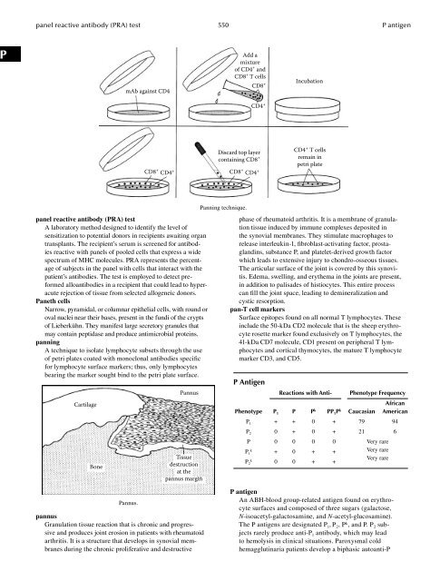
- Page 550 and 551: nonresponder 535 normal lymphocyte
- Page 552 and 553: nuclear matrix protein 537 nutritio
- Page 554 and 555: O125 (ovarian celomic) Nonmucinous
- Page 556 and 557: oncogene 541 one-turn recombination
- Page 558 and 559: oral tolerance 543 Orthoclone OKT®
- Page 560 and 561: Oudin, Jacques (1908-1986) 545 ovar
- Page 562 and 563: owl eye appearance 547 oxygen-depen
- Page 566 and 567: palivizumab (injection) 551 PAP (pe
- Page 568 and 569: papain hydrolysis 553 paracrine fac
- Page 570 and 571: parasite immunity 555 parietal cell
- Page 572 and 573: passive agglutination test 557 pass
- Page 574 and 575: Pasteurella immunity 559 pathogen-a
- Page 576 and 577: PCP 561 pemphigus foliaceus PCP Abb
- Page 578 and 579: penicillin hypersensitivity 563 pep
- Page 580 and 581: Percoll® 565 peripheral lymphoid o
- Page 582 and 583: persistent generalized lymphadenopa
- Page 584 and 585: phagocyte disorders 569 phagocytosi
- Page 586 and 587: phagolysosome 571 phorbol ester(s)
- Page 588 and 589: phytoimmunity 573 pituitary autoant
- Page 590 and 591: plaque-forming cells 575 plasma cel
- Page 592 and 593: plasmid 577 platelet antigens plasm
- Page 594 and 595: PMN 579 POEMS syndrome PMN Abbrevia
- Page 596 and 597: polyagglutination 581 polyclonal ly
- Page 598 and 599: polymeric Ig 583 polymyositis (PM)
- Page 600 and 601: positive induction apoptosis 585 po
- Page 602 and 603: preactivation 587 pre-B cell recept
- Page 604 and 605: precipitation in gel media 589 prec
- Page 606 and 607: Pressman, David 591 primary biliary
- Page 608 and 609: primary tumor 593 private specifici
- Page 610 and 611: progressive transformation of germi
- Page 612 and 613: prostatic acid phosphatase (PAP) 59
- Page 614 and 615:
protein S 599 protoplast protein S
- Page 616 and 617:
pseudoallergy 601 psoriasis vulgari
- Page 618 and 619:
purine nucleoside phosphorylase (PN
- Page 620 and 621:
Q fever An acute disease caused by
- Page 622 and 623:
R5 viruses CCR5-binding HIV strains
- Page 624 and 625:
adioimmunodiffusion test 609 radiol
- Page 626 and 627:
Raji cell assay for CIC 611 rapamyc
- Page 628 and 629:
RB200 613 reactive oxygen species (
- Page 630 and 631:
ecombinant vaccine 615 refractory c
- Page 632 and 633:
elative risk (RR) 617 reptile immun
- Page 634 and 635:
estitope 619 reticulin autoantibodi
- Page 636 and 637:
etrovirus 621 reverse transcriptase
- Page 638 and 639:
hesus blood group system 623 rhesus
- Page 640 and 641:
heumatoid arthritis cell (RA cell)
- Page 642 and 643:
Ricinus communis 627 RNA polymerase
- Page 644 and 645:
Rood, J. J. van 629 Rose-Waaler tes
- Page 646:
RPR (rapid plasma reagin) test 631
- Page 649 and 650:
Sabin-Feldman dye test 634 SAMS loc
- Page 651 and 652:
sarcoma 636 scavenger receptors Ope
- Page 653 and 654:
schlepper 638 scratch test schleppe
- Page 655 and 656:
secondary immune response 640 secre
- Page 657 and 658:
secretory piece 642 selective IgA d
- Page 659 and 660:
self antigen 644 senescent cell ant
- Page 661 and 662:
sequential determinant 646 serology
- Page 663 and 664:
serum sickness 648 severe combined
- Page 665 and 666:
Sézary syndrome 650 Shwartzman (or
- Page 667 and 668:
sia test (historical) 652 side chai
- Page 669 and 670:
signal sequence 654 single cysteine
- Page 671 and 672:
Sips distribution 656 Sjögren’s
- Page 673 and 674:
skin-reactive factor (SRF) 658 slow
- Page 675 and 676:
smooth muscle antibodies (SMAs) 660
- Page 677 and 678:
soluble cytokine receptors 662 spec
- Page 679 and 680:
spectrotype 664 splenic cords spect
- Page 681 and 682:
spontaneous cancer 666 SS-A/Ro in t
- Page 683 and 684:
steric hindrance 668 Streptococcus
- Page 685 and 686:
Strongyloides immunity 670 superant
- Page 687 and 688:
suppressor T cells (Ts cells) 672 S
- Page 689 and 690:
SXY-CIITA regulatory system 674 syn
- Page 691 and 692:
systemic lupus erythematosus (SLE)
- Page 694 and 695:
T1DM Abbreviation for type 1 diabet
- Page 696 and 697:
Tamm-Horsfall glycoprotein (uromodu
- Page 698 and 699:
T cell antigen-specific suppressor
- Page 700 and 701:
T cell receptor (TCR) 685 T cell re
- Page 702 and 703:
T cell tolerance 687 telencephalin
- Page 704 and 705:
terminal deoxynucleotidyl transfera
- Page 706 and 707:
tetanus toxoid 691 Th cells tetanus
- Page 708 and 709:
T h1 cells 693 Theiler’s virus my
- Page 710 and 711:
three-signal model of lymphocyte ac
- Page 712 and 713:
thymic humoral factors (THFs) 697 t
- Page 714 and 715:
thymus 699 thymus Marrow stem cell
- Page 716 and 717:
thymus cell differentiation 701 thy
- Page 718 and 719:
thymus-independent (TI) antigen 703
- Page 720 and 721:
tissue transglutaminase autoantibod
- Page 722 and 723:
T lymphocyte antigen receptor (TCR)
- Page 724 and 725:
tolerosome 709 tonsil Susumu Tonega
- Page 726 and 727:
toxin-1 (TSST-1) 711 TRALI (transfu
- Page 728 and 729:
TRANCE (RANK ligand) 713 transformi
- Page 730 and 731:
transgenes 715 translation chills,
- Page 732 and 733:
T reg cells 717 triple response of
- Page 734 and 735:
tuberculin hypersensitivity 719 tum
- Page 736 and 737:
tumor-infiltrating lymphocytes (TIL
- Page 738 and 739:
tumor necrosis factor (TNF) recepto
- Page 740 and 741:
type I cytokine receptors 725 type
- Page 742 and 743:
type II interferon 727 type III imm
- Page 744:
typhus vaccination 729 tyrosine kin
- Page 747 and 748:
ulcerative colitis (immunologic col
- Page 749 and 750:
US28 734 uveitis Rhus toxicodendron
- Page 751 and 752:
valence 736 varicella valence The n
- Page 753 and 754:
vascular permeability factors 738 V
- Page 755 and 756:
venom 740 V gene Ileum Lumen the fi
- Page 757 and 758:
Videx® 742 viral hemagglutination
- Page 759 and 760:
vitamin B and immunity 744 vitronec
- Page 761 and 762:
Vpr (HIV) 746 V T region of lysis o
- Page 763 and 764:
warm antibody 748 Wegener’s granu
- Page 765 and 766:
Whipple’s disease 750 Winn assay
- Page 767 and 768:
Wu-Kabat plot 752 W, X, Y boxes (MH
- Page 769 and 770:
xenotype 754 X-linked lymphoprolife
- Page 772:
Rosalyn Sussman Yalow. Yalow, Rosal
- Page 775 and 776:
zinc and immunity 760 zinc and immu
- Page 778 and 779:
Appendix I Expression On most cells
- Page 780 and 781:
Appendix I 765 Mouse CD Chart Antig
- Page 782 and 783:
Appendix I 767 Mouse CD Chart Antig
- Page 784 and 785:
Appendix I 769 Mouse CD Chart Antig
- Page 786 and 787:
Appendix I 771 Mouse CD Chart Antig
- Page 788 and 789:
Appendix I 773 Mouse CD Chart Antig
- Page 790 and 791:
Appendix I 775 Mouse CD Chart Antig
- Page 792 and 793:
Appendix I 777 Mouse CD Chart Antig
- Page 794:
Activation unit Appendix II Complem
- Page 797 and 798:
III Appendix III 782 Cytokines and
- Page 800 and 801:
Appendix IV Chemokines and Their Re
- Page 802 and 803:
Appendix V Human Leukocyte Differen
- Page 804 and 805:
Appendix V 789 Human Leukocyte Diff
- Page 806 and 807:
Appendix V 791 Human Leukocyte Diff
- Page 808 and 809:
Appendix V 793 Human Leukocyte Diff
- Page 810 and 811:
Appendix V 795 Human Leukocyte Diff
- Page 812 and 813:
Appendix V 797 Human Leukocyte Diff
- Page 814 and 815:
Appendix V 799 Human Leukocyte Diff
- Page 816:
Appendix V 801 Human Leukocyte Diff
- Page 819:
Editorial Staff Jeanann Lovell Sugg