Array CGH Hybridization Protocol - UCSF Helen Diller Family ...
Array CGH Hybridization Protocol - UCSF Helen Diller Family ...
Array CGH Hybridization Protocol - UCSF Helen Diller Family ...

Create successful ePaper yourself
Turn your PDF publications into a flip-book with our unique Google optimized e-Paper software.
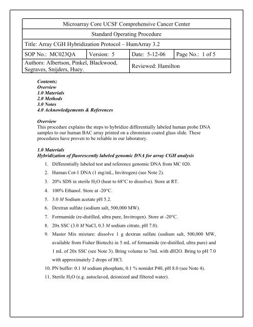
Microarray Core <strong>UCSF</strong> Comprehensive Cancer Center<br />
Standard Operating Procedure<br />
Title: <strong>Array</strong> <strong>CGH</strong> <strong>Hybridization</strong> <strong>Protocol</strong> – Hum<strong>Array</strong> 3.2<br />
SOP No.: MC023QA Version: 5 Date: 5-12-06 Page No.: 1 of 5<br />
Authors: Albertson, Pinkel, Blackwood,<br />
Segraves, Snijders, Huey.<br />
Contents;<br />
Overview<br />
1.0 Materials<br />
2.0 Methods<br />
3.0 Notes<br />
4.0 Acknowledgements & References<br />
Reviewed: Hamilton<br />
Overview<br />
This procedure explains the steps to hybridize differentially labeled human probe DNA<br />
samples to our human BAC array printed on a chromium coated glass slide. These<br />
procedures have proven to be reliable in our laboratory.<br />
1.0 Materials<br />
<strong>Hybridization</strong> of fluorescently labeled genomic DNA for array <strong>CGH</strong> analysis<br />
1. Differentially labeled test and reference genomic DNA from MC 020.<br />
2. Human Cot-1 DNA (1 mg/mL, Invitrogen) (see Note 2).<br />
3. 20% SDS in sterile H2O (heat to 68°C to dissolve). Store at RT.<br />
4. 100% Ethanol. Store at -20°C.<br />
5. 3.0 M Sodium acetate pH 5.2.<br />
6. Dextran sulfate (sodium salt, 500,000 MW).<br />
7. Formamide (re-distilled, ultra pure, Invitrogen). Store at -20°C.<br />
8. 20x SSC (3.0 M NaCl, 0.3 M sodium citrate, pH 7.0).<br />
9. Master Mix mixture: dissolve 1 g dextran sulfate (sodium salt, 500,000 MW,<br />
available from Fisher Biotech) in 5 mL of formamide (re-distilled, ultra pure) and<br />
1 mL of 20x SSC (see Note 3). Bring volume to 7mL with dH2O. Bring to pH 7.0<br />
with approximately 2 drops of HCl.<br />
10. PN buffer: 0.1 M sodium phosphate, 0.1 % nonidet P40, pH 8.0 (see Note 4).<br />
11. Sterile H2O (e.g. autoclaved, deionized and filtered water).
12. UV Stratalinker 2400 (available from Stratagene) capable of producing 130,000 x<br />
100μJoules UV.<br />
13. Very slow rocking table (~ 1 rpm) inside a 37°C incubator (e.g. a VWR brand<br />
Rocker, Model 100).<br />
14. Rubber cement (Ross, American Glue Corporation).<br />
15. Silicone gasket (Press-to-Seal, 2mm thick, #62-6508-24, PGC Scientific).<br />
16. 100% Glycerol.<br />
17. 10x PBS: 1.4 M NaCl, 0.027 M KCl, 0.1 M Na2HPO4, 0.018 M KH2PO4, adjusted<br />
to pH 7.4 with HCl.<br />
18. Stereomicroscope.<br />
19. 10 mL Syringe.<br />
20. 200 μL Disposable pipet tip without a filter.<br />
21. Binder clips, medium size.<br />
2.0 Methods<br />
<strong>Hybridization</strong> of fluorescently labeled genomic DNA for array <strong>CGH</strong> analysis<br />
1. Preparation of array for hybridization:<br />
a. Expose a printed array to 260,000 μJoules (2,600 x 100uJoules) of UV by<br />
using a Stratalinker (see Note 5).<br />
b. Fill a 10 mL syringe with rubber cement and fit a 200 μL pipet tip on the<br />
syringe outlet. You may have to cut 1-2mm off the wide end of the pipet<br />
tip for it to fit well. Apply a rubber cement ring around each array on the<br />
slide using a stereomicroscope to observe the area of the array. Air-dry the<br />
rubber cement and apply a second thick layer of rubber cement on top of<br />
the first layer. Air-dry the rubber cement.<br />
2. Preparation of samples for hybridization:<br />
a. Combine 30 μL labeled test genomic DNA, 30 μL labeled reference<br />
genomic DNA, (see MC 022QA) and 75 μg of human Cot-1 DNA. The<br />
volume of Cot1 will depend on the Cot1 concentration. Precipitate the<br />
DNA sample mixture by adding 2.5 volumes of ice-cold 100% ethanol and<br />
0.1 volume of 3 M sodium acetate (pH 5.2). Vortex the solution briefly but
thoroughly and collect the precipitate by centrifugation at 14,000 rpm for<br />
45 minutes at 4°C.<br />
b. Carefully aspirate and discard the supernatant. Wipe the excess liquid<br />
from the tube with a chem-wipe paper tissue, being careful not to disturb<br />
the pellet. Air-dry the pellet for approximately 5-10 minutes. Dissolve the<br />
pellet in 7μL dH2O, 14μL 20% SDS, and 49 μL Master mix mixture (for<br />
Master Mix preparation see Note 3). The pellet may need to incubate at<br />
room temperature on the bench for an hour to completely re-suspend.<br />
3. Denature the DNA sample solution from step 2.2.b in a water bath at 73°C for 13<br />
minutes and then incubate at 37°C for 60 to 120 minutes to allow the Cot1 DNA<br />
to anneal to repetitive sequences on both the sample and reference DNA.<br />
4. Place <strong>Array</strong> on a heat block set at 37°C for 5 minutes to warm array.<br />
5. Apply the combined sample/reference hybridization solution onto the array (see<br />
Note 7). Keep sample/reference hybridization solution at 37°C until just before<br />
application to the array to reduce non-specific binding of the probe to the array<br />
surface. Place a silicon gasket around the edge of the slide and lay a clean glass<br />
slide on top, aligning the edges with the gasket. Clamp the assembly together<br />
using binder clips. Incubate the array for 48 to 68 hours at 37°C on a slowly<br />
rocking table (~1rpm).<br />
6. Disassemble the slide assembly and rinse the hybridization solution from the slide<br />
under a stream of PN buffer. It is preferred to leave the rubber cement on the<br />
array at this time, as it will not affect the rinsing steps that follow.<br />
7. Wash the slides once in 50% formamide, 2x SSC, pH7 for 15 minutes at 45°C,<br />
followed by a 15 minute wash in PN buffer at room temperature. The washes can<br />
conveniently be done in slide staining jars (coplin jars) placed in water baths.<br />
8. At the bench, carefully remove the rubber cement with forceps, while keeping the<br />
array moist with PN buffer.<br />
9. Mount the slide in a DAPI solution to stain the array spots (90% glycerol, 10%<br />
PBS, 1 μM DAPI).<br />
10. <strong>Array</strong>s are ready for imaging (see Note 7).
3.0 Notes<br />
1. DNA concentrations should be determined using a Fluorometer, rather than a<br />
spectrophotometer to obtain more accurate measurements.<br />
2. The DNA concentration of each new lot of Cot-1 DNA should be determined<br />
using a Fluorometer and should measure 500 ng/μL or greater.<br />
3. Distribute 1 g of dextran sulfate over the entire length of a 15 mL tube. While<br />
holding the tube horizontally, squirt in 5 mL of formamide. Close the tube and<br />
shake vigorously for 30 seconds. Add 1 mL of 20x SSC and shake vigorously for<br />
30 seconds. Add 1 mL of dH2O and mix thoroughly. Bring pH to 7.0 using<br />
approximately 2 drops of HCl. Dissolve overnight at room temperature. The final<br />
volume should be approximately 7 mL.<br />
4. Prepare approximately 16 L of 0.1 M Na2HPO4, 0.1% nonidet P40 pH9. While<br />
continuously measuring the pH, adjust the pH to 8.0 with 0.1 M NaH2PO4, 0.1 %<br />
nonidet P40 (approximately 1 L). Make sure not to go below pH 8.0. Store at<br />
room temperature.<br />
5. Place the slide(s) in the Stratalinker, arrays facing up. The arrays should be given<br />
a fixed amount of energy (260,000 μJ or 2,600 x 100 μJ) instead of other<br />
available options the Stratalinker might have, such as autocrosslink or time. Overcrosslinking<br />
the slide might result in a decrease in fluorescent hybridization<br />
signal.<br />
6. Place about 10 μL of the hybridization solution in each corner of the array, and<br />
the remaining 30 μL in the center. Tilt slide until entire surface of array is wet<br />
with hybridization solution. Remove any air bubbles with needle or other sharp<br />
object.<br />
7. Images can be acquired using commercially available CCD or laser scanning<br />
imaging systems (e.g. an Axon scanner 4000B). Image analysis and quantification<br />
can be done using commercially available image analysis programs (e.g. GenePix<br />
from Axon) or the <strong>UCSF</strong> program Spot.<br />
4.0 Acknowledgements & References<br />
1. R. Segraves, A. Snijders, and B. Huey were instrumental in preparing and<br />
proof reading this protocol.
2. Snijders, A., Segraves, R., Blackwood, S., Pinkel, D., Albertson, D., (2001) BAC<br />
Microarray-based Comparative Genomic <strong>Hybridization</strong>, Comprehensive Cancer<br />
Center, Cancer Research Institute and Department of Laboratory Medicine, <strong>UCSF</strong>,<br />
San Francisco.