Ph.D. Thesis - Physics
Ph.D. Thesis - Physics
Ph.D. Thesis - Physics

Create successful ePaper yourself
Turn your PDF publications into a flip-book with our unique Google optimized e-Paper software.
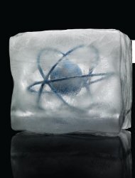
Figure 7-24: Images of ion crystals of two and four ions in Uraniborg 2. The principal axes<br />
ˆx and ˆy of the trap are shown by depicting the corresponding ion-ion spacings dx and dy in<br />
each figure.<br />
way, and this wire forms a part of the resonant circuit that drives the trap. Therefore,<br />
some voltage differential is expected. In some papers, the rf voltage is treated as a fit<br />
parameter since it is difficult to directly measure [SCR + 06, CLBC09]. However, in this<br />
case the frequencies are not off by a constant factor, so this is not a completely satisfactory<br />
explanation. It is possible that some combination of the two effects explains the discrepancy:<br />
the dc fields at the ion location are unknown, as is the magnitude of the rf field.<br />
A more interesting question is how well the ion crystal structures match the theory for<br />
the measured frequencies, which we explore next.<br />
7.5.3 Ion crystal structure<br />
Ion crystals consisting of between one and four ions were observed in Uraniborg 2. To<br />
obtain these images, it was necessary to readjust the imaging optics repeatedly to reduce<br />
aberrations, to set the dc voltages carefully, and to carefully coalign and focus the detection<br />
lasers. Without these conditions being met, somewhat misshapen crystals of two ions (but<br />
no more) were observed. Unfortunately, it was difficult to cool more than four ions into a<br />
crystalline state in this trap.<br />
Although ion crystals were observed, the signal was fairly weak and long exposure times<br />
of up to 5 s were required, using the HIGH resolution of the SBIG camera, in which each<br />
individual pixel is displayed (no binning). Images of crystals are plotted in Fig. 7-24.<br />
Images were analyzed as follows: the centers of each ion were determined from a Gaus-<br />
sian fit of the intensity. The magnification was calibrated to the spacing of two ions. The<br />
image of four ions was processed in the same way, using this magnification. Errors on the<br />
spacing of ions were determined from the standard deviation on the centers of the ions. For<br />
two ions, the calculated spacing is 16.5 µm, and the spacing between ions is 11±3 pixels,<br />
giving a magnification of 4.5, or 1.5 µm per pixel.<br />
For four ions, we calculate the spacing between the ions along the ˆx and ˆy directions.<br />
182