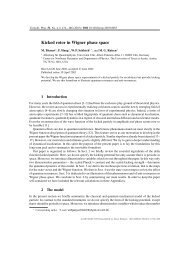
Ph.D. Thesis - Physics
Ph.D. Thesis - Physics
Ph.D. Thesis - Physics

You also want an ePaper? Increase the reach of your titles
YUMPU automatically turns print PDFs into web optimized ePapers that Google loves.
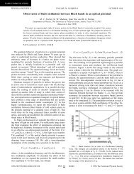
Figure 6-13: A diagram of the setup showing the position and orientation of the ablation<br />
target relative to the ion trap. The surface of the ablation target is approximately 25 mm<br />
from the trap center and is orthogonal to the direction to the ion trap. Not to scale.<br />
Attractive as laser ablation loading is, there is much that is not well-understood about<br />
it, especially on a practical level. This motivates the following research questions:<br />
1. How shallow a trap may be loaded with direct laser ablation, and how does that value<br />
compare to other techniques, including electron impact ionization and photoioniza-<br />
tion?<br />
2. How does the loading efficiency depend upon the composition of the material that is<br />
ablated, and upon the ablation laser power?<br />
3. How does the number of ions loaded depend on the ablation laser power?<br />
4. Is the buildup of stray charge an issue when using this method, especially with a<br />
surface-electrode trap?<br />
In the remainder of this section we present an experimental study of the ablation loading<br />
of ion traps, focusing on our experimental setup and results, with a goal of evaluating the<br />
utility of the technique for loading ion traps in view of the above questions.<br />
6.5.1 Experimental setup<br />
The trap is driven with an rf voltage with amplitude 200-600 V at 8 MHz. The dc voltages,<br />
not discussed here in detail, are chosen to provide sufficient ˆz confinement (with depth at<br />
least equal to that in the ˆx and ˆy directions) and rough compensation. The ablation laser<br />
is a frequency-tripled pulsed Nd:YAG laser (Continuum Minilite) at 355 nm. It produces<br />
pulses from 1-10 mJ at a duration of 4 ns. The 422 nm and 1092 nm lasers are directed in<br />
a direction along the ˆz and ˆx directions, while the ablation laser is along ˆz; this is depicted<br />
in Fig. 6-13. As in the last section, a CCD camera and PMT are used for ion detection.<br />
145