Ph.D. Thesis - Physics
Ph.D. Thesis - Physics
Ph.D. Thesis - Physics

Create successful ePaper yourself
Turn your PDF publications into a flip-book with our unique Google optimized e-Paper software.
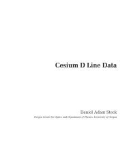
Figure 6-7: <strong>Ph</strong>otograph of the experimental setup for measurements on the San Quentin<br />
trap. The spherical octagon vacuum chamber with 4 1/2 in. CF viewport is visible top<br />
center, and the top plate PCB is seen within it. Above it is the 2 in. diameter lens tube.<br />
Connection of the rf signal is top left, while connections to the dc electrodes and the e-gun<br />
are on the small box (bottom right). Connection to the oven is top right. The 422 and<br />
1092 nm lasers are collimated by Thorlabs collimation packages, are coaligned on a dichroic<br />
mirror, and are then sent through the 1 1/3 in. CF viewport to the trap. Vacuum pumps<br />
and the leak valve are outside the frame of this photograph.<br />
(PI) loading, but at the same time species-specific loading is not possible, and “dark ions,”<br />
particles other than the desired one that are trapped but do not fluoresce, are quite com-<br />
mon. The key disadvantage of the technique for surface-electrode traps became evident in<br />
our work; the e-gun deposits a stray electric charge on the exposed dielectric that is an<br />
unavoidable part of the PCB trap. The stray fields resulting from this trapped charge can<br />
become high enough to prevent any trapping in UHV conditions.<br />
Our vacuum system includes a leak valve for introducing helium buffer gas. The colli-<br />
sional cooling provided by the helium allows large samples (100’s of ions) to be trapped and<br />
cooled even without stray field compensation or optimal alignment of the cooling lasers. In<br />
this trap, a current in the oven of 8 A and a voltage across the filament of about 3.5 V was<br />
used. The filament was also biased at -20 V with respect to ground to increase the flux of<br />
emitted electrons. A photograph of our experimental setup is shown in Fig. 6-7.<br />
Trapped ions were detected using both an electron-multiplying CCD camera (Princeton<br />
Instruments <strong>Ph</strong>otonMax) and a photomultiplier tube (Hamamatsu H6780-04). The 422 nm<br />
and 1092 nm lasers used for inducing fluorescence were locked to low-finesse cavities. Typical<br />
laser powers in this experiment were 1.2 mW of 1092 and 20-50 µW of 422. The width of<br />
the 1092 was about 1 mm, so that the entire trap region would be illuminated by it, while<br />
the 422 was focused to a 60 µm spot. The smaller spot size is useful in our measurement<br />
of the position of the ion cloud. Ions were loaded prior to nulling the stray electric fields<br />
138