File #1 to be Downloaded - UCSF Radiation Oncology
File #1 to be Downloaded - UCSF Radiation Oncology
File #1 to be Downloaded - UCSF Radiation Oncology

You also want an ePaper? Increase the reach of your titles
YUMPU automatically turns print PDFs into web optimized ePapers that Google loves.
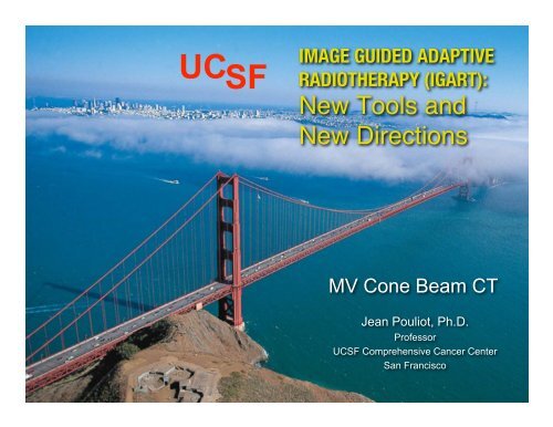
UC SF<br />
IMAGE GUIDED ADAPTIVE<br />
RADIOTHERAPY (IGART):<br />
New Tools and<br />
New Directions<br />
MV Cone Beam CT<br />
Jean Pouliot, Ph.D.<br />
Professor<br />
<strong>UCSF</strong> Comprehensive Cancer Center<br />
San Francisco
IMAGE GUIDED ADAPTIVE<br />
RADIOTHERAPY (IGART):<br />
New Tools and<br />
New Directions<br />
Objectives:<br />
• Basic principles of MV CBCT<br />
• Workflow of IGRT with MV CBCT<br />
• Range of applications of MV CBCT<br />
– Patient positioning<br />
– moni<strong>to</strong>ring of ana<strong>to</strong>mical changes, tumor tracking<br />
– planning purposes and in-situ dose calculation<br />
– Tomosynthesis<br />
• Future possibilities of MV CBCT for Dose-Guided<br />
<strong>Radiation</strong> Therapy (DGRT).
<strong>UCSF</strong><br />
• Michelle Aubin<br />
• Jean-Francois Aubry<br />
• Kara Bucci<br />
• Al<strong>be</strong>rt Chan<br />
• Josephine Chen<br />
• Hong Chen<br />
• Cynthia Chuang<br />
• Martina Descovitcz<br />
• Bruce Faddegon<br />
• Amy Gillis<br />
• Olivier Morin<br />
• Mack Roach III<br />
• Joycelyn Speight<br />
• Lynn Verhey<br />
• Ping Xia<br />
Main Collabora<strong>to</strong>rs<br />
Siemens<br />
• Ali Bani-Hashemi<br />
• Fahard Ghelmansarai<br />
• Paco Hernandez<br />
• Dimitre Histrov<br />
• Bijumon Gangadharan<br />
• Matthias Mitschke<br />
This work is supported<br />
by<br />
Siemens O.C.S.
Basic Principles of MV CBCT<br />
Fan <strong>be</strong>am CT Cone-<strong>be</strong>am CT<br />
1 slice per rotation Entire volume in 1 rotation
Basic Principles of MV CBCT<br />
• MV CBCT generates a 3D<br />
image of the patient ana<strong>to</strong>my<br />
from the same x-ray <strong>be</strong>am<br />
(6MV) used for treatment.<br />
• Image and x-ray <strong>be</strong>am share<br />
the same isocenter.<br />
• Patient 3D ana<strong>to</strong>my in<br />
treatment position, moments<br />
<strong>be</strong>fore dose delivery.
MV CBCT: Main Features<br />
• Very low dose-rate linac <strong>be</strong>am (0.005 M.U. per degree)<br />
• Beam Pulse Triggered Acquisition Mode<br />
(Synchronized pulse-panel readout)<br />
• High sensitivity a-Si Panel EPID (Optimized for MV)<br />
• Integrated workstation: EPID deployment, image<br />
acquisition, reconstruction, fusion with CT, patient<br />
alignment, treatment delivery
Basic Characteristics<br />
of MV CBCT<br />
• Half rotation: 200 degrees<br />
• Acquisition ~ 45 seconds<br />
• Acquisition + Reconstruction < 2 min.<br />
• 27 cm x 27 cm x 27 cm Field of View<br />
• Volume of 256 x 256 x 270<br />
• Pixel size (0.5 mm)3<br />
• Typical dose: 2 <strong>to</strong> 8 cGy<br />
• Accurate Electron Density
MV CBCT
MV CBCT: Post processing<br />
Diffusion filter reduces<br />
noise and maintains edges
Future Beam Line for MV CBCT: LowZ tgt + No FF + 4.5 MV<br />
Current BL Future BL Current BL Future BL<br />
Courtesy of Bruce Faddegon, <strong>UCSF</strong>
Workflow of IGRT with MV CBCT<br />
CT Dose Plan Patient alignment Dose Delivery<br />
1: Image<br />
Acquisition<br />
2: Image<br />
Reconstruction<br />
S<br />
!<br />
o<br />
r<br />
F<br />
X (voxel)<br />
x (pixel)<br />
3: Image<br />
Registration<br />
4: Patient<br />
Alignment<br />
X-offset<br />
Y-offset<br />
Z-offset
Workflow of IGRT with MV CBCT<br />
• 1) The patient and the cone<strong>be</strong>am<br />
acquisition mode are<br />
selected at the treatment<br />
console.<br />
• 2) The linac gantry is placed<br />
in starting position, namely<br />
270 degrees.
Workflow of IGRT with MV CBCT<br />
• 3) During the acquisition, the<br />
gantry rotates 200 degrees until<br />
it reaches its final position, 110<br />
degrees.<br />
During the rotation, a portal<br />
image is acquired at each<br />
degree. The reconstruction of<br />
the cone-<strong>be</strong>am image starts<br />
immediately after the first portal<br />
image has <strong>be</strong>en acquired.
Workflow of IGRT with MV CBCT<br />
• 3) During the acquisition, the<br />
gantry rotates 200 degrees until it<br />
reaches its final position, 110<br />
degrees.<br />
During the rotation, a portal<br />
image is acquired at each<br />
degree. The reconstruction of the<br />
cone-<strong>be</strong>am image starts<br />
immediately after the first portal<br />
image has <strong>be</strong>en acquired.
Workflow of IGRT with MV CBCT<br />
• 4) Upon completion of the<br />
reconstruction image, the<br />
cone-<strong>be</strong>am image is<br />
au<strong>to</strong>matically loaded in the<br />
Adaptive Targeting<br />
Sofware TM , and the CB <strong>to</strong><br />
CT image registration is<br />
performed au<strong>to</strong>matically in<br />
few seconds using a mutual<br />
information algorithm.
MV CBCT - CT Registration
Workflow of IGRT with MV CBCT<br />
• 5) Proper alignment is<br />
validated and manual<br />
alignment can <strong>be</strong> performed<br />
when fine-tuning is required.<br />
• 6) The couch translation offset<br />
values required <strong>to</strong> obtain<br />
the <strong>be</strong>st alignment of the<br />
patient at isocenter are<br />
displayed. The couch is<br />
moved remotely from the<br />
treatment console according<br />
<strong>to</strong> these values and the<br />
patient is ready for treatment.
Setup Methods<br />
1. Conventional CT 2. Treatment Planning System<br />
CT<br />
2D method: DRR with EPI<br />
DRR<br />
3. Patient Setup<br />
CT, Points & Con<strong>to</strong>urs<br />
3D method: CT with MV CBCT
y = 1.00x + 0.00<br />
R 2 = 0.99<br />
Phan<strong>to</strong>m Study<br />
3 em<strong>be</strong>dded gold seeds imaged at different locations:<br />
MVCBCT Shift (cm)<br />
Alignment of 3 Gold Seeds<br />
2.00<br />
1.50<br />
1.00<br />
0.50<br />
0.00<br />
-2.00 -1.50 -1.00 -0.50 0.00 0.50 1.00 1.50 2.00<br />
-0.50<br />
IMAGE GUIDED ADAPTIVE<br />
RADIOTHERAPY (IGART):<br />
New Tools and<br />
New Directions<br />
-1.00<br />
-1.50<br />
-2.00<br />
EPI Shift (cm)
Dose Delivered<br />
2 - 8 cGy @ isocenter<br />
3-5 cGy : daily alignment<br />
6-8 cGy : planning purpose
Dose Delivered
Compensation of dose delivered<br />
Tx 96% * Tx + 40 daily 10 MU MV CBCT
Compensation of dose delivered<br />
These DVHs demonstrate the possibility <strong>to</strong> nearly eliminate the<br />
extra dose delivered <strong>to</strong> the patient for daily MV CBCT imaging.
Clinical applications of MV CBCT<br />
• Patient positioning<br />
• Head & Neck<br />
• Lung<br />
• Spine<br />
• Chest<br />
• Prostate<br />
• Moni<strong>to</strong>ring of ana<strong>to</strong>mical changes<br />
• Target delineation with non-compatible CT objects<br />
• Dose calculation from MV CBCT <strong>to</strong> assess dosimetrical<br />
impact (DGRT1)<br />
• Dose-Guided <strong>Radiation</strong> Therapy (DGRT2)
Patient Setup and Prostate Alignment<br />
3D Approach
IMAGE GUIDED ADAPTIVE<br />
RADIOTHERAPY (IGART):<br />
New Tools and<br />
New Directions<br />
Daily Prostate Alignment<br />
EPID + Markers
Prostate Alignment with MV CBCT<br />
Reference CT MV CBCT 50% Blend CT-MV CBCT<br />
Provides additional information over EPID+markers:<br />
- Rectum, bladder, etc.<br />
- Prostate con<strong>to</strong>urs
Patient Setup: Prostate Bed<br />
Irradiation of postprostatec<strong>to</strong>my patient<br />
Markers are<br />
distinguished from<br />
surgical clips for daily<br />
alignment
The presence of a hip prosthesis often makes the<br />
markers difficult <strong>to</strong> see on the lat EPI.<br />
LAT<br />
Patient Setup: Prostate<br />
AP<br />
MV CBCT could <strong>be</strong> used for marker detection and<br />
prostate alignment with less dose than the regular<br />
2D approach.<br />
MV CBCT with only<br />
1.8 M.U. (1.3 cGy)
Hip Prosthesis<br />
In 2005 in US and Europe,<br />
more than 500,000 people had a hip replaced.<br />
IMAGE GUIDED ADAPTIVE<br />
RADIOTHERAPY (IGART):<br />
New Tools and<br />
New Directions<br />
MV CBCT
Patient setup: Head & Neck<br />
6 mm
Moni<strong>to</strong>r Weight Loss
Day 1<br />
Moni<strong>to</strong>r Weight Loss<br />
3D rendering of CT 3D rendering of MV CBCT<br />
Day 23
Aligned with MV<br />
CBCT<br />
Lung<br />
Aligned<br />
according <strong>to</strong> EPI
Paraspinous Tumors<br />
• Surgery + supporting hardware + post-op IMRT<br />
• Spinal cord <strong>to</strong>lerance limits Dx (palliative)<br />
• Image hardware artifact<br />
– impairs target delineation<br />
– hinders treatment verification<br />
• MV CBCT -> Target definition<br />
-> Daily 3D patient alignment<br />
-> Improved confidence Dx (curative)
Treatment of Paraspinous Tumors<br />
in the Presence of Orthopedic Hardware
Paraspinous Tumors: Patient Setup<br />
Case Report<br />
Mean magnitude of setup errors:<br />
- Lateral: 3.6 mm<br />
(95% C.I., 2.6-4.6 mm)<br />
- Longitudinal: 4.1 mm<br />
(95% C.I., 3.2-5.0 mm)<br />
- Vertical: 1.0 mm<br />
(95% C.I., 0.6-1.3 mm)
Paraspinous Tumors: Dosimetric Impact<br />
4.1 mm<br />
Dose<br />
1.0 mm<br />
3.6 mm<br />
Without daily corrections:<br />
(simulated in TPS)<br />
Maximum dose (D 0.1cc ) <strong>to</strong> Spinal cord<br />
would have increased from 51 <strong>to</strong> 61 Gy.<br />
The CTV coverage decreased by 5 Gy.<br />
Daily setup errors detected via MV CBCT were critical for both protection of<br />
spinal cord <strong>to</strong>lerance and maintenance of CTV coverage.
Dose Calculation using MV CBCT
Dose Calculation using MV Cone-Beam CT
Dosimetrical Impact of Weight Loss<br />
Week 1<br />
IMAGE GUIDED ADAPTIVE<br />
RADIOTHERAPY (IGART):<br />
New Tools and<br />
New Directions<br />
Week 3<br />
Differences in dose<br />
distribution due <strong>to</strong><br />
ana<strong>to</strong>mical changes<br />
Δ(%) >5%<br />
>10%
Dose-Guided <strong>Radiation</strong> Therapy<br />
(DGRT)<br />
DGRT is an extension of adaptive radiation therapy where<br />
dosimetric considerations constitute the basis of<br />
treatment modification<br />
• Improved confidence that the dose is delivered as<br />
planned<br />
• Allow more aggressive and definitve treatment<br />
(dose escalation)<br />
• Better understanding of the outcome of treatments
Dose Plan<br />
In-vivo<br />
Dosimetry<br />
Dose-Guided <strong>Radiation</strong> Therapy<br />
In-situ<br />
Dose<br />
Calculation<br />
(DGRT1)<br />
In-situ<br />
Exit<br />
Dosimetry<br />
In-situ<br />
Dose<br />
Reconstruction<br />
(DGRT2)
MV CBCT: Summary<br />
• Provide 3D ana<strong>to</strong>my of patient in treatment position<br />
-> Patient Setup, Moni<strong>to</strong>ring of ana<strong>to</strong>mical changes<br />
• Accurate electron density for dose calculation<br />
-> Assess dosimetric Impact<br />
• Facilitate dose reconstruction for DGRT<br />
• Specialized imaging <strong>be</strong>am line and MV adapted Flat<br />
panel EPID will further improve image quality
• MV Cone-Beam CT:<br />
Thank You<br />
– Pouliot et al., IJROBP, 61(2); 238-246, 2005<br />
– Morin et al., Med. Dosi., 31(1);51-61, 2006<br />
– Pouliot et al., Cancer et Radiothérapie, June 2006<br />
• Exit Dosimetry with EPID:<br />
– Chen et al., Med. Phys., 33(3); 584-594, 2006<br />
• Dose Calculation with MV CBCT:<br />
– Morin et al., submitted <strong>to</strong> IJROBP, 2006<br />
• DGRT with MV CBCT:<br />
– Chen et al., Brit. J. Radiol, June 2006<br />
• Use of MV CBCT <strong>to</strong> complement CT for target definition in pelvic radiotherapy<br />
in the presence of hip replacement:<br />
– Aubin et al., submitted <strong>to</strong> Brit. J. Radiol., 2006<br />
• IGRT using MV CBCT for Treatment of Paraspinous Tumors<br />
in the Presence of Orthopedic Hardware<br />
– Hansen et al., submitted <strong>to</strong> IJROBP, 2006