Advances in Brachytherapy and IORT - UCSF Radiation Oncology
Advances in Brachytherapy and IORT - UCSF Radiation Oncology
Advances in Brachytherapy and IORT - UCSF Radiation Oncology

You also want an ePaper? Increase the reach of your titles
YUMPU automatically turns print PDFs into web optimized ePapers that Google loves.
<strong>Advances</strong> <strong>in</strong><br />
<strong>Brachytherapy</strong> <strong>and</strong><br />
<strong>IORT</strong><br />
Physics Residents Talks
Overview<br />
Change <strong>in</strong> brachytherapy from orig<strong>in</strong>s to<br />
present<br />
Sources / Equipment<br />
Plann<strong>in</strong>g<br />
<strong>IORT</strong>
<strong>Radiation</strong> Sources<br />
Isotopes used<br />
Radium / Radon<br />
Cesium-137<br />
Iridium-192<br />
Iod<strong>in</strong>e-125<br />
Palladium-103
Radium / Radon<br />
Part of U-238 decay<br />
226 Ra 222 Rn 218 Po<br />
206 Pb<br />
1622 y, α<br />
210 Po<br />
3.8 days, α<br />
Radium/Radon <strong>in</strong> equilibrium<br />
Shield<strong>in</strong>g needed to remove particles<br />
1 mg ~ 1 mCi<br />
210 Bi<br />
214 Pb<br />
210 Pb<br />
214 Bi+β - +γ<br />
214 Po+β - +γ
Cesium-137<br />
Product of U-235 fission<br />
Decays to Ba-137<br />
Long half-life (30 yrs)<br />
β - , 8%<br />
1.17 MeV<br />
137 Cs 30yr<br />
β - , 92%<br />
0.51 MeV<br />
137 Ba<br />
0.662 MeV
Cesium-137<br />
Can substitute for radium<br />
Easier to shield than radium<br />
Used <strong>in</strong> the form of tubes <strong>and</strong> needles<br />
Also used for manual <strong>and</strong> remote<br />
afterload<strong>in</strong>g
Iridium-192<br />
Produced by neutron<br />
bombardment of Ir-<br />
191<br />
Source is <strong>in</strong> the form<br />
of iridium/plat<strong>in</strong>um<br />
core with th<strong>in</strong> (~0.1-<br />
0.2 mm) cladd<strong>in</strong>g.<br />
Available as wire,<br />
hairp<strong>in</strong>, or seed<br />
192 Ir<br />
74 days<br />
β - (0.08-0.7<br />
MeV)<br />
192 Pt<br />
0.2-1.06 MeV<br />
photons<br />
(E eff =.37 MeV)
Cobalt-60<br />
Produced by neutron activation of Co-59<br />
Used for HDR <strong>and</strong> teletherapy units<br />
Half-life: 5.27 years<br />
Emission: Decays to Ni-60 via β- decay<br />
followed by 1.17 MeV <strong>and</strong> 1.33 MeV<br />
photons.
Permanent Implant Isotopes<br />
Radon-222<br />
Iod<strong>in</strong>e-125<br />
Gold-198<br />
Palladium-103
Iod<strong>in</strong>e-125<br />
Half-life: 59.4 days<br />
Decay: I-125 decays by EC to Te-125<br />
Emissions: characteristic x-rays with mean<br />
energy ~27 keV.
Palladium-103<br />
Half-life: 17 days<br />
Decay: EC to excited states of ruthenium-<br />
103<br />
Emissions: <strong>in</strong>ternal conversion result <strong>in</strong><br />
characteristic x-rays with mean energy of<br />
21 keV.
Gold-198<br />
Half-life: 2.7 days.<br />
Emissions: beta decay with 0.41 MeV<br />
photons.<br />
Can be used as radon substitute for<br />
implants. 1 mCi gold = 0.284 mg Ra<br />
Used <strong>in</strong> solutions <strong>in</strong> body cavities.
Desirable Properties<br />
Half-life: Long t 1/2 = lower specific activity,<br />
but less frequent source replacement. Not<br />
desirable for permanent implants.<br />
Solid (not gas) w/ solid decay products<br />
Emissions<br />
High energy: shield<strong>in</strong>g issues<br />
Low energy: tissue attenuation may become<br />
important<br />
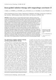
Other
Electronic <strong>Brachytherapy</strong> Source<br />
50 kV, 0.3 mA, 15 W<br />
Water cooled<br />
Can be used like HDR<br />
source<br />
Rivard et al, Med Phys.<br />
33(11), 2006.
Afterload<strong>in</strong>g<br />
First used for radiation<br />
safety purposes<br />
Manual vs. Remote<br />
Remote<br />
LDR: 12 Gy/hr
Remote Afterloaders<br />
Applicator<br />
Afterloader
Other afterloader designs<br />
Afterloader sorts<br />
spherical Cs-137 <strong>and</strong><br />
spacers to create<br />
“source tra<strong>in</strong>s”
Applicators<br />
Used to hold radioactive sources <strong>in</strong> the<br />
correct position<br />
Can be used with various afterload<strong>in</strong>g<br />
techniques (LDR, HDR, manual)
Applicators<br />
Fletcher-Suit<br />
Vag<strong>in</strong>al Cyl<strong>in</strong>ders<br />
Intralum<strong>in</strong>al Catheters<br />
Interstitial Implants
MammoSite<br />
Used for Accelerated Partial Breast Irradiation<br />
Fluid filled balloon placed dur<strong>in</strong>g surgery<br />
Attached to HDR afterloader (e.g., Nucletron) with<br />
Ir-192 source<br />
Patient treated <strong>in</strong> 5 fx.<br />
http://www.mammosite.com
GliaSite<br />
Used to treat bra<strong>in</strong> tumors<br />
Balloon filled with I-125<br />
conta<strong>in</strong><strong>in</strong>g solution<br />
Example: used to treat<br />
glioblastoma multiformae<br />
to 50 Gy followed by EBRT<br />
boost
Source Strength / Dose Specification<br />
Radium equivalent (mg-Ra)<br />
Activity (Ci)<br />
Exposure rate (R/hr)<br />
Air kerma strength (Gy/hr/m 2 )
Traditional Dose Calculation<br />
Dose calculation approximated source as a po<strong>in</strong>t<br />
A app =apparent activity<br />
f med = exposure to dose conversion<br />
(Γ δ ) x =exposure rate constant for radionuclide<br />
T(r) is tissue attenuation<br />
φ an is anisotropy factor
TG-43 formalism<br />
S k: Air kerma strength<br />
Λ: Dose rate constant<br />
G L(r, θ): geometric<br />
function<br />
g L(r): radial dose<br />
function<br />
F(r, θ): anisotropy<br />
function
Plann<strong>in</strong>g Systems<br />
Paterson-Parker / Manchester (1934)<br />
Quimby (1944)<br />
Paris (1966)
Manchester system<br />
Surface applicators<br />
Planar implants<br />
Volume implants<br />
Uter<strong>in</strong>e implants
Surface Applicators
Surface Applicator Distributions
Planar Implants<br />
Used for tumors too deep for<br />
surface applicators<br />
Insert radium needles beneath sk<strong>in</strong>
Volume Implants<br />
Volume implants used<br />
when target can’t be<br />
covered by two<br />
planes<br />
Rules are given for<br />
various shapes
Uter<strong>in</strong>e Implants<br />
Treatment of cervical <strong>and</strong><br />
other gynecological cancers<br />
Treat cervix, vag<strong>in</strong>a, or<br />
uterus
Uter<strong>in</strong>e Implants<br />
Dose at Po<strong>in</strong>t A represents<br />
limit<strong>in</strong>g dose<br />
Dose to Po<strong>in</strong>t B represents<br />
dose to pelvic lymph nodes
Uter<strong>in</strong>e implants<br />
Bladder def<strong>in</strong>ed<br />
by balloon of<br />
Foley catheter<br />
Rectal reference<br />
po<strong>in</strong>t is 0.5 cm fro<br />
posterior vag<strong>in</strong>al<br />
wall
Imag<strong>in</strong>g<br />
Reconstruction us<strong>in</strong>g orthogonal<br />
radiographs<br />
3D imag<strong>in</strong>g allows more sophisticated<br />
plann<strong>in</strong>g<br />
Functional imag<strong>in</strong>g (MRSI, PET, SPECT)<br />
can also be used
Other techniques<br />
Intravascular<br />
Radio-immunotherapy<br />
Intraoperative radiation therapy
Intravascular <strong>Brachytherapy</strong><br />
Coronary artery disease caused<br />
by occlusion of cardiac vessels<br />
IVB used to prevent re-stenosis<br />
after angioplasty<br />
<strong>Radiation</strong> delivered either with<br />
temporary implant or radioactive<br />
stent
IVBT<br />
Lum<strong>in</strong>al diameter:<br />
3-5 mm<br />
Target volume: 2-3<br />
cm length , 2 mm<br />
from radial center<br />
Mostly replaced by<br />
drug releas<strong>in</strong>g<br />
stents
Radioimmunotherapy<br />
Use radiolabeled antibody targeted to tumor<br />
cells
Intraoperative <strong>Radiation</strong> Therapy<br />
Started <strong>in</strong> the early 1900’s.<br />
Attractive for deep tumors because the sk<strong>in</strong><br />
dose was limit<strong>in</strong>g prior to the <strong>in</strong>vention of<br />
megavoltage accelerators.<br />
Rega<strong>in</strong>ed popularity <strong>in</strong> Japan <strong>in</strong> 1970’s for<br />
treatment of gastric cancer.<br />
Applications <strong>in</strong>clude: retroperitoneal<br />
sarcoma, pancreatic cancer, rectal cancer.
<strong>IORT</strong> Methods<br />
L<strong>in</strong>ac<br />
Dedicated systems<br />
IntraOp Mobetron<br />
Hitesys Novac7<br />
HDR based<br />
Intrabeam
IntraOp Mobetron<br />
X-b<strong>and</strong> l<strong>in</strong>ac (9300<br />
MHz vs. 2856 MHz)<br />
Electron energies: 4,<br />
6, 9, 12 MeV<br />
Soft dock<strong>in</strong>g system<br />
for applicators<br />
Typical dose: ~15 Gy<br />
<strong>in</strong> one fraction
INTRABEAM<br />
Compact x-ray source: max<br />
energy 50 kVp<br />
Spherical applicator<br />
(1.5-5 cm diameter) placed<br />
<strong>in</strong> cavity dur<strong>in</strong>g surgery<br />
Patient treated <strong>in</strong>traoperatively<br />
<strong>in</strong> 1 fraction<br />
(~15-20 Gy)<br />
http://www.targittrial.com/whatisiort.htm
Summary<br />
Radioisotopes: mostly radium → many<br />
reactor produced isotopes<br />
Equipment: manually <strong>in</strong>sert<strong>in</strong>g sources →<br />
remote afterload<strong>in</strong>g<br />
Plann<strong>in</strong>g: “System” based, planar imag<strong>in</strong>g<br />
→ computer optimization & 3D imag<strong>in</strong>g
References<br />
Johns <strong>and</strong> Cunn<strong>in</strong>gham<br />
“Pr<strong>in</strong>ciples <strong>and</strong> Practice of <strong>Brachytherapy</strong><br />
Us<strong>in</strong>g Afterload<strong>in</strong>g Systems,” edited by<br />
Josl<strong>in</strong>, Flynn, <strong>and</strong> Hall.<br />
Rivard et al, Medical Physics, 36(6), 2009.<br />
Van Dyk, ‘Modern Technology of <strong>Radiation</strong><br />
Therapy”