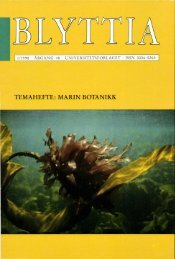
Bulletin of the British Museum (Natural History)
Bulletin of the British Museum (Natural History)
Bulletin of the British Museum (Natural History)

Create successful ePaper yourself
Turn your PDF publications into a flip-book with our unique Google optimized e-Paper software.
LICHEN GENUS MICAREA IN EUROPE 31<br />
This difference can be due to <strong>the</strong> much branched and entangled apices <strong>of</strong> <strong>the</strong> paraphyses, or<br />
(and) deposits <strong>of</strong> pigment in <strong>the</strong> gel-matrix, or on (or in) <strong>the</strong> walls <strong>of</strong> <strong>the</strong> paraphyses. The upper<br />
layer <strong>of</strong> <strong>the</strong> hymenium differentiated in such ways is perhaps more correctly termed <strong>the</strong><br />
'epihymenium' (Poelt, 1974a) or 'pseudoepi<strong>the</strong>cium' (Korf, 1973). In its strict (original) sense<br />
an 'epi<strong>the</strong>cium' refers to a layer <strong>of</strong> branches <strong>of</strong> paraphyses that overtop <strong>the</strong> asci. Such a layer is<br />
approached in some species <strong>of</strong> Micarea and is best exemplified by M. contexta and M.<br />
melanobola. In Micarea <strong>the</strong> difference between an 'epihymenium' and an 'epi<strong>the</strong>cium' is not<br />
clear-cut, and so I have chosen to employ <strong>the</strong> latter term which has been long and widely<br />
employed in lichenology.<br />
The height <strong>of</strong> <strong>the</strong> hymenium in Micarea species is sometimes difficult to measure accurately<br />
because <strong>the</strong> hymenium <strong>of</strong>ten merges ra<strong>the</strong>r imperceptibly into <strong>the</strong> hypo<strong>the</strong>cium. Measurements<br />
are most accurately made by mounting thin sections in Lugol's iodine, in which <strong>the</strong> strongly<br />
amyloid (dark blue) hymenium contrasts strongly with <strong>the</strong> non-amyloid (or ± so) hypo<strong>the</strong>cium.<br />
Even when accurately determined <strong>the</strong> height <strong>of</strong> <strong>the</strong> hymenium is rarely a useful character in<br />
distinguishing similar species. The overall range in height is about 23-90 /am, and in most cases is<br />
within 35-50 /xm. Shallow hymenia (rarely exceeding 35 /am) are characteristic <strong>of</strong> many <strong>of</strong> <strong>the</strong><br />
species with small spores, viz.: M. hedlundii, M. melanobola, M. misella, M. myriocarpa, M.<br />
nigella, M. olivacea, M. osloensis, M. rhabdogena, and M. tuberculata. Likewise, tall hymenia<br />
are characteristic <strong>of</strong> species with large spores, and <strong>the</strong> tallest hymenium (65-90 /u,m) belongs to<br />
<strong>the</strong> species with <strong>the</strong> largest spores, namely M. subleprosula. The height <strong>of</strong> <strong>the</strong> hymenium<br />
sometimes increases with <strong>the</strong> age <strong>of</strong> <strong>the</strong> apo<strong>the</strong>cium, and this is particularly true in M.<br />
bauschiana and M. sylvicola: small, yet mature, apo<strong>the</strong>cia usually have a hymenium c. 40 /zm<br />
tall, but as <strong>the</strong> apo<strong>the</strong>cium enlarges and increases in convexity, <strong>the</strong> hymenium <strong>of</strong>ten increases in<br />
height up to about 60 /xm; at <strong>the</strong> same time <strong>the</strong> paraphyses appear to 'stretch' and become<br />
thinner (especially in <strong>the</strong> lower part <strong>of</strong> <strong>the</strong> hymenium).<br />
The hymenium in Micarea never contains hyaline, crystalline inclusions that adhere to<br />
paraphyses (as found in some species <strong>of</strong> Lecidella, and many Graphidaceae) or numerous oil<br />
droplets (as found in several species <strong>of</strong> Buellia and Caloplaca). Epi<strong>the</strong>cial granules that dissolve<br />
in K and <strong>of</strong>ten give <strong>the</strong> apo<strong>the</strong>cia a pruinose appearance are also never found in Micarea. Minute<br />
granules <strong>of</strong> blue-violet (K+aeruginose) pigment, like those found in Lecidea hypnorum, are<br />
occasionally observed in <strong>the</strong> hymenium <strong>of</strong> M. contexta and M. lignaria.<br />
The colour <strong>of</strong> <strong>the</strong> hymenium and epi<strong>the</strong>cium is an important character in <strong>the</strong> identification <strong>of</strong><br />
Micarea species. A discussion <strong>of</strong> <strong>the</strong> various pigments involved is given in <strong>the</strong> sections on<br />
'chemistry'. The hymenium may be dilutely and ± evenly coloured throughout, or <strong>the</strong><br />
colouration may be more intense in <strong>the</strong> upper part. In many species <strong>the</strong> hymenium is <strong>of</strong>ten<br />
intersected by dark vertical streaks, a feature usually due to dense pigment surrounding<br />
individual, or small fascicles <strong>of</strong> stout paraphyses (see p. 61).<br />
Asci<br />
The asci <strong>of</strong> Micarea species are clavate or cylindrical-clavate in shape and belong to <strong>the</strong><br />
Lecanora-type <strong>of</strong> Honegger (1978). When mounted in Lugol's iodine (following treatment in<br />
10% KOH) <strong>the</strong> ascus is seen to have a non-amyloid wall surrounded by an amyloid outer-layer<br />
('fuzzy coat') and an internal staining amyloid apical dome (Fig. 6). Micarea has sometimes<br />
been placed in <strong>the</strong> Arthoniaceae (e.g. Lamb, 1953) but <strong>the</strong> ascus <strong>of</strong> members <strong>of</strong> that family does<br />
not have an amyloid outer-layer and <strong>the</strong> apical dome is not deeply amyloid. In a few species<br />
<strong>of</strong> Arthonia and Artho<strong>the</strong>lium I have observed a faint bluing in <strong>the</strong> part <strong>of</strong> <strong>the</strong> apical dome<br />
immediately adjacent to <strong>the</strong> ocular chamber. In some Arthonia species a tiny amyloid ring is<br />
apparent above <strong>the</strong> apex <strong>of</strong> <strong>the</strong> ocular chamber, and, despite statements to <strong>the</strong> contrary by<br />
Eriksson (1981), such a ring is found in <strong>the</strong> asci <strong>of</strong> ^. radiata (type species oi Arthonia Ach.) and<br />
A. fuscopurpurea. The same type <strong>of</strong> ring was also described for Bryostigma leucodontis by Poelt<br />
& Dobbeler (1979). Such aspects <strong>of</strong> ascus structure in <strong>the</strong> Arthoniaceae clearly merit more<br />
detailed investigation. The ascus structure in Micarea supports my opinion that <strong>the</strong> genus should<br />
be placed in <strong>the</strong> Lecideaceaes. str., at least for <strong>the</strong> time being.<br />
The asci <strong>of</strong> all European Micarea species are 8-spored. The North American Lecidea populina