Ankyloblepharon filiforme adnatum - Swiss Society of Neonatology
Ankyloblepharon filiforme adnatum - Swiss Society of Neonatology
Ankyloblepharon filiforme adnatum - Swiss Society of Neonatology

Create successful ePaper yourself
Turn your PDF publications into a flip-book with our unique Google optimized e-Paper software.
SWISS SOCIETY OF NEONATOLOGY<br />
<strong>Ankyloblepharon</strong> <strong>filiforme</strong><br />
<strong>adnatum</strong><br />
May 2013
Theodoropoulou K, Panchard MA, Service de Pédiatrie, Hôpital<br />
Riviera, Vevey, Switzerland<br />
© <strong>Swiss</strong> <strong>Society</strong> <strong>of</strong> <strong>Neonatology</strong>, Thomas M Berger, Webmaster<br />
2
3<br />
<strong>Ankyloblepharon</strong> <strong>filiforme</strong> <strong>adnatum</strong> (AFA) is a rare<br />
congenital malformation characterized by partial or<br />
complete fusion <strong>of</strong> the eyelids. It may be present as<br />
an isolated finding, in association with other anoma-<br />
lies, or as part <strong>of</strong> a well-defined syndrome. We report<br />
a case <strong>of</strong> AFA in a female newborn and describe its<br />
management.<br />
This female neonate was born to a 32-year-old G2/<br />
P2 at 41 4/7 weeks <strong>of</strong> gestation by a vaginal delivery<br />
following an unremarkable pregnancy. The baby girl<br />
adapted without difficulties with Apgar scores <strong>of</strong> 7,<br />
9 and 10 at 1, 5 and 10 minutes, respectively. Her<br />
birth weight was 3430 g. The neonatal examination<br />
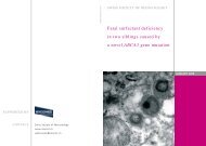
revealed a single band <strong>of</strong> tissue vertically attached to<br />
right upper and lower eyelids, covering the pupil and<br />
preventing full opening <strong>of</strong> the eyelid (Fig. 1 A, B). A de-<br />
tailed systemic pediatric assessment failed to identify<br />
any other congenital abnormalities. There was no si-<br />
milar congenital malformation in the family, no family<br />
history <strong>of</strong> eye or systemic diseases, and the mother<br />
denied taking any drugs. The band <strong>of</strong> tissue was di-<br />
vided by one cut using scissors, without necessitating<br />
either sedation or administration <strong>of</strong> a local anesthetic<br />
(Fig. 2). No bleeding occurred and the patient showed<br />
no signs <strong>of</strong> distress. Eye examination performed by an<br />
ophthalmologist did not identify any underlying ab-<br />
normalities.<br />
INTRODUCTION<br />
CASE REPORT
Fig. 1<br />
A<br />
A fibrous band connects the upper and lower eye lids<br />
(A) and prevents opening <strong>of</strong> the eye (B).<br />
4
B<br />
5
Fig. 2<br />
Appearance <strong>of</strong> the right eye following division <strong>of</strong> the<br />
band <strong>of</strong> tissue.<br />
6
7<br />
AFA is defined by partial or complete eyelid adhesion<br />
with a single or multiple bands <strong>of</strong> fibrous tissue verti-<br />
cally attached to upper and lower eyelids. The develo-<br />
ping eyelid margins remain fused until the fifth month<br />
<strong>of</strong> gestation but may not be completely separated<br />
until the seventh month (1). The etiology <strong>of</strong> AFA is<br />
unknown, but failure <strong>of</strong> apoptosis at a critical stage in<br />
eyelid development has been suggested (2).<br />
Usually, AFA constitutes a solitary malformation, as in<br />
our case, with sporadic occurrence and an incidence<br />
<strong>of</strong> 4.4 per 100.000 births (3, 4). However, it can be<br />
associated with several disorders such as trisomy 18<br />
(Edward’s syndrome) (5), Hay-Wells syndrome (a va-<br />
riant <strong>of</strong> the ectodactyly-ectodermal dysplasia-cleft lip<br />
palate syndrome) (6), popliteal pterygium syndrome<br />
(characterised by intercrural webbing <strong>of</strong> the lower<br />
limbs) (7), CHANDS (curly hair-ankyloblepharon-nail<br />
dysplasia) (8) and cleft lip and palate (9). Other associ-<br />
ations may include hydrocephalus, meningomyelo-<br />
coele, imperforate anus (10), bilateral syndactyly (7),<br />
infantile glaucoma (11), and cardiac problems such as<br />
patent ductus arteriosus and ventricular septal defects<br />
(7).<br />
AFA is diagnosed clinically and the treatment consists<br />
<strong>of</strong> simple surgical resection <strong>of</strong> the fibrous bands. Ozy-<br />
azgan et al. (12) have described treating AFA under<br />
intravenous sedation, and Ioannides et al. (13) with<br />
the aid <strong>of</strong> topical anesthesia. In our patient, no seda-<br />
DISCUSSION
tion or local anesthetic was necessary, as described<br />
previously by Williams et al. (5) and Gruener et al. (4).<br />
Surgical correction should be performed promptly to<br />
minimise any risk <strong>of</strong> occlusion amblyopia, enable full<br />
examination <strong>of</strong> the eye, alleviate parental stress and<br />
for neonatal comfort.<br />
8
9<br />
1. Sharkey D, Marlow N, Stokes J. <strong>Ankyloblepharon</strong> <strong>filiforme</strong><br />
<strong>adnatum</strong>. J Pediatr 2008;152:594<br />
2. Mohamed YH, Gong H, Ameniya T. Role <strong>of</strong> apoptosis in eyelid<br />
development. Exp Eye Res 2003;76:115-123<br />
3. Cizmeci MN, Kanbuoroglu MK, Akelma AZ, Talti MM. stitched<br />
Eye in the newborn: ankyloblepharon <strong>filiforme</strong> <strong>adnatum</strong>.<br />
J Pediatr 2012;162:211-212<br />
4. Gruener AM and Mehat MS. A newborn with ankyloblepharon<br />
<strong>filiforme</strong> <strong>adnatum</strong>: a case report. Cases J 2009;2:8146<br />
5. Williams MA, White ST, McGinnity G. <strong>Ankyloblepharon</strong> <strong>filiforme</strong><br />
<strong>adnatum</strong>. Arch Dis Child 2007;92:73-74<br />
6. Vanderho<strong>of</strong>t SL, Stephan MJ, Sybert VP. Severe skin erosions<br />
and scalp infections in AEC syndrome. Pediatr Dermatol<br />
1993;10:334-340<br />
7. Akkermans CH, Stern LM. <strong>Ankyloblepharon</strong> <strong>filiforme</strong> <strong>adnatum</strong>.<br />
Br J Ophthalmol 1979;63:129-131<br />
8. Toriello HV, Lindstrom JA, Waterman DF, Baughman FA.<br />
Re-evaluation <strong>of</strong> CHANDS. J Med Genet 1979;16:316-317<br />
9. Long JC, Blandford SE. <strong>Ankyloblepharon</strong> <strong>filiforme</strong> <strong>adnatum</strong> with<br />
cleft lip and palate. Am J Ophthalmol 1962;53:126-129<br />
10. Kazarian EL, Goldstein P. <strong>Ankyloblepharon</strong> <strong>filiforme</strong> <strong>adnatum</strong><br />
with hydrocephalus, meningomyelocele, and imperforate anus.<br />
Am J Ophthalmol 1977;84:355-357<br />
11. Scott MH, Richard JM, Farris BK. <strong>Ankyloblepharon</strong> <strong>filiforme</strong><br />
<strong>adnatum</strong> associated with infantile glaucoma and iridogoniodys-<br />
genesis. J Pediatr Ophthalmol Strabismus 1994;31:93-95<br />
REFERENCES
12. Özyazgan Ī, Eskitaşcioǧlu T, Dűndar M, et al. Hereditary isola-<br />
ted ankyloblepharon <strong>filiforme</strong> <strong>adnatum</strong>. Plast Reconstr Surg<br />
2005;115:363-364<br />
13. Ioannides A, Georgakarakos ND. Management <strong>of</strong><br />
ankloblepharon <strong>filiforme</strong> <strong>adnatum</strong>. Eye 2011;25:823<br />
10
SUPPORTED BY<br />
CONTACT<br />
<strong>Swiss</strong> <strong>Society</strong> <strong>of</strong> <strong>Neonatology</strong><br />
www.neonet.ch<br />
webmaster@neonet.ch<br />
concept & design by mesch.ch