Nano-C60 cytotoxicity is due to lipid peroxidation
Nano-C60 cytotoxicity is due to lipid peroxidation
Nano-C60 cytotoxicity is due to lipid peroxidation

Create successful ePaper yourself
Turn your PDF publications into a flip-book with our unique Google optimized e-Paper software.
7590<br />
% Dead<br />
100<br />
80<br />
60<br />
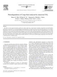
concentration on a per cell bas<strong>is</strong> between controls or<br />
cells exposed <strong>to</strong> 2400 ppb nano-<strong>C60</strong> (Fig. 2).<br />
In the presence of nano-<strong>C60</strong>, there appears <strong>to</strong> be<br />
significant membrane d<strong>is</strong>ruption. The membrane permeability<br />
was evaluated for HDF, HepG2, and NHA cells<br />
using fluorescent dextrans. Results for all three cell lines<br />
show that cells grown under normal conditions, without<br />
the addition of the fullerene species, uptake of fluorescent<br />
dextran was only observed with the smallest<br />
(10 kDa) dye. However, the cells dosed with 240 ppb<br />
nano-<strong>C60</strong> were permeable <strong>to</strong> the 10, 70, and 500 kDa<br />
dyes (Fig. 1).<br />
Membrane oxidation was qualitatively examined<br />
using two separate methodologies. In the first method,<br />
<strong>lipid</strong> <strong>peroxidation</strong> was indirectly measured by moni<strong>to</strong>ring<br />
the production of MDTA from the oxidation of<br />
malondialdehyde in the presence of cells in culture. A<br />
gradual increase of MDTA, which corresponds <strong>to</strong> an<br />
increase of peroxyradicals on membranes, was observed<br />
for cells dosed with 0.24–4.8 ppb; and a dramatic<br />
increase of MDTA was observed for cells dosed with<br />
240 and 2400 ppb (Fig. 3a). Additionally, the concentration<br />
of glutathione, a natural-occurring antioxidant,<br />
noticeably increases at the 240 ppb nano-C 60 concentration<br />
(Fig. 3b) in HDF, HepG2, and NHA cells. We<br />
believe th<strong>is</strong> demonstrates an increase in glutathione<br />
synthes<strong>is</strong> by the cells in response <strong>to</strong> oxidative stress. The<br />
ARTICLE IN PRESS<br />
C.M. Sayes et al. / Biomaterials 26 (2005) 7587–7595<br />
10-2 100 102 104 40<br />
1.2<br />
20<br />
0<br />
1.0<br />
0.8<br />
0.6<br />
Control 0.24 ppb 2.4 ppb 4.8 ppb 24 ppbr240 ppb<br />
(A) nano-<strong>C60</strong> Concentration (ppb) (B)<br />
nano-<strong>C60</strong> Concentration<br />
HDF, control HDF, nano-<strong>C60</strong> ABS (normalized)<br />
(C) (D)<br />
1.8<br />
1.6<br />
1.4<br />
HDF<br />
HepG 2<br />
NHA<br />
Fig. 1. The response of aggregated water-soluble fullerene species on HDF, HepG2, and NHA are dependant on nanoparticle dose and surface<br />
chem<strong>is</strong>try. All assays were performed in culture. (A) The dose response curve of the nano-C 60 species on (’) HDF, ( ) HepG2, and (m) NHA cells.<br />
A non-linear curve fit was applied <strong>to</strong> three separate data sets. For some data points, the symbol <strong>is</strong> <strong>to</strong>o large for errors bars <strong>to</strong> be seen. (B) Death of<br />
HDF, HepG2, and NHA cells via nano-<strong>C60</strong> was confirmed with the lactate dehydrogenase release. Dextran fluorescein (500,000 kDa) was added <strong>to</strong><br />
(C) normal healthy cells and (D) cells dosed with 10 ppm nano-C 60. Part C shows data for HDF cells only.<br />
cells could be exporting oxidized glutathione <strong>to</strong> the<br />
media, as well as synthesizing intracellular reduced<br />
glutathione.<br />
Further evidence of <strong>lipid</strong> <strong>peroxidation</strong> <strong>is</strong> shown in<br />
Fig. 4. The second method of detecting <strong>lipid</strong> <strong>peroxidation</strong><br />
utilizes the lipophilic fluorescent dye C11-BODI-<br />
PY 581/591 . Th<strong>is</strong> fluorescence assay was developed for<br />
localization and quantification of <strong>lipid</strong> oxidation in<br />
living cells [17]. The oxidative sensitivity of C11-<br />
BODIPY 581/591 provides indication that conditions<br />
favoring oxidation of the dye ex<strong>is</strong>t at the surface of<br />
the membrane. Once oxidized, the fluorescence of th<strong>is</strong><br />
dye shifts from red <strong>to</strong> green. The probe incorporates<br />
readily in<strong>to</strong> cellular membranes and <strong>is</strong> about twice as<br />
sensitive <strong>to</strong> oxidation compared <strong>to</strong> probes such as c<strong>is</strong>parinaric<br />
acid [18]. Using confocal microscopy, the<br />
oxidation of C11-BODIPY 581/591 can be v<strong>is</strong>ualized at<br />
the cellular level. Over time, in the presence of normal<br />
cellular membranes, the C11-BODIPY 581/591 fluorophore<br />
maintained red fluorescence (610 nm) with no<br />
green fluorescence (510 nm). However, in the presence of<br />
nano-<strong>C60</strong>, the BODIPY fluorophore decreased in red<br />
fluorescence and increased in green fluorescence, indicating<br />
membrane oxidation.<br />
In an additional set of studies, L-ascorbic acid was<br />
added <strong>to</strong> the cells during exposure <strong>to</strong> nano-<strong>C60</strong> as a test<br />
<strong>to</strong> determine if the oxidative damage <strong>to</strong> HDFs could be