Nano-C60 cytotoxicity is due to lipid peroxidation
Nano-C60 cytotoxicity is due to lipid peroxidation
Nano-C60 cytotoxicity is due to lipid peroxidation

Create successful ePaper yourself
Turn your PDF publications into a flip-book with our unique Google optimized e-Paper software.
Abstract<br />
Biomaterials 26 (2005) 7587–7595<br />
<strong>Nano</strong>-C 60 <strong>cy<strong>to</strong><strong>to</strong>xicity</strong> <strong>is</strong> <strong>due</strong> <strong>to</strong> <strong>lipid</strong> <strong>peroxidation</strong><br />
Chr<strong>is</strong>tie M. Sayes a , Andre M. Gobin b , Kevin D. Ausman c , Joe Mendez a ,<br />
Jennifer L. West b,c, , Vicki L. Colvin a,c<br />
a Department of Chem<strong>is</strong>try, 6100 Main St. MS-60, Rice University, Hous<strong>to</strong>n, TX 77005, USA<br />
b Department of Bioengineering, 6100 Main St. MS-142, Rice University, Hous<strong>to</strong>n, TX 77005, USA<br />
c Center for Biological and Environmental <strong>Nano</strong>technology, 6100 Main St. MS-63, Rice University, Hous<strong>to</strong>n, TX 77005, USA<br />
Received 8 April 2005; accepted 16 May 2005<br />
Available online 11 July 2005<br />
Th<strong>is</strong> study examines the biological effects of water-soluble fullerene aggregates in an effort <strong>to</strong> evaluate the fundamental<br />
mechan<strong>is</strong>ms that contribute <strong>to</strong> the <strong>cy<strong>to</strong><strong>to</strong>xicity</strong> of a classic engineered nanomaterial. For th<strong>is</strong> work we used a water-soluble fullerene<br />
species, nano-C 60, a fullerene aggregate that readily forms when pr<strong>is</strong>tine C 60 <strong>is</strong> added <strong>to</strong> water. <strong>Nano</strong>-C 60 was cy<strong>to</strong><strong>to</strong>xic <strong>to</strong> human<br />
dermal fibroblasts, human liver carcinoma cells (HepG2), and neuronal human astrocytes at doses X50 ppb (LC50 ¼ 2–50 ppb,<br />
depending on cell type) after 48 h exposure. Th<strong>is</strong> water-soluble nano-<strong>C60</strong> colloidal suspension d<strong>is</strong>rupts normal cellular function<br />
through <strong>lipid</strong> <strong>peroxidation</strong>; reactive oxygen species are responsible for the membrane damage. Cellular viability was determined<br />
through live/dead staining and LDH release. DNA concentration and mi<strong>to</strong>chondrial activity were not affected by the nano-<strong>C60</strong><br />
inoculations <strong>to</strong> cells in culture. The integrity of cellular membrane was examined by moni<strong>to</strong>ring the peroxy-radicals on the <strong>lipid</strong><br />
bilayer. Subsequently, glutathione production was measured <strong>to</strong> assess the cell’s reaction <strong>to</strong> membrane oxidation. The damage <strong>to</strong> cell<br />
membranes was observed both with chemical assays, and confirmed physically by v<strong>is</strong>ualizing membrane permeability with high<br />
molecular weight dyes. With the addition of an antioxidant, L-ascorbic acid, the oxidative damage and resultant <strong>to</strong>xicity of nano-<strong>C60</strong><br />
was completely prevented.<br />
r 2005 Elsevier Ltd. All rights reserved.<br />
Keywords: <strong>Nano</strong>particle; Cy<strong>to</strong><strong>to</strong>xicity; <strong>Nano</strong>-C 60; Membrane oxidation<br />
1. Introduction<br />
Water-soluble fullerene systems are prom<strong>is</strong>ing candidates<br />
for many medical technologies and have been<br />
proposed as crucial components for emerging electronic,<br />
optical and mechanical materials [1,2]. Given the widespread<br />
applications and their impending commercialization,<br />
both humans and environmental systems will be<br />
increasingly exposed <strong>to</strong> materials like <strong>C60</strong> in the near<br />
future; thus, early evaluations of their health effects are<br />
valuable [3]. Previously, we have evaluated the differential<br />
<strong>cy<strong>to</strong><strong>to</strong>xicity</strong> of a series of water-soluble fullerene<br />
Corresponding author. 6100 Main Street, MS-142, Rice University,<br />
Hous<strong>to</strong>n, TX 77005, USA. Tel.: +1 713 348 5955.<br />
E-mail address: jwest@rice.edu (J.L. West).<br />
0142-9612/$ - see front matter r 2005 Elsevier Ltd. All rights reserved.<br />
doi:10.1016/j.biomaterials.2005.05.027<br />
ARTICLE IN PRESS<br />
species and concluded that changes in the fullerene cage<br />
structure had substantial impact on in vitro <strong>cy<strong>to</strong><strong>to</strong>xicity</strong><br />
[4]. As the number of hydroxyl or carboxyl groups on<br />
the surface of the fullerene cage was increased,<br />
<strong>cy<strong>to</strong><strong>to</strong>xicity</strong> decreased over seven orders of magnitude.<br />
The series of water-soluble fullerenes included<br />
nano-<strong>C60</strong>, tr<strong>is</strong>-malonic acid-<strong>C60</strong> (or C3), Na + 2 3[<strong>C60</strong>O7 9<br />
(2 3)<br />
(OH)12 15]<br />
www.elsevier.com/locate/biomaterials<br />
, and <strong>C60</strong>(OH)24. Using two separate<br />
methods, we determined that the nano-<strong>C60</strong> generated<br />
substantially more reactive oxygen species (ROS) than<br />
the other species under cell-free conditions. The ROS<br />
generation mono<strong>to</strong>nically decreased with increasing<br />
derivatization of the fullerene cage. The dramatic<br />
<strong>cy<strong>to</strong><strong>to</strong>xicity</strong> observed for nano-C 60 warranted further<br />
evaluation as presented here. Additional studies were<br />
also conducted <strong>to</strong> probe the mechan<strong>is</strong>ms governing the
7588<br />
<strong>cy<strong>to</strong><strong>to</strong>xicity</strong> of nano-<strong>C60</strong> and confirm the importance of<br />
oxidation.<br />
Water-soluble fullerene derivatives are most commonly<br />
formed by deliberate synthetic methodologies<br />
and typically have altered cage chem<strong>is</strong>try and high<br />
(4100 ppm) water solubility. In contrast, the nano-C 60<br />
colloid investigated here <strong>is</strong> only sparingly soluble; it <strong>is</strong><br />
produced by the addition of organic soluble <strong>C60</strong> <strong>to</strong> water<br />
[5–8]. The same substance <strong>is</strong> also found when solid <strong>C60</strong> <strong>is</strong><br />
stirred in tap water for 2 months [9]. Because of its ease<br />
of formation, and stability in water, nano-<strong>C60</strong> <strong>is</strong> likely <strong>to</strong><br />
be an important form of <strong>C60</strong> in natural aqueous systems.<br />
The full characterization of the nano-C 60 water<br />
suspension <strong>is</strong> publ<strong>is</strong>hed in two separate reports [4,10].<br />
Proof of the presence of <strong>C60</strong> in the aqueous suspension<br />
include electron diffraction via cryo-TEM, chroma<strong>to</strong>graphic<br />
profiles and signature spectroscopic peaks<br />
identify the presence of <strong>C60</strong> in the aqueous solution. In<br />
addition, microscopy images show the size, shape and<br />
crystallinity of the <strong>C60</strong> colloids. While the method of<br />
nano-C 60 preparation may leave intercalated THF, mass<br />
spectroscopy and liquid chroma<strong>to</strong>graphy prove that<br />
more than 90% by weight of the suspension <strong>is</strong> <strong>C60</strong>, i.e.<br />
o10% of the suspension <strong>is</strong> residual solvent. Viability<br />
controls confirm that th<strong>is</strong> residual solvent does not<br />
contribute <strong>to</strong> cell death or generation of ROS. The<br />
structural of the nano-<strong>C60</strong> colloid cons<strong>is</strong>ts of a pr<strong>is</strong>tine<br />
<strong>C60</strong> core (of 10–1000 <strong>C60</strong> molecules, depending on size of<br />
crystal) surrounded by a low derivatized C 60 layer that<br />
forces the aggregate <strong>to</strong> be m<strong>is</strong>cible in the aqueous phase.<br />
The extent of the derivatization <strong>is</strong> 3 groups or less,<br />
composed of either hydroxylated or oxidized fullerenes<br />
(confirmed by nuclear magnetic resonance and Fourier<br />
transform infrared spectroscopy). The negative surface<br />
charge of the aggregate <strong>is</strong> further evidence of a<br />
hydrophilic surface derivation. Because of th<strong>is</strong> low<br />
degree of derivatization, we cannot fully identify the<br />
exact chemical composition of these groups.<br />
Previous reports by Oberdorster suggest the brain and<br />
liver of largemouth bass produce changes in glutathione<br />
production once exposed <strong>to</strong> nano-<strong>C60</strong>. Therefore, we<br />
hypothesize that the human cell lines hDF, Human liver<br />
carcinoma cells (HepG2), and NHA might be affected<br />
by nano-<strong>C60</strong>. More specifically, within these cell lines,<br />
we concentrated on examining the effect of nano-C 60 on<br />
the membrane of the cell, since we previously reported<br />
that nano-<strong>C60</strong> produces oxygen radicals in water.<br />
2. Materials and methods<br />
All chemicals were purchased through Sigma Aldrich at<br />
highest purity unless otherw<strong>is</strong>e stated and experiments were<br />
performed minimally in triplicate. Data are presented as mean<br />
7 standard deviation, and a student’s t-test was used <strong>to</strong><br />
determine significance.<br />
ARTICLE IN PRESS<br />
C.M. Sayes et al. / Biomaterials 26 (2005) 7587–7595<br />
2.1. <strong>Nano</strong>-C 60 preparation<br />
C 60 was suspended in water using the method of Deguchi et<br />
al. [7] <strong>C60</strong> (99.5%, MER Corporation), was d<strong>is</strong>solved in<br />
tetrahydrofuran (THF, F<strong>is</strong>her Scientific) at a concentration of<br />
100 mg/L. The solution was sparged with nitrogen and stirred<br />
overnight in the dark. The solution was then filtered through a<br />
0.22 mm nylon filter (Osmonics, F<strong>is</strong>her Scientific). MilliQ water<br />
was added <strong>to</strong> an equal volume of C 60 in THF at a rate of 1 L/<br />
min. Th<strong>is</strong> solution was evaporated <strong>to</strong> eliminate the THF using<br />
a rotary evapora<strong>to</strong>r (Buchii). Mass spectroscopy of solvent<br />
after th<strong>is</strong> procedure finds no residual THF in solution. A 1 L<br />
mixture was evaporated <strong>to</strong> 450 mL, refilled <strong>to</strong> 550 mL with<br />
MilliQ water, and then again evaporated <strong>to</strong> 450 mL. The<br />
volume was adjusted <strong>to</strong> 550 mL again, and then evaporated <strong>to</strong><br />
500 mL. Th<strong>is</strong> final solution was s<strong>to</strong>red overnight and then<br />
filtered through a 0.22 mm nylon filter <strong>to</strong> yield a suspension of<br />
aggregated <strong>C60</strong> in water referred <strong>to</strong> as nano-<strong>C60</strong>. <strong>C60</strong>(OH)24<br />
was obtained from MER Corporation at 99.8% purity.<br />
2.2. Cy<strong>to</strong><strong>to</strong>xicity/viability<br />
Human dermal fibroblasts (HDF) (Cambrex) were cultured<br />
in Dulbecco’s Modification of Eagles Media (DMEM) with<br />
10% fetal bovine serum and 2 mM L-glutamine, 100 U/mL<br />
penicillin, and 1 mg/mL and strep<strong>to</strong>mycin. HepG2 (American<br />
Type Culture Collection) were cultured in minimal essential<br />
media (MEM) with 10% fetal bovine serum and 2 mM Lglutamine,<br />
100 U/mL penicillin, and 1 mg/mL and strep<strong>to</strong>mycin.<br />
Neuronal human astrocytes (NHA) (Cambrex) were<br />
cultured in Astrocyte Basal Medium (ABM TM ) derived from<br />
MCBB131 classical media with 10% fetal bovine serum and<br />
1.5% rhEGF, 2.5% insulin, 1% ascorbic acid, 1% gentamicin<br />
sulfate and amphotericin-B, and 10% L-glutamine. For all<br />
three cell types, passage numbers 2–10 were used in the<br />
experiments. 5 mL calcein AMand 20 mL ethidium homodimer<br />
(Molecular Probes) in 10 mL phosphate buffer solution were<br />
used <strong>to</strong> determine cell viability following 48 h exposure <strong>to</strong><br />
fullerenes. Fullerenes (nano-<strong>C60</strong>) suspended in ultrapure water<br />
and sterilized via filtration (0.22 mm) were added <strong>to</strong> cells,<br />
grown in 24 well plates, at concentration of 0.24, 2.4, 24, 240<br />
and 2400 ppb. For each experiment, the viability of untreated<br />
HDF, HepG2, and NHA cells, as well as cells treated with<br />
phosphate buffered saline (PBS) solution, was evaluated as<br />
well. Cell viability was evaluated via fluorescence microscopy<br />
(Ze<strong>is</strong>s Axiovert 135). The experiment was repeated six times.<br />
2.3. Lactate dehydrogenase release<br />
The release of lactate dehydrogenase (LDH) was also<br />
moni<strong>to</strong>red over the nano-C 60 concentration range described<br />
above. Cells (HDF, HepG2, NHA) were seeded in 24-well<br />
plates, exposed <strong>to</strong> nano-<strong>C60</strong>, and incubated for 48 h. After<br />
incubation, the cell media was transferred <strong>to</strong> 15 mL centrifugation<br />
tubes (<strong>to</strong>tal volume 4 mL) and were centrifuged at 300g<br />
for 4 min. The media was decanted and analyzed for LDH<br />
release as previously described [11,12]. Upon addition of assay<br />
solutions, the media was protected from the light for 30 min.<br />
During th<strong>is</strong> inoculation time, NAD <strong>is</strong> reduced <strong>to</strong> NADH using<br />
the LDH released in the medium. 400 mL of1N HCl was then
added <strong>to</strong> each sample <strong>to</strong> terminate the reduction of NADH.<br />
The resulting absorbance was measured at 490 nm.<br />
2.4. Mi<strong>to</strong>chondrial activity<br />
The 1-(4,5-dimethylthiazol-2-yl)-3,5-diphenylformazan (MTT)<br />
assay (Sigma) was used <strong>to</strong> evaluate mi<strong>to</strong>chondrial activity [13].<br />
Cells, grown in 24-well plates, were exposed <strong>to</strong> fullerenes as<br />
described above. After 48 h, 150 mL of M TT (5mg/mL) was<br />
added <strong>to</strong> each well and incubated for 4 h. Afterwards, 850 mL<br />
of the MTT solubilization solution (10% Tri<strong>to</strong>n X-100 in 0.1 N<br />
HCl in anhydrous <strong>is</strong>opropanol) was added <strong>to</strong> each well. The<br />
24-well plate was gently mixed on a gyra<strong>to</strong>ry shaker <strong>to</strong> solubilize<br />
the formazan crystals. After solubilization in acidic<br />
<strong>is</strong>opropanol, the product was quantified by measuring absorbance<br />
at 570 nm.<br />
2.5. DNA content<br />
Using the fluorescent DNA-binding dye PicoGreen s<br />
(Molecular Probes), the concentration of DNA in normal<br />
HDF, HepG2, and NHA cells and cells dosed with 240 ppb<br />
nano-C 60 was measured. A TE buffer solution (10 mM Tr<strong>is</strong>-<br />
HCl, 1 mM EDTA, pH 7.5) was used <strong>to</strong> prepare the Pico-<br />
Green s dsDNA quantitation solution. An equal volume of the<br />
PicoGreen s quantitation solution was added <strong>to</strong> each HDF,<br />
HepG2, and NHA lysed sample solution. After 5 min of<br />
incubation, absorbance was measured using a spectrofluorometer<br />
(excitation 480 nm, em<strong>is</strong>sion 520 nm). A standard curve<br />
(1 ng/mL–1 mg/mL) was made using a DNA s<strong>to</strong>ck solution.<br />
Total protein was measured in cellular homogenates using the<br />
Bradford method [14,15], and DNA content was normalized <strong>to</strong><br />
protein content.<br />
2.6. Permeability of the plasma membrane<br />
The integrity of the plasma membrane was examined by<br />
moni<strong>to</strong>ring the uptake of fluorescein-derivatized dextrans<br />
(Molecular Probes) of increasing molecular weights<br />
(10,000–500,000 Da). Cells were treated with fullerenes as<br />
described above. After cells were washed with phosphatebuffered<br />
saline (PBS, pH 7.4), 2.5 mg/mL dextran was added<br />
<strong>to</strong> each well and then incubated at 37 1C/5% CO 2 for 30 min.<br />
Cells were washed with PBS then fixed with 5% glutaraldehyde<br />
(Sigma) and observed via fluorescent and phase contrast<br />
microscopy (Ze<strong>is</strong>s Axiovert 135).<br />
2.7. Lipid <strong>peroxidation</strong><br />
Lipid <strong>peroxidation</strong> was measured in HDF, HepG2, and<br />
NHA cells using the thiobarbituric acid (TBA) assay for<br />
malondialdehyde [16] and normalized <strong>to</strong> <strong>to</strong>tal protein. Cells<br />
were homogenized in 50 mM HEPES-buffered saline (pH 7.2)<br />
with 0.1 mM phenylmethanesulfonyl fluoride (PMSF) using a<br />
glass t<strong>is</strong>sue tearor. The homogenate was centrifuged at 14,000g<br />
for 5 min. 1.4 mL of 0.02% butylated hydroxy<strong>to</strong>luene (BHT)<br />
and 0.375% TBA were incubated with 100 mL of each<br />
homogenate for 15 min at 100 1C. The samples were cooled,<br />
centrifuged (5900g for 5 min), and the appearance of malondialdehyde<br />
was measured in the supernatant at 532 nm and<br />
ARTICLE IN PRESS<br />
C.M. Sayes et al. / Biomaterials 26 (2005) 7587–7595 7589<br />
compared <strong>to</strong> a standard curve containing 0–3.2 mM malondialdehyde.<br />
Total protein was measured in t<strong>is</strong>sue homogenates<br />
using the Bradford method [14,15] and <strong>lipid</strong> <strong>peroxidation</strong> was<br />
normalized <strong>to</strong> protein content.<br />
To confirm the evidence of <strong>lipid</strong> <strong>peroxidation</strong>, HDF,<br />
HepG2, and NHA cells were dosed with the nano-<strong>C60</strong> solution<br />
as described above; and C11-BODIPY 581/591<br />
(Molecular<br />
Probes) was used <strong>to</strong> moni<strong>to</strong>r <strong>lipid</strong> <strong>peroxidation</strong> in cultured<br />
cells [17]. Th<strong>is</strong> lipophilic fluorophore changes its fluorescence<br />
when it interacts with peroxyradicals. The oxidation of C11-<br />
BODIPY 581/591 was evaluated via fluorescence microscopy<br />
(Ze<strong>is</strong>s Axiovert 135). The degree of peroxidized <strong>lipid</strong>s was<br />
quantified by spectrometrically measuring the loss of red<br />
fluorescence and gain of green fluorescence (Molecular Devices<br />
SpectraMax Plus 384 ).<br />
2.8. Glutathione production<br />
Glutathione production was moni<strong>to</strong>red for HDF, HepG2,<br />
and NHA cells exposed <strong>to</strong> nano-C 60 at varying concentrations<br />
(0.24–2400 ppb). Control cells were treated with an equivalent<br />
volume of MilliQ water. After 48 h, cells were homogenized in<br />
5% sulfosalicylic acid (SSA) via glass t<strong>is</strong>sue tearor. The<br />
homogenate was centrifuged at 14,000g for 5 min. After 10 min<br />
incubation at 37 1C, 15 mL of 50 U/mL glutathione reductase<br />
was added <strong>to</strong> each sample. Absorbance at 412 nm was then<br />
measured. Glutathione standards (0–4 mM) with 100 mL<br />
NADPH, 15 mL 5,5 0 -dithiob<strong>is</strong>(2-nitrobenzoic acid) (DTNB),<br />
and 25 mL SSA were evaluated similarly for compar<strong>is</strong>on. Total<br />
protein was measured in t<strong>is</strong>sue homogenates using the<br />
Bradford method [14,15] and <strong>to</strong>tal glutathione was normalized<br />
<strong>to</strong> protein content.<br />
2.9. Addition of ascorbic acid<br />
A 1mM L-ascorbic acid solution was prepared. HDFs<br />
were incubated in appropriate media and inoculated with<br />
60 mL of 50 mg/L nano-<strong>C60</strong>, as described above. After<br />
40 h, 240 mL of1mML-ascorbic acid was added <strong>to</strong> the HDFs.<br />
8 h later, the <strong>lipid</strong> <strong>peroxidation</strong> assay was performed as<br />
described above.<br />
3. Results<br />
Cell viability was determined following 48 h exposure<br />
<strong>to</strong> fullerenes. We previously found that LDH release<br />
does not occur at 1, 12, or 30 h after inoculation with the<br />
nano-<strong>C60</strong>. For th<strong>is</strong> reason, all LDH measurements were<br />
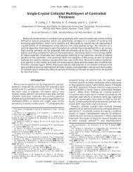
performed after 48 h. The LC50 values, determined from<br />
the dose response curves shown in Fig. 1, are as follows:<br />
for the HDF cells, 20 ppb; for HepG2, 50 ppb; and for<br />
NHA cells, 2 ppb. Since substantial <strong>cy<strong>to</strong><strong>to</strong>xicity</strong> was<br />
observed for nano-<strong>C60</strong>, the mechan<strong>is</strong>ms by which nano-<br />
C 60 damages cells in culture were examined. Using the<br />
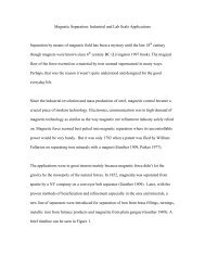
MTT assay, we found that mi<strong>to</strong>chondrial activity was<br />
unchanged. Using the PicoGreen s DNA assay, we<br />
determined that there was no difference in DNA
7590<br />
% Dead<br />
100<br />
80<br />
60<br />
concentration on a per cell bas<strong>is</strong> between controls or<br />
cells exposed <strong>to</strong> 2400 ppb nano-<strong>C60</strong> (Fig. 2).<br />
In the presence of nano-<strong>C60</strong>, there appears <strong>to</strong> be<br />
significant membrane d<strong>is</strong>ruption. The membrane permeability<br />
was evaluated for HDF, HepG2, and NHA cells<br />
using fluorescent dextrans. Results for all three cell lines<br />
show that cells grown under normal conditions, without<br />
the addition of the fullerene species, uptake of fluorescent<br />
dextran was only observed with the smallest<br />
(10 kDa) dye. However, the cells dosed with 240 ppb<br />
nano-<strong>C60</strong> were permeable <strong>to</strong> the 10, 70, and 500 kDa<br />
dyes (Fig. 1).<br />
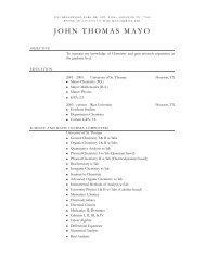
Membrane oxidation was qualitatively examined<br />
using two separate methodologies. In the first method,<br />
<strong>lipid</strong> <strong>peroxidation</strong> was indirectly measured by moni<strong>to</strong>ring<br />
the production of MDTA from the oxidation of<br />
malondialdehyde in the presence of cells in culture. A<br />
gradual increase of MDTA, which corresponds <strong>to</strong> an<br />
increase of peroxyradicals on membranes, was observed<br />
for cells dosed with 0.24–4.8 ppb; and a dramatic<br />
increase of MDTA was observed for cells dosed with<br />
240 and 2400 ppb (Fig. 3a). Additionally, the concentration<br />
of glutathione, a natural-occurring antioxidant,<br />
noticeably increases at the 240 ppb nano-C 60 concentration<br />
(Fig. 3b) in HDF, HepG2, and NHA cells. We<br />
believe th<strong>is</strong> demonstrates an increase in glutathione<br />
synthes<strong>is</strong> by the cells in response <strong>to</strong> oxidative stress. The<br />
ARTICLE IN PRESS<br />
C.M. Sayes et al. / Biomaterials 26 (2005) 7587–7595<br />
10-2 100 102 104 40<br />
1.2<br />
20<br />
0<br />
1.0<br />
0.8<br />
0.6<br />
Control 0.24 ppb 2.4 ppb 4.8 ppb 24 ppbr240 ppb<br />
(A) nano-<strong>C60</strong> Concentration (ppb) (B)<br />
nano-<strong>C60</strong> Concentration<br />
HDF, control HDF, nano-<strong>C60</strong> ABS (normalized)<br />
(C) (D)<br />
1.8<br />
1.6<br />
1.4<br />
HDF<br />
HepG 2<br />
NHA<br />
Fig. 1. The response of aggregated water-soluble fullerene species on HDF, HepG2, and NHA are dependant on nanoparticle dose and surface<br />
chem<strong>is</strong>try. All assays were performed in culture. (A) The dose response curve of the nano-C 60 species on (’) HDF, ( ) HepG2, and (m) NHA cells.<br />
A non-linear curve fit was applied <strong>to</strong> three separate data sets. For some data points, the symbol <strong>is</strong> <strong>to</strong>o large for errors bars <strong>to</strong> be seen. (B) Death of<br />
HDF, HepG2, and NHA cells via nano-<strong>C60</strong> was confirmed with the lactate dehydrogenase release. Dextran fluorescein (500,000 kDa) was added <strong>to</strong><br />
(C) normal healthy cells and (D) cells dosed with 10 ppm nano-C 60. Part C shows data for HDF cells only.<br />
cells could be exporting oxidized glutathione <strong>to</strong> the<br />
media, as well as synthesizing intracellular reduced<br />
glutathione.<br />
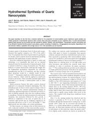
Further evidence of <strong>lipid</strong> <strong>peroxidation</strong> <strong>is</strong> shown in<br />
Fig. 4. The second method of detecting <strong>lipid</strong> <strong>peroxidation</strong><br />
utilizes the lipophilic fluorescent dye C11-BODI-<br />
PY 581/591 . Th<strong>is</strong> fluorescence assay was developed for<br />
localization and quantification of <strong>lipid</strong> oxidation in<br />
living cells [17]. The oxidative sensitivity of C11-<br />
BODIPY 581/591 provides indication that conditions<br />
favoring oxidation of the dye ex<strong>is</strong>t at the surface of<br />
the membrane. Once oxidized, the fluorescence of th<strong>is</strong><br />
dye shifts from red <strong>to</strong> green. The probe incorporates<br />
readily in<strong>to</strong> cellular membranes and <strong>is</strong> about twice as<br />
sensitive <strong>to</strong> oxidation compared <strong>to</strong> probes such as c<strong>is</strong>parinaric<br />
acid [18]. Using confocal microscopy, the<br />
oxidation of C11-BODIPY 581/591 can be v<strong>is</strong>ualized at<br />
the cellular level. Over time, in the presence of normal<br />
cellular membranes, the C11-BODIPY 581/591 fluorophore<br />
maintained red fluorescence (610 nm) with no<br />
green fluorescence (510 nm). However, in the presence of<br />
nano-<strong>C60</strong>, the BODIPY fluorophore decreased in red<br />
fluorescence and increased in green fluorescence, indicating<br />
membrane oxidation.<br />
In an additional set of studies, L-ascorbic acid was<br />
added <strong>to</strong> the cells during exposure <strong>to</strong> nano-<strong>C60</strong> as a test<br />
<strong>to</strong> determine if the oxidative damage <strong>to</strong> HDFs could be
ABS (normalized)<br />
(A)<br />
µg DNA/mg protein<br />
(B)<br />
2.0<br />
1.8<br />
1.6<br />
1.4<br />
1.2<br />
1.0<br />
0.8<br />
0.6<br />
4.0<br />
3.5<br />
3.0<br />
2.5<br />
2.0<br />
1.5<br />
1.0<br />
0.5<br />
0.0<br />
prevented (Fig. 5). After 48 h, the cells were moni<strong>to</strong>red<br />
for viability and <strong>lipid</strong> <strong>peroxidation</strong>. Results show that<br />
no oxidative damage occurred when both nano-<strong>C60</strong> and<br />
L-ascorbic acid was added <strong>to</strong> HDF cells. Further, cell<br />
viability was unchanged from controls when cells were<br />
treated with both nano-<strong>C60</strong> and L-ascorbic acid.<br />
4. D<strong>is</strong>cussion<br />
HDF<br />
HepG 2<br />
NHA<br />
Control 0.24 ppb 2.4ppb 4.8 ppb 24 ppb 240 ppb<br />
nano-<strong>C60</strong> Concentration<br />
nano-C 60<br />
control<br />
HDF HepG 2 NHA<br />
Fig. 2. <strong>Nano</strong>-C 60 does not d<strong>is</strong>rupt the mi<strong>to</strong>chondrial activity or DNA<br />
concentration of HDF, HepG2, and NHA. (A) The concentration of<br />
mi<strong>to</strong>chondrial dehydrogenase does not increase as nano-<strong>C60</strong> concentration<br />
increases. (B) Also, the concentration of DNA in HDF,<br />
HepG2, and NHA cells before dosing <strong>is</strong> equivalent <strong>to</strong> DNA in cells<br />
after dosing and fits within the standard curve. All cells were seeded at<br />
70% confluency. Cells were exposed <strong>to</strong> nano-<strong>C60</strong> for 48 h.<br />
We investigated the effect of water-soluble fullerene<br />
aggregates, nano-<strong>C60</strong>, on HDF, HepG2, and NHA cells<br />
in culture. <strong>Nano</strong>-C 60 demonstrated significant <strong>to</strong>xicity in<br />
previous cell culture studies, while a highly hydroxylated,<br />
water-soluble fullerene, <strong>C60</strong>(OH)24 did not [4]. In<br />
these studies, we have determined that <strong>lipid</strong> peroxida-<br />
ARTICLE IN PRESS<br />
C.M. Sayes et al. / Biomaterials 26 (2005) 7587–7595 7591<br />
µM MDTA/mg protein<br />
(A)<br />
µM GSH/mg protein<br />
(B)<br />
25<br />
20<br />
15<br />
10<br />
5<br />
0<br />
800<br />
700<br />
600<br />
500<br />
400<br />
300<br />
200<br />
100<br />
0<br />
HDF<br />
HepG 2<br />
NHA<br />
Control 0.24 ppb 2.4 ppb 4.8 ppb 24 ppb 240 ppb 2400 ppb<br />
nano-<strong>C60</strong> Concentration<br />
900 HDF<br />
HepG 2<br />
NH<br />
Control 0.24 ppb 2.4 ppb 4.8 ppb 24 ppb 240 ppb 2400 ppb<br />
nano-<strong>C60</strong> Concentration<br />
Fig. 3. The proposed mechan<strong>is</strong>m in which nano-C 60 d<strong>is</strong>rupts normal<br />
cellular function. The oxy-radical produced from the solvent extraction<br />
of <strong>C60</strong> from the organic solvent <strong>to</strong> water attacks the <strong>lipid</strong> bilayer of the<br />
cy<strong>to</strong>plasmic membrane, producing a peroxyradical resulting in holes in<br />
the bilayer. There <strong>is</strong> no indication of protein oxidation in any cell lines.<br />
(A) However, HDF, HepG2, and NHA cells exhibit a hysteretic <strong>lipid</strong><br />
<strong>peroxidation</strong> curve in the form of eventual increase in mM MDTA<br />
concentration. (B) However, with the addition of ascorbic acid <strong>to</strong> the<br />
system, <strong>lipid</strong> <strong>peroxidation</strong> <strong>is</strong> prevented. In addition, the glutathione<br />
production also dramatically increases at the 240 ppb mark for the<br />
dermal and liver carcinoma, and astrocyte cell lines.<br />
tion and resultant membrane damage are responsible for<br />
the <strong>cy<strong>to</strong><strong>to</strong>xicity</strong> of nano-<strong>C60</strong>. In addition, the oxidative<br />
damage and <strong>to</strong>xicity of nano-<strong>C60</strong> were prevented by<br />
addition of L-ascorbic acid <strong>to</strong> the culture medium as an<br />
antioxidant.<br />
Our findings on derivatized fullerenes agree with<br />
previous studies already establ<strong>is</strong>hed in the literature, but<br />
add additional insight in<strong>to</strong> the mechan<strong>is</strong>ms that dictate<br />
the differential <strong>cy<strong>to</strong><strong>to</strong>xicity</strong> of various water-soluble<br />
fullerene species. Th<strong>is</strong> <strong>is</strong> the first study that investigates<br />
the interaction of nano-<strong>C60</strong> with human cells in culture.<br />
Further, the few cell culture experiments involving other
7592<br />
types of nanoparticles, metal oxides and quantum dots,<br />
have identified particles within or around the mi<strong>to</strong>chondria<br />
[4,19–21]. We have found that the water-soluble<br />
fullerene nanoparticles used in th<strong>is</strong> study do not affect<br />
mi<strong>to</strong>chondrial activity, but have not specifically evaluated<br />
intracellular localization of the nano-<strong>C60</strong>.<br />
Our observation of membrane oxidation <strong>is</strong> not<br />
unexpected given that ROS have been observed in<br />
aqueous solutions of water-soluble fullerenes [22].<br />
However, our <strong>cy<strong>to</strong><strong>to</strong>xicity</strong> <strong>is</strong> several orders of magnitude<br />
higher than that seen for other forms of fullerenes<br />
including C 3, C 60(OH) 12, and C 60(OH) 18 25O 3 7<br />
[23–25]. We believe that the difference can be accounted<br />
for by the substantial fraction of pr<strong>is</strong>tine <strong>C60</strong> in the<br />
interior of the nanocrystalline aggregates. In its under-<br />
RFU<br />
RFU<br />
RFU<br />
60<br />
50<br />
40<br />
30<br />
20<br />
10<br />
0<br />
60<br />
50<br />
40<br />
30<br />
20<br />
10<br />
0<br />
60<br />
50<br />
40<br />
30<br />
20<br />
10<br />
0<br />
HDF<br />
HepG 2<br />
NHA<br />
ARTICLE IN PRESS<br />
C.M. Sayes et al. / Biomaterials 26 (2005) 7587–7595<br />
0 50 100<br />
Time (min)<br />
150 200<br />
before nano-C 60 after 30 min after 1.5 h<br />
Fig. 4. The <strong>peroxidation</strong> of the <strong>lipid</strong> bilayer of cells inoculated with nano-<strong>C60</strong>: HDF (<strong>to</strong>p), HepG2 (middle), and NHA (bot<strong>to</strong>m). The oxidation of<br />
C11-BODIPY 581/591 fluorophore <strong>is</strong> moni<strong>to</strong>red simultaneously at 510 nm and 610 nm. Red fluorescence (’) 581/610 nm; green fluorescence ( ) 484/<br />
510 nm. Arrows indicate points of nano-C 60 inoculation.<br />
ivatized form, <strong>C60</strong> has a very high reduction potential<br />
and would be a good electron accep<strong>to</strong>r in biological<br />
media.<br />
These data unambiguously show that membrane<br />
oxidation occurs in a wide variety of cell lines when<br />
exposed <strong>to</strong> nano-<strong>C60</strong>. Given our prior data in cell free<br />
systems [4], which show these species capable of<br />
generating superoxide anions, we speculate that nano-<br />
<strong>C60</strong> itself <strong>is</strong> the origin of the oxygen radicals, and hence a<br />
prooxidant. Th<strong>is</strong> behavior <strong>is</strong> d<strong>is</strong>tinctive from the<br />
antioxidant behavior found for other types of watersoluble<br />
fullerenes [26–33]. We can reconcile these two<br />
observations by first realizing that the chemical structure<br />
of nano-<strong>C60</strong> <strong>is</strong> different from intentionally water<br />
solubilized materials. Th<strong>is</strong> should lead <strong>to</strong> different
µM MDTA/mg protein<br />
20<br />
15<br />
10<br />
5<br />
HDF<br />
HepG 2<br />
NHA<br />
0<br />
Control 0.24 ppb2.4 ppb 4.8 ppb 24 ppb 240 ppb 2400 ppb<br />
nano-<strong>C60</strong> Concentration<br />
Control<br />
chemical properties, such as oxidant behavior in<br />
biological systems. Additionally, we note that many<br />
common antioxidants, including nitric oxide and vitamin<br />
E, will also act as prooxidants in certain biological<br />
environments [26,28–31,33].<br />
Antioxidants, such as a-<strong>to</strong>copherol, uric acid and Lascorbic<br />
acid, typically prevent cellular damage that <strong>is</strong><br />
caused by oxygen radicals [27,32]. With the addition of<br />
the antioxidant, L-ascorbic acid, the oxidative damage<br />
caused by ROS associated with the nano-<strong>C60</strong> suspension<br />
was eliminated. In most of our experiments, the cell<br />
culture media itself contains antioxidants at relatively<br />
low concentrations. It may be that peroxidative<br />
processes begin at time zero, and that it takes more<br />
than 30 h <strong>to</strong> deplete the antioxidants in the cell culture<br />
media. In th<strong>is</strong> interpretation, protection <strong>is</strong> extended<br />
beyond 48 h when L-ascorbic acid <strong>is</strong> added. Additional<br />
ascorbic acid <strong>is</strong> needed <strong>to</strong> protect the astrocytes, which<br />
are <strong>lipid</strong> rich cells. NHA were grown in media containing<br />
1% ascorbic acid which protects these cells from<br />
oxidative stress generated during normal incubation.<br />
Th<strong>is</strong> initial concentration of L-ascorbic acid <strong>is</strong> substantially<br />
lower than the L-ascorbic acid added during the<br />
48 h of nano-C 60 exposure.<br />
To date, there have no reports on long-term exposure<br />
<strong>to</strong> engineered nanomaterials, including fullerenes or<br />
fullerene derivatives. The only in vivo studies have<br />
ARTICLE IN PRESS<br />
C.M. Sayes et al. / Biomaterials 26 (2005) 7587–7595 7593<br />
Ascorbic acid<br />
% Dead<br />
(A) (B)<br />
100<br />
80<br />
60<br />
40<br />
20<br />
0<br />
Control<br />
nano-C 60<br />
focused on the short-term effects of nanomaterials<br />
instilled in<strong>to</strong> the lungs of rodents. In that work carbon<br />
nanostructures, single-walled carbon nanotubes, were<br />
found <strong>to</strong> aggregate after exposure and apparently could<br />
clog small airways [21]. Additionally, short-term exposure<br />
of fullerenes with 2 or more covalently bonded<br />
water-solubilizing groups <strong>to</strong> cells in culture has also<br />
been reported [34–43].<br />
C 60 <strong>is</strong> alternately described in the literature as a<br />
nanomaterial and as a molecule. However, nano-<strong>C60</strong>, a<br />
crystalline aggregate, <strong>is</strong> clearly an example of an<br />
engineered nanomaterial. Because of its size, a nanoparticle<br />
<strong>is</strong> potentially biologically active and could<br />
d<strong>is</strong>rupt normal cellular processes; therefore, it <strong>is</strong><br />
important <strong>to</strong> examine the potential biological effects of<br />
nanoparticles on the cellular level. Th<strong>is</strong> study, as well as<br />
our previous work, demonstrates that the biological<br />
behavior of nanoscale structures differs from both<br />
molecules and bulk materials, much in the same way<br />
that these materials exhibit unique chemical, physical<br />
and electronic properties [4].<br />
5. Conclusion<br />
Ascorbic acid nano-<strong>C60</strong> nano-<strong>C60</strong> +<br />
ascorbic acid<br />
nano-C 60 and<br />
ascorbic acid<br />
Fig. 5. Lipid <strong>peroxidation</strong> prevention and ascorbic acid <strong>to</strong>xicity. (A) MDTA does not increase at the 240 and 2400 ppb nano-<strong>C60</strong> concentration when<br />
equal amounts of L-ascorbic acid <strong>is</strong> added <strong>to</strong> the cells. (B) Cell viability was determined in control cells, cells dosed with 240 ppb L-ascorbic acid,<br />
240 ppb nano-C 60, and L-ascorbic acid/nano-C 60. (Bot<strong>to</strong>m) Fluorescent micrographs of the viability of cells dosed with ascorbic acid, nano-C 60, and<br />
ascorbic acid/nano-<strong>C60</strong>.<br />
The response of a cell <strong>to</strong> a nanomaterial can aid in the<br />
evaluation of the material for medical applications and
7594<br />
environmental fate. Given the enormous range of<br />
nanoparticle types, morphologies and surface chem<strong>is</strong>tries,<br />
as well as the uncertain form of nanoparticles in<br />
future applications, <strong>to</strong>xicological testing that only<br />
provides a measure of hazard <strong>is</strong> not useful. Instead,<br />
<strong>to</strong>xicology in th<strong>is</strong> emerging area must provide a bas<strong>is</strong> for<br />
predicting systematically how a nanoparticle’s biological<br />
behavior relates <strong>to</strong> its structure, composition and<br />
morphology. In vitro testing provides a cost-effective<br />
means for such studies, and as th<strong>is</strong> report illustrates, cell<br />
culture experiments are well suited for developing<br />
mechan<strong>is</strong>tic models <strong>to</strong> inform material development.<br />
We hope th<strong>is</strong> work will set a standard for future efforts<br />
<strong>to</strong> characterize the environmental and health impacts of<br />
other classes of engineered nanoparticles. Ultimately,<br />
such proactive environmental and <strong>to</strong>xicological studies<br />
will be vital <strong>to</strong> ensure the nanomaterials design process<br />
yields both effective and safe technologies.<br />
Acknowledgements<br />
We thank Marcella Estrella for technical ass<strong>is</strong>tance<br />
with the cell cultures; Prof. Jane Grande-Allen for<br />
instrument use; and John D. Fortner, Delina Lyon, and<br />
Adina M. Boyd for supplying the nano-<strong>C60</strong> sample. Th<strong>is</strong><br />
work was financially supported by the Center for<br />
Biological and Environmental <strong>Nano</strong>technology (NSF<br />
EEC-0118007).<br />
References<br />
[1] Da Ros T, Pra<strong>to</strong> M. Medicinal chem<strong>is</strong>try with fullerenes and<br />
fullerene derivatives. Chem Commun 1999;8:663–9.<br />
[2] Da Ros T, Spallu<strong>to</strong> G, Bou<strong>to</strong>rine A, Pra<strong>to</strong> M. Fullerene<br />
derivatives as potential DNA pho<strong>to</strong>probes. Aust J Chem<br />
2001;54:223–4.<br />
[3] Colvin VL. The potential environmental impact of engineered<br />
nanomaterials. Nat Biotechnol 2003;21:1166–70.<br />
[4] Sayes C, Fortner J, Lyon D, Boyd A, Ausman K, Tao Y,<br />
Sitharaman B, Wilson L, West J, Colvin VL. The differential<br />
<strong>cy<strong>to</strong><strong>to</strong>xicity</strong> of water soluble fullerenes. <strong>Nano</strong> Lett 2004;4:1881.<br />
[5] Andrievsky GV, Klochkov VK, Karyakina EL, McHedlov-<br />
Petrossyan NO. Studies of aqueous colloidal solutions of fullerene<br />
C-60 by electron microscopy. Chem Phys Lett 1999;300:392–6.<br />
[6] Andrievsky GV, Klochkov VK, Bordyuh AB, Dovbeshko GI.<br />
Comparative analys<strong>is</strong> of two aqueous-colloidal solutions of C-60<br />
fullerene with help of FTIR reflectance and UV-V<strong>is</strong> spectroscopy.<br />
Chem Phys Lett 2002;364:8–17.<br />
[7] Deguchi S, Alargova RG, Tsujii K. Stable d<strong>is</strong>persions of<br />
fullerenes, C-60 and C-70, in water. Preparation and characterization.<br />
Langmuir 2001;17:6013–7.<br />
[8] Scrivens W, Tour J, Creek K, Pir<strong>is</strong>i L. Synthes<strong>is</strong> of 14C-labeled<br />
<strong>C60</strong>, its suspensions in water, and its uptake by human<br />
keratinocytes. J Am Chem Soc 1994;116:4517–8.<br />
[9] Cheng XK, Kan AT, Tomson MB. Naphthalene adsorption and<br />
desorption from Aqueous C-60 fullerene. J Chem Eng Data<br />
2004;49:675–83.<br />
ARTICLE IN PRESS<br />
C.M. Sayes et al. / Biomaterials 26 (2005) 7587–7595<br />
[10] Fortner J, Lyon D, Falkner J, Boyd A, Hotze E, Sayes C, Tao Y,<br />
Guo W, Ausman K, Colvin V, et al. <strong>C60</strong> in water: nanocrystal<br />
formation and biological effects. Env Sci Technol 2005, in press.<br />
[11] Legrand C. Lactate dehydrogenase (LDH) activity of the number<br />
of dead cells in the medium of cultured eukaryotic cells as marker.<br />
J Biotechnol 1992;25:231–43.<br />
[12] Decker T, Lohmann-Matthes M-L. A quick and simple method<br />
for the quantitation of lactate dehydrogenase release in measurements<br />
of cellular <strong>cy<strong>to</strong><strong>to</strong>xicity</strong> and tumor necros<strong>is</strong> fac<strong>to</strong>r (TNF)<br />
activity. J Immunol Methods 1988;15:61–9.<br />
[13] Mossman T. Rapid colorimetric assay for cellular growth and<br />
survival: application <strong>to</strong> proliferation and <strong>cy<strong>to</strong><strong>to</strong>xicity</strong> assays. J<br />
Immunol Methods 1983;65:55–63.<br />
[14] S<strong>to</strong>scheck C. Quantitation of protein. Methods Enzymol<br />
1990;182:50–69.<br />
[15] Bradford M. A rapid and sensitive for the quantitation of<br />
microgram quantitites of protein utilizing the principle of proteindye<br />
binding. Anal Biochem 1976;72:248–54.<br />
[16] Aust S. CRC Handbook of methods for oxygen radical research.<br />
Boca Ra<strong>to</strong>n, Fl: CRC Press; c1985.<br />
[17] Pap EHW, Drummen GPC, Winter VJ, Kooij TWA, Rijken P,<br />
Wirtz KWA, Op den Kamp JAF, Hage WJ, Post JA. Ratiofluorescence<br />
microscopy of <strong>lipid</strong> oxidation in living cells using<br />
C11-BODIPY 581/591. FEBS Lett 1999;453:278–82.<br />
[18] Drummen GPC, Op den Kamp JAF, Post JA. Validation of the<br />
peroxidative indica<strong>to</strong>rs, c<strong>is</strong>-parinaric acid and parinaroyl-phospho<strong>lipid</strong>s,<br />
in a model system and cultured cardiac myocytes. BBA-<br />
Mol Cell Biol Lipids 1999;1436:370–82.<br />
[19] Derfus AM, Chan WCW, Bhatia SN. Probing the <strong>cy<strong>to</strong><strong>to</strong>xicity</strong> of<br />
semiconduc<strong>to</strong>r quantum dots. <strong>Nano</strong> Lett 2004;4:11–8.<br />
[20] Shvedova AA, Castranova V, K<strong>is</strong>in ER, Schwegler-Berry D,<br />
Murray AR, Gandelsman VZ, Maynard A, Baron P. Exposure <strong>to</strong><br />
carbon nanotube material: assessment of nanotube <strong>cy<strong>to</strong><strong>to</strong>xicity</strong><br />
using human keratinocyte cells. J Toxicol Env Health-Part A<br />
2003;66:1909–26.<br />
[21] Warheit DB, Laurence BR, Reed KL, Roach DH, Reynolds<br />
GAM, Webb TR. Comparative pulmonary <strong>to</strong>xicity assessment of<br />
single-wall carbon nanotubes in rats. Toxicol Sci 2004;77:117–25.<br />
[22] Cusan C, Da Ros T, Spallu<strong>to</strong> G, Foley S, Jan<strong>to</strong> J, Seta P,<br />
Larroque C, Tomasini M, An<strong>to</strong>nelli T, Ferraro L, et al. A new<br />
multi-charged C-60 derivative: synthes<strong>is</strong> and biological properties.<br />
Eur J Org Chem 2002;17:2928–34.<br />
[23] Corona-Morales AA, Castell A, Escobar A, Drucker-Colin R,<br />
Zhang LM. Fullerene C-60 and ascorbic acid protect cultured<br />
chromaffin cells against levodopa <strong>to</strong>xicity. J Neurosci Res<br />
2003;71:121–6.<br />
[24] Dugan LL, Gabrielsen JK, Yu SP, Lin TS, Choi DW. Buckminsterfullerenol<br />
free radical scavengers reduce exci<strong>to</strong><strong>to</strong>xic and apop<strong>to</strong>tic<br />
death of cultured cortical neurons. Neurobiol D<strong>is</strong> 1996;3:129–35.<br />
[25] Dugan LL, Turetsky DM, Du C, Lobner D, Wheeler M, Almli<br />
CR, Shen CK-F, Luh TY, Choi DW, Lin TS. Carboxyfullerenes<br />
as neuroprotective agents. Proc Natl Acad Sci USA<br />
1997;94:9434–9.<br />
[26] Bowry VW, Ingold KU, S<strong>to</strong>cker R. Vitamin-E in human lowdensity-lipoprotein—when<br />
and how th<strong>is</strong> antioxidant becomes a<br />
prooxidant. Biochem J 1992;288:341–4.<br />
[27] Bur<strong>to</strong>n GW, Ingold KU. Au<strong>to</strong>xidation of biological molecules. 1.<br />
The antioxidant activity of vitamin-E and related chain-breaking<br />
phenolic antioxidants invitro. J Am Chem Soc 1981;103:6472–7.<br />
[28] Cillard J, Cillard P, Griffon B, Sinbandhit S, Sergent O. Nitric<br />
oxide as antioxidant and prooxidant in iron-mediated oxidative<br />
stress. Free Radical Biol Med 2004;36:S51.<br />
[29] d’Eril GVM, De Luca G, Albertini R. An analogue of vitamin E<br />
has either antioxidant or prooxidant activity during oxidation of<br />
low-density lipoprotein: a descriptive and mechan<strong>is</strong>tic study. Clin<br />
Chem 2001;47:A53.
[30] Hiramo<strong>to</strong> K, Ohkawa T, Oikawa N, Kikugawa K. Is nitric oxide<br />
(NO) an antioxidant or a prooxidant for <strong>lipid</strong> <strong>peroxidation</strong>?<br />
Chem Pharm Bull 2003;51:1046–50.<br />
[31] Laskin JD, Heck DE, Gardner CR, Laskin DL. Prooxidant and<br />
antioxidant functions of nitric oxide in liver <strong>to</strong>xicity. Antioxid<br />
Redox Signal 2001;3:261–71.<br />
[32] Packer JE, Slater TF, Willson RL. Direct observation of a freeradical<br />
interaction between vitamin-E and vitamin-C. Nature<br />
1979;278:737–8.<br />
[33] Ponthier JL, Joshi MS, Lancaster JR. Mechan<strong>is</strong>ms of nitric<br />
oxide’s antioxidant and prooxidant effects on oxidative killing of<br />
cells. Free Radical Biol Med 1999;27:S84.<br />
[34] Tsao N, Luh TY, Chou CK, Chang TY, Wu JJ, Liu CC, Lei HY.<br />
In vitro action of carboxyfullerene. J Antimicrob Chemother<br />
2002;49:641–9.<br />
[35] Yang XL, Fan CH, Zhu HS. Pho<strong>to</strong>-induced <strong>cy<strong>to</strong><strong>to</strong>xicity</strong> of<br />
malonic acid C-60 fullerene derivatives and its mechan<strong>is</strong>m.<br />
Toxicol Vitro 2002;16:41–6.<br />
[36] Huang SS, Chen YH, Chiang LY, Tsai MC. Effects of<br />
hexasulfobutylated C-60 on the thalamic neurons in neonatal<br />
rat in vitro. Fullerene Sci Technol 1999;7:551–71.<br />
[37] Sakai A, Yamakoshi Y, Miyata N. V<strong>is</strong>ible light irradiation of 60<br />
fullerene causes killing and initiation of transformation in BALB/<br />
3T3 cells. Fullerene Sci Technol 1999;7:743–56.<br />
ARTICLE IN PRESS<br />
C.M. Sayes et al. / Biomaterials 26 (2005) 7587–7595 7595<br />
[38] Chen HCC, Huang YT, Pang VF, Liang SC, Chiang LY. Watersoluble<br />
C-60 and macrophages: morphologic features of FC4Streated<br />
peri<strong>to</strong>neal macrophages in vitro and in vivo—a preliminary<br />
report. Fullerene Sci Technol 1999;7:505–17.<br />
[39] Lin AMY, Chyi BY, Wang SD, Yu HH, Kanakamma PP,<br />
Luh TY, Chou CK, Ho LT. Carboxyfullerene prevents ironinduced<br />
oxidative stress in rat brain. J Neurochem 1999;72:<br />
1634–40.<br />
[40] Zhang LM, Zhou LY, Martinez-Garcia M, Mendoza D, Drucker-<br />
Colin R. Effects of short-term and subchronical application of<br />
fullerene C-60 compound on guinea pig <strong>is</strong>olated myocyte<br />
electrical activity and rat chromaffin cell differentiation and<br />
proliferation. Fullerene Sci Technol 1998;6:815–25.<br />
[41] Qian KX, Yan QF, Huang WD, Li WZ. Studies on the<br />
pho<strong>to</strong>action effect of C-60 on cancer cells in vitro and its<br />
mechan<strong>is</strong>m. Prog Biochem Biophys 1997;24:237–41.<br />
[42] Tsuchiya T, Oguri I, Yamakoshi YN, Miyata N. Novel harmful<br />
effects of 60 fullerene on mouse embryos in vitro and in vivo.<br />
FEBS Lett 1996;393:139–45.<br />
[43] Yamago S, Tokuyama H, Nakamura E, Kikuchi K, Kanan<strong>is</strong>hi S,<br />
Sueki K, Nakahara H, Enomo<strong>to</strong> S, Ambe F. In-vivo biological<br />
behavior of a water-m<strong>is</strong>cible fullerene—C-14 labeling, absorption,<br />
d<strong>is</strong>tribution, excretion and acute <strong>to</strong>xicity. Chem Biol 1995;2:<br />
385–9.