

Otolaryngology Advances - Cleveland Clinic
Otolaryngology Advances - Cleveland Clinic
Otolaryngology Advances - Cleveland Clinic

Create successful ePaper yourself
Turn your PDF publications into a flip-book with our unique Google optimized e-Paper software.
<strong>Otolaryngology</strong><br />
<strong>Advances</strong><br />
Fall 2007<br />
Genetic Testing and<br />
Counseling Available for<br />
Patients with Head and<br />
Neck Paraganglioma p. 4<br />
Laser/Cryoablation for the<br />
Treatment of Early Glottic<br />
Carcinoma p. 6<br />
a Physician’s newsletter<br />
From the head and neck institute<br />
Researchers Bridge Gap<br />
Between Asthma and<br />
Sinus Disease p. 8<br />
Dendritic cells in laryngeal transplantation<br />
In 1998, Marshall Strome, M.D., and his team<br />
at <strong>Cleveland</strong> <strong>Clinic</strong> successfully performed a total<br />
laryngeal transplant in a human laryngeal trauma<br />
victim. Many other patients have lost their larynges<br />
too – not just to trauma, but also to cancer. While<br />
most patients are potential and willing laryngeal<br />
allograft candidates, the increased risk of malignancy<br />
associated with post-transplant immunosuppression<br />
remains problematic.<br />
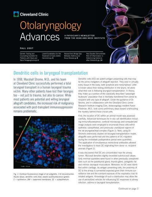
Fig. 1: Confocal fluorescence image of rat subglottis, 3-D reconstruction.<br />
Nuclei (blue), dendritic cells (red), elastin autofluorescence (green).<br />
E = epithelium, BM = basement membrane, SE = subepithelium.<br />
New Studies Demonstrate<br />
Role of Inflammation in<br />
Determining Outcome<br />
after Ototoxicity p. 14<br />
Dendritic cells (DC) are potent antigen presenting cells that may<br />
be the prime instigators of allograft rejection. They exist in virtually<br />
every tissue in the body, both lymphoid and nonlymphoid. Little<br />
is known about their resting distribution in the larynx, let alone<br />
what their role is following laryngeal transplantation. In theory,<br />
they make up a portion of the classically described “passenger<br />
leukocyte” population that is inevitably transferred from donor to<br />
recipient with any type of allograft. Under the guidance of Dr.<br />
Strome, and in collaboration with the <strong>Cleveland</strong> <strong>Clinic</strong> Lerner<br />
Research Institute Imaging Core, otolaryngology resident Aaron<br />
Friedman, M.D., took some preliminary steps toward unshrouding<br />
the mystery behind these crucial cells.<br />
First, the location of DC within an animal model was assessed<br />
carefully. Advanced techniques for in situ cell identification including<br />
immunofluorescence, confocal microscopy and computerized<br />
image analysis were employed to enumerate these cells within<br />
epithelial, subepithelial, and previously unanalyzed regions of<br />
the rat laryngotracheal complex (Figure 1). Next, using Dr.<br />
Strome’s extensively studied rat laryngeal transplantation model,<br />
allografts were performed and the patterns of DC migration<br />
during the immediate postoperative period were quantified.<br />
The application of simultaneous monoclonal antibodies allowed<br />
the investigators to track DC originating from donor vs. recipient<br />
animals (Figure 2).<br />
It was discovered that DC are concentrated near the airway<br />
lumen. Mucosal densities slightly exceeded submucosal values.<br />
Only minimal quantities were found in other previously unexplored<br />
sites such as the parathyroid gland, thyroid gland, paraglottic fat<br />
and intrinsic laryngeal musculature. Moreover, no DC were identified<br />
within cartilage. As antigen-presenting cells, the proximity of<br />
DC to the airway is somewhat expected given their immune surveillance<br />
role and the constant exposure of the respiratory tract to<br />
inhaled antigens. Knowledge of such a distribution may allow the<br />
use of aerosolized vehicles for influencing DC responses to airway<br />
infection, asthma or laryngeal transplantation.<br />
Continued on page 3
Marshall strome, M.D., M.s., F.a.C.s.<br />
TribuTe To Marshall sTroMe, M.D.<br />
By Tom I. Abelson, M.D., Medical Editor, <strong>Otolaryngology</strong> <strong>Advances</strong><br />
This space always has been filled with a yearly update from Marshall Strome, M.D., Chairman of the<br />
Head and Neck Institute. Dr. Strome announced last year he would step down from this position, and<br />
Michael S. Benninger, M.D., has been chosen to be the institute’s next Chairman (see back cover). It is<br />
with sadness over Dr. Strome’s departure and pride in his accomplishments that both this column and<br />
the entire issue of <strong>Otolaryngology</strong> <strong>Advances</strong> are dedicated to him.<br />
After Dr. Strome joined <strong>Cleveland</strong> <strong>Clinic</strong>’s Department of <strong>Otolaryngology</strong> and Communicative<br />
Disorders in August 1993, the department grew dramatically and eventually earned the designation<br />
Head and Neck Institute. The growth has been manifested in sections specifically designed to<br />
encompass the breadth and depth of staff expertise, which allow both productivity and mentoring<br />
of junior faculty. The institute has been a model for the four cornerstones of <strong>Cleveland</strong> <strong>Clinic</strong>’s<br />
mission: patient care (clinical and hospital), research and education.<br />
Below is a summary of some of Dr. Strome’s accomplishments, but he would probably say that<br />
he is most proud of his family and the family of people who work in the institute. And he knows<br />
the people. He knows the names of the custodial and maintenance staff and most others whose<br />
faces frequent our floors. He has actively supported and promoted the careers and professional<br />
development of his physician and research staff, as well as nurses and other support staff.<br />
During Dr Strome’s tenure, the staff has increased from seven to 32. There are now 133 people<br />
working within the institute.<br />
One of the cornerstones of Dr. Strome’s mission for the institute focused on research by residents,<br />
faculty and dedicated researchers. One year of the residency has been dedicated entirely to research,<br />
and our residents have won the most prestigious research awards in the country. Many of them<br />
later sought careers in academic medicine. In addition, Dr. Strome initiated a competitive resident<br />
research day as part of the two days of events during graduation ceremonies each spring.<br />
In the last two years, members of the institute have produced 240 publications, and the faculty<br />
has had more than 1,000 academic contacts, including publications, courses, visiting lectureships,<br />
professorships, awards and honors.<br />
Dr. Strome is especially proud of having expanded the <strong>Otolaryngology</strong> Regional Practice staff,<br />
opening offices in <strong>Cleveland</strong> <strong>Clinic</strong> family health centers in six <strong>Cleveland</strong> suburban locations and in<br />
Florida. His successful integration of faculty from these facilities to the main campus is a model<br />
for <strong>Cleveland</strong> <strong>Clinic</strong>.<br />
Dr. Strome’s professional accomplishments are well-known. He performed the first laryngeal<br />
transplant, developed many other significant innovations, authored more than 200 scientific<br />
publications, received a multitude of national and international awards and led many professional<br />
organizations and meetings.<br />
While we will miss the prodigious scientific and leadership qualities that Dr. Strome possesses, it is<br />
Dr. Strome the caring, engaged person that the <strong>Cleveland</strong> <strong>Clinic</strong> family will miss the most.
Dendritic cells in laryngeal transplantation continued from cover<br />
12 Hours<br />
7 Days<br />
H&E IMMUNOFLUORESCENCE<br />
DC also were distributed in an increasing craniocaudal gradient,<br />
with tracheal values exceeding supraglottic densities. The glottis,<br />
however, exhibited dramatically fewer DC. This may explain why<br />
croup is clinically confined to specific regions of the airway.<br />
Following laryngeal transplantation in rats, donor DC migrated to<br />
recipient cervical lymph nodes as early as 12 hours postoperatively<br />
and reached a nadir within 3 to 5 days. These cells also were<br />
identified in cervical lymph nodes of recipient animals from 12<br />
hours to 5 days postoperatively, ensuring that migration (rather<br />
than cell death or loss of cell surface markers) was occurring.<br />
Conversely, recipient DC infiltrated the laryngeal allograft, reaching<br />
a maximal density by day 7. The timing of these events helps to<br />
explain the investigators’ previously documented result that a<br />
week-long course of postoperative immunosuppression followed<br />
by no additional anti-rejection medication prevents significant<br />
laryngeal allograft rejection in rats for several months. This data,<br />
in turn, may translate into more optimized immunosuppression<br />
regimens for future human laryngeal transplant patients.<br />
In the setting of head and neck malignancy, however, laryngeal<br />
transplantation following total laryngectomy will only take root if<br />
systemic immunosuppression can be minimized or if donorspecific<br />
immunosuppression can be achieved. Dr. Strome and his<br />
research team are continuing in these efforts. Now that the early<br />
OX62 (Total DC) OX3 (Donor MHC II) OX6 (All MHC II) Combined Image<br />
Fig. 2: Immunofluorescence (IF) staining of 3 monoclonal antibodies in laryngotracheal allograft recipients sacrificed at 12 hours<br />
and 7 days postoperatively. Co-staining of cells allowed for differentiation of donor vs. recipient-derived DC. Hematoxylin and<br />
eosin (H & E) stained slides are 16 μm away from the corresponding IF slide. Images shown are from tracheal ring 4.<br />
kinetics of postoperative DC migration have been elucidated,<br />
steps toward manipulating this process can begin. By inhibiting<br />
donor DC efflux, preventing recipient DC influx, or perhaps a<br />
combination of the two, laryngeal allograft tolerance in the<br />
absence of generalized immunosuppression may become a reality.<br />
Moreover, the culture and preoperative administration of donor<br />
DC to the recipient has shown promise in other allograft systems,<br />
and adaptation into the laryngeal model is currently under way.<br />
Key Points:<br />
• Dendritic cells (DC) are potent antigen-presenting cells that<br />
may be the prime instigators of allograft rejection.<br />
• Laryngeal transplantation following total laryngectomy<br />
will only take root if systemic immunosuppression<br />
can be minimized or if donor-specific immunosuppression<br />
can be achieved.<br />
• By inhibiting donor DC efflux, preventing recipient DC influx,<br />
or perhaps a combination of the two, laryngeal allograft<br />
tolerance in the absence of generalized immunosuppression<br />
may become a reality.<br />
Head and Neck Institute 3
Genetic Testing and Counseling are Now Available<br />
for Patients with Head and Neck Paraganglioma<br />
Physicians caring for patients with head and neck<br />
paraganglioma are encouraged to refer them to<br />
<strong>Cleveland</strong> <strong>Clinic</strong> for genetic testing. At most, a<br />
positive result can be lifesaving because it will tell<br />
caregivers that a patient’s relatives may be at risk<br />
for familial paragangliomas — including vagal<br />
paragangliomas and glomus jugulare, glomus<br />
tympanicum and carotid body tumors. At the least,<br />
a negative result can put a family’s mind at ease.<br />
In either case, the benefit is worth the effort.<br />
Drs. Eng and Lorenz examining MRIs of a patient with multiple<br />
head and neck paragangliomas.<br />
4 Head and Neck Institute<br />
“The point of this simple blood test is to look for specific genetic<br />
mutations that are associated with hereditary paragangliomas,”<br />
says Robert Lorenz, M.D., Section Head of Head and Neck<br />
Oncologic Surgery. “When we find such a mutation in a patient,<br />
we can alert his or her family members that they may be at risk.<br />
If the relative is also positive, we can begin a monitoring program.<br />
If the relative does develop the disease, we will be able to detect<br />
it at the earliest possible moment.”<br />
Dr. Lorenz and colleague Charis Eng, M.D., Ph.D., Head of<br />
<strong>Cleveland</strong> <strong>Clinic</strong>’s Genomic Medicine Institute, established the<br />
screening program in spring 2007, and they have received a<br />
favorable response. Says Dr. Lorenz, “Patients have thanked us<br />
and told us things like, ‘I never knew this test existed. I’ve been<br />
living in fear that my kids will get this tumor, too.’ Well, there’s<br />
no point in fearing the unknown. It’s better to know for sure.”<br />
“For people who test positive, we can set up a schedule for regular<br />
magnetic resonance imaging scans, urine screens, other follow-up<br />
tests and family screening,” he continues. “On the other hand,<br />
people who test negative won’t ever have to worry about this again,<br />
nor will their children.”<br />
So far, Drs. Lorenz and Eng have screened 20 patients or family<br />
members of patients, and they have identified a positive result in 6.<br />
all testing includes counseling<br />
Counseling is an integral part of the screening process. Before<br />
any blood is drawn, the individual meets with Dr. Eng and a<br />
genetic counselor. Another meeting takes place when the results<br />
become known.<br />
“Pre- and post-test counseling are crucial,” says Dr. Lorenz. “You<br />
should never just perform a test and disclose the results without<br />
having adequately prepared a patient. You need to have a qualified<br />
person lead them down the road. A counselor makes the patient<br />
aware of all the possible outcomes and the implications of each.<br />
When a test is negative, the counselor can explain exactly what that<br />
means. When a test is positive, the counselor can initiate the longterm<br />
monitoring process.”
Characteristic splaying of the<br />
internal and external carotid<br />
areas on MRI: the telltale<br />
radiologic sign of a carotid<br />
body paraganglioma.<br />
the nature of the mutations<br />
The mutation responsible for paragangliomas occurs in one of<br />
the genes that encodes certain succinate dehydrogenase (SDH)<br />
subunits. SDH is an enzyme involved in the mitochondrial<br />
respiratory chain complex II, and it has four subunits (A, B, C,<br />
and D). Mutations in three of these subunits (B, C, and D) have<br />
been reported in individuals with hereditary paragangliomas and<br />
pheochromocytomas. These tumors may also be seen in<br />
association with syndromes caused by mutations in the VHL,<br />
RET, or NF1 genes.<br />
While a standard history may identify only 10% of patients<br />
with hereditary paraganglioma, studies have shown that testing<br />
for the SDH gene will demonstrate a mutation in up to 28%<br />
of cases. In addition, patients positive for a mutation in SDH<br />
subunit C are at increased risk for thyroid and kidney cancer<br />
at a young age.<br />
These mutations are passed on in families by autosomal<br />
dominant transmission. As a result, each child of a carrier has<br />
a 50% chance of inheriting the abnormal copy. A maternal<br />
imprinting effect has been observed in families with SDH<br />
subunit D mutations, so the children of a woman with an<br />
SDH-D mutation will not be at increased risk of developing a<br />
paraganglioma, even if they inherit the mutation and can<br />
pass it on to their own children.<br />
expanding the database<br />
As part of a research project, Drs. Lorenz and Eng are also<br />
offering genetic testing to more than 400 untested<br />
paraganglioma patients who were previously treated at the<br />
<strong>Cleveland</strong> <strong>Clinic</strong> Head and Neck Institute.<br />
“If you have a patient with a paraganglioma and you don’t know<br />
if it’s hereditary or not, please give us a call,” says Dr. Lorenz.<br />
“This is especially important if your patient is younger than 50 or<br />
has multiple tumors.”<br />
Key Points:<br />
• Genetic testing for patients with head and neck<br />
paraganglioma can be lifesaving.<br />
• Pre- and post-test counseling are an integral part of the<br />
screening process.<br />
• Testing for the SDH gene will demonstrate a mutation in up<br />
to 28% of cases.<br />
Head and Neck Institute 5
hot-and-Cold running success: laser/Cryoablation Procedure<br />
is superior to Traditional Treatment for early Glottic Carcinoma<br />
Thanks to a combined procedure, patients undergoing treatment for early-stage glottic carcinoma enjoy<br />
significantly better and quicker voice restoration.<br />
Preoperative videoendoscopy shows<br />
a T1 lesion of the right vocal fold.<br />
The procedure, which was pioneered by Marshall Strome, M.D.,<br />
Chairman of <strong>Cleveland</strong> <strong>Clinic</strong> Head and Neck Institute, involves<br />
the addition of cryoablation to standard CO2 laser resection.<br />
Data accumulated thus far reveal that patients treated with the<br />
combination procedure experience less perioperative inflammation,<br />
less scarring and less deposition of collagen. What’s more, the<br />
collagen that is present is less dense and histoarchitecturally<br />
amenable to vocal fold mobility. As a result, patients end up with<br />
more pliable vocal folds and better phonation. Moreover, the<br />
incidence of anterior glottic web formation in patients with anterior<br />
commissure involvement is lower. Finally, cure rates are comparable<br />
to those of laser therapy alone and radiotherapy (~90%).<br />
The addition of cryoablation adds only a few minutes to this<br />
outpatient procedure. “First, the tumor is lasered endoscopically<br />
until negative margins are confirmed by frozen-section biopsies,”<br />
says Claudio F. Milstein, Ph.D., a speech-language pathologist at<br />
<strong>Cleveland</strong> <strong>Clinic</strong> Voice Center. “Then the cryoprobe is applied to<br />
the tumor bed and slightly beyond. The tissue freezes instantly<br />
and thaws shortly thereafter. Patients are sent home with no<br />
restrictions on eating or speaking.”<br />
6 Head and Neck Institute<br />
Videoendoscopy 3 months postoperatively<br />
shows the larynx at rest and during<br />
phonation.<br />
An unexpected benefit<br />
Note the healthy appearance of the<br />
right vocal fold.<br />
“Dr. Strome initially developed this combined procedure thinking<br />
that it would result in better disease control,” Dr. Milstein continues.<br />
“But we were pleasantly surprised to discover that the cryoablation<br />
resulted in a quicker recovery and better voice quality than we had<br />
expected. We started seeing excellent clinical outcomes, but the<br />
reason was not clear.”<br />
To understand the mechanism behind these findings, Dr. Strome<br />
and colleagues conducted an experiment on 12 dogs. In each<br />
animal, one vocal fold was treated with laser therapy alone and<br />
the other was treated with laser and cryotherapy. Then histologic<br />
samples were analyzed and compared.<br />
“What we found was very interesting,” Dr. Milstein says. “In the<br />
cryoablated tissue, we saw less inflammation, less keratinization,<br />
and more deposition of hyaluronic acid than we saw in the tissues<br />
treated with laser alone. Also, we found that freezing alters the<br />
histoarchitecture of the collagen fibers, as they are more organized—that<br />
is, cross-linked in a basket-weave pattern. These<br />
are key findings that revealed why the pliability of the vocal fold<br />
tissue is enhanced and less scar formation is observed.”<br />
“As a result of our animal study, we started to understand why<br />
our surgical outcomes were so good. The combination of less<br />
inflammation, less collagen deposition, less-dense collagen and<br />
cross-linked fibers are all important for vocal fold vibration,<br />
mucosal waves and the resulting voice quality.”
Postoperative assessments<br />
For 1 year postoperatively, patients are scheduled for follow-up<br />
every 2 months because if a recurrence does develop, it is most<br />
likely to do so within the first year. Thereafter, patients are seen<br />
every 3 or 4 months through year 4, and then twice a year<br />
during year 5.<br />
“We keep a close eye on all our patients,” Dr. Milstein says.<br />
“Follow-up assessments are critical for early detection of possible<br />
recurrences. Even those patients who live outside the <strong>Cleveland</strong><br />
area are asked to make a commitment to return for regular<br />
follow-ups.”<br />
The postoperative assessments include videostroboscopy, a<br />
perceptual voice evaluation, and a patient self-rating:<br />
• During the videostroboscopic examination, clinicians look<br />
for minute changes in vibratory patterns and mucosal<br />
waves.<br />
• During the perceptual assessment, the patient’s voice<br />
quality is evaluated by staff clinicians.<br />
• Finally, patients rate their own voice quality.<br />
Findings are compared with preoperative baseline values, and<br />
the results are also tracked by serial evaluations throughout the<br />
course of follow-up.<br />
Follow-up study<br />
Thus far, the combined procedure has been performed on 48<br />
patients—45 men and three women, aged 46 to 96 years<br />
(mean: 66.2). Of this group, eight patients (16.7%) had already<br />
failed previous radiation treatment. Also, 17 patients (35.4%)<br />
had involvement of the anterior commissure, which is noteworthy<br />
because these patients generally fare poorly with the traditional<br />
laser procedure. Ten patients (20.8%) had carcinoma in situ, 19<br />
patients (39.6%) had category T1a disease, six patients (12.5%)<br />
were at category T1b, and 13 patients (27.1%) had a category<br />
T2 tumor. Follow-up ranged from 3 to 118 months (mean: 23).<br />
results<br />
The greatest improvement in subjectively appraised voice quality<br />
occurred between months 3 and 6, and improvement continued<br />
for up to 18 months in some cases. On the self-rating, 36 patients<br />
(75.0%) said their voices were “much better” and seven<br />
(14.6%) said they were “somewhat better” than they were before<br />
the procedure. Only three patients (6.3%) reported “no change,”<br />
and two (4.2%) said they were “somewhat worse.” “Overall,”<br />
says Dr. Milstein, “patient satisfaction has been very high.”<br />
The subjective findings mirrored the results of videostroboscopy.<br />
At 3 to 6 months, vocal fold vibrations and the resulting mucosal<br />
waves were much improved. Very little improvement was seen<br />
prior to the 3-month mark.<br />
The recurrence rate with combined laser/cryotherapy is consistent<br />
with the rates for both laser alone and radiation alone. Five of<br />
our patients (10.4%) experienced a recurrence. All of these<br />
developed within the first year postoperatively, and all patients<br />
were re-treated with the same therapy. All five remain tumor-free<br />
today. It is noteworthy that none of these recurrences arose in any<br />
of the eight patients who had previously failed radiation therapy.<br />
Finally, only two of the 17 patients (11.8%) with involvement of<br />
the anterior commissure developed an anterior glottic web, and<br />
one of those patients was already predisposed to keloid formation.<br />
“This is a significant finding because the literature shows that<br />
most patients with anterior commissure involvement who undergo<br />
laser surgery alone tend to develop a web,” says Dr. Milstein.<br />
“Our finding that cryotherapy is associated with a dramatic reduction<br />
in anterior glottic webs is yet another important advantage<br />
of combination treatment.<br />
“In conclusion,” he says, “the combined procedure is not just<br />
a viable alternative to laser ablation alone or a 7-week course<br />
of radiotherapy. We believe that it will eventually become the<br />
primary modality for treating early glottic carcinoma.”<br />
Key Points:<br />
• The addition of cryoablation to laser surgery results in<br />
better and quicker voice restoration than does treatment<br />
with laser surgery alone or radiation.<br />
• Combined laser/cryotherapy is available only at<br />
<strong>Cleveland</strong> <strong>Clinic</strong>.<br />
• Not only are voice outcomes better, disease-control<br />
rates are comparable to those of traditional treatments.<br />
• The incidence of anterior glottic web formation in patients<br />
with anterior commissure involvement is much lower with<br />
the combined procedure.<br />
Head and Neck Institute 7
Head and Neck Institute Researchers Bridge<br />
the Gap Between Asthma and Sinus Disease<br />
Researchers at the <strong>Cleveland</strong> <strong>Clinic</strong> Head and Neck Institute (HNI) have been studying the links between asthma<br />
and sinusitis during the last few years. Projects related to sinonasal polyposis have sought to uncover inflammatory<br />
pathways known to be active in asthma. Steven Cannady, M.D., Pete Batra, M.D., and Martin Citardi,<br />
M.D., collaborated with Serpil C. Erzurum, M.D., of the <strong>Cleveland</strong> <strong>Clinic</strong> Lerner Research Institute.<br />
The effort represents the first such collaboration between the<br />
HNI and Pathobiology and Pulmonary, Allergy and Critical Care<br />
Medicine Departments. “This project was a unique opportunity to<br />
take the lessons learned from intensive research into the mechanisms<br />
of asthma inflammation and apply them to our field,” says<br />
Dr. Cannady. Dr. Batra added, “With this project, we have begun<br />
to narrow the gap in understanding between the asthmatic airway<br />
and the chronic inflammation associated with sinus disease.”<br />
one airway hypothesis<br />
Asthma is a major public health problem with a long history of<br />
research funding that has allowed a legacy of important discoveries<br />
into its pathophysiology. More recently, basic research into the<br />
mechanisms of inflammation in the chronic sinusitis sufferer has<br />
been conducted. It has long been recognized that asthma and<br />
chronic nasal conditions such as rhinitis, sinusitis and nasal polyposis<br />
occur together more frequently than by chance. In addition,<br />
many of the standard treatments for asthma mirror those utilized<br />
in chronic sinus disease. It stands to reason that some of the<br />
mechanisms that initiate and sustain asthmatic airway inflammation<br />
could be active in sinusitis. Proving the presence in nasal<br />
polyps of molecular signaling cascades present in asthma is an<br />
important initial step in linking the two diseases.<br />
“The identification of common inflammatory mechanisms in<br />
asthma and sinusitis is paramount to understanding the nasal<br />
airway’s response to infection, chronic inflammation, and the<br />
origins of sinusitis,” says Dr. Citardi.<br />
translational research approach<br />
Through NIH funding obtained by Drs. Cannady, Citardi and Batra<br />
through the <strong>Cleveland</strong> <strong>Clinic</strong> General <strong>Clinic</strong>al Research Center, and<br />
an institutional grant, a translational research project was conducted.<br />
Healthy volunteers were recruited and compared with nasal<br />
polyp patients. <strong>Clinic</strong>al data, laboratory studies and tissue samples<br />
were collected and compared on all patients enrolled in the study.<br />
8 Head and Neck Institute<br />
“Translational research is a great way to apply science from the<br />
lab bench to help understand patient disease in the clinic,” says<br />
Dr. Erzurum. “Given the large population of patients suffering from<br />
chronic sinusitis and followed at the Head and Neck Institute, this<br />
type of project was bound to result in some interesting data.”<br />
More than 20 patients with chronic rhinosinusitis and nasal<br />
polyps were enrolled, as well as 10 healthy volunteers. Each<br />
patient enrolled was subjected to the sinonasal outcomes survey,<br />
had routine blood testing including tests for allergy, and underwent<br />
nasal brushings, lavage and biopsy (of polyp tissue or middle<br />
turbinate for disease and volunteers, respectively).<br />
New findings uncovered in nasal polyposis<br />
Signal transducers are responsible for translating a cells’ reaction<br />
to its environment into gene expression. As such, the Signal<br />
Transducers Activated on Transcription (STAT) class of tranducers<br />
are a relatively new family found to be important in the body’s inflammatory<br />
and anti-proliferative response. STAT1 is a relatively<br />
specific responder to infection and cytokines linked to infection.<br />
It was found to be upregulated in the tissue of nasal polyp patients<br />
when compared with healthy nasal tissue. Immunostaining localized<br />
this expression to the epithelium and endothelium of polyps,<br />
suggesting that inflammation is active at the mucosa-air interface,<br />
and the site of inflammatory cell recruitment-blood vessels (Figure).<br />
“STAT1 has previously been shown to be expressed at high levels<br />
in asthmatic airways, and in patients with cystic fibrosis. As a<br />
central signaling event, its activation results in gene expression<br />
and protein translation of inflammatory chemokines and cytokines<br />
found to be present in nasal polyposis. In some ways, it represented<br />
the discovery of the most upstream event in the polyp inflammatory<br />
cascade discovered to date,” says Dr. Cannady.<br />
From there, a thorough investigation into downstream events<br />
was conducted, representing the first complete evaluation of these<br />
pathways in polyp tissue. Highlights of the findings included<br />
confirmation of low nitric oxide (NO) in polyp irrigations despite<br />
high iNOS enzyme production.
Figure: Immunostaining demonstrated pSTAT1 staining was<br />
confined to the epithelium and endothelium of polyp tissue.<br />
Representative sections of H&E (A), cytokeratin (B), pSTAT1 (C),<br />
and von Willebrand factor (D) aided in localizing the cell types<br />
staining positively for pSTAT1.<br />
“The paradox represented by low NO in the setting of activated<br />
machinery to produce the gas has always puzzled researchers in<br />
this field. Thus, we sought to determine why,” says Dr. Batra.<br />
The substrates required for NO production were analyzed for the<br />
first time in nasal polyp tissue and found to be comparable to<br />
the levels in healthy volunteers.<br />
“Once we established that the reason for low NO was not a<br />
supply problem, we needed to assess what was happening to<br />
the NO produced that could contribute to its low measured<br />
values,” Dr. Citardi says.<br />
Expanding upon previous work by Dr. Citardi, nitrotyrosine, a<br />
marker of nitrosative damage to amino acids, was found to be<br />
elevated in polyps. In addition, superoxide dismutase (SOD), a<br />
critical enzyme for deactivation of reactive gases such as NO,<br />
was decreased in polyps. These findings comprised a plausible<br />
explanation for low NO despite STAT1 activation and iNOS<br />
expression. The NO gas was causing oxidative damage to<br />
the tissue of the nose with unchecked ability to counteract it<br />
with SOD.<br />
“It is very interesting just how similar the findings uncovered<br />
during Dr. Cannady’s time in my laboratory are with those we<br />
have found in asthma. They are nearly mirror images,” says<br />
Dr. Erzurum.<br />
“Interestingly, loss of NO by any mechanism has been associated<br />
with cell proliferation, and at least on the surface, could explain<br />
some of the proliferative phenotype seen in polyposis,” says<br />
Dr. Cannady.<br />
Significance of findings<br />
Identification of an upstream event such as STAT1 activation is<br />
an exciting finding. Not only does it provide evidence for similar<br />
biologic mechanisms in asthma and sinonasal polyposis, it<br />
provides a potential target for therapy.<br />
“More specific therapy for sinonasal polyposis is on the horizon.<br />
The identification of STAT1 activation is one event that may allow<br />
us to target therapy in a more specific manner for airway inflammatory<br />
diseases,” Dr. Citardi says.<br />
With the basic understanding afforded by this research, a clearer<br />
picture of the abnormalities involved in chronic nasal inflammation<br />
has emerged. “Other studies have identified portions of the<br />
pathway we have defined, but have not examined all factors that<br />
may affect it in one study. This was lacking in the literature up<br />
until now,” Dr. Cannady says.<br />
Future studies are currently planned to assess whether external<br />
factors activate STAT1 or whether the transducer is activated constitutively.<br />
“The next set of studies of polyp tissue will seek to understand<br />
if STAT1 represents a response of mucosa to its environment<br />
now, or a change in cellular mechanics that occurred distantly<br />
in the past,” says Dr. Batra.<br />
“In asthma, one hypothesis of the disease pathogenesis is that of<br />
perfectly timed viral insult can alter cell signaling and setting up a<br />
perpetual inflammatory environment. The same could be true of<br />
sinus sufferers,” says Dr. Erzurum.<br />
Regardless of what is found next, the findings already uncovered<br />
have unlocked a new avenue of investigation for rhinologists<br />
seeking to understand the pathogenesis of sinus disease.<br />
Key Points:<br />
• The Signal Transducers Activated on Transcription (STAT)<br />
class of transducers are a relatively new family found to<br />
be important in the body’s inflammatory and<br />
anti-proliferative response.<br />
• Nitrotyrosine, a marker of nitrosative damage to amino<br />
acids, was found to be elevated in polyps. Superoxide<br />
dismutase, a critical enzyme for deactivation of<br />
reactive gases such as NO, was decreased in polyps.<br />
• The identification of STAT1 activation may allow us to<br />
target therapy in a more specific manner for airway<br />
inflammatory diseases.<br />
• Future studies are currently planned to assess whether<br />
external factors activate STAT1 or whether the transducer<br />
is activated constitutively.<br />
Head and Neck Institute 9
Treatment of Benign Paroxysmal Positional<br />
Vertigo Continues to Evolve and Improve<br />
According to Judith White, M.D., Ph.D., Section Head of Vestibular and Balance Disorders, office maneuvers<br />
to diagnose benign paroxysmal positional vertigo (BPPV) have improved. The Dix-Hallpike maneuver is now<br />
routinely performed with little or no neck extension. This technique improves tolerance by older patients who<br />
may have cervical arthritis or other limitations. Alternately, the “side-lying” test may be used, which is the<br />
first position in the Semont maneuver. The patient lies on his side and points his nose to the ceiling. No neck<br />
extension is needed and good stimulation of the posterior semicircular canal of the undermost ear is<br />
achieved. Visualization of the nystagmus can be aided by the use of infrared video or optical Frenzel lenses,<br />
which eliminate visual fixation.<br />
Fig. 1: In left lateral geotropic semicircular canal benign paroxysmal<br />
positional vertigo, horizontal nystagmus beats toward the<br />
undermost ear in supine head turns.<br />
Treatment efficacy has been well proven in randomized controlled<br />
trials. Approximately 90% of BPPV cases involve the<br />
posterior semicircular canal. Canalith repositioning is performed<br />
while the patient is still reclined in the diagnostic Dix-Hallpike<br />
position. Meta-analysis suggests symptoms resolve in 72% of<br />
these patients following a single office maneuver and in 99%<br />
after repeated maneuvers, in contrast to a 33% resolution rate<br />
at three weeks without treatment. BPPV is believed to be selflimiting,<br />
although Baloh and Honrubia reported symptoms that<br />
persisted for more than one year in one-third of their 240 patients<br />
with BPPV. Several controlled randomized trials have<br />
found no efficacy of vestibular suppressants in BPPV.<br />
10 Head and Neck Institute<br />
lateral canal involvement is more frequently recognized<br />
The lateral (horizontal) canal is the second most common canal<br />
affected by BPPV. Lateral semicircular canal benign paroxysmal<br />
positional vertigo (LSC-BPPV) is characterized by nystagmus<br />
provoked by supine bilateral head turns and beating toward the<br />
undermost ear. There are two distinct subtypes of LSC-BPPV<br />
based on the direction of horizontal nystagmus during supine<br />
head turns: geotropic and apogeotropic. Geotropic LSC-BPPV<br />
beats toward the undermost ear on supine positional testing and<br />
is characterized by short latency and prolonged duration of horizontal<br />
nystagmus with poor fatiguability. Apogeotropic LSC-BP-<br />
PV is characterized by similar short latency and prolonged-duration<br />
horizontal nystagmus, but the nystagmus beats away from<br />
the undermost ear on supine positional testing. These two<br />
types may reflect the position of the otoconial debris in the lateral<br />
canal (Figures 1 and 2).<br />
The diagnosis of lateral canal BPPV is improved by performing<br />
supine head turns. While Dix-Hallpike positioning is highly<br />
sensitive to posterior semicircular canal BPPV, it lacks sensitivity<br />
in LSC-BPPV. Positioning testing should include both Dix-Hallpike<br />
and supine positional testing in the head-centered supine,<br />
right-ear-down and left-ear-down positions. We generally perform<br />
the supine testing first, because the nystagmus observed when<br />
initially lying supine may be very helpful in identifying the affected<br />
canal. The head is then turned to the left and right while supine,<br />
and then the patient returns to sit and the Dix-Hallpike positioning<br />
is performed. The Dix-Hallpike alone was entirely negative in two<br />
published patient reports where horizontal nystagmus with lateral<br />
supine head turns reached 12 d/s and 16 d/s due to LSC-BPPV.<br />
The identification of the involved ear in lateral semicircular<br />
canal BPPV can be especially difficult because the canals are<br />
coplanar, and nystagmus is seen in both lateral supine positions.<br />
Order effect and head tilt may affect the direction of nystagmus.<br />
In geotropic LSC-BPPV, the nystagmus is worse with the<br />
affected ear down. Treatment for geotropic LSC-BPPV consists<br />
of 360° roll maneuvers toward the unaffected ear beginning
Fig. 2: Conversion of posterior semicircular canal BPPV to lateral canal following canalith repositioning. The particle leaves the posterior canal<br />
and travels across the utricle to enter the long arm of the lateral canal or to abut directly against the cupula of the lateral canal. The nystagmus<br />
seen is geotropic horizontal in the first case and apogeotropic in the second case.<br />
with the patient in the supine position with the head flexed 0 to<br />
30° and laterally rotated toward the affected ear, and proceeding in<br />
90° increments every 30 to 60 seconds toward the unaffected ear.<br />
The Gufoni maneuver is also highly effective and is performed with<br />
the patient beginning in the sitting position and lying quickly to the<br />
unaffected side and then rotating the head 45° downward, maintaining<br />
the position for two to three minutes as described in Casani.<br />
treatment for apogeotropic lsc-BPPV consists<br />
of a variety of maneuvers<br />
Identification of the affected ear can be more challenging in<br />
apogeotropic LSC-BPPV. Nystagmus is usually worse with the<br />
affected ear uppermost, and nystagmus is occasionally seen on<br />
assuming the supine position, which usually beats toward the<br />
involved side. The modified Gufoni maneuver can be performed<br />
with the patient beginning in the sitting position and lying quickly<br />
to the affected side and then rotating the head 45° upward,<br />
maintaining the position for 2 to 3 minutes, as described by<br />
Appiani. Anterior semicircular canal BPPV is a controversial<br />
entity. Some investigators suggest that torsional nystagmus has<br />
a downbeat component, in contrast to posterior canal, which<br />
has an upbeat component. Since the same maneuvers used to<br />
treat posterior canal BPPV appear effective for possible anterior<br />
canal involvement, the question may have more theoretical than<br />
clinical relevance, and has rarely been a consideration in our<br />
clinical practice.<br />
Central nystagmus may be positional. Several cases of<br />
multiple sclerosis have presented to the vestibular section with<br />
paroxysmal downbeat nystagmus in supine testing. MRI<br />
studies revealed the diagnosis. Central pathology should always<br />
be a consideration in atypical, vertical nystagmus or in patients<br />
with persistent BPPV despite multiple repositioning attempts.<br />
In another case, persistent apogeotropic horizontal positional<br />
nystagmus was associated with an acoustic neuroma.<br />
recurrence is common after successful canalith<br />
repositioning for BPPV<br />
Treatment is commonly effective in eliminating the current<br />
episode, but does not prevent additional episodes. Although the<br />
average recurrence rate is approximately 15% per year, reported<br />
rates have ranged from 5% per year to 45% at 30 weeks.<br />
Recurrence usually affects the same ear as the original BPPV.<br />
Conversion between canals can occasionally occur, usually<br />
between posterior and lateral canals when the patient is retested<br />
with Dix-Hallpike positioning after canalith repositioning has been<br />
performed. It is heralded by the development of brisk horizontal<br />
nystagmus, and responds well to an immediate supine head roll<br />
maneuver toward the unaffected side.<br />
Several studies have suggested that post-procedure instructions<br />
to keep the head elevated do not increase treatment efficacy,<br />
although conflicting reports are also published. Numerous centers<br />
continue to recommend post-procedure restrictions based<br />
on anecdotal experience.<br />
For references, please e-mail the editor.<br />
Key Point:<br />
• Office maneuvers to diagnose BPPV have improved, along<br />
with treatment efficacy.<br />
Head and Neck Institute 11
studying Novel Techniques for Detection of hPV infectivity in squamous<br />
Cell Carcinoma Patients without a history of alcohol or Tobacco use<br />
Tobacco and alcohol use are well-known and established risk factors for head and neck squamous cell carcinoma<br />
(HNSCC). However, there is a subset of HNSCC patients who do not report a history of tobacco or<br />
alcohol use. These patients comprise a unique population, and other etiologies for their HNSCC have been<br />
investigated. One such area of research is the role of human papillomavirus (HPV).<br />
A recent article in the New England Journal of Medicine has<br />
shown HPV to be a risk factor for HNSCC. Furthermore, numerous<br />
reports have demonstrated a survival outcome advantage<br />
in patients with HPV-positive tumors. With the recent approval<br />
of the HPV multivalent vaccine for cervical cancer, research is<br />
ongoing as to the utility of such a vaccine against HNSCC in<br />
this patient population. Rapid and accurate diagnosis of patients<br />
with HPV-associated HNSCC will be critical to any therapeutic<br />
intervention targeting the virus.<br />
HPV serotypes that have been associated with cancer<br />
development are 16 and 18. The incidence of these patients<br />
with HPV varies widely. Across published reports, HPV positivity<br />
varies from 20-90%. This variation may be due to the variety<br />
and sensitivity of the methods used to identify HPV. One of the<br />
most common methods is polymerase chain reaction (PCR).<br />
However, this method is known to be sensitive to contamination,<br />
and may be prone to false negatives. If accurate investigation of<br />
HPV-associated tumors is to be done, then it will be necessary<br />
to provide an accurate and sensitive method of determining<br />
HPV involvement.<br />
Walter Lee, M.D., and colleagues in the Department of<br />
Pathology and the Head and Neck Institute, have recently<br />
presented research on detecting HPV using a new method in<br />
surgical samples at an international meeting in Barcelona,<br />
Spain. A series of 22 patients without a history of alcohol or<br />
tobacco use underwent detection of HPV using novel and<br />
newly available well-validated low-and high-risk probe sets<br />
(Ventana Medical Systems, Tucson, Ariz.). Both low- and<br />
high-risk serotypes were screened in pathological specimens.<br />
This technique allows for accurate and rapid detection of HPV<br />
infectivity in pathological samples. Furthermore, it allows for<br />
direct visualization of HPV in HNSCC cells. The high-risk<br />
Family 16 probe cocktail has an affinity to high-risk HPV<br />
genotypes 16, 18, 31, 33, 35, 39, 51, 52, 56, 58, and 66.<br />
The low-risk Family 6 probe cocktail has an affinity to HPV<br />
genotypes 6 and 11. The slides were examined for the presence<br />
of integrated HPV using light microscopy. Positive signal is<br />
indicated by dark blue staining. Copy number can be estimated<br />
by comparison to the controls, which are xenograft controls<br />
that have known copy numbers. Internal negative controls were<br />
examined for each tissue section.<br />
12 Head and Neck Institute<br />
There were a total of 22 patients in this series. Three patients<br />
had multiple samples due to persistent or recurrent tumor and,<br />
thus, the total number of samples tested was 25. There were<br />
14 males and 8 females. The average age of the patients was<br />
64 years. In no case did the multiple samples have a different<br />
HPV status. The most common anatomic sites included tongue<br />
(8 cases), tonsil (7 cases) and larynx (7 cases). The Tumor<br />
(T) staging of these 25 samples were: T1=5, T2=8, T3=4<br />
and T4=5. The Neck (N) staging was: N0=17, N1=2, N2b=5,<br />
N2c=1. 24 samples were M0 while one sample was stage M1.<br />
Two of 22 (10%) patients had positive detection for high risk<br />
HPV probes. Both of these samples were tonsillar tumors,<br />
thus resulting in 29% of tonsil samples having high-risk HPV.<br />
No tumors were positive for low-risk HPV.<br />
The study demonstrates the utility of new, commercially<br />
available, well-validated probe sets for low- and high-risk HPV<br />
determination that can be used in clinical paraffin embedded<br />
tissue samples. The significant advantages of the ISH technique<br />
are visualization and localization of HPV and a high sensitivity<br />
that is coupled with a high specificity.<br />
For references, please e-mail the editor.<br />
Key Points:<br />
• Detection of HPV in head and neck squamous cell cancer<br />
may be critical in some patients without a history of tobacco<br />
or alcohol use.<br />
• A new technique available at <strong>Cleveland</strong> <strong>Clinic</strong> provides for<br />
accurate and rapid detection of HPV infectivity.
H & E stain of squamous cell carcinoma of the tonsil. Positive ISH for high-risk HPV in same sample. The blue demonstrates<br />
The Hearing Implant Program (HIP) to date has done approximately<br />
80 bilateral cochlear implants; approximately 25 have<br />
been simultaneous and the rest sequential, according to Peter<br />
Weber, M.D., Co-Director of HIP.<br />
The advantages that bilateral cochlear implantation provide<br />
are superior hearing in conditions where background noise is<br />
prevalent, as well as better hearing in situations where there<br />
is quiet. In addition, a significant number of patients are able<br />
to obtain sound localization skills and the squelch effect is<br />
averted. The squelch effect occurs when the head blocks sound<br />
coming from the opposite side (i.e., the side that does not have<br />
the microphone for the cochlear implant attached). Thus, the<br />
patient has to constantly turn his/her head in the direction<br />
of the sound in order for the microphone to pick it up.<br />
Every adult patient who has undergone a bilateral implant in<br />
a sequential fashion has demonstrated significant improvement<br />
in hearing abilities when wearing both implants. The same is<br />
true for children who are old enough to test. Interestingly, the<br />
vast majority of adult patients actually do better with the new<br />
implant than with their old implant. Part of this result may be<br />
due to new technology or we may be implanting the “dominant<br />
ear.” This includes patients who had their first implant up to 11<br />
years before their second implant. Our simultaneous group is<br />
demonstrating impressive results with scores and testing abilities<br />
that far outpace what we expect from a single implant user and,<br />
in some instances, outpacing children with normal hearing.<br />
HPV in situ.<br />
bilateral Cochlear implantation Program Proves successful<br />
We are proud that one of our patients, whom we implanted on<br />
her first birthday, will be one of the subjects featured in a new<br />
movie about bilateral implants.<br />
We also have evaluated surgical complications associated with<br />
bilateral cochlear implants and have not seen any significant<br />
complications. Surgical complications evaluated were facial<br />
paralysis (0 instances), vertigo (about 5% incidence - only<br />
transient for few days postop, no long-term problems) dysguesia<br />
(0%), TM perforations (0%), infection including meningitis (0%),<br />
long-term pain or headache (0%), and bleeding (0%). Increased<br />
time of surgery for simultaneous bilateral CI was on average<br />
about 60 minutes; this increase did not necessitate any other<br />
treatment such as placement of a Foley catheter. Thus, we are<br />
demonstrating the significant advantages of bilateral cochlear<br />
implantation as well as the relative safety of this implantation.<br />
Key Points:<br />
• Bilateral cochlear implantation provides superior hearing in<br />
conditions where background noise is prevalent, as well as<br />
better hearing in situations where there is quiet. In addition,<br />
a significant number of patients are able to obtain sound<br />
localization skills and avert the squelch effect.<br />
• We have not seen any significant surgical complications<br />
associated with bilateral cochlear implants.<br />
Head and Neck Institute 13
New Studies Demonstrate Role of Inflammation<br />
in Determining Outcome after Ototoxicity<br />
Researchers in <strong>Cleveland</strong> <strong>Clinic</strong>’s auditory neuroscience<br />
laboratory have recently demonstrated a new<br />
association between the role of inflammation and the<br />
effect of ototoxic drugs on hair cell survival and hearing.<br />
This work represents the first study in which inflammation<br />
has been shown to play a role in the outcome<br />
after drug toxicity in the ear.<br />
In the case of ototoxicity, the primary determinants of cell survival<br />
or cell death are likely due to direct effects of the drug on hair<br />
cells and their function. Studies in aminoglycoside ototoxicity<br />
have demonstrated that hair cells do uptake the drug, the drug is<br />
incorporated in lysosomes of the hair cells, and proapoptotic<br />
pathways are initiated via activation of oxygen-free radicals and<br />
stimulation of caspase-mediated cell death pathways. However,<br />
it is possible that other influences that are derived from outside<br />
the cochlea also play a role in determining which cells eventually<br />
recover and which cells succumb to toxicity. This is the first<br />
study that shows that inflammation in the cochlea can adversely<br />
affect cell survival after ototoxic injury.<br />
“We used a transgenic mouse that expresses green fluorescent<br />
protein in the place of an important signalling molecule called<br />
fractalkine receptor,” says Keiko Hirose, M.D., Section Head of<br />
Pediatric <strong>Otolaryngology</strong>. Fractalkine receptor, abbreviated CX3-<br />
CR1, is expressed in monocytes, macrophages, microglia, some<br />
T cells and NK cells. CX3CR1 was found to be important in<br />
protecting hair cells and in preserving residual hearing after<br />
kanamycin exposure in mice. The cells that express CX3CR1<br />
are the cochlear inflammatory cells, called cochlear macrophages.<br />
These macrophages have been observed in the past in the<br />
cochlea in various types of damage including noise damage and<br />
drug-induced damage. Macrophages are primarily seen in the<br />
structures of the cochlea called the spiral ligament and spiral<br />
limbus, sites known to manifest cellular loss following a number<br />
of different hearing insults.<br />
14 Head and Neck Institute<br />
The discovery of the protective role of this receptor carried by<br />
macrophages suggests that hair cell damage and hearing loss<br />
can be rendered far worse by a poorly regulated inflammatory<br />
response after primary insult to the ear.<br />
Immunohistochemistry has shown that the population of green<br />
fluorescent CX3CR1-expressing cells are best described as<br />
macrophages for the following reasons: one, they express other<br />
surface markers consistent with tissue macrophages, and two,<br />
they are observed engulfing particles of cellular debris that<br />
result from the primary tissue damage exerted by the ototoxic<br />
medication. Both immunostaining and detailed light microscopic<br />
studies have confirmed these two aspects of the inflammatory<br />
cell population in the cochlea. The recruitment of these inflammatory<br />
cells may in part be a means to clear the cochlea of toxic<br />
substances unleashed by dead and dying cells. The inflammatory<br />
cells could be capable of exerting some protective effects or<br />
may be laying the foundation for repair.<br />
“Based on our recent studies, it is clear that when inflammatory<br />
cells lose their ability to signal through CX3CR1, these cells<br />
can exacerbate the primary damage mediated by ototoxic drugs<br />
such as kanamycin, an antibiotic, and furosemide, a diuretic,”<br />
Dr. Hirose says. “If inflammation plays an important role in the<br />
repair process after destructive hearing loss, we may be able to<br />
intervene with agents that modulate the inflammatory response<br />
and thereby prevent or perhaps attenuate loss of hearing.”<br />
This research was presented at the International Meeting of<br />
Neuroimmunology in Nagoya, Japan.<br />
Key Points:<br />
• If inflammation plays an important role in the repair process<br />
after destructive hearing loss, we may be able to intervene<br />
with agents that modulate the inflammatory response and<br />
thereby prevent or perhaps attenuate loss of hearing.
Head and Neck Institute Staff Directory<br />
Chairman<br />
Audiology<br />
Center for Surgery Research<br />
Marshall Strome, M.D., M.S., F.A.C.S.<br />
Professor and Chairman,<br />
Head and Neck Institute<br />
<strong>Clinic</strong>al interests: head and neck surgery<br />
with special interest in laryngology; thyroid<br />
and parotid surgery<br />
Ph: 216.444.6691 FX: 216.445.9409<br />
Craig Newman, Ph.D.<br />
Main Campus<br />
Vice Chairman and Section Head<br />
<strong>Clinic</strong>al interests: adult audiologic rehabilitation;<br />
tinnitus; evoked potentials; hearing aids;<br />
outcomes research<br />
Ph: 216.444.6691 FX: 216 .445.9409<br />
Donald Goldberg, Ph.D.<br />
Hillcrest Hospital Atrium<br />
Co-Director, Hearing Implant Program<br />
<strong>Clinic</strong>al interests: audiologic (aural) management/<br />
treatment; cochlear implants; auditory-verbal therapy;<br />
pediatric and educational audiology; communication<br />
assessment of children and adults who are deaf<br />
or hard of hearing<br />
Ph: 440.312.3681 FX: 440.312.8810<br />
Sharon Sandridge, Ph.D.<br />
Main Campus<br />
Director, Audiology <strong>Clinic</strong>al Services<br />
<strong>Clinic</strong>al interests: electrophysiologic assessment;<br />
state-of-the-art amplification options including<br />
assistive listening devices and digital hearing aids;<br />
tinnitus and older adults<br />
Ph: 216.444.6691 FX: 216.445.9409<br />
Suyu Shu, Ph.D.<br />
Main Campus<br />
research interests: cellular immunology;<br />
cancer immunotherapy; molecular biology<br />
Ph: 216.445.3800 FX: 216.445.3805<br />
Community <strong>Otolaryngology</strong><br />
Robert Katz, M.D.<br />
Solon Family Health Center<br />
Section Head<br />
<strong>Clinic</strong>al interests: pediatric otolaryngology;<br />
otology; head and neck surgery; general<br />
otolaryngology<br />
Ph: 440.519.6950 FX: 440.519.1364<br />
Tom Abelson, M.D.<br />
Beachwood Family Health and Surgery Center<br />
Solon Family Health Center<br />
<strong>Clinic</strong>al interests: voice medicine; pediatric<br />
otolaryngology; sinus disease; general<br />
otolaryngology<br />
Ph: 216.839.3740 FX: 216.839.3749<br />
Steven Ball, M.D.<br />
Strongsville Family Health and Surgery Center<br />
<strong>Clinic</strong>al interests: adult and pediatric general<br />
otolaryngology; chronic sinusitis and sinus surgery;<br />
thyroid and salivary gland surgery; adult and<br />
pediatric neck masses<br />
Ph: 440.878.2500 FX: 440.878.2666<br />
Edward Fine, M.D., Ph.D.<br />
Westlake Family Health Center<br />
<strong>Clinic</strong>al interests: laryngology; sinonasal disease;<br />
facial cosmetics and reconstruction<br />
Ph: 440.899.5630 FX: 440.899.5636<br />
Richard Freeman, M.D., Ph.D.<br />
Westlake Family Health Center<br />
<strong>Clinic</strong>al interests: general otolaryngology;<br />
head and neck surgery; sinonasal disease<br />
Ph: 440.899.5630 FX: 440.899.5636<br />
Daniel Knott, M.D.<br />
Hillcrest Hospital Atrium<br />
<strong>Clinic</strong>al interests: general otolaryngology; facial<br />
plastic and reconstructive surgery<br />
Ph: 440.312.3681 FX: 440.312.8810
Staff directory continued<br />
George Ozbardakci, M.D.<br />
Lorain Family Health and Surgery Center<br />
<strong>Clinic</strong>al interests: sinus problems; hearing loss; hearing<br />
aids; snoring; sleep apnea; tonsils and adenoids<br />
Ph: 440.204.7400 FX: 440.204.7396<br />
Facial Aesthetic and Reconstructive Surgery<br />
Daniel Alam, M.D.<br />
Main Campus, Beachwood Family Health and Surgery<br />
Center<br />
Section Head<br />
<strong>Clinic</strong>al interests: plastic and reconstructive surgery;<br />
facial plastic surgery; head and neck microvascular<br />
reconstruction; facial paralysis; rhinoplasty<br />
Ph: 216.444.6691 FX: 216.445.9409<br />
Michael Fritz, M.D.<br />
Main Campus, Independence Family Health Center<br />
<strong>Clinic</strong>al interests: pediatric and adult facial plastic<br />
surgery; functional and cosmetic rhinoplasty; soft tissue<br />
and bony facial reconstruction for complex head and<br />
neck wounds following surgery or trauma; microvascular<br />
reconstruction<br />
Ph: 216.444.6691 FX: 216.445.9409<br />
General <strong>Otolaryngology</strong><br />
Head and Neck Surgery<br />
Alan Kominsky, M.D.<br />
Main Campus, Beachwood Family Health Center<br />
Vice Chairman and<br />
Quality Review Officer<br />
<strong>Clinic</strong>al interests: adult and pediatric general otolaryngology;<br />
sleep apnea; snoring; sinonasal disease<br />
Ph: 216.444.6691 FX: 216.445.9409<br />
Catherine Henry, M.D.<br />
Main Campus<br />
<strong>Clinic</strong>al interests: chronic cough; reflux disease;<br />
medical management of sinus disease; hearing loss;<br />
balance disorders<br />
Ph: 216.444.6691 FX: 216.445.9409<br />
Robert Lorenz, M.D.<br />
Main Campus<br />
Section Head<br />
<strong>Clinic</strong>al interests: vocal cord paralysis; head and neck<br />
oncology; laryngotracheal reconstruction; skull base<br />
tumors; benign and malignant tumors of the neck,<br />
thyroid and salivary glands<br />
Ph: 216.444.6691 FX: 216.445.9409<br />
Laryngology<br />
Walter Lee, M.D.<br />
Main Campus<br />
<strong>Clinic</strong>al interests: salivary gland tumors; oral cavity<br />
lesions; laryngeal tumors; immunotherapy research<br />
Ph: 216.444.6691 FX: 216.445.9409<br />
Joseph Scharpf, M.D.<br />
Main Campus<br />
<strong>Clinic</strong>al interests: head and neck cancer<br />
and reconstructive surgery; general adult<br />
and pediatric otolaryngology<br />
Ph: 216.444.6691 FX: 216.445.9409<br />
Marshall Strome, M.D.<br />
Main Campus<br />
<strong>Clinic</strong>al interests: head and neck surgery with special<br />
interest in laryngology; thyroid and parotid surgery<br />
Ph: 216.444.6691 FX: 216.445.9409<br />
Benjamin Wood, M.D.<br />
Main Campus<br />
<strong>Clinic</strong>al interests: head and neck benign and malignant<br />
tumors; thyroid and parotid tumors; sinus tumors and<br />
chemodectomas<br />
Ph: 216.444.6691 FX: 216.445.9409<br />
Marshall Strome, M.D.<br />
Main Campus<br />
<strong>Clinic</strong>al interests: head and neck surgery with special<br />
interest in laryngology; thyroid and parotid surgery<br />
Ph: 216.444.6691 FX: 216.445.9409<br />
Nasal and Sinus Disorders<br />
Martin Citardi, M.D.<br />
Main Campus, Beachwood Family Health<br />
and Surgery Center<br />
Section Head and<br />
Associate Residency Program Director<br />
<strong>Clinic</strong>al interests: revision sinus surgery; frontal sinus<br />
surgery; sinonasal neoplasia; computer-aided sinus surgery;<br />
endoscopic orbital decompression; endoscopic CSF<br />
leak repair<br />
Ph: 216.444.6691 FX: 216.445.9409
Staff directory continued<br />
Otology<br />
Pete Batra, M.D.<br />
Main Campus, Beachwood Family Health<br />
and Surgery Center<br />
<strong>Clinic</strong>al interests: revision endoscopic sinus surgery;<br />
medical and surgical management of sinonasal polyposis;<br />
sinonasal tumors; CSF leak repair; endoscopic orbital<br />
and DCR surgery; image-guided surgery; inhalant allergy<br />
Ph: 216.444.6691 FX: 216.445.9409<br />
Gordon Hughes, M.D.<br />
Main Campus, Hillcrest Hospital Atrium<br />
Section Head<br />
<strong>Clinic</strong>al interests: ear surgery for deafness and<br />
infection; facial paralysis; immunology of the ear;<br />
pediatric ear diseases; vertigo diagnosis and management;<br />
tumors of the ear<br />
Ph: 216.444.6691 FX: 216.445.9409<br />
Peter Weber, M.D.<br />
Main Campus, Hillcrest Hospital Atrium,<br />
Beachwood Family Health and Surgery Center<br />
Co-Director, Hearing Implant Program<br />
Residency Program Director<br />
<strong>Clinic</strong>al interests: surgery for pediatric and adult ear<br />
disease including cochlear implants; implantable hearing<br />
aids; infectious cholesteatomas; acoustic neuromas; ear<br />
tumors; skull bone lesions; facial nerve disorders and vertigo<br />
Ph: 216.444.6691 FX: 216.445.9409<br />
Pediatric <strong>Otolaryngology</strong><br />
Keiko Hirose, M.D.<br />
Main Campus<br />
Section Head, Pediatric <strong>Otolaryngology</strong><br />
<strong>Clinic</strong>al interests: pediatric ear surgery; hearing loss<br />
evaluation; cochlear implantation; basic science research<br />
in causes of deafness; general pediatric otolaryngology<br />
Ph: 216.444.6691 FX: 216.445.9409<br />
Chris Discolo, M.D.<br />
Main Campus, Hillcrest Hospital Atrium<br />
<strong>Clinic</strong>al interests: pediatric otolaryngology; diagnosis<br />
and management of complex airway problems; head and<br />
neck masses; speech and swallowing disorders;<br />
management of children with cleft lip and palate<br />
Ph: 216.444.6691 FX: 216.445.9409<br />
Paul Krakovitz, M.D.<br />
Main Campus, Beachwood Family Health and Surgery<br />
Center, Independence Family Health Center<br />
<strong>Clinic</strong>al interests: malignant head and neck disease;<br />
pediatric and adult thyroid/parathyroid disorders; voice<br />
surgery and velopharyngeal insufficiency; general<br />
pediatric otolaryngology<br />
Ph: 216.444.6691 FX: 216.445.9409<br />
Speech and Language Pathology/Voice Center<br />
Vestibular and Balance Disorders<br />
Douglas Hicks, Ph.D.<br />
Main Campus<br />
Section Head<br />
Director, Voice Center<br />
<strong>Clinic</strong>al interests: voice science; voice disorders; care<br />
of the professional voice<br />
Ph: 216.444.6691 FX: 216.445.9409<br />
Claudio Milstein, Ph.D.<br />
Main Campus<br />
<strong>Clinic</strong>al interests: voice disorders;<br />
care of the professional voice; aerodigestive<br />
tract disorders; laryngeal physiology; functional<br />
dysphonia; vocal cord dysfunction<br />
Ph: 216.444.6691 FX: 216.445.9409<br />
Judith White, M.D., Ph.D.<br />
Main Campus, Beachwood Family Health<br />
and Surgery Center<br />
Section Head<br />
<strong>Clinic</strong>al interests: vestibular disorders; dizziness and<br />
balance; hearing problems; ear disease; vertigo<br />
Ph: 216.444.6691 FX: 216.445.9409<br />
beachwood Ph: 216.839.3740<br />
<strong>Cleveland</strong> <strong>Clinic</strong> in Florida<br />
Gilberto Alemar, M.D.<br />
Weston<br />
<strong>Clinic</strong>al interests: surgery of the nose<br />
and sinuses; sinusitis; voice and swallowing disorders;<br />
head and neck tumor surgery; sleep apnea and snoring;<br />
surgery for airway reconstruction<br />
Ph: 954.659.5786 FX: 954.659.5787<br />
Eloy Villasuso III, M.D.<br />
Weston<br />
<strong>Clinic</strong>al interests: Hearing loss; dizziness/balance<br />
disorders; chronic ear infections; pediatric ear disease;<br />
acoustic neuromas; lateral skull base tumors; facial<br />
nerve disorders; cochlear implants; otosclerosis<br />
Ph: 954.659.5178 FX: 954.659.5787
New staff<br />
Daniel Knott, M.D.<br />
Daniel Knott, M.D., recently joined<br />
the staff of <strong>Cleveland</strong> <strong>Clinic</strong>’s Head and<br />
Neck Institute. Dr. Knott specializes<br />
in general otolaryngology and facial<br />
plastic and reconstructive surgery.<br />
Dr. Knott earned his medical degree from the University of California San Diego<br />
School of Medicine. He has a bachelor of arts in international politics and<br />
economics from Middlebury College in Middlebury, Vermont. Dr. Knott completed<br />
a general surgery internship and a residency in otolaryngology/head and neck<br />
surgery at <strong>Cleveland</strong> <strong>Clinic</strong>. He completed his fellowship in facial plastic and<br />
reconstructive surgery at UCLA Medical Center in Los Angeles.<br />
Dr. Knott recently won the American Head and Neck Society’s Robert Maxwell<br />
Byers Award for his evaluation of hardware-related complications in vascularized<br />
bone grafts with locking mandibular reconstruction plate fixation. He has received<br />
several additional awards and honors during his education and residency.<br />
Dr. Knott has authored numerous papers and given presentations across<br />
the country. He has applied for a patent, along with several co-inventors, for<br />
tyramine-based hydrogels for vocal cord repair and augmentation.<br />
Dr. Knott is seeing patients at Hillcrest Hospital. His appointment number<br />
is 440.312.3681.<br />
18 Head and Neck Institute<br />
U.S.News ranks <strong>Cleveland</strong> <strong>Clinic</strong><br />
one of america’s Top hospitals<br />
head and Neck institute ranked among Top 10<br />
<strong>Cleveland</strong> <strong>Clinic</strong> has been ranked among America’s top hospitals since<br />
U.S.News & World Report began its annual survey of “America’s Best<br />
Hospitals” in 1990. The 2007 survey recognized <strong>Cleveland</strong> <strong>Clinic</strong> as one<br />
of the nation’s best hospitals overall, ranking the hospital as No. 4 in the<br />
country. The magazine’s “America’s Best Hospitals” survey ranks our<br />
otolaryngology program among the top 10 in the nation.<br />
For more details, visit clevelandclinic.org.<br />
Staff Awards<br />
Keiko Hirose, M.D., is First Recipient<br />
of George Adams Award<br />
Keiko Hirose, M.D., Head of Pediatric <strong>Otolaryngology</strong>, is the first<br />
recipient of the George Adams Award for general research. The<br />
Triologic Society presented the award and honored Dr. Hirose in<br />
recognition of her dedication to academic medicine and research<br />
in otolaryngology. George L. Adams, M.D., who passed away<br />
last year, was the Chair of <strong>Otolaryngology</strong> at the University of<br />
Minnesota. His wife created the award in his memory. Dr. Hirose<br />
was nominated by <strong>Cleveland</strong> <strong>Clinic</strong> Head and Neck Institute<br />
Chairman Marshall Strome, M.D.<br />
Section of Audiology Awarded for Program<br />
The Audiology Foundation of America recently awarded the<br />
Excellence in Education Award to the Section of Audiology in<br />
the Head and Neck Institute at <strong>Cleveland</strong> <strong>Clinic</strong>. The award<br />
was presented for the Doctor of Audiology (Au.D.) program as<br />
part of the Northeast Ohio Au.D. Consortium (NOAC). NOAC<br />
is a joint program of <strong>Cleveland</strong> <strong>Clinic</strong>, The University of Akron<br />
and Kent State University that allows students to gain experience<br />
and education through an internationally recognized healthcare<br />
provider and at universities recognized nationally for strong<br />
audiology programs.<br />
Craig Newman, Ph.D., Receives<br />
Presidential Award<br />
Craig Newman, Ph.D., Head of Audiology, received the<br />
Presidential Award at the recent meeting of the American<br />
Academy of Audiology in Denver. The award was presented<br />
to him for his service to the academy and to the profession<br />
of audiology.
outcomes Data available<br />
The latest edition of outcomes data from the<br />
<strong>Cleveland</strong> <strong>Clinic</strong> Head and Neck Institute is<br />
available. Our outcomes booklet also offers<br />
summary reviews of medical and surgical trends<br />
and approaches. Charts, graphs and data illustrate<br />
the scope and volume of procedures performed<br />
in our department each year. To view outcomes<br />
booklets for the Head and Neck Institute, as well as<br />
many other <strong>Cleveland</strong> <strong>Clinic</strong> medical and surgical<br />
disciplines, visit clevelandclinic.org/quality.<br />
Visit Us Online<br />
To learn more about <strong>Cleveland</strong> <strong>Clinic</strong>’s Head and Neck Institute, including<br />
information about our research and educational programs, visit our Web site:<br />
clevelandclinic.org/headandneck.<br />
online access to your Patient’s treatment Progress<br />
Whether you are referring from near or far, our e<strong>Cleveland</strong> <strong>Clinic</strong> service,<br />
Drconnect, can streamline communication from <strong>Cleveland</strong> <strong>Clinic</strong> physicians<br />
to your office. This new online tool offers you secure access to your patient’s<br />
treatment progress at <strong>Cleveland</strong> <strong>Clinic</strong>. With one-click convenience, you can<br />
track your patient’s care using the secure Drconnect Web site. To establish<br />
a Drconnect account, visit eclevelandclinic.org or e-mail drconnect@ccf.org.<br />
Head and Neck Institute 19
<strong>Otolaryngology</strong><br />
<strong>Advances</strong><br />
Fall 2007<br />
<strong>Otolaryngology</strong> <strong>Advances</strong> offers information from <strong>Cleveland</strong> <strong>Clinic</strong><br />
otolaryngologists, speech pathologists and audiologists about<br />
state-of-the-art medical, surgical and rehabilitative techniques.<br />
It is written for physicians and should be relied upon for medical<br />
education purposes only. It does not provide a complete overview<br />
of the topics covered, and should not replace the independent<br />
judgment of a physician about the appropriateness or risks of a<br />
procedure for a given patient.<br />
© The <strong>Cleveland</strong> <strong>Clinic</strong> Foundation 9/2007<br />
Tom Abelson, M.D.<br />
Medical Editor<br />
Christine Harrell<br />
Managing Editor<br />
Irwin Krieger<br />
Art Director<br />
Dino Rhodes<br />
Marketing Manager<br />
Please DireCT CorresPoNDeNCe To:<br />
Head and Neck Institute/A-71<br />
The <strong>Cleveland</strong> <strong>Clinic</strong> Foundation<br />
9500 Euclid Avenue<br />
<strong>Cleveland</strong>, Ohio 44195<br />
In addition to providing general adult and pediatric otolaryngology<br />
services, the <strong>Cleveland</strong> <strong>Clinic</strong> Head and Neck Institute offers highly<br />
specialized tertiary care services including audiology, facial aesthetic<br />
and reconstructive surgery, head and neck surgery, laryngotracheal<br />
reconstruction, voice disorder treatment, nasal and sinus disorders<br />
treatment, otology-neurotology, speech-language pathology, and<br />
vestibular and balance disorders treatment. The Head and Neck<br />
Institute is part of <strong>Cleveland</strong> <strong>Clinic</strong>, an independent, not-for-profit,<br />
multispecialty academic medical center.<br />
The <strong>Cleveland</strong> <strong>Clinic</strong> Foundation<br />
Head and Neck Institute / A71<br />
9500 Euclid Avenue<br />
<strong>Cleveland</strong>, OH 44195<br />
Michael S. Benninger, M.D.<br />
Michael S. Benninger, M.D., has been appointed<br />
Chairman of the Head and Neck Institute, effective<br />
January 1, 2008. Marshall Strome, M.D., Chairman of<br />
the Head and Neck Institute since 1993, announced<br />
last year he would be retiring from his position.<br />
Dr. Benninger is the Chair of the Department of<br />
<strong>Otolaryngology</strong>-Head and Neck Surgery and is the<br />
Cummings-Brush Chair in Surgical Education for the<br />
Henry Ford Health System in Detroit. He is the Chair of<br />
the Board of Governors of the Henry Ford Medical Group<br />
and Hospital. He serves as Director of the hospital’s<br />
Medical Center for the Performing Artist and is Co-Director of its Professional Voice <strong>Clinic</strong>.<br />
In addition to his work at the hospital, Dr. Benninger has been involved in regional, national<br />
and international medical organizations. He serves on the Board of Directors of the American<br />
Academy of <strong>Otolaryngology</strong>-Head and Neck Surgery, and has been a former Vice President<br />
and Chairman of the Board of Governors of that organization. He is a Past-President of the<br />
American Rhinologic Society and the Michigan Oto-Laryngological Society. He is the past<br />
Editor-in-Chief of <strong>Otolaryngology</strong>-Head and Neck Surgery. He is the Treasurer and on the<br />
Executive Council of the American Laryngologic Association. Dr. Benninger is on the Board<br />
of Directors of the Voice Foundation and the International Association of Phonosurgeons, and<br />
has recently been on the Executive Council of the Triological Society. He has served on the<br />
Residency Review Committee for <strong>Otolaryngology</strong> and as a member of the Medical Advisory<br />
Board for WebMD. He is the Past-Chairman of the Steering Committee for the Sinus and<br />
Allergy Health Partnership.<br />
Dr. Benninger has authored or edited five books, including his most recent, The Performer’s<br />
Voice. He also has written numerous book chapters and scientific articles, focusing primarily<br />
on voice care and laryngology, nasal and sinus disease and healthcare management. He has<br />
lectured extensively across the country and throughout the world.<br />
A graduate of Harvard University, Dr. Benninger received his medical degree from Case<br />
Western Reserve University in <strong>Cleveland</strong>. He completed his residency at <strong>Cleveland</strong> <strong>Clinic</strong>.


