Chapter 61-88 (English - pdf - 3775 Kb) - Malaysian Paediatric ...
Chapter 61-88 (English - pdf - 3775 Kb) - Malaysian Paediatric ...
Chapter 61-88 (English - pdf - 3775 Kb) - Malaysian Paediatric ...

You also want an ePaper? Increase the reach of your titles
YUMPU automatically turns print PDFs into web optimized ePapers that Google loves.
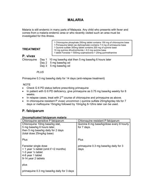
MALARIA<br />
Malaria is still endemic in many parts of Malaysia. Any child who presents with fever and<br />
comes from a malaria endemic area or who recently visited such an area must be<br />
investigated for this illness.<br />
TREATMENT<br />
P. vivax<br />
Chloroquine Day 1 10 mg base/kg stat then 5 mg base/kg 6 hours later<br />
Day 2 5 mg base/kg od<br />
Day 3 5 mg base/kg od<br />
PLUS<br />
Primaquine 0.3 mg base/kg daily for 14 days (anti-relapse treatment)<br />
NOTE<br />
• Check G 6 PD status before prescribing primaquine<br />
• In patient with G 6 PD deficiency, give primaquine as 0.75 mg base/kg weekly for 8<br />
weeks.<br />
• In relapse cases, treat with 2 nd course of chloroquine and primaquine as above.<br />
• In chloroquine resistant P vivax( uncommon ) quinine sulfate 25mg/kg/day tds for 7<br />
days or mefloquine 15mg/kg followed by 10mg/kg 8-12hrs later can be used.<br />
P. falciparum<br />
Uncomplicated falciparum malaria<br />
Chloroquine sensitive P falciparum Chloroquine resistant P falciparum<br />
Chloroquine 10mg base/kg stat,<br />
Quinine 8 mg base/kg/dose every 8 hourly<br />
5 mg base/kg 6 hours later,<br />
for 7 days.<br />
then 5 mg base/kg daily for 2 days<br />
(total dose 25mg/kg base)<br />
Plus<br />
Fansidar single dose<br />
< 1 year ¼ tablet (omit if
If sensitive to chloroquine, asexual parasitaemia should begin to decrease within 24-36<br />
hours and be eliminated within 3-5 days, When sexual forms are present, a 3 days<br />
course of primaquine 0.3mg base/kg/day will result in clearance of gametocytes<br />
(chloroquine and quinine has no gametocidal effect)<br />
P. malariae<br />
• Treatment same as P. vivax except primaquine is not required<br />
Complicated malaria/severe malaria<br />
• almost always due to P. falciparum<br />
• always suspects mixed infections if vivax / malariae malaria appear more severe<br />
than usual<br />
Signs of severe malaria<br />
• changes in behaviour<br />
• impaired consciousness<br />
• parasitaemia > 5%<br />
• jaundice<br />
• hyperpyrexia<br />
• continued vomiting<br />
• oliguria<br />
• severe metabolic acidosis<br />
Complications of falciparum malaria<br />
• cerebral malaria<br />
• renal failure<br />
• pulmonary edema / ARDS<br />
• haemorrhage due to DIVC<br />
• severe anemia ( Hb < 5 g% )<br />
• shock<br />
• haemoglobulinemia<br />
• hypoglycaemia<br />
Treatment of complicated falciparum malaria/severe malaria<br />
Intravenous quinine loading of 20mg base/kg in 5%Dextrose over 4 hour followed by<br />
10mg base/kg every 8 hourly (loading dose reduced parasite clearance time and<br />
duration of fever)<br />
Change to oral quinine as soon as patient can take orally and complete 7 days of<br />
treatment. If unable to take orally after 48 hours of intravenous therapy, reduce quinine<br />
dose by 1/3 to 1/2.<br />
Omit loading dose if patient was given quinine within last 24 hours<br />
Monitor blood glucose<br />
Monitor BP closely<br />
Monitor for arrhythmia<br />
Cerebral malaria<br />
Diagnosis criteria: ( 1 ) Unarousable coma. ( 2 ). Positive BFMP. ( 3 ). Exclusion of other<br />
causes of acute encephalopathies.<br />
Other features:<br />
• Focal or generalized seizure is common.<br />
• Abnormal neurological signs may be present – decerebrate / decorticate posturing,<br />
symmetrical limbs rigidity, sometimes opisthotonus, extensor plantar reflexes.<br />
• Accompanying multisystem dysfunction.<br />
• Retinal haemorrhages are common.
Management:<br />
• IV quinine as soon as possible. (refer treatment for severe malaria)<br />
• Ventilation most likely needed.<br />
• Prevention and correction of hypoglycemia, hypoxia, electrolyte abnormalities.<br />
• Careful fluid balance. Correct dehydration but avoid overhydration. (increased risk of<br />
pulmonary edema )<br />
• Control seizure. Caution in using phenobarbitone for prophylaxis as a recent studies<br />
showed phenobarbitone increased mortality in children with cerebral malaria.<br />
• No role of dexamethasone.<br />
• Use of mannitol controversial.<br />
• Exchange transfusion may be beneficial in severe malaria when parasitemia<br />
exceeds > 10%<br />
• Coma nursing.<br />
Outcome:<br />
• Mortality rate 10 – 20%<br />
• 5 – 10% of survivors have neurological sequelae<br />
Severe multidrug resistant P. falciparum<br />
Treatment<br />
Intravenous artesunate 2.4 mg/kg (loading dose ) followed by 1.2 mg/kg at 12 hour and<br />
24 hour later then 1.2 mg/kg daily for 6 days plus mefloquine 25 mg/kg single dose.<br />
(artemisinin drugs should be combined with a second antimalarial to prevent high<br />
recrudescent rate)<br />
OR<br />
Intramuscular artemether 3.2mg/kg (loading dose) followed by 1.6 mg/kg daily for 6<br />
days. Give mefloquine as above.<br />
REFERENCES<br />
1. Newton, Charles RJC, Hien, Tran Tinh, White, Nicholas. Cerebral malaria. Journal of Neurology, Neurosurgery &<br />
Psychiatry. 69 ( 4 ): pg 433 –441. OCT 1, 2000.<br />
2. White NJ. Current concepts: The treatment of malaria. NEJM, 1996, 335 ( 11 ), pg 800 – 806.<br />
3. Molyneux, Malcolm, Fox, Ray. Regular review: Diagnosis and treatment of malaria. BMJ 306 : pg 1175 – 1180. May<br />
1, 1993.<br />
4. J. Crawley. Malaria: new challenges, new treatments. Current paediatrics. Vol 9, No 1, June 1999. Pages 34 – 41.<br />
5. Report of an informal consultation Geneva, WHO, Division of control of tropical diseases. Management of<br />
uncomplicated malaria and the use of antimalaria drugs for the protection of travellers. 2000.<br />
6. J. Crawley, Warairu C, Mithwanis, et.al. Effect of phenobarbital on seizure frequency and mortality in childhood<br />
cerebral malaria: a randomised controlled intervention study. Lancet 2000, 355, pg 701 – 706.<br />
7. W.H.O Geneva 2000. Management of severe malaria –2 nd edition<br />
8. A.Omari,Severe life threathening malaria in endemic areas. BMJ 2004;328:154<br />
9. Treatment of malaria pg 705-710. Red Book 2000
1. Definition of TB Disease<br />
Tuberculosis<br />
• The presence of symptoms, signs and/or radiographic findings caused by MTB<br />
complex (M. tuberculosis or M. bovis).<br />
• Disease may be pulmonary or extrapulmonary, [e.g. central nervous system<br />
(CNS), disseminated (miliary), lymph node, bone & joint] or both.<br />
2. Clinical Features<br />
• Pulmonary disease is commonest. Symptoms include fever, cough, weight loss,<br />
night sweats, respiratory distress.<br />
• Extrapulmonary disease may manifest as prolonged fever, apathy, weight loss,<br />
enlarged lymph nodes (cervical, supraclavicular, axillary), headache, vomiting,<br />
increasing drowsiness, infants may stop vocalising. Swellings and loss of function<br />
may suggest bone, joint or spinal TB.<br />
• Phlyctenular conjuctivitis, erythema nodosum and pleural effusions are considered<br />
hypersensitivity reactions of TB disease.<br />
3. Diagnosis of TB Disease<br />
Diagnosis in children is usually difficult to make. Features suggestive of tuberculosis<br />
are:<br />
1. Recent contact with a person (usually adult) with active tuberculosis, This<br />
constitutes one of the strongest evidence of TB in a child who has symptoms and<br />
x-ray abnormalities suggestive of TB.<br />
2. Symptoms and Signs<br />
Symptoms and signs suggestive of TB are as listed above.<br />
Infants are more likely to have non specific symptoms like low-grade fever, cough,<br />
weight loss, failure to thrive, and signs like wheezing, reduced breath sounds,<br />
tachypnoea and occasionally frank respiratory distress.<br />
3. Positive Mantoux test (of >10 mm induration at 72 hours using tuberculin strength<br />
of 10 IU PPD).<br />
4. Chest X-ray<br />
• Enlarged hilar Iymph nodes +/- localised obstructive emphysema<br />
• Persistent segmental collapse consolidation not responding to conventional<br />
antibiotics.<br />
• Pleural effusion.<br />
• Calcification in Iymph nodes, this usually develops more than 6 months after<br />
infection.
5. Laboratory Tests<br />
Presence of AFB on smears of clinical specimens and positive histopathology or<br />
cytopathology on tissue specimens are highly suggestive of TB.<br />
Isolation of M. tuberculosis by culture from appropriate specimens is confirmatory.<br />
4. Diagnostic Work-up<br />
• Efforts should be made to collect clinical specimens for AFB smear, cytopathology<br />
or histopathology, special stains and AFB culture to assure confirmation of<br />
diagnosis and drug susceptibility.<br />
• If the source case is known, it is important to utilize information from the source<br />
such as culture and susceptibility results to help guide therapy.<br />
• The diagnostic work-up for TB disease is tailored to the organ system most likely<br />
affected. The tests to consider include but are not limited to the following:<br />
Pulmonary TB<br />
• Chest radiograph<br />
• Early morning gastric aspirates §<br />
• Sputum (if >12 years and able to expectorate sputum) §<br />
• Pleural fluid §<br />
• Pleural biopsy §<br />
CNS TB<br />
• CSF for FEME , AFB smear and TB culture §<br />
• CT head with contrast<br />
Abdominal TB<br />
• CT abdomen with contrast<br />
• Biopsy of mass / mesenteric lymph node §<br />
TB osteomyelitis<br />
• CT/MRI of affected limb<br />
• Biopsy of affected site §<br />
TB adenitis<br />
• Excisional biopsy or fine needle aspirate §<br />
Miliary / Disseminated TB<br />
• As for pulmonary TB<br />
• Early morning urine §<br />
• CSF §<br />
§ These specimens should be sent for AFB smear and TB culture and<br />
susceptibility testing. Cytopathology or histopathology should be carried out on<br />
appropriate specimens.
In addition, all children evaluated for TB disease require a chest radiograph to rule<br />
out pulmonary disease.<br />
5. Treatment of TB disease<br />
• Antimicrobial therapy for TB disease requires a multidrug treatment regimen. Drug<br />
selection is dependent on drug susceptibility seen in the area the TB is acquired,<br />
disease burden and exposure to previous TB medications.<br />
• Therapeutic choices are best made according to drug susceptibility of the<br />
organism cultured from the patient.<br />
• Almost all recommended treatment regimens have 2 phases, an initial intensive<br />
phase and a second continuation phase.<br />
• For any one patient, the treatment regimen would depend on the diagnosis<br />
(pulmonary or extrapulmonary), severity and history of previous treatment.<br />
• Directly observed therapy is recommended for treatment of active disease.<br />
Tuberculosis Chemotherapy in Children<br />
Drug Daily Dose Intermittent Dose<br />
(Biweekly)<br />
Intermittent Dose<br />
(Thrice Weekly)<br />
Mg/kg/day Maximum Mg/kg/day Maximum Mg/kg/day Maximum<br />
(mg)<br />
(mg)<br />
(mg)<br />
Streptomycin S 15-30 1000 15 1000 15 1000<br />
Isoniazid H 5-10 300 15 1200 10 900<br />
Rifampicin R 10 600 10 600 10 600<br />
Pyrazinamide Z 20-40 2000 Not used Not used - -<br />
Ethambutol E 15-25 2500 Not used Not used 30-50<br />
Short Course Therapy<br />
• This consists of a 6 month regimen, an initial 2 month intensive and subsequent 4<br />
month continuation phase. Short course therapy is suitable for pulmonary<br />
tuberculosis and non-severe extrapulmonary tuberculosis. It is not recommended<br />
for drug resistant TB. The short course consists of:<br />
a. Intensive Phase (2 months)<br />
• Daily Isoniazid, Rifampicin and Pyrazinamide<br />
• A 4th drug (either Ethambutol of Streptomycin) is added when initial drug<br />
resistance may be present or the burden of organisms is high.<br />
b. Maintenance Phase (4 months)<br />
• Isoniazid and rifampicin for the remaining 4 months.<br />
• This may be given daily (preferred) or biweekly or thrice weekly.
• WHO does not recommend a twice weekly regimen but advocates a thrice<br />
weekly regimen for intermittent dosing.<br />
• All intermittent dose regimens must be directly supervised.<br />
Pulmonary TB and Less Severe Extrapulmonary TB<br />
• Recommended regimen is short course therapy as above.<br />
• Less severe extrapulmonary TB include lymph node disease, unilateral pleural<br />
effusion, skin, and bone / joint (single site) excluding spine.<br />
Extrapulmonary TB (Severe Forms)<br />
• Severe forms of extrapulmonary TB include meningeal and CNS TB, spinal TB,<br />
abdominal TB, bilateral pleural effusion, pericardial effusion and bone and joint TB<br />
(> 1 site) and disseminated disease.<br />
• For these cases, intensive phase is as above but followed by a longer continuation<br />
phase from 7 to 10 months.<br />
Corticosteroids<br />
• Indicated for children with TB meningitis<br />
• May be considered for children with pleural and pericardial effusion (to hasten<br />
reabsorption of fluid), severe miliary disease (if hypoxic) and endobronchial<br />
disease<br />
• Steroids should be given only when accompanied by appropriate antituberculous<br />
therapy<br />
• Dosage: prednisolone 1-2mg/kg per day for first 3-4 weeks, then taper over 3-4<br />
weeks.<br />
6. Monitoring of Drug Toxicity<br />
• Indications for baseline and routine monitoring of serum transaminases and<br />
bilirubin are recommended for:<br />
1. Severe TB disease<br />
2. Clinical symptoms of hepatotoxicity<br />
3. Underlying hepatic disease<br />
4. Use of other hepatotoxic drugs (especially anticonvulsants)<br />
5. HIV infection<br />
• Routine testing of serum transaminases in healthy children with none of the above<br />
risk factors is not necessary.
• Children on Ethambutol should be monitored for visual acuity and colour<br />
discrimination.<br />
7. Breast-feeding and the Mother with Pulmonary Tuberculosis<br />
• Tuberculosis treatment in lactating mothers is safe as the amount of drug ingested<br />
by the baby is minimal. Hence if the mother is already on treatment and is noninfective,<br />
the baby can be breastfed.<br />
• Women who are receiving isoniazid and are breastfeeding should recieve<br />
pyridoxine.<br />
• If the mother is diagnosed to have active pulmonary TB and is still infective,<br />
The newborn should be separated from the mother for at least two weeks<br />
while the mother is being treated.<br />
Breast feeding is best avoided during this period, however, expressed<br />
breast milk can be given .<br />
The infant should be evaluated for congenital TB. If this is excluded, BCG<br />
is deferred and the baby should receive isoniazid for 3 months and then<br />
tuberculin tested. If tuberculin negative and mother has been adherent to<br />
treatment and non-infectious, isoniazid can be discontinued and BCG<br />
given. If tuberculin positive, the infant should be reassessed for TB disease<br />
and if disease is not present, isoniazid is continued for total of 9 months<br />
and BCG given at the end of treatment.<br />
Other close household contacts should be evaluated for TB.<br />
• Congenital TB is rare but should be suspected if the infant born to a tuberculous<br />
mother fails to thrive or is symptomatic.
FLOWCHART FOR MANAGEMENT OF CHILDREN WITH POSITIVE HISTORY<br />
OF CONTACT WITH TUBERCULOSIS.<br />
asymptomatic<br />
> 5<br />
years<br />
old<br />
> 10mm<br />
CXR<br />
normal<br />
Follow-up<br />
Chemoprophylaxis<br />
< 5<br />
years<br />
old<br />
abnormal<br />
Symptoms<br />
suggestive<br />
of TB<br />
Treat as<br />
tuberculosis<br />
CHILD<br />
(CONTACT)<br />
MANTOUX TEST<br />
symptomatic<br />
CXR<br />
Reference: Clinical Practice Guidelines on Tuberculosis.<br />
BCG LYMPHADENITIS<br />
• BCG lymphadenitis refers to cases where the lymph nodes have become large<br />
enough to be easily palpable and a cause of concern for the parents.<br />
• Most of the cases appear within 6 months of the BCG.<br />
• Ipsilateral axillary glands are involved in more than 95% of the cases, though the<br />
supraclavicular or cervical glands may occasionally be enlarged in isolation or<br />
association.<br />
• Two forms of lymphadenitis can be recognized, non-suppurative or simple which<br />
may resolve spontaneously within a few weeks, or suppurative which is marked by<br />
the appearance of fluctuation with erythema and oedema of the overlying skin.<br />
• Once suppuration has occurred, the subsequent course is usually one of<br />
spontaneous perforation, discharge and sinus formation. Healing eventually takes<br />
place through cicatrization and closure of the sinus, the process taking several<br />
months.<br />
Management<br />
1. BCG lymphadenitis without suppuration (no fluctuation)<br />
• Drugs are not required.<br />
• Reassurance and follow-up is advised.<br />
• Several controlled trials and a recent metaanalysis (Cochrane database) have<br />
suggested that drugs such as antibiotics (e.g. erythromycin) or antituberculous<br />
drugs neither hasten resolution nor prevent its progression into suppuration.<br />
2. BCG lymphadenitis with suppuration (fluctuation)<br />
• Needle aspiration is recommended. Usually one aspiration is effective, but<br />
repeated aspirations may be needed for some patients.<br />
• Surgical excision may be needed when needle aspiration has failed (as in the<br />
case of matted and multiloculated nodes) or when suppurative nodes have<br />
already drained with sinus formation.<br />
• Surgical incision is not recommended.<br />
Needle aspiration:<br />
prevents spontaneous perforation and associated complications<br />
shortens the duration of healing<br />
safe
3. Persistent Lymphadenitis<br />
• In patients with large and persistent or recurrent lyphadenopathy, possibility of<br />
underlying immunodeficency should be investigated. Thus all infants<br />
presenting with BCG lymphadenitis should be followed up till resolution.<br />
References:<br />
1. Singla A, Surjit S, Goraya S, Radhika S et al. The natural course of non-suppurative Calmette-Guerin<br />
bacillus lymphadenitis. Pediatr Infect Dis J 2002:21:446-448<br />
2. Goraya JS, Virdi VS. Treatment of Calmette-Guerin bacillus adenitis, a metaanalysis. Pediatr Infect<br />
Dis J 2001;20:632-4 (also inCochrane Database of Systematic Reviews 2004; Vol 2.)<br />
3. Banani SA, Alborzi A. Needle aspiration for suppurative post-BCG adenitis. Arch Dis Child<br />
1994;71:446-7<br />
BCG Vaccination<br />
Development of the normal BCG papule and scar<br />
• A small papule with induration should appear in most infants within 3-4 weeks.<br />
• The papule may increase in size for a few weeks (sometimes up to 10mm in<br />
diameter).<br />
• This subsides gradually, followed by a local lesion that may ulcerate 6-8 weeks<br />
later.<br />
• The lesion will heal spontaneously and leave a small flat scar 3-6 months after<br />
immunization<br />
Correct technique to give BCG:<br />
Needle: Short (10mm) 26-27 gauge needle with a short bevel using a BCG or<br />
insulin syringe<br />
Site: Left arm at Deltoid insertion<br />
Dose: 0.05 mls for infants (< 1 year of age)<br />
0.1 ml for children > 1 year.<br />
Route: Intradermal<br />
Do not BCG at other sites where the lymphatic drainage makes subsequent<br />
lymphadenitis difficult to diagnose and dangerous (especially on buttock where<br />
lymphatic drains to inguinal and deep aortic nodes).
Dengue Haemorrhagic Fever & Dengue Shock Syndrome<br />
CLINICAL SPECTRUM OF DENGUE INFECTION<br />
Dengue virus infection<br />
Asymptomatic Symptomatic<br />
Dengue hemorrhagic<br />
fever<br />
Undifferented Dengue fever (plasma leakage)<br />
Fever syndrome<br />
Without With unusual No shock Dengue shock<br />
haemorrhage haemorrhage syndrome<br />
Pointers to clinical diagnosis of Dengue infection<br />
Dengue Fever Dengue haemorrhagic fever<br />
1. High fever of 3 or more days duration<br />
2. Presence of petechiael haemorrhage/positive tourniquet test with other bleeding<br />
tendencies<br />
3. Hepatomegaly<br />
4. Pleural effusion or ascites<br />
5. Shock<br />
6. Fall in platelet count that precedes/ simultaneous with rise in haematocrit<br />
7. Normal/low WBC with relative lymphocytosis<br />
8. Rash –generalised flushing /maculopapular<br />
NB: All criteria need not be present at the same time<br />
Atypical Presentations<br />
• Acute abdominal pain, diarrhoea, severe gastro-intestinal haemorrhage (GIH)<br />
• Severe headache, convulsions, altered consciousness<br />
• Encephalitis<br />
• Hepatic failure, obstructive jaundice, raised liver enzymes, Reye’s syndrome<br />
• Acute renal failure, haemolytic uraemic syndrome<br />
• Disseminated intravascular coagulation (DIC)<br />
• Vertical transmission in newborns.
WHO grading of DHF /DSS<br />
Grade 1 Fever accompanied by non-specific constitutional symptoms.<br />
The only haemorrhagic manifestation is a positive Hess test.<br />
Grade 2 Spontaneous bleeding (usually skin ± other bleeds) in addition to<br />
manifestations of grade 1<br />
Grade 3 Circulatory failure (rapid weak pulse with pulse pressure < 20mmHg but<br />
systolic BP still normal<br />
Grade 4 Profound shock (Hypotension or undetectable BP and PR).<br />
NB:<br />
Grade 3 and 4 = Dengue Shock Syndrome<br />
Presence of thrombocytopenia and haemoconcentration (↑ PCV by 5 g%) differentiates<br />
Grade 1 and 2 DHF from DF.<br />
Because clinical differentiation of grade 1 and 2 DHF from DF is not always clear cut due<br />
to variation in baseline haematocrit, all patients ill enough to require IVD should be<br />
notified as DHF if baseline haematocrit is unknown<br />
WHO case definition of DHF<br />
ALL of the following criteria must be present:<br />
1. Fever. High grade and continuous for 2-7 days duration.<br />
2. Haemorrhagic diathesis /Positive tourniquet test except in shock.<br />
3. Thrombocytopenia (less than 100,000/mm 3 )<br />
4. Haemoconcentration (HCT 20% or more relative to baseline) or evidence of<br />
plasma leakage.<br />
Other clinical manifestations suggestive of DHF:<br />
Hepatomegaly<br />
Circulatory disturbances (cool extremities, capillary refill >2sec, tachycardia.)<br />
A fall in haematocrit following volume replacement.<br />
Hess test:<br />
BP cuff pressure maintained between SBP and DBP for 5 min. Positive test if > 20 petechiae<br />
per 2.5 cm 2 area.<br />
In clinical practice, the following classification of dengue infection is proposed:<br />
Dengue Fever Without increased vascular permeability<br />
Dengue Haemorrhagic fever (DHF) Increased vascular permeability and<br />
fragility<br />
Evidence of pleural effusion or ascites or<br />
haemoconcentration > 20%
DHF can be further graded as follows:<br />
1. DHF with no shock<br />
2. DHG with shock (DSS) which can be further graded into:<br />
a) DHF with compensated shock<br />
Signs of shock – tachycardia out of proportion to body<br />
temperature, decreased tissue percussion as evidenced by cool<br />
extremities, increased capillary refill time, narrowing of pulse<br />
pressure, weak distal pulses, oliguria and altered conscious level.<br />
Systolic pressure within the normal range (See Appendix 4 for<br />
details of hypotension, and normal blood pressure in relation to<br />
age, height and weight)<br />
b) DHF with decompensated shock<br />
Signs of shock – tachycardia, cool extremities, increase capillary<br />
refill time, weak or absent pulses, oliguria and altered conscious<br />
level<br />
Systolic hypotension<br />
Assessment of Circulation<br />
1. Fluid intake for previous 1-2 days, vomiting<br />
losses.<br />
2. Urine output for past 24 hours and time of<br />
last micturation.<br />
3. Bleeding and amount.<br />
4. Degree of dehydration.<br />
5. Peripheral circulation<br />
temperature and colour of extremities<br />
capillary refill<br />
distal pulses<br />
pulse volume<br />
6. Mental Status: headache, irritability, combativeness, drowsiness, coma, seizures<br />
(may indicate reduce cerebral perfusion, cerebral oedema or encephalopathy,<br />
intracranial bleed).<br />
7. Pleural effusion and ascites (third space loss).<br />
8. Abdominal pain: may indicate GIT bleed, acute liver enlargement, and<br />
hypovolaemia with intestinal ischaemia (shock).<br />
9. Hypotension is a late sign.<br />
Management<br />
Grade 1 and 2 DHF<br />
Laboratory investigations<br />
FBC/Platelet<br />
PCV<br />
BUSE, Creatinine<br />
LFT<br />
PT/PTT<br />
GXM - FFP, Platelet concentrates<br />
and whole blood.<br />
Blood culture<br />
Dengue Blot Test<br />
Hess Test *<br />
1. Admit.<br />
2. IV Cannula.<br />
3. Encourage oral fluids. IV fluids using 1/2 NS + D5% if unable to take orally and<br />
patients with evidence of plasma leakage.<br />
4. Paracetamol for fever. Avoid NSAIDS.<br />
5. Monitoring.<br />
Clinical (circulation): pulses, Temp., PR, RR, and BP.<br />
I/O Chart
Urine SG<br />
PCV, platelet, Hb 8-12 hourly<br />
Observations are continued till temperature returns to normal for 1-2 days. Critical<br />
period occurs during the transition from febrile to afebrile phase (usually after third<br />
day).<br />
Haemoconcentration usually precedes changes in pulse pressure and rate.<br />
Dengue Shock Syndrome<br />
1. Admit to ICU.<br />
2. Obtain IV access.<br />
3. Resuscitation: refer Fluid Therapy flow chart for DSS<br />
• 0.9% NaCl/Hartmann’s solution at 10-20 ml/kg, run as rapidly as possible.<br />
Dose is repeated till peripheral circulation, pulse volume and BP return to<br />
normal. 2 – 3 boluses may be needed in profound shock.<br />
• If no definite improvement and haematocrit remains high, use colloids e.g.<br />
haemecel or gelafundin<br />
• If no definite improvement and haematocrit is low or has decreased,<br />
transfuse blood this signifies bleeding, occult or obvious.<br />
• Avoid Dextrose containing solution during initial resuscitation. Circulation<br />
and fluid therapy must be assessed frequently.<br />
4. Monitor:<br />
Vital signs, peripheral perfusion.<br />
BP hourly till stable<br />
PCV or HCT<br />
Urine output.<br />
Platelet count 6 hrly.<br />
BUSE and serum creatinine.<br />
ABG<br />
5. Fluid maintenance:<br />
• Following fluid resuscitation, continue with 0.45%saline 5% dextrose at 1-2<br />
times maintenance, guided by haematocrit, urine output and vital signs.<br />
• In general, the duration of vascular permeability lasts 1-2 days following<br />
onset of shock, after which further infusion of large volume of fluids may result<br />
in pulmonary oedema and pleural effusion.<br />
6. Electrolyte and metabolic disturbances:<br />
Hyponatremia and metabolic acidosis occur in DSS. Isotonic fluids and<br />
restoration of tissue perfusion correct both problems. Correct hypoglycaemia<br />
that may occur in liver failure<br />
7. Transfusion of blood and blood products.<br />
A. Blood transfusion :Indications<br />
Significant haemorrhage<br />
Persistant shock despite crystalloid replacement in presence of low<br />
or declining haematocrit<br />
Fresh whole blood is preferable.
B. Platelet concentrate :Indications<br />
Platelet count
HCT falls<br />
PR, BP stable<br />
Urine output rises<br />
Reduce IV fluid therapy to 1X<br />
maintenance 5%D ½ NS ± KCl<br />
Reduce IV therapy to ½ X maintenance<br />
5%D ½NS ± KCl<br />
Discontinue IV therapy after 24-48 hrs.<br />
Vital signs & HCT stable<br />
adequate diuresis<br />
Fluid Therapy for Patients with DHF and DSS<br />
SIGNS OF SHOCK<br />
Compensated / decompensated shock٭<br />
Establish 2 IV lines<br />
Line 1: replacement fluid- rapid fluid bolus of normal saline (10-20ml/kg or 20ml/kg)<br />
Line 2: maintenance fluid 5% dextrose ½ normal saline ± KCl<br />
Total volume of IV fluid = 1½-2 X maintenance٭ ٭<br />
FBC, BUSE,RBS , GXM<br />
PCV 1-2 hrly<br />
HCT or PR rises, or<br />
Signs of shock, or<br />
Pulse pressure < 25mmHg, or<br />
Urine output falls<br />
Administer 2 nd rapid fluid bolus٭ of NS<br />
(10-20 ml/kg or 20ml/kg<br />
Maintenance fluid 5%D ½NS ± KCl<br />
Unstable vital signs<br />
Urine output falls<br />
Signs of shock still present٭ ٭ ٭<br />
HCT rises<br />
Rapid bolus with IV<br />
colloids eg. Haemaccel or<br />
Gelafundin 20ml/kg<br />
HCT falls<br />
Transfusion of<br />
blood/blood<br />
products<br />
٭ Rapid<br />
* Rapid<br />
fluid bolus<br />
fluid bolus: In<br />
-in<br />
decompensated<br />
decompensated<br />
shock,<br />
shock,<br />
give<br />
give<br />
20ml/kg<br />
20ml/kg<br />
fast;<br />
fast<br />
in compensated shock give 10-20ml/kg over 30-60 minutes,<br />
- in compensated shock give 10-20ml/kg over 30-60 minutes if patients is warming up<br />
slower if patient is warming up or presence of pulmonary edema.<br />
٭ ٭ use ** weight In obese adjusted patients, to height use weight centile adjusted for age to to height calculate centile the for volume age. of maintenance fluids<br />
٭ ٭ ٭ ensure good IV, check urinary catheter<br />
Yes No<br />
IMPROVEMENT<br />
If improvement occurs<br />
If further improvement occurs<br />
If improvement occurs<br />
No improvement<br />
CONDITION DETERIORATES<br />
Unstable vital signs or HCT rises<br />
Yes<br />
IMPROVEMENT<br />
PICU<br />
No improvement<br />
No
Special Notes<br />
1. Insertion of NG tube carries risk of trauma and bleeding. If gastric tube required,<br />
use oral route.<br />
2. Blood and blood product transfusion to correct thrombocytopenia or coagulopathy<br />
carry risk of disease transmission. Avoid if vital signs are stable.<br />
3. Insertion of chest tubes carries risk of haemorrhage. Careful titration of iv fluids<br />
with small doses of frusemide 0.25-0.5 mg/kg 4-6 hourly for 1-2 doses should<br />
make it possible to avoid chest tube insertion.<br />
4. Insertion of central venous line also carries significant risk of bleeding.<br />
Intraosseous route is acceptable.<br />
5. Use of steroids and immunoglobulin in DSS has no beneficial effect.<br />
Laboratory Diagnosis<br />
1. SEROLOGY<br />
Dengue IgM Dot Enzyme Immunoassay –available in all central laboratories<br />
in each state<br />
Interpretation of results must be considered against clinical suspicion and<br />
not taken in isolation<br />
Serology may be negative in early blood specimen. A second specimen<br />
should be sent in 10 days to confirm the diagnosis.<br />
2. VIRUS ISOLATION<br />
The most definitive diagnostic test. Availability limited.<br />
If patient dies soon after admission, a liver biopsy specimen sent in viral transport<br />
media may be useful in confirming the diagnosis.<br />
3. DETECTION OF DENGUE RIBONUCLEIC ACID<br />
The use of PCR reaction to detect dengue RNA is indicated when there is a<br />
diagnostic problem.<br />
REFERENCES:<br />
Ministry of Health Malaysia 2004 - Clinical practice guidelines: Dengue Fever in Children<br />
WHO Geneva 1997 - Dengue Haemorrhagic Fever- diagnosis, treatment, prevention and control<br />
Aspects of management of DHF /DSS –Dr Lucy Lum, University Malaya Medical Centre.
Congenital Syphilis<br />
Decision to treat an infant for congenital syphilis depends on<br />
i) Identification of presence of maternal syphilis<br />
ii) Adequacy of maternal treatment<br />
iii) Evidence of clinical, laboratory or radiographic syphilis in the infant<br />
iv) Comparison of infant’s/ cord VDRL with maternal’s VDRL<br />
The following infants require treatment<br />
1) Infants suggestive of congenital syphilis<br />
a. clinical – non immune hydrops, IUGR, jaundice, hepatosplenomegaly, rhinitis, skin<br />
rash, pseudoparalysis of extremity<br />
b. laboratory – cord blood VDRL (4X) higher than maternal level<br />
2) Infants with presumed congenital syphilis<br />
a. infant with positive cord blood VDRL<br />
b. mothers – untreated/unknown/inadequate treatment<br />
treatment > 38 weeks gestation (or treatment ≤ 4 weeks before<br />
delivery)<br />
treatment with erythromycin<br />
treated but VDRL did not decrease at least 4 fold<br />
Further Investigations:<br />
a. Infants with Signs of Congenital syphilis.<br />
i) Lumbar puncture: CSF for counts, proteins and VDRL status.<br />
ii) X-ray of long bones, CXR<br />
b. Check VDRL/TPHA status of both mother/father and any other partner involved. If<br />
father’s VDRL is negative, please check TPHA.<br />
c. TPHA titres (not necessary)<br />
Treatment<br />
a) Infants with congenital syphilis and presumed congenital syphilis<br />
i) PROCAINE PENICILLIN 50,000 units/kg IM daily x 10 - 14 days<br />
ii) IV CRYSTALLINE PENICILLIN 50,000 units/kg/dose 12hrly X 1 st 7 days then<br />
8hrly for total of 10 - 14 days<br />
iii) Alternative:<br />
IV/IM Ceftriaxone 75mg/kg daily < 30 days old<br />
100mg/kg daily > 30 days old<br />
If >1 day of treatment is missed, the entire course should be restarted<br />
b) Mother with positive VDRL but infant’s cord blood VDRL negative:<br />
IM BENZATHINE PENICILLIN 50,000 units/kg single dose
Note:<br />
c) Refer the parents to the STD clinic for management.<br />
1. Tetracycline, doxycycline or erythromycin does not have an established<br />
and well-evaluated high rate of success as injection penicillin in the<br />
treatment of syphilis.<br />
2. Penetration of tetracycline, doxycycline and erythromycin into the CSF is<br />
poor.<br />
Notification (Notify only cases which fulfil the following criteria):<br />
Follow-up<br />
Reference<br />
a. Infants with clinical features of syphilis.<br />
b. Infants with VRDL titres > 4 fold that of the mother’s.<br />
a. Clinical examination and serological tests at intervals for a total period of two<br />
years; every 3 months until VDRL non reactive or decrease by 4 fold (should<br />
decline by 3 months and non reactive by 6 months)<br />
b. Retreatment is indicated if: -<br />
i) Clinical signs and symptoms persist or recur.<br />
ii) Four-fold rise of titre in VDRL.<br />
iii) Failure of VDRL titre to decrease 4 fold within one year for early<br />
syphilis cases.<br />
WHO Guidelines on Treatment of Sexually Transmitted Diseases 1998<br />
The Sanford guide to antimicrobial therapy, 34 th edition, 2004
OPHTHALMIA NEONATORUM<br />
Conjunctivitis occurring within the 1st 4 weeks of life<br />
Aetiology;<br />
1. Bacterial<br />
a. Gonococcal<br />
Bilateral purulent conjunctival discharge within first few days of life.<br />
Treatment: Systemic:<br />
• Ceftriaxone 50mg/kg (max. 125mg) IV or IM once<br />
daily for 2-3 days<br />
OR<br />
• Cefotaxime 50mg/kg/day IV in two divided doses<br />
Q12H for 2-3 days<br />
Disseminated infections : Duration for 7 days<br />
Documented meningitis : 10-14 days<br />
Local: Irrigate eyes with sterile normal saline and at least hourly<br />
as long as necessary to eliminate discharge. Topical antibiotics<br />
optional.<br />
Refer to ophthalmologist for assessment.<br />
• Check VDRL of the infant to exclude associated congenital syphilis and<br />
screen for C. trachomatis and HIV.<br />
• Screen both parents for Gonococcal infections, syphilis and HIV. Parents<br />
should be referred to STD clinic for further management.<br />
• On discharge, infants should be seen at 2 weeks with a repeat eye swab<br />
gram stain and C&S<br />
b. Non- Gonococcal<br />
• Include Staphylococcus aureus, Streptococcus viridans, Haemophilus,<br />
E.coli and Pseudomonas<br />
• Treatment: Local – Neomycin eye ointment 0.5% after feed, both eyes<br />
(Change according to sensitivity ,duration according to response)<br />
OR<br />
Ceftazidime 5% bd to qid for a week
2. Chlamydial<br />
References<br />
Unilateral or bilateral conjunctivitis with peak incidence at 2 weeks of life<br />
Treatment:<br />
During 1 st week of life :<br />
• 2000g – Erythromycin 30mg/kg/d PO in divided doses<br />
>1 week to 1 mth: Erythromycin 40mg/kg/d PO in divided doses<br />
>1 month: Erythromycin 40 mg/kg/d PO in divided doses<br />
Duration of treatment = 14 days<br />
Local Rx: tetracycline ointment 1% q6H for 7-14 days<br />
• Systemic treatment is essential. Local treatment may be unnecessary if<br />
systemic treatment is given.<br />
• Refer both parent to STD clinic for further management<br />
• Refer to ophthalmologist for assessment of ocular complications<br />
Gonococcal Infections in Older Children<br />
• Suspect child abuse.<br />
• Children over 45 kg or 12 years old should receive adult regimens.<br />
• For children < 45 kg or < 12 years old with uncomplicated vulvovaginitis and<br />
urethritis:-<br />
Ceftriaxone 125mg IM single injection or<br />
Spectinomycin 40mg/kg IM single injection<br />
1. Canadian STD Guidelines 1998<br />
2. MMWR Recomm Rep 1998;47(RR-1) : 1-127. 1998 Guidelines for treatment of sexually transmitted<br />
diseases.<br />
3. Red Book 2000.<br />
4. WHO Guidelines for the management of sexually transmitted infections , 2001.<br />
5. Teoh DL,Reynolds S. Diagnosis and management of pediatric conjunctivitis. Pediatric Emerg Care<br />
2003;19: 48-55.<br />
Input from Dr Joseph Alagaratnam , Consultant Opthalmologist HKL , is acknowledged.
ATOPIC DERMATITIS<br />
DEFINITION:<br />
Common chronic relapsing inflammatory skin disease, characterized by intense<br />
itching, dry skin, inflammation and exudation.<br />
First symptoms commonly develop in infancy and 50% of cases are diagnosed by 1<br />
year of age. The disease is often familial and frequently associated with asthma, food<br />
allergy, allergic rhinitis and recurrent secondary skin infections. The prevalence of<br />
atopic dermatitis is 10-15% of children under 5 years of age. It is typically a long term<br />
condition with at least 1/3 of patients have persistent of disease throughout adulthood.<br />
DIAGNOSIS CRITERIA FOR ATOPIC DERMATITIS<br />
MAJOR FEATURES (MUST HAVE THREE)<br />
Hanifin and Rajka criteria<br />
1. Pruritus<br />
2. Typical morphology and distribution<br />
• Facial and extensor involvement during infancy and early childhood.<br />
• Flexural lichenification and linearity by adolescence.<br />
3. Chronic or chronically relapsing dermatitis.<br />
4. Personal or family history of atopy (asthma, allergic rhinoconjuctivitis, atopic<br />
dermatitis).<br />
MINOR/LESS SPECIFIC FEATURES<br />
1. Xerosis<br />
2. Preauricular fissures<br />
3. Icthyosis / palmar hyperlinearity / keratosis pilaris<br />
4. Ig E reactivity<br />
5. Hand/foot dermatitis<br />
6. Cheilitis<br />
7. Scalp dermatitis (cradle cap)<br />
8. Susceptibility to cutaneous infection (e.g. Staph. aureus and Herpes simplex)<br />
9. Perifollicular accentuation (esp. in pigmented races)<br />
TRIGGERING FACTORS<br />
1. Infection, bacterial, viral or fungal<br />
2. Emotional stress<br />
3. Sweating & itching<br />
4. Irritant - hand washing soap/detergent<br />
5. Extremes of weathers<br />
6. Allergen<br />
• food – egg, peanuts, milk, fish, soy, and wheat.<br />
• Aeroallergens – House dust mite, pollen, animal dander and molds
MANAGEMENT<br />
• The goal of therapy is control of skin inflammation, pruritus, and secondary<br />
infection.<br />
• At present there is no 100% life-long cure for atopic eczema.<br />
• Management comprise combining adjuvant basic therapy, anti-inflammatory<br />
measurements and identification and avoidance of triggering factors.<br />
• Major factor in successful management is COMPLIANCE and proper<br />
COMMUNICATION between doctor and patient.<br />
MEASURES<br />
A. BATH & EMOLLIENTS<br />
1. Baths are helpful in soothing itching and removing crusting. They should be<br />
lukewarm and limited to 10 minutes duration. Avoid soaps. Use soap substitute<br />
e.g. aqueous cream or emulsifying ointment instead of soap.<br />
2. Moisturizers work to reduce dryness in the by trapping in moisture. They<br />
should be applied to normal and abnormal skin. They should be applied at<br />
least twice a day and more frequently in severe cases. Emollients are best<br />
applied after bath. E.g. aqueous cream, ung. emulcificans, and urea cream<br />
N.B. Different classes of moisturiser are based on their mechanism of action,<br />
including occlusives, humectants, emollients and protein rejuvenators.<br />
In acute exudative form KMNO4 1:10,000 solution or normal saline daps or<br />
soak is useful - as mild disinfectant and desiccant.<br />
TOPICAL CORTICOSTEROIDS<br />
Topical corticosteroid is an anti-inflammatory agent and the mainstay of treatment for<br />
atopic eczema. Topical steroid are often prescribed intermittently for short term<br />
reactive treatment of acute flares and supplemented by emollients.<br />
• Choice depends on a balance between efficacy and side-effects.<br />
• The more potent the steroid, the more the side-effect<br />
• Apply steroid cream twice daily.<br />
• Potent steroid can be used initially but only on a short term or intermittent<br />
basis.<br />
• Avoid sudden discontinuation to prevent rebound phenomenon.<br />
• Use milder steroids for face, flexures and scalp<br />
Amount of topical steroid to be used – the finger tip (FTU) is convenient way of<br />
indicating to patients how much of a topical steroid should be applied to skin at any<br />
one site. 1 FTU is the amount of steroid expressed from the tube to cover the length<br />
of the flexor aspect of the terminal phalanx of the patient’s index finger.<br />
Number of FTU required for the different body areas.
1 hand / foot / face 1 FTU<br />
1 arm 3 FTU<br />
1 leg 6 FTU<br />
Front and back of trunk 14 FTU<br />
Adverse effect results from prolonged use of potent topical steroids. Local effects<br />
include skin atrophy, telangiectasia, purpura, striae, acne, hirsutism and secondary<br />
infections. Systemic effects are adrenal axis suppression, Cushing syndrome<br />
SYSTEMIC THERAPY<br />
Consists of:-<br />
• relief of pruritus,<br />
• treatment of secondary infection and<br />
• treatment of refractory cases.<br />
1. RELIEF OF PRURITUS (ANTITIHISTAMINES)<br />
1. For sedation and as anti-pruritus.<br />
2. Helpful to reduce scratching.<br />
2. TREATMENT OF SECONDARY INFECTION<br />
Secondary bacterial skin infection is common & may cause acute exacerbation of<br />
eczema. Systemic antibiotics are necessary when there is evidence of extensive<br />
infection.<br />
1. Predominant pathogen is Staphyloccus aureus.<br />
2. Useful in exudative form where superinfection occurs.<br />
3. Choice:<br />
• Syr. erythromycin 125mg tds 6 hourly for 5 days.<br />
• Suspected resistant case use cloxacillin/ cephalosporin.<br />
• May need prolonged antibiotic (1 month) if pustular infection occur over the<br />
extremities.<br />
4. Secondary infection can arise from Herpes simplex virus causing Eczema<br />
Herpeticum. Treatment using antiviral e.g. Acyclovir may be necessary.<br />
4. REFRACTORY CASES<br />
Refractory cases are those who do not response to conventional topical therapy<br />
and with extensive eczema. Refractory cases are referred to Dermatologist for<br />
treatment and monitoring.<br />
1. Systemic steroid<br />
2. Cyclosporin A<br />
3. Interferon<br />
4. Azathioprine<br />
5. Phototherapy
OTHER MEASURES<br />
• Avoid woollen toys, clothes, bedding.<br />
• Keep nails short.<br />
• Reduce use of detergent (esp. biological).<br />
• Double rinse clothes of patient.<br />
• BCG contraindicated till skin improves.<br />
• Tar/UV light might be useful.<br />
• Swimming useful (MUST apply moisturiser immediately upon exiting pool).<br />
Avoid Aggravating Factors<br />
FOR RELAPSE: -<br />
1. Check compliance.<br />
2. Suspect secondary infection - send for skin swab; start antibiotics.<br />
3. Exclude scabies.<br />
4. For severe eczema, emollient and topical steroid can be applied under<br />
occlusion with ‘ wet wrap ’. This involves the use of a layer of wet, followed by<br />
a layer of dry Tubifast to the affected areas i.e. limbs and trunk. The benefits<br />
are probably due to cooling by evaporation, relieving pruritus, enhanced<br />
absorption of the topical steroid and physical protection of the skin from<br />
excoriation.<br />
PROGNOSIS<br />
1. Tendency towards improvement throughout childhood.<br />
2. Two third will clear by adolescence.
IMPETIGO<br />
Definition:<br />
Superficial, contagious skin infection occurring in the epidermis and / or dermis. It is<br />
associated with formation of blisters. It is the most common skin infection in children.<br />
There are two types of impetigo, a bullous form caused by infection with Staphylococcus<br />
aureus and a non-bullous form caused by infection with group A Streptococci and may<br />
have secondary infection with Staphylococcus Aureus. The causative organism should<br />
be identified by taking skin swabs from affected sites.<br />
Clinical features<br />
Crusted lesions, usually yellow in colour, most commonly on the face.<br />
Typically there may be scattered surrounding lesions, known as ‘satellite lesions’.<br />
Usually patients are asymptomatic.<br />
Commonly spread to other areas of the body if not treated.<br />
It is contagious and can be passed to other family members.<br />
Treatment<br />
Localised infection may be adequately treated with topical mupirocin ointment which is<br />
active against infection due to both Staphylococcus and Streptococcus.<br />
More severe or generalised cases should be treated with systemic antibiotics according<br />
to the sensitivities to the causal organism but Erythromycin or Cloxacillin are generally<br />
suitable.
SCABIES<br />
Definition:<br />
Infestation caused by the mite Sarcoptes scabiei. Any part of the body may be affected, and<br />
transmission is by skin to skin contact.<br />
Clinical features<br />
Symptoms<br />
• Mites burrow into the skin where they lay eggs. The resulting offspring crawl out onto the<br />
skin and makes new burrows.<br />
• The absorption of mite excrement into skin capillaries generates a hypersensitivity<br />
reaction. The main symptom, which may take four to six weeks to develop, is<br />
generalised itch – especially at night.<br />
Signs<br />
• Characteristic silvery lines may be seen in the skin where mites have burrowed.<br />
• Classic sites include the interdigital folds, the wrists and elbows, umbilical area, genital<br />
area and feet.<br />
• Nodular Scabies- Papules or nodules seen at the site of mite infestation often affect the<br />
scrotum, axillae, back, or feet of children.<br />
• Crusted or Norwegian scabies- Seen in young infants or immunosuppressed patients.<br />
Wide- spread mite infestation causing a hyperkeratotic and / or crusted generalized.<br />
Diagnosis<br />
• The clinical appearance is usually typical, but there is often diagnostic confusion with<br />
other itching conditions such as eczema.<br />
• Scrapings taken from burrows may be examined under light microscopy to reveal mites.<br />
Management<br />
General advice<br />
• Parents should be given a detailed explanation of their condition, and clear and accurate<br />
written information on applying the treatment.<br />
• Treat everyone in the household or in close contact at same time. Ignore the plea that<br />
someone does not itch; infected people can be without symptoms and re-infect<br />
household members.<br />
• Change bedding, nightclothes & towels on night of treatment and clean them in a hot<br />
wash & hot iron after.<br />
Treatment<br />
• Benzyl Benzoate lotion (EBB) 25% for children more than 10 years old, 12.5% for<br />
children between 6 to 9 years<br />
• EBB should be applied to the whole body from the neck downwards, and washed off<br />
after 12 hours, usually overnight for 3 consecutive days.<br />
• Sulpha with Calamine in children between 1 to 5 years, and crotamiton for infants.<br />
• Itch may persist for several weeks.<br />
• Application of crotamiton cream may give symptomatic relief.<br />
• Antihistamines may also be helpful in relieving itch.<br />
• Mites separated from the human host die after 72 hours.
STEVEN–JOHNSON SYNDROME (SJS) / TOXIC EPIDERMAL<br />
NECROLYSIS (TEN)<br />
DEFINITIONS:<br />
SJS - Severe erosions of at least two mucosal surfaces with extensive necrosis of lips<br />
and mouth, and a purulent conjunctivitis. Epidermal detachment may occur in SJS, but<br />
less than 10% of the body surface area is involved. Morbidity with this disease is high,<br />
and can include photophobia, burning eyes, visual impairment and blindness.<br />
TEN – severe exfoliative disease associated with systemic reaction characterized by<br />
rapid onset of widespread erythema and epidermal necrolysis. Involves more than<br />
30% loss of epidermis.<br />
Aim of treatment: To remove the causative and prevent complications<br />
A. Drugs aetiology<br />
i) Antibiotics – Sulphonamides, Amoxycillin, Ampicillin,<br />
Ethambutol, Isoniazid<br />
ii) Anticonvulsant – Phenobarbitone, carbamazepine, phenytoin<br />
iii) NSAID – Phenylbutazone, Salicylates.<br />
B. Herpes infection<br />
C. Mycoplasma Pneumonia<br />
D. Many viruses, enteroviruses, adenoviruses, measles, mumps<br />
E. Bacteria, Streptococcus, typhoid fever<br />
Salient features<br />
• Acute prodromal flu-like symptoms, fever, conjunctivitis, malaise<br />
• Skin tenderness, morbilliform to diffuse or macular erythema target lesions,<br />
vesicles progressing to bullae. Blisters on the face, and upper trunk, then<br />
exfoliation with wrinkled skin which peels off by light stroking, Nikolksy’ sign.<br />
• Buccal mucosa involvement may precede skin lesion by up to 3 days in 30% of<br />
cases. Less commonly the genital areas, perianal area, nasal and conjuctival<br />
mucosa.<br />
• In the GIT, esophageal sloughing is very common, and can cause bleeding<br />
and diarrhoea.<br />
• In the respiratory tract, tracheobronchial erosions can lead to hyperventilation,<br />
interstitial oedema, and acute respiratory disease syndrome.<br />
• Skin biopsy of TEN - Extensive eosinophilic necrosis of epidermis with<br />
surabasal cleavage plane.<br />
• Renal profile – raised blood urea, hyperkalaemia and creatinine,<br />
Glucose - hypoglycaemia
MANAGEMENT OF SJS / TEN<br />
Supportive Care:<br />
• Admit to isolate or room where possible<br />
• May need IV fluid resuscitation for shock<br />
• Good nursing care (Barrier Nursing and hand washing)<br />
• Use of air fluidized bed, avoid bed sores<br />
• Adequate nutrition – nasogastric tubes, IV lines, parenteral nutrition if severe<br />
mucosal involvement.<br />
Specific treatment:<br />
• Eliminate suspected drugs<br />
• Human Intravenous Immunoglobulin at a dose of 0.4mg/kg/per day for 5 days.<br />
IVIG is a safe and effective in treatment for SJS/TEN in the paediatric patient<br />
(JAAD 2002;47:548-52). It arrests the progression of the disease and help<br />
complete re-epithelialization of lesions.<br />
Monitoring:<br />
• Maintenance of body temperature. Avoid excessive cooling or overheating<br />
• Careful monitoring of fluids and electrolytes – BP/PR<br />
• Intake / output charts, daily weighing & renal profile<br />
Prevent Complication:<br />
Skin care<br />
• Cultures of skin, mucocutaneous erosions, tips of Foley’s catheter.<br />
• Treat infections with appropriate antibiotics<br />
• Topical antiseptic preparations:<br />
Saline wash followed by topical bacitracin or 10% Chlorhexidine wash<br />
• Dressings of denuded areas with paraffin gauze / soffra-tulle<br />
• Surgery may be needed to remove necrotic epidermis<br />
Eye care<br />
• Frequent eye assessment<br />
• Antibiotic or antiseptic eye drops 2 hourly<br />
• Synechiea should be disrupted<br />
Oral care<br />
• Good oral hygiene aimed at early restoration of normal feeds.
General Approach to Inborn Errors of Metabolism (IEM)<br />
Broadly speaking, there are 2 situations where IEM are encountered:<br />
• Acute emergency in a sick child (sometimes in an adult)<br />
• Chronic problems involving either single or multiple organs, either recurrent or<br />
progressive, or permanent<br />
1. In an acutely ill child, IEM should be considered a differential diagnosis along with<br />
other diagnoses:<br />
• in all neonates with unexplained, overwhelming, or progressive disease<br />
particularly after a normal pregnancy or birth, but deteriorates after feeding<br />
• in all children with acute encephalopathy, particularly preceded by vomiting,<br />
fever or fasting<br />
• in all children with symptoms and signs of acidosis or hypoglycemia<br />
Appropriate history and physical examination should be taken in order to elicit diagnostic<br />
clues from the patient.<br />
Appropriate diagnostic and therapeutic measures must be initiated as soon as possible to<br />
prevent long term damage. (refer to algorithm for sick infant)<br />
1.1 Initial phase: basic investigations in emergency situation and first line treatment<br />
Insert i.v. line and sample blood for urgent analysis (results available within 30 to 60<br />
minutes) of:<br />
• Blood gas<br />
• Blood urea serum electrolytes (including chloride), creatinine, uric acid<br />
• Glucose<br />
• Liver function test (including AST, ALT), CPK<br />
• Full blood count<br />
• Ammonia and Lactate<br />
• Plasma amino acid (lithium heparin bottle, spin down), carnitine (IMR, HKL)<br />
• Filter paper card with dried blood spots for acylcarnitine, amino acids (IMR), or<br />
DNA studies.<br />
• Store the rest of the sample in either EDTA bottle/plain bottle/lithium heparin for<br />
possible additional tests<br />
Sample 1 st urine passed after rehydration or i.v. fluid for:<br />
• colour or odour<br />
• ketone, glucose/reducing substances (Clinistix and Clinitest), protein, pH<br />
• organic acid analysis<br />
• orotic acid (in hyperammonemia)<br />
• storage for additional test in freezer<br />
Correct shock with boluses of rehydration fluid. Then give dextrose 10% with appropriate<br />
electrolytes at about 1 ½ maintenance; achieving at least 60 to 80 kcal/kg/day. Glucose<br />
supplied at this rate approximates the normal hepatic glucose production and is usually<br />
sufficient for disorders of fasting intolerance (MCAD, Glycogenoses).
However, it may not be sufficient in disorders exacerbated by catabolism like urea cycle<br />
defects or organic acidurias.<br />
It may be potentially dangerous for mitochondrial defects as it enhances lactic acidosis.<br />
Other investigations as indicated: ECG, echocardiography, Cranial MRI/CT<br />
1.2 Later phase: treatment and investigations according to initial findings<br />
If emergency investigation shows……<br />
• hypoglycemia…(see approach to hypoglycemia)<br />
• hyperammonemia……(see approach to hyperammonemia)<br />
• metabolic acidosis……(see approach to metabolic acidosis)<br />
• lactic acidemia<br />
• severe liver disease<br />
If results inconclusive but metabolic disease remains a possibility:<br />
• continue glucose infusion<br />
• review history and physical signs<br />
• call up the metabolic centre for advice<br />
• send relevant investigations for the diagnosis of treatable metabolic disorders<br />
(results of urgent amino acids and organic acids should be available within 24<br />
to 48 hours receiving the sample)<br />
• additional testing e.g.; CSF for amino acids, lactate, biochemistry<br />
(sugar/protein), microscopy<br />
Maintain electrolytes, glucose, lactate, ammonia, ketones and acid base status within<br />
normal limit<br />
2. In children with chronic disease or organ dysfunction, detailed history including family<br />
history and prenatal history and physical examination are important to delineate the<br />
patient’s problem before further testing. Referral to metabolic center should only be made<br />
after initial investigations suggesting a possible metabolic cause.<br />
2.1 Chronic encephalopathy/psychomotor retardation<br />
• decide clinically if gray matter (affecting cognitive function, vision, causing fits)<br />
or white matter (long tract signs) or both are involved<br />
• decide if there is regression<br />
• decide the specific areas of involvement – pyramidal, extrapyramidal, cerebellar,<br />
basal ganglia<br />
• decide if there are other organ involvement<br />
• MRI brain may be the first investigation<br />
• CSF analysis<br />
• Neurophysiological testing as appropriate: EEG, EMG, nerve conduction test<br />
• Skeletal survey if there is skeletal involvement clinically
2.1.1. Laboratory test in isolated mental retardation without dysmorphism:<br />
Basic Investigations:<br />
• Lactate/ammonia, glucose, creatinine kinase, liver function test,<br />
electrolytes, calcium, phosphate<br />
• Thyroid function test<br />
• Genetic testing: Fragile X DNA testing, MECP2 mutation analysis for<br />
Rett syndrome<br />
Additional test if other neurological findings are positive beside mental retardation:<br />
• Urine organic acid, mucopolysaccharide, oligosaccharide (IMR)<br />
• Plasma amino acid<br />
• Filter paper for biotinidase activity if there is eczema and hair loss<br />
(overseas)<br />
• Urine purine and pyrimidine (overseas)<br />
• Serum carbohydrate deficient transferrin assay - congenital<br />
glycosylation disorders (overseas) if there is squint, scoliosis,<br />
abnormal supragluteal fat pad<br />
2.1.2. Additional testing in mental retardation and dysmorphism<br />
• Plasma sterol assay (overseas)<br />
• Very long chain fatty acid/phytanic acid (IMR)<br />
• Screening for subtelomeric deletion (overseas/HKL)<br />
• Copper level if there is hair abnormality (pili torti) for Menkes disease<br />
2.1.3 Psychomotor retardation and<br />
• Regression with or without organomegaly consider lysosomal<br />
disease. Do MRI to look for leukodystrophy. Refer to metabolic centre<br />
• Multi-system disorder, consider mitochondrial disease and refer to<br />
metabolic center<br />
• Liver disease - refer to approach to metabolic liver disease<br />
• Visual deterioration/lens dislocation: serum total homocyteine (IMR)<br />
• Cardiomyopathy (see cardiomyopathy below)<br />
• Abnormal hair: light microscopy for hair abnormality:<br />
o pili torti - Menkes kinky hair disease<br />
o Trichorrhexis nodosa - arginosuccinic aciduria<br />
• Seizure: see epileptic encephalopathy below<br />
• Macrocephaly: urine organic acid to rule out Glutaric aciduria I and<br />
Canavan disease<br />
MRI brain<br />
Urine mucopolysaccharides<br />
2.2 Epileptic encephalopathy<br />
• obtain detailed history and EEG to classify the type of seizure<br />
• basic laboratory tests: blood gas, electrolytes, calcium/<br />
phosphate/magnesium, sugar, liver function test<br />
• Fresh urine for sulphite (bedside labstix testing) to screen for sulphite<br />
oxidase defect
• Full blood picture to rule out neuroacanthocytosis (Refsum disease)<br />
• Urine for organic acid<br />
• Urine for purine/pyrimidine (overseas), guadinioacetate (overseas)<br />
• Plasma amino acid<br />
• CSF protein, glucose, lactate, amino acids (IMR/HKL), neurotransmitter<br />
(overseas)<br />
• Filter paper for biotinidase assay<br />
• WBC electromicroscopy to rule out neuronal ceroid lipofuscinosis<br />
• Further investigations should be discussed with or referred to the metabolic<br />
center<br />
Trial of<br />
• Pyridoxine 100 mg daily oral (watch out for apnoea which occurs in responsive<br />
cases).<br />
• Folinic acid (3 mg/kg i.v.), after CSF sampled for amino acid and neurotransmitter<br />
assay<br />
(Discuss with metabolic geneticist)<br />
2.3 Cardiomyopathy – refer to metabolic center for further investigation after baseline<br />
investigations<br />
2.4 Dysmorphic features- refer to genetic center for further investigation after baseline<br />
investigations.<br />
2.5 Fetal hydrops – contact metabolic center for advice<br />
3. Post-mortem investigations:<br />
If a child suddenly dies of an unknown, possibly genetic disease, it is essential to collect<br />
post-mortem samples and discuss their analysis with a metabolic specialist. Without a<br />
definite diagnosis, genetic counseling of the parents and reliable risk assessment for<br />
future children is impossible.<br />
Following samples should be collected:<br />
• serum (plain tube) and plasma (lithium heparin tube), a few cc. should be<br />
centrifuged and stored frozen<br />
• dried blood spots on filter paper card<br />
• urine to be frozen immediately, consider bladder washout with NaCl<br />
• DNA from 3 to 10 ml of EDTA whole blood<br />
• Skin biopsy for fibroblast culture (stored in ambient temperature for 1 to 2 days in<br />
viral culture medium or 0.9% NaCl)<br />
• CSF (several 1 cc samples, freeze immediately at 70 ºC)<br />
• Organ biopsy (discuss with metabolic specialist, freeze immediately for enzyme<br />
analysis, keep in glutaraldehyde for electron microscopy, etc)<br />
Always consider blood and urine sampling prior to expected death, because autolysis<br />
after death causes intracellular fluid to mix with extracellular fluid, resulting in misleading<br />
changes.<br />
Basic investigations: plasma amino acids, urine organic acids, acylcarnitine in dried filter<br />
paper spots.
“SICK” INFANT: AN ALGORITHM TO SCREEN FOR TREATABLE INBORN ERRORS OF<br />
METABOLISM<br />
Premature<br />
Low birth weight<br />
Not suggestive<br />
but possibility of<br />
fortuitous<br />
association<br />
e.g. Transient<br />
hyperammonemia<br />
of newborn<br />
Acute<br />
encephalopathy<br />
( intoxication type )<br />
Affected infant usually<br />
appear normal at birth,<br />
symptoms develop<br />
hours to days resulted<br />
from toxic effects of<br />
gradually accumulating<br />
metabolites.<br />
Predominant<br />
seizures<br />
exclude<br />
exclude<br />
exclude<br />
exclude<br />
Inborn errors<br />
of<br />
metabolism<br />
‘Traumatism’<br />
‘Accidental’<br />
Hypoxia<br />
Intracranial<br />
injury<br />
Infection<br />
Neonatal hepatic<br />
syndrome<br />
- Jaundice<br />
- Hepatocellular<br />
dysfunction<br />
Full term neonates<br />
Major electrolytes<br />
disturbance<br />
-hypo/hypercalcemia<br />
-hypo/hypernatremia<br />
-hypo/hyperkalemia<br />
Isolated/multiple<br />
malformations<br />
‘dysmorphic’<br />
First think of treatable disorders<br />
Emergency treatment must be undertaken in parallel with investigation<br />
e.g. Correction of hypoglycemia, metabolic acidosis, hyperammonemia etc<br />
Cardiac syndrome<br />
- Cardiac failure<br />
- Cardiomyopathy<br />
- Arrhythmias<br />
Chest X-ray<br />
Cranial<br />
sonography<br />
Septic screen<br />
Renal profiles<br />
Serum<br />
calcium<br />
Hormonal<br />
investigation<br />
Radiological<br />
investigations<br />
Echography<br />
Karyotype<br />
Genetic<br />
advice<br />
Persistent<br />
hypoglycemia<br />
A B<br />
C<br />
D<br />
E
A<br />
B<br />
C<br />
Check NH3,<br />
lactate,<br />
arterial blood<br />
gases & acidbase<br />
status,<br />
anion gap,<br />
urine/serum<br />
ketones<br />
Evaluation may<br />
include EEG,<br />
therapeutic trial of<br />
vitamins ( B6, biotin,<br />
folate, folinic acid ),<br />
lumbar puncture, urine<br />
sulphite test etc<br />
Evaluation may include<br />
urine reducing sugar,<br />
RBC’s galactose –1phosphate,<br />
RBC’s<br />
Glucose-1-phosphate<br />
uridyl transferase activity,<br />
urine organic acids<br />
analysis, acylcarnitine<br />
profile, α-1-antitrypsin,<br />
serum transferrin isoelectric<br />
focusing, bile acid<br />
analysis etc<br />
NH3 increased<br />
Respiratory alkalosis<br />
Lactate normal<br />
Urine ketones negative<br />
Metabolic acidosis<br />
Increased anion gap<br />
Massive ketonuria<br />
NH3 normal or<br />
increased<br />
Lactate normal or<br />
increased<br />
Ketosis<br />
NH3 normal or mildly<br />
increased<br />
Lactate normal or mildly<br />
increased<br />
No or mild metabolic<br />
acidosis<br />
Predominant<br />
lactate acidosis<br />
UREA CYCLE<br />
DEFECT<br />
(Diagnostic test: plasma<br />
amino acids, urine orotate )<br />
(c ORGANIC ACIDEMIA<br />
(c ( Diagnostic test: urine<br />
organic acid analysis, serum<br />
carnitine )<br />
MAPLE SYRUP URINE<br />
DISEASE<br />
( Diagnostic test: plasma<br />
amino acids )<br />
MITOCHONDRIOPATHY<br />
( Further tests: Multiorgan<br />
assessment, urine organic acid,<br />
mt DNA analysis, respiratory<br />
chain enzymes analysis etc )<br />
Vitamin responsive seizure ( B6, biotin, folate, folinic<br />
acid )<br />
Non ketotic hyperglycinemia ( raised CSF/plasma<br />
ratio of glycine )<br />
Glucose transporter protein ( GLUT 1 ) deficiency<br />
( reduced CSF/blood ratio of glucose )<br />
Sulphite oxidase defect<br />
Galactosaemia<br />
Fructose intolerance<br />
Tyrosinemia ( presence of succinylaccetone<br />
in urine organic acid analysis )<br />
Fatty acids oxidation disorders<br />
Congenital disorders of glycosylation 1b<br />
( with diarrhea )<br />
Bile acid synthesis defects<br />
( with cholestasis )<br />
α-1-antitrypsin deficiency<br />
mitochondrial hepatopathy
D<br />
E<br />
Evaluation may<br />
include acylcarnitine<br />
profile, serum<br />
carnitine, urine<br />
organic acid<br />
analysis, blood<br />
counts, echography,<br />
ECG etc<br />
Causes include<br />
-Glycogenosis<br />
-Hyperinsulinism<br />
-Fatty acids oxidation<br />
disorders<br />
-Hyperinsulinism<br />
hyperammonemia<br />
syndrome ( HIHA)<br />
Fatty acids oxidation disorders<br />
Systemic carnitine deficiency<br />
Pompe disease<br />
Barth’s syndrome<br />
( with neutropenia )<br />
Mitochondrial cardiomyopathies<br />
Congenital disorders of glycosylation<br />
( with pericardial effusion )<br />
Refer algorithm on investigation of<br />
hypoglycemia
Approach to Hyperammonemia<br />
1. NH3 values Neonates Healthy 200 umol/L<br />
After neonatal period Healthy 50-80umol/L<br />
Suspect IEM >100umol/L<br />
NH3 must be measure in every sick child who is encephalopathic for an<br />
apparently unknown cause, otherwise hyperammonemia may be missed and the<br />
child deprived of an efficient treatment.<br />
Blood sample must be taken as uncuffed venous (or arterial) sample, kept on ice and<br />
analysed immediately.<br />
The test must be available 24 hour round the clock in order to be useful.<br />
Caution: False elevations of NH3 are common<br />
2. Causes<br />
IEM:<br />
• Urea cycle defects<br />
• Organic acidemia<br />
• Fatty acid oxidation defects<br />
• Pyruvate carboxylase deficiency<br />
Non-IEM<br />
• Severe liver failure<br />
• Reye syndrome (check history for application of medicated oil with salicylate on<br />
umbilical cord or abdomen in newborn)<br />
• Transient hyperammonemia of newborn (due to open ductus venosus in<br />
neonates)<br />
• Drugs: Valproate, Asparagines in leukemia therapy<br />
• UTI: (particularly with urinary stasis and urease positive organism such as<br />
Proteus sp.)<br />
• Portohepatic fistula.<br />
• Increased muscle activity during assisted ventilation, respiratory distress<br />
syndrome, shortly after seizures ( Hyperammonemia due to increased muscle<br />
activity is rarely above 180umol/L but can be very high in other non-IEM causes<br />
listed above. )<br />
3. Emergency investigation<br />
It is important to reach the diagnosis as soon as possible. Contact the metabolic<br />
geneticist by phone and<br />
Send sample by courier services.<br />
- basic investigations: blood gas (often show respiratory alkalosis as NH3<br />
stimulates the respiratory center)<br />
Urea and electrolytes, full blood count, liver function tests, CK, glucose,<br />
Urine ketone, Urinalysis
- plasma amino acids<br />
- urine orotic acid<br />
- urine organic acids<br />
- acylcarnitine in dried filter paper spots<br />
- Doppler ultrasound to rule out portohepatic fistula<br />
4. Treatment<br />
Principle:<br />
• Stop protein intake, provide adequate calories to reduce catabolism which<br />
increases NH3<br />
• Remove NH3 by drugs or extracorporeal detoxification.<br />
• Replenish urea cycle intermediates (argininine)<br />
• Increase urinary ammonia excretion by generous fluid intake.<br />
Organize all treatment options as soon as hyperammonemia is confirmed.<br />
Extracorporeal detoxification must be promptly initiated. Contact the metabolic geneticist<br />
and arrange for transfer if possible.<br />
Otherwise admit the child to intensive care unit. Insert a central line and an arterial line.<br />
While awaiting for anti-hyperammonemia cocktail, infuse i.v. Dextrose 10% with an<br />
appropriate concentration of sodium and potassium at a rate of 1 to 1.25 maintenance<br />
(120-150 mls/kg/day). Mannitol may be needed concurrently if the child has signs of<br />
gross cerebral oedema. Intralipid may be used to increase the calorie intake if fatty acid<br />
oxidation defects have been excluded. Infection must also be treated as appropriate.<br />
If NH3 is > 200umol/L and patient is symptomatic / encephalopathic:<br />
Start intravenous anti-hyperammonemia cocktail:<br />
• Arginine hydrochloride 300mg/kg<br />
• Sodium benzoate 250mg/kg<br />
• Sodium phenylbutyrate 250mg/kg<br />
All diluted and mixed in 30mls/kg of dextrose 10% and run over 24 hours in addition to<br />
the child’s maintenance fluid (total fluid for administration of above cocktail ~ 1/4<br />
maintenance)<br />
Side effects of the medications: nausea and vomiting, consider the use of ondansetron<br />
or granisetron<br />
If NH3 is > 400umol/L and patient is symptomatic / encephalopathic:<br />
Above cocktail should be given as bolus in 2 hours. Followed by 24 hours continuous<br />
infusion of the same cocktail.<br />
Recheck the ammonia 2 hours later. If the level remain high and patient is<br />
encephalopathic, hemodialysis or hemofiltration should be instituted immediately to<br />
remove the ammonia effectively<br />
Peritoneal dialysis can be carried out as an alternative but may not be that effective.<br />
Contact the nephrologists for the dialysis if the service is available.<br />
Monitor the ammonia levels 6 hourly.<br />
Exchange transfusion should not be carried out as it increases the protein and<br />
NH3 load.
Blood transfusion or transfusion of any blood products should be deferred as it<br />
increases the protein load and NH3 levels. Drugs known to impair liver function or<br />
increase NH3 levels should also be avoided.<br />
Contact the metabolic geneticist for long term maintenance therapy<br />
The prognosis is poor if there has been prolonged coma >36 hours before the start of<br />
therapy, or more specifically if the concentration of NH3 multiply by the duration of coma<br />
(in days) exceeds 4000 umol/L.<br />
Ketotic<br />
Organic acidemia<br />
Citrulinaemia<br />
Metabolic acidosis<br />
5. Algorithm for Hyperammonemia<br />
Non-ketotic<br />
High ammonia<br />
Fatty acid oxidation defects<br />
Blood gas, ketone, plasma amino acids<br />
Urine orotic acid, organic acids<br />
No metabolic acidosis but respiratory<br />
alkalosis (low CO2, normal HCO3)<br />
Citruline<br />
High Normal<br />
Low<br />
Arginosuccinic acid<br />
Arginosuccinic aciduria<br />
Orotic Acid<br />
Negative Positive<br />
Normal / Low Elevated<br />
N- Acetylglutamine<br />
Synthetase defect<br />
or<br />
Carbamyl phosphate<br />
Synthetase Defect<br />
Ornithine<br />
Transcarbamylase<br />
Deficiency
1. Introduction<br />
Approach to Hypoglycemia<br />
Definition: blood glucose 10 mg/kg/min indicates hyperinsulinism unless there is marked loss in<br />
urine<br />
Rule out: Septicemia, severe systemic illness, small for gestation age, maternal diabetes<br />
2. Laboratory tests during symptomatic hypoglycemia:<br />
Adequate laboratory tests must be done to identify the cause, or else the diagnosis may be<br />
missed. Ensure samples are taken before correcting the hypoglycemia.<br />
2.1 Essential Tests<br />
• Ketone (serum or urine): normal or low indicate fatty acid oxidation defects<br />
• Free fatty acids (if available)<br />
• Acylcarnitine (dried blood spots or plasma): diagnostic of fatty acid oxidation<br />
defects or organic academia<br />
• Lactate – elevated in sick child with poor circulation (commonest cause), liver<br />
damage, impaired glycogenosis/gluconeogenesis, after seizures, difficult<br />
sampling<br />
• Urine organic acids - diagnostic of various IEM causes hypoglycemia<br />
• One spare tube (serum or plasma) kept frozen for anything below or forgotten or<br />
lost<br />
• Serum insulin (less than 2 to 5 mU/L in hypoglycemia) and cortisol (normal<br />
>270 nmol/L)<br />
2.2 Others<br />
• Blood gas, blood count, C – Reactive Protein (CRP), electrolytes, Liver function<br />
and renal function tests, Creatine Kinase (CK),<br />
• Uric acid, cholesterol/triglyceride<br />
• Carnitine<br />
• Growth hormone<br />
• Ammonia (liver damage)<br />
• Amino acid<br />
• Consider toxicology tests (C-peptide)
3. Treatment<br />
• Glucose i.v. 7 to 10 mg/kg/min or glucose 10% 110-150 ml/kg/day, keep blood sugar ><br />
5.5 mmol/L<br />
• Hourly blood sugar until the level is normalized<br />
• Ensure good i.v. line without interrupt<br />
• If bolus glucose is needed, do not give more than 200 mg/kg or glucose 10% 2 ml/kg<br />
• Await results of special investigations mentioned above<br />
• Consult metabolic geneticist or endocrinologist if necessary<br />
Note:<br />
• Hypoglycemia due to metabolic disorders is easily corrected with i.v. glucose but may<br />
recur if the underlying metabolic defect is not treated.<br />
• In contrast, hypoglycemia due to endocrine disorders especially hyperinsulinism is<br />
persistent and difficult to control requiring agents such as<br />
o glucagon (1 mg/day or 5 to 10 ug/kg/hour, i.v. continuously over 2 to 3 days),<br />
o diazoxide (15mg/kg/day in 3 doses, may take up to 5 days to work, may cause<br />
cardiac failure which may require hydrochlorothiazide 2mg/kg/day in 2 doses),<br />
o somatostatin (1 to 5 ug/kg/hour i.v.),<br />
o octreotide (3 to 20 ug/kg/day in 3 to 4 does) for long term treatment,<br />
o nifedipine (0.5 - 2 mg/kg/day) may be justified in selective cases.
Algorithm for Hypoglycemia<br />
Rule out severe systemic illness<br />
Sepsis, asphyxia<br />
SGA<br />
IDM<br />
Drugs<br />
Urine reducing substances<br />
Positive Negative<br />
Galactosaemia<br />
Tyrosinemia<br />
Hereditary fructose intolerance<br />
Liver failure<br />
Urine ketone<br />
High Low/Negative<br />
Big liver No big liver Insulin<br />
Amino acidopathies<br />
MSUD<br />
Organic acidemia<br />
Glycogenosis<br />
Gluconeogenesis<br />
Defects<br />
Growth hormone<br />
deficiency<br />
Cortisol deficiency<br />
Hypopituitarism<br />
Fatty acid oxidation<br />
defects<br />
Low High<br />
Congenital Hyperinsulinism<br />
(nesidoblastosis)<br />
Congenital hyperinsulinism<br />
variants<br />
- 4 types<br />
[including hyperinsulinism<br />
hyperammonemia syndrome,<br />
HIHA, often leucine sensitive]<br />
Beckwith Wiedemann<br />
syndrome, etc.
Approach to Metabolic Acidosis<br />
Metabolic acidosis is characterized by decreased pH, HCO3 - , and PaCO2. By far, the<br />
commonest cause of metabolic acidosis in a sick child is poor circulatory status associated with<br />
anaerobic respiration due to tissue hypoxia, dehydration, catabolism and sepsis. Metabolic<br />
acidosis due to inborn errors of metabolism is usually associated with an increase in<br />
anion gap.<br />
Anion gap = [Na + + K + ] – [Cl - + HCO3 - ], normal upper limit: 15-20 mmol/L<br />
1. Differential Diagnosis:<br />
a) Renal loss of bicarbonate<br />
o normal anion gap, increased Cl - , urinary pH>5, may be accompanied by other<br />
signs of renal tubular dysfunction<br />
b) Intestinal loss of bicarbonate<br />
o normal anion gap, increased Cl - , urinary pH may be elevated because of<br />
hypokalemia and secondary increase of urinary ammonium;<br />
c) Organic aciduria - Increased anion gap, lactate or ketones<br />
Ketosis is a physiological response to fasting, catabolic state or ketogenic diet. In some<br />
children ketosis is associated with nausea and vomiting. “Ketonemic” vomiting of infants with<br />
normal blood sugar is rarely caused by a primary IEM. Permanent ketosis may in rare cases<br />
indicate a ketolytic defect. Ketosis in addition to other metabolic abnormalities is frequently<br />
found in organic aciduria or mitochondrial respiratory chain disorders.<br />
Ketonuria in neonate is often indicative of a primary IEM but may be physiological in late<br />
infancy or older children.<br />
But ketosis is abnormal if it causes acidosis.<br />
2. Investigations<br />
Lactate<br />
Plasma ketone, urine ketones<br />
Urinary organic acids<br />
Plasma amino acids<br />
Carnitine<br />
Acylcarnitine (dry blood spots)<br />
Lactate<br />
Normal value: Blood
Lactic acidosis is most commonly secondary to:<br />
- the use of a tourniquet or difficulty in drawing the blood<br />
- muscular activity, assisted ventilation, seizures<br />
- severe systemic disease: central or peripheral hypoxia, ischemia, shock, cardiac failure,<br />
liver failure, renal failure septicemia, diabetes mellitus<br />
IEM associated with lactic acidosis:<br />
- respiratory chain disorders or mitochondrial cytopathy<br />
- fatty acid oxidation defects<br />
- organic aciduria<br />
- glycogen storage disease, gluconeogenesis defects<br />
3. Treatment<br />
Treatment of metabolic acidosis or lactic acidosis secondary to IEM is treatment of the primary<br />
disorders. Sodium bicarbonate may be used to treat severe acidosis uncontrolled by the<br />
treatment of underlying disorder. It’s use is discouraged if there is hyperammonemia as it may<br />
increase cellular influx of ammonia in the brain.<br />
Stop all protein intake if organic aciduria or amino acidopathies are suspected.<br />
If mitochondrial respiratory chain defects are suspected, vitamin or cofactors may be useful as<br />
antioxidants, and in an effort to improve energy production:<br />
• Coenzyme Q 4 - 10 mg/kg/day<br />
• Vitamin C 100 mg/day<br />
• Riboflavin 100 mg/day<br />
• Thiamine 100 mg/day<br />
If organic aciduria is suspected, addition of vitamin cocktail may be useful while awaiting for<br />
confirmation of diagnosis:<br />
• Oral Thiamin 100 mg daily<br />
• Oral Riboflavin 100 mg daily<br />
• Oral Biotin 10 mg daily<br />
• IV Vitamin B12 1 mg daily .
Algorithm for metabolic acidosis<br />
Metabolic acidosis<br />
Anion gap<br />
Normal Increased<br />
-<br />
Renal loss of HCO3<br />
accumulation of fixed acids<br />
-<br />
Intestinal loss HCO3<br />
Ketosis No ketosis<br />
Hyperglycemia Normoglycemia Hypoglycemia High lactate Normal lactate<br />
NH3 Lactate Lactate Sugar normal sugar<br />
High Normal High Normal High Normal Normal Low<br />
Organic<br />
aciduria;<br />
MMA;<br />
PA;<br />
IVA.<br />
Diabetes;<br />
Ketolytic<br />
Defects<br />
Mitochondrial<br />
disease<br />
Organic aciduria;<br />
MSUD;<br />
Ketolytic Defects<br />
Respiratory chain<br />
disease;<br />
Gluconeogenesis<br />
defects<br />
MSUD;<br />
Organic aciduria;<br />
Adrenal insufficiency<br />
PDH<br />
deficiency<br />
MMA= Methylmalonic Acidemia, PA= Propionic Acidemia, IVA= Isovaleric Acidemia,<br />
MSUD= Maple Syrup Urine Disease, PDH= Pyruvate Dehydrogenase<br />
FAOD= Fatty Acid Oxidation Defects, GSD1= Glycogen Storage Disease type I<br />
FAOD;<br />
GSD1<br />
Fructosemia<br />
Pyroglutamic<br />
aciduria
A. Medical problems<br />
DOWN SYNDROME<br />
Newborn<br />
• Cardiac defects (40 to 60%) AVSD, VSD, ASD, TOF<br />
or PDA<br />
• Gastrointestinal (12%)<br />
Commonest Duodenal atresia, others include<br />
Tracheo-oesophageal fistula, Anorectal malformation<br />
Pyloric stenosis and Hirshsprung disease.<br />
• Vision: Congenital cataracts (3%), Glaucoma.<br />
• Hypotonia<br />
• Feeding problems,<br />
• Congenital hypothyroidism<br />
• Congenital dislocation of the hips<br />
Infancy and Childhood<br />
• Delayed developmental milestones<br />
• Mild to moderate intellectual impairment (IQ25 to 50)<br />
• Hearing loss due to glue ear or sensorineural<br />
deafness<br />
• Visual Impairment –squint, nystagmus, glaucoma, refractive errors (70%)<br />
• Seizure disorders (10%)<br />
• Recurrent respiratory infections<br />
• Upper airway obstruction –due to hypertrophy of tonsils & adenoids may lead to sleep<br />
apnoea and cor pulmonale.<br />
• Leukaemia (15 to 20 times greater risk).<br />
Incidence 1%.<br />
• Atlantoaxial instability<br />
• Hypothyroidism – prevalence increases with<br />
age. Affects 10-20%<br />
• Short stature – modified centile charts<br />
available.<br />
Adolescence and Adulthood<br />
• Puberty - In Girls menarche only slightly delayed. Fertility presumed.<br />
- Boys are usually infertile due to low testosterone levels.<br />
• Increased risk of dementia /Alzheimer disease in adult life.<br />
• Shorter life expectancy<br />
Incidence: 1 in 660 newborns<br />
Maternal Age-Specific Risk for<br />
Trisomy 21 at livebirth*<br />
20 yrs. 1 in1500<br />
30 yrs. 1 in 900<br />
35 yrs. 1 in 350<br />
40 yrs. 1 in 100<br />
41 yrs 1 in 70<br />
42 yrs 1 in 55<br />
43 yrs 1 in 40<br />
44 yrs 1 in 30<br />
45 yrs 1 in 25<br />
*Source Hecht and Hook ‘94<br />
Karyotype<br />
Full trisomy 21 94%<br />
Translocation 4%<br />
Mosaicism 1%<br />
B. Initial Management<br />
• Breaking news of diagnosis should be done by a senior medical officer or<br />
specialist.<br />
• Complete examination to look for complications.<br />
• The cardiac status of every child must be established by age 6 weeks by neonatal<br />
paediatric examination and ECHO
• Even with ECHO an occasional lesion is 'missed'. Therefore constant clinical<br />
vigilance essential.<br />
• Chromosomal analysis to confirm diagnosis.<br />
• Baseline T4 /TSH at birth or by 1-2 weeks of life.<br />
• The following information should be conveyed before discharge from hospital:<br />
Information on Down Syndrome.<br />
Contact with local parent support group.(see local resources.)<br />
Every child with down syndrome should have access to an Early<br />
Intervention Programme as soon as possible.<br />
Advice on potential recurrence risk and contraception until a chromosomal<br />
diagnosis is available.<br />
RECURRENCE RISK FOR DOWN SYNDROME IN SUBSEQUENT PREGNANCY<br />
Karyotype Recurrence Risk<br />
Regular Trisomy 47(XX or XY)+ 21 1% (>37 yrs - 2x age related risk)<br />
Translocation<br />
- Both Parents normal 2-3%<br />
- Mother carrier t(14;21) 12%<br />
- Father carrier t(14;21) 3%<br />
- Either parent t(21q;21q) 100%<br />
Mosaics < 1%<br />
NB: The recurrence risks for t(13;21), (15;21) and( 21;22)are the same as that for t(14;21).<br />
KEY POINTS ON MEDICAL MANAGEMENT. (Based on DSMIG guidelines)<br />
Cervical Spine Instability<br />
• People with Down syndrome have a small risk for major neurological damage<br />
caused by cervical spine instability.<br />
• While the need for Cervical spine X-Rays is still debated, the American Academy<br />
of <strong>Paediatric</strong>s recommends for radiographs to be taken at 3 to 5 years of age and<br />
repeated every 10 years. These studies are more important for children who may<br />
participate in contact sports and are clearly indicated in those who are<br />
symptomatic.<br />
Lateral cervical radiographs in the neutral, flexed, and extended positions should<br />
be taken. The space between the posterior segment of the anterior arch of C1 and<br />
the anterior segment of the odontoid process of C2 should be measured.<br />
Measurements of less than 5 mm are normal; 5 to 7 mm indicates instability, and<br />
greater than 7 mm is grossly abnormal. The cervical canal width should also be<br />
measured.<br />
• Children with Down's syndrome should not be barred from taking part in sporting<br />
activities.
• Clinical symptoms - often mild - are currently the most useful predictors of future<br />
risk and merit urgent cervical spine X-Rays and specialist referral.<br />
• Symptoms of spinal cord compression may include neck pain, unusual posturing of<br />
the head and neck (torticollis), change in gait, loss of upper body strength,<br />
abnormal neurological reflexes, and change in bowel/bladder functioning.<br />
Growth<br />
• Appropriate growth monitoring is essential. Those who are excessively short or<br />
underweight may have additional pathology that requires investigation and<br />
treatment.<br />
• Down's specific growth charts provide essential reference values. The possibility of<br />
additional pathology should be considered for those falling in the lower centiles<br />
who do not have congenital heart disease.<br />
• Overweight/obesity is not inevitable and should always be thoroughly assessed.<br />
Hearing problems<br />
• 6-10 months – hearing assessment should include Auditory thresholds, Impedance<br />
tests and Otoscopy.<br />
• Aim to establish whether or not there is permanent hearing loss by 10 months and<br />
instigate intervention where necessary.<br />
C. Follow-up<br />
• At 6 weeks: Review chromosomal results and provide nondirective<br />
counselling for next pregnancy.<br />
Cardiac assessment<br />
Review T4 /TSH results.<br />
Confirm involvement in parent support group and EIP<br />
Discuss parental concerns.<br />
• First year: Developemental assessment<br />
Review 3 monthly Growth<br />
Hearing assessment at 6 to 10 months.<br />
Visual assessment for squints etc<br />
Fill in forms for registration with welfare dept.<br />
• 1- 6 years: Regular developemental assessment.<br />
Annual hearing and visual assessments<br />
Annual Thyroid function tests.<br />
Begin Dental checks at 2 years and continue 6 monthly thereafter<br />
Check on EIP. Encourage entry into normal kindergarten.<br />
Plan school entry by age 5<br />
• 7- 12 years: Check on school performance and placement.<br />
See 1-2 yearly Dental checks 6 monthly<br />
Annual Hearing, Visual and Thyroid function tests.<br />
• Adolescent: Discuss sexuality and employment.
Useful websites<br />
1. The Down Syndrome Medical Interest Group (UK): www.dsmig.org.uk<br />
2. Down Syndrome: Health Issues (up to date health information for professionals<br />
and parents): www.ds-health.com<br />
3. Growth charts for children with Down Syndrome: www.growthcharts.com<br />
4. Educational issues: www.downsed.org<br />
5. Kiwanis Down Syndrome Foundation: www.kdsf.netmyne.com<br />
Recommendations for Medical Surveillance for children with Down Syndrome*<br />
The following are suggested ages for health checks. Check at any other time if there are<br />
parental or other concerns<br />
Thyroid<br />
blood<br />
tests<br />
Growth<br />
monitoring<br />
Eye<br />
check<br />
Hearing<br />
check<br />
Heart<br />
check<br />
and<br />
other<br />
advice<br />
Birth to<br />
6 weeks<br />
T4 & TSH<br />
6 - 10<br />
months<br />
12 months<br />
T4 & TSH<br />
including<br />
antibodies<br />
Length, weight and head circumference<br />
should be checked regularly and plotted<br />
on Down's syndrome growth charts.<br />
Visual<br />
behaviour.<br />
Check for<br />
congenital<br />
cataract<br />
Neonatal<br />
screening, if<br />
locally available<br />
Echocardiogram<br />
0-6 weeks<br />
Visual<br />
behavior.<br />
Check for<br />
squint.<br />
Full<br />
audiological<br />
review<br />
(hearing,<br />
impedance,<br />
otoscopy)<br />
<strong>Paediatric</strong> review Annually<br />
Visual<br />
behaviour.<br />
Check for<br />
squint.<br />
18 months<br />
to 2½ years<br />
3 - 3½<br />
years<br />
T4 & TSH<br />
including<br />
antibodies<br />
4 - 4½ years<br />
Length and weight should be checked<br />
at least annually and plotted on<br />
Down's syndrome growth charts.<br />
Orthoptic<br />
examination,<br />
refraction<br />
and<br />
ophthalmic<br />
examination.<br />
Visual<br />
acuity,<br />
refraction<br />
and<br />
ophthalmic<br />
examination.<br />
Full audiological review<br />
(hearing, impedance, otoscopy) annually<br />
dental advice<br />
FROM AGE 5 TO 19 YEARS<br />
Hearing 2 yearly audiological review (as above)<br />
Vision / Orthoptic check 2 yearly<br />
Thyroid blood tests At age 5 years, then with 2 yearly<br />
* adapted from Down Syndrome Medical Interest Group (DSMIG) guidelines<br />
References<br />
1. Medical Management of Children with Down Syndrome. E Marder & J Dennis. Current <strong>Paediatric</strong>s<br />
(2001) 11, 57-63<br />
2. Health Supervision for Children with Down Syndrome. American Academy of <strong>Paediatric</strong>s. Committee<br />
on Genetics. 2000 – 2001<br />
3. The Down Syndrome Medical Interest Group (UK). Guidelines for Essential Medical Surveillance for<br />
Children with Down Syndrome
Down Syndrome: Local Resources<br />
o This is an online parents support group.<br />
http://groups.yahoo.com/group/DownSynd<br />
romeMalaysia<br />
o For links and information on local resources<br />
for Down's Syndrome, visit website<br />
http://www.geocities.com/nia14_my/dsres<br />
ources/ds_list.html<br />
o Kiwanis Down Syndrome Foundation:<br />
www.kdsf.netmyne.com<br />
o KDSF National Centre<br />
Lot 13490, Jln. Jenjarum, Off Jalan SS 23/1,<br />
Taman SEA, 47400 PetalingJaya,<br />
Tel: 03-78030179 Fax 03-78064862<br />
E-mail: us_kdsf@tm.net.my<br />
o KDSF Klang<br />
Lot 2492 Jalan Bukit Kuda 41300 Klang<br />
Tel/ Fax: 03-3428259<br />
o KDSF Seremban<br />
1129 Blossom Heights Jalan Tok Ungku<br />
70100 Seremban<br />
Tel: 06-6325595<br />
o KDSF Johor Baru<br />
2C Jalan Yahya Awal,81100 Johor Bahru<br />
Tel: 07-2242336<br />
E-mail: kdscjb@pd.jaring.my<br />
o KDSF Kota Kinabalu<br />
P.O.Box 21040,Pos Luyang,<strong>88</strong>767 Kota<br />
Kinabalu,Sabah<br />
Tel: 089-423508 Fax: 089-420512<br />
o KDSF Melaka<br />
24-G Jalan Permai,Taman Perkota,Bukit<br />
Sebukor,75350 Melaka<br />
Tel: 06-2824742<br />
o Persatuan Sindrom Down Malaysia:<br />
http://www.mysindromdown.org/<br />
No. 79, Lingkungan U-Thant, 55000 Kuala<br />
Lumpur,<br />
Tel: +603 4257 9818<br />
Email: psdm@tm.net.my<br />
o Penang Down Syndrome Association<br />
http://www.dsapenang.org/<br />
CO-15-UP ,Jln. Scotland<br />
Komplex Masyarakat Penyayang<br />
10450 Georgetown, Pulau Pinang<br />
o Association of the Network for the Needs<br />
of Children with Disability (Perak)<br />
148, Jln R.C.M.Rayan, Ipoh Garden,31400<br />
Ipoh,Perak.<br />
Tel: 05-545 1878 Fax: 05-545 1878<br />
Email: needs@pd.jaring.my<br />
o Pibakat (Pertubuhan Bagi Ibu-Bapa Kanak<br />
Cacat Sarawak)<br />
3 Jalan Bisaya; Off Jalan Ong Thian Swee<br />
93200 Kuching, Sarawak<br />
Tel: 082-251336; 082-254360<br />
Fax. 242971<br />
o Miri Sunflower Center<br />
Miri, Sarawak<br />
085-420722<br />
o Sibu Special Childrens Clinic<br />
Lau King Howe Polyclinic<br />
Sibu, Sarawak<br />
084-322281<br />
o Asia Community Service:<br />
http://www.asiacommunityservice.org/con<br />
tact_us.htm<br />
CO-23-UP ,Caring Society Complex, Jalan<br />
Utama, 10450 Penang,<br />
Tel : 604 – 6585396 Fax : 604 - 6597852<br />
email : acspen@po.jaring.my<br />
o PEKAKA, (Persatuan Bantuan Pendidikan<br />
Kanak-Kanak Khas), is a Parents' Support<br />
Group for Children with Special Needs in<br />
Sungai Petani.<br />
65B Taman Cempaka, Jalan Kolam<br />
Air,08000 Sungai Petani, Kedah Darulaman,<br />
Tel: 04-4255936 Fax: 04-4255423<br />
email: pekaka<strong>88</strong>@tm.net.my<br />
o <strong>Malaysian</strong> Care: www.mcare.org.my<br />
15, Jalan 3/146, Metro Centre, Bandar Tasik<br />
Selatan, 57000 Kuala Lumpur,<br />
Tel : 03-90582102 Fax :03-90584057<br />
Email : mcare@po.jaring.my<br />
o Jabatan Pendidikan Khas<br />
http://www.moe.gov.my/jpk.htm<br />
Bangunan SPPK, 46, Jalan Dungun,<br />
Damansara Heights,50604 Kuala Lumpur<br />
Tel : 03-2544129/ 2544130 ; Faks : 03-<br />
2542644<br />
o *Contact your Local Education Department<br />
for a list of special education schools in your<br />
locality.<br />
o Contact your local Welfare Department for a<br />
list of Community Based Rehabilitation (CBR)<br />
or Pemulihan Dalam Komuniti (PDK) Centres<br />
in your locality. http://www.kempadu.gov.my/
Appendicitis<br />
Appendicitis is the most common surgical condition of the abdomen in children over the<br />
age of 4 years and yet can be a challenge to diagnose and manage. Although diagnosis<br />
and treatment have improved over the years, it continues to cause considerable<br />
morbidity and even mortality in Malaysia. The deaths appear to be due to delay and<br />
difficulty in diagnosis, inadequate perioperative fluid replacement and sepsis.<br />
Clinical Features<br />
Abdominal pain – Lower abdominal pain is an early and almost invariable feature.<br />
Usually the pain starts in the epigastrium or periumbilical region before localising to the<br />
lower abdomen or the right iliac fossa. However the younger child may not be able to<br />
localise the pain. If there is free pus, the pain is generalised.<br />
Nausea and vomiting occurs in about 90% of children and is an early symptom. Most<br />
children have a loss of appetite. A hungry child rarely has appendicitis.<br />
Diarrhoea is more common in the younger age group causing confusion with<br />
gastroenteritis. It can be due to pelvic appendicitis or collection of pus within the pelvis.<br />
Dysuria and frequency are also commonly present in the child with pelvic appendicitis or<br />
perforated appendicitis<br />
Physical Findings<br />
General – the child is usually quiet and may be dehydrated.<br />
Dehydration must be actively sought for especially in the obese child and the child with<br />
perforated appendicitis. A history of vomiting, tachycardia, poor urine output and poor<br />
perfusion are indicators of dehydration.<br />
Tenderness on palpation or percussion is essential for the diagnosis. However it may<br />
be localised to the right iliac fossa or be generalised. The tenderness may also be mild<br />
initially and difficult to elicit in the obese child or if the appendix is retrocaecal. Rebound<br />
tenderness is usually not required to make the diagnosis and can cause unnecessary<br />
discomfort.<br />
Guarding signifies peritonitis but may be subtle especially if the child is toxic and very<br />
dehydrated.<br />
Rectal examination is only required if other diagnosis are suspected e.g. ovarian or<br />
adnexal pathology.<br />
Investigations<br />
• Full blood count – The total white blood cell count may be elevated but a normal<br />
count does not exclude appendicitis<br />
• Blood Urea and Serum Electrolytes – The sodium level may be apparently<br />
normal if the child is dehydrated
• Serum Amylase – If pancreatitis cannot be ruled out<br />
• Ultrasound and CT scan have been suggested to improve the diagnostic<br />
accuracy in doubtful cases. So in our setting the recommendation is that the<br />
children need to be assessed by a specialist preoperatively.<br />
Complications<br />
Perforation can occur within 36 hours of the onset of symptoms. Perforation rate<br />
increases with the duration of symptoms and delayed presentation is an important factor<br />
in determining perforation rate.<br />
Perforation rate - Adolescent age group - 30-40%<br />
Younger child - up to about 70%.<br />
However, “active observation” with adequate fluid resuscitation and preoperative<br />
antibiotics before embarking upon surgery has not shown an increase in morbidity or<br />
mortality. Delaying surgery for both perforated and non perforated appendicitis till the<br />
daytime did not significantly affect the perforation rate, complications or operating time.<br />
Appendicular abscess, mass and perforation may be treated with intravenous<br />
antibiotics to settle the inflammatory and infectious process. If the child settles, this can<br />
then be followed by an interval appendicectomy which needs to be done within 14 weeks<br />
of the original disease process as recurrent appendicitis has been reported between 10-<br />
46 %.<br />
Management<br />
1) Children with appendicitis (suspected or confirmed) should be reviewed by a<br />
specialist.<br />
2) Dehydration should be actively looked for in a child with appendicitis especially if it is<br />
advanced and if they have a history of vomiting and diarrhoea. The heart rate,<br />
perfusion and the urine output should be closely monitored. The blood pressure is<br />
usually maintained in the children until they have decompensated.<br />
3) Rehydration must be aggressive using 20 mls/kg boluses of normal saline or<br />
Hartmann’s solution given fast up to over 2 hours. The child should be reviewed after<br />
each bolus and the rehydration continued until the child’s heart rate, perfusion and<br />
urine output and electrolytes are within normal limits. Maintenance fluid – ½ saline +<br />
5% D/W<br />
4) Antibiotics must be started soon after the diagnosis is made.<br />
5) Inotropes may need to be started early if the child is in severe sepsis
6) There is no rush to go to take the child to the operating theatre and it is<br />
recommended that appendicectomies not be performed after 11 pm especially in the<br />
sick child. However, the time should be utilised to continue the resuscitation and<br />
antibiotics with close monitoring of the child.<br />
7) At surgery, a thorough peritoneal washout with copious amount of normal saline is<br />
done after the appendicectomy. No drains are required and the skin can be closed<br />
with a subcuticular suture.<br />
References:<br />
1. Fyfe, AHB (1994) Acute Appendicitis, Surgical Emergencies in Children , Butterworth- Heinemann<br />
2. Anderson KD, Parry RL (1998) Appendicitis, <strong>Paediatric</strong> Surgery Vol 2, 5 th Edition Mosby<br />
3. Pledger HG, Fahy LT, et al: Deaths in children with a diagnosis of acute appendicitis in England and<br />
Wales 1980-4. BMJ 1987; 295:1233-5<br />
4. Pledger HG, Stringer MD: Childhood deaths from acute appendicitis in England and Wales 1963-97:<br />
observational population based study. BMJ 2001; 323:430-1<br />
5. Williams N, Bello M: Perforation rate relates to delayed presentation in childhood acute appendicitis.<br />
J.R.Coll.Surg.Edinb. Apr 1998; 43:101-2<br />
6. Lelli JL, Drongowski RA et al: Historical changes in the postoperative treatment of appendicitis in children:<br />
Impact on medical outcome. J Pediatr. Surg Feb 2000; 35:239-245<br />
7. Bachoo P, Mahomed AA, Ninan GK, Youngson GG: Acute appendicitis: the continuing role for active<br />
observation. Pediatr Surg Int 2001 17(2-3): 125-8<br />
8. Meier DE, Guzzetta PC, et al: Perforated Appendicitis in Children: Is there a best treatment? J Pediatr.<br />
Surg Oct 2003; 38:1520-4<br />
9. Surana R, Feargal Q, Puri P: Is it necessary to perform appendicectomy in the middle of the night in<br />
children? BMJ 1993;306:1168<br />
10.Yardeni D, Coran AG et al: Delayed versus immediate surgery in acute appendicitis: Do we need to<br />
operate during the night? J Pediatr. Surg March 2004; 39:464-9<br />
11. Cappendijk VC, Hazebroek FWJ: The impact of diagnostic delay on the course of acute appendicitis.<br />
Arch Dis Child 2000; 83:64-66<br />
12. Chung CH, Ng CP, Lai KK: Delays by patients, emergency physicians and surgeons in the management<br />
of acute appendicitis: retrospective study HKMJ Sept 2000; 6:254-9<br />
13:Mazziotti MV, et al: Histopathologic analysis of interval appendectomy specimens: support for the role of<br />
interval appendectomy. J Pediatr. Surg June 1997; 32:806-809<br />
14. Horwitz JR, Gursoy M, et al: Importance of diarrhea as a presenting symptom of appendicitis in very<br />
young children. Am J Surg 1997; 173:80-82
Persistent Vomiting in the Neonate and Child<br />
• Vomiting in the child is NOT normal.<br />
• Bilious vomiting is ALWAYS significant until otherwise proven<br />
1. Gastro-oesophageal Reflux<br />
• More common in infancy than generally recognized<br />
• Majority (>90%) resolve spontaneously within the first year of life<br />
• Small percentage develop complications<br />
Mechanisms preventing Reflux<br />
Anatomical<br />
• Length and pressure of the lower<br />
and intra-abdominal oesophagus<br />
• Angle of His<br />
• Hiatal pinch of cock effect<br />
Physiological<br />
• Coordinate effective peristaltic<br />
clearance<br />
• Normal gastric emptying<br />
Pathophysiology<br />
• Aspiration<br />
• Recurrent<br />
LRTI<br />
Acid pepsin reflux<br />
• Erythema<br />
• Hemorrhage<br />
• Ulceration<br />
• Stricture<br />
Factors causing Reflux<br />
• Immature lower oesophageal<br />
sphincter (LES)<br />
• Increased intra-gastric pressure,<br />
e.g.: pyloric stenosis<br />
• Associated anomalies, e.g.: hiatal<br />
hernia<br />
• Neurologically impaired children<br />
Clinical Presentation<br />
Infants Child<br />
• Vomiting<br />
• Vomiting<br />
• Failure to thrive<br />
• Heartburn<br />
• Repeated otitis media<br />
• Regurgitation<br />
• Oesophagitis- crying, irritability, • Haematemesis<br />
anemia<br />
• Dysphagia<br />
• Stricture - dysphagia<br />
• Aspiration<br />
• Aspiration - recurrent infections,<br />
asthma<br />
• Sandifer’s Syndrome<br />
• Apnoeic spells, SIDS<br />
• Vagal stimulation<br />
• Apnoea<br />
• Bronchospasm<br />
• SIDS
Investigation<br />
• High index of suspicion<br />
• Barium swallow and meal<br />
• 24-hour pH monitoring<br />
• Endoscopy and biopsy<br />
Treatment<br />
Medical<br />
• Small frequent feeds<br />
• Thickened feeds - cornstarch, cereal, carobel<br />
• Position- > 30 degree prop-up for 24 hours a day<br />
• H2 antagonists/ proton pump blockers<br />
Surgical<br />
• Fundoplication<br />
• Correction of associated anomalies<br />
Complications of fundoplication<br />
• Recurrence<br />
• Gas bloat<br />
• Inability to vomit<br />
• Dysphagia<br />
2. Pyloric Stenosis<br />
• Cause- unknown<br />
• Usually first born baby boy usually presenting at the 2 nd to 8 th week of life<br />
• Strong familial pattern<br />
Clinical Features<br />
• Vomiting -Frequent, forceful, non-bilious with/without haematemesis. The child is<br />
keen to feed but unable to keep the feed down<br />
• Failure to thrive<br />
• Dehydration<br />
• Constipation<br />
• Seizures<br />
Physical Examination<br />
• Dehydrated<br />
• A test feed can be given with the child in the mother’s left arm and visible gastric<br />
peristalsis (left to right) observed for. The doctor’s left hand then palpates beneath<br />
the liver feeling for a palpable olive sized pyloric tumour against the vertebra.<br />
Investigation<br />
• Investigation to confirm diagnosis usually unnecessary<br />
o Ultrasound<br />
o Barium meal
• Pre-operative assessment is very important<br />
o Metabolic alkalosis is the first abnormality<br />
o Hypochloremia < 100 mmol/l<br />
o Hyponatremia < 130 mmol/l<br />
o Hypokalaemia < 3.5 mmol/l<br />
o Hypocalcaemia
Investigations<br />
Plain Abdominal X-ray<br />
- All the small bowel is to the right side<br />
- Dilated stomach +/- duodenum with rest of abdomen being gasless<br />
Barium meal and follow through<br />
- Duodeno-jejunal junction to the right of the spine<br />
- Duodenal obstruction, often with spiral or corkscrew appearance of barium flow<br />
- Presence of small bowel mainly on the right side<br />
Treatment<br />
Pre-operative Management<br />
- Rapid rehydration with correction of electrolytes<br />
- Orogastric or nasogastric tube with 4 hourly aspiration and free flow<br />
- Antibiotics ( + inotropes) if septic<br />
Operative<br />
- Reduction of volvulus ± resection (aim to preserve maximum bowel) (KIV 2 nd look<br />
operation) with division of Ladd’s bands
BILIOUS VOMITING IN A BABY OR CHILD – PLEASE REFER EARLY<br />
Causes of persistent vomiting: numerous<br />
Neonate<br />
- General<br />
o Sepsis<br />
Meningitis<br />
Hydrocephalus/ neurological<br />
disorder<br />
Urinary tract infection<br />
o Motility disorder<br />
o Poor feeding techniques<br />
- Swallowing disorder - incoordinate<br />
- Oesophageal<br />
o Atresia<br />
o Webs<br />
- Stomach<br />
o Gastro-oesophageal reflux<br />
o Duodenal atresia/ stenosis<br />
- Small intestines<br />
o Malrotation<br />
o Stenosis/ atresia<br />
o Adhesions<br />
o Meconium peritonitis/ ileus<br />
o Enterocolitis<br />
- Large intestine/ rectum<br />
o Stenosis/ atresia<br />
o Hirschprung’s disease<br />
o Anorectal malformation<br />
Infant<br />
- General<br />
o Sepsis<br />
Meningitis<br />
Hydrocephalus/ neurological<br />
disorder<br />
Urinary tract infection<br />
o Tumours<br />
o Metabolic disease<br />
- Oesophageal stricture<br />
- Stomach<br />
o Gastro-oesophageal reflux<br />
o Pyloric stenosis<br />
- Small intestines<br />
o Malrotation/ volvulus<br />
o Adhesions<br />
o Meckel’s diverticulum<br />
- Appendix- rare<br />
- Large intestines<br />
o Intussusception<br />
o Hirschprung’s disease<br />
o Enterocolitis/gastroenteritis<br />
Older Child<br />
- General<br />
o Sepsis<br />
o Neurological disorder<br />
o Tumours<br />
o Metabolic disease<br />
- Oesophageal stricture<br />
- Stomach<br />
o Gastro-oesophageal stricture/ reflux<br />
o Peptic ulcer disease<br />
o Pyloric stenosis<br />
o Gastric volvulus<br />
- Small intestines<br />
o Malrotation/ volvulus<br />
o Adhesions<br />
o Meckel’s diverticulum<br />
o Foreign body<br />
- Appendicitis/ peritonitis<br />
- Large intestines<br />
o Intussusception<br />
o Worm infestation<br />
o Constipation: habitual
Intussusception<br />
Intussusception is the invagination of one portion of intestine into another with 80%<br />
involving the ileocaecal junction. The mortality and morbidity from intussusception in<br />
Malaysia is still high due to delay in diagnosis, inadequate intravenous fluid therapy<br />
and surgical complications.<br />
It is the most common form of intestinal obstruction in infancy and early childhood<br />
with the peak age group being 2 months to 2 years. Majority of the children in this<br />
age group have no pathological lead point. Lymphoid hyperplasia has been<br />
implicated. The children may also have a preceding viral illness.<br />
Common lead points (usually in the age group outside the above)<br />
• Structural – Meckel’s diverticulum, duplication cysts,<br />
• Neoplastic – Lymphoma, polyps, vascular malformations,<br />
• Vascular – Henoch-Schonlein purpura, leukaemia<br />
• Miscellaneous – Foreign body<br />
Clinical Features<br />
• Previously healthy or preceding viral illness<br />
• Pain - Sudden onset ,severe intermittent cramping pain lasting seconds to<br />
minutes<br />
• During the time in between attacks lasting between 5 to 30 minutes, the child<br />
may be well or quiet.<br />
• Vomiting – Early reflex vomiting consists of undigested food but if the child<br />
presents late, the vomiting is bilious due to obstruction.<br />
• Stools- Initially normal, then become dark red and mucoid (redcurrant jelly)<br />
• Note that small bowel intussusception may have an atypical presentation<br />
Physical Findings<br />
• Well- looking/ drowsy/ dehydrated/fitting<br />
(due to hyponatremia) depending on the<br />
stage of presentation<br />
• Abdominal mass may be difficult to palpate<br />
in a distended abdomen<br />
• Abdominal distension is a late sign<br />
Investigations<br />
Plain abdominal X-ray – Target sign,<br />
absence of caecal gas, loss of<br />
visualization of the tip of the liver,<br />
paucity of bowel gas in the right lower<br />
quadrant, small bowel obstruction (late sign)<br />
Ultrasound – Useful diagnostic tool.<br />
Characteristic signs - target sign on<br />
transverse section and pseudokidney sign on<br />
longitudinal section. May also help to identify<br />
lead points if present.<br />
Plain abdominal X-ray<br />
showing dilated loops of<br />
small bowel
Barium enema – for diagnosis and<br />
reduction if present<br />
Management<br />
Resuscitation<br />
Aggressive rapid rehydration with<br />
boluses of 20 mls/kg of Normal<br />
saline/Hartmann’s solution till<br />
parameters are normal<br />
Do NOT proceed to enema reduction<br />
or surgery till fully resuscitated<br />
Close monitoring of vital signs and<br />
urine output<br />
Antibiotics and inotropes as required<br />
Non operative reduction<br />
Should be attempted in most patients, if<br />
there are trained radiologists and<br />
surgeons available, as successful<br />
reduction rate is about 80-90%.<br />
Types<br />
• Barium enema reduction<br />
• Air/Oxygen reduction<br />
• Ultrasound guided saline reduction<br />
The younger child who has been sick for a<br />
longer duration of more than 36 hours and<br />
has complete bowel obstruction is at risk<br />
of colonic perforation during attempted<br />
enema reduction.<br />
Delayed repeat enemas done after 30 minutes<br />
or more after the initial unsuccessful reduction<br />
Ultrasound showing target sign<br />
enema may improve the outcome of a select group of patients. These patients are<br />
clinically stable and the initial attempt had managed to move the intussusceptum.<br />
Contraindications to enema<br />
reduction<br />
• Peritonitis<br />
• Bowel Perforation<br />
• Severe Shock<br />
• Neonates or children more<br />
than 3 years old<br />
• History more than 48 hours<br />
Barium enema reduction showing<br />
intussusceptum (“claw sign”)<br />
Indications for surgery<br />
• Failed non-operative<br />
reduction<br />
• Bowel Perforation<br />
• Suspected lead point<br />
• Small bowel<br />
intussusception
Recurrence of intussusception<br />
• Rate – 5-10% with lower rates after surgery<br />
• Success rate for non operative reduction in recurrent intussusception being<br />
about 30-60%<br />
Successful management of intussusception depends on high index of<br />
suspicion, early diagnosis, adequate resuscitation and prompt reduction.<br />
References:<br />
POMR Bulletin Vol 22 (<strong>Paediatric</strong> Surgery) 2004<br />
Navarro OM et al: Intussusception: Use of delayed, repeated reduction attempts and the<br />
management of pathologic lead points in paediatric patients. AJR 182(5): 1169-76, 2004<br />
Lui KW et al: Air enema for the diagnosis and reduction of intussusception in children: Clinical<br />
experience and fluoroscopy time correlation. J Pediatr Surg 36:479-481, 2001<br />
Calder FR et al: Patterns of management of intussusception outside tertiary centres. J Pediatr<br />
Surg 36:312-315, 2001<br />
DiFiore JW: Intussusception.Seminars in <strong>Paediatric</strong> Surgery Vol 6, No 4:214-220, 1999<br />
Hadidi AT, Shal N El: Childhood intussusception: A comparative study of nonsurgical<br />
management. J Pediatr Surg 34: 304-7, 1999<br />
Fecteau et al: Recurrent intussusception: Safe use of hydrostatic enema. J Pediatr Surg<br />
31:859-<strong>61</strong>, 1996<br />
Ein SH et al: Intussusception: Towards less surgery? J Pediatr Surg 29:433-5,1994<br />
Saxton V: Intussusception: A repeat delayed gas enema increases the non operative reduction<br />
rate. J Pediatr Surg 29:5<strong>88</strong>-9, 1994<br />
Stringer MD, Pledger G, Drake DP: Childhood deaths from intussusception in England and<br />
Wales, 1984-9. BMJ 304(6829): 737-9,1992<br />
Ein SH : Colon perforation during attempted barium reduction of intussception. J Pediatr Surg<br />
16:313-5, 1981<br />
Ein SH: Recurrent Intussusception in children. J Pediatr Surg 10: 751-5, 1975
INGUINAL HERNIA/HYDROCOELE<br />
Both are due to a patent processus vaginalis peritonei. The patent communication in the<br />
hydrocoele is smaller and so sac contains only fluid. The hernial sac can contain bowel,<br />
omentum or ovaries.<br />
1. Inguinal Hernia<br />
Incidence – 0.8%-4.4% in children, but 16-25% in premature babies<br />
Boys:girls = 6 : 1<br />
Site: 60% right side but 10% may be bilateral<br />
Presentation<br />
• Bulge in groin – extends into<br />
scrotum when crying/straining.<br />
Reducible.<br />
• With complications<br />
• Lump in groin (girls) – sliding<br />
hernia containing ovary (rule out<br />
testicular feminization syndrome<br />
if bilateral)<br />
Complications<br />
• Incarceration/Irreducibility –<br />
Highest incidence (2/3) before<br />
the age of one year<br />
• Testicular atrophy<br />
• Torsion of ovary<br />
Management<br />
Hernia: operate as soon as possible<br />
o premature: before discharge (corrected age-44 to 60 week)<br />
o infant: as soon as possible<br />
o older child: on waiting list<br />
Operation: herniotomy<br />
Incarcerated hernia<br />
- Attempt manual reduction as soon as possible to relieve compression on the<br />
testicular vessels. The child is rehydrated and then given intravenous analgesic<br />
with sedation. Constant gentle manual pressure is applied in the direction of the<br />
inguinal canal to reduce the hernia. The sedated child can also be placed in a<br />
Trendelenburg position for an hour to see if the hernia will reduce spontaneously.<br />
- Herniotomy is performed 24 to 48 hours later<br />
2. Hydrocoele<br />
- Usually present since birth. May be communicating or encysted<br />
- Is typically a soft bluish swelling which is not reducible but may fluctuate in size<br />
Management<br />
- The patent processus closes spontaneously within the first year of life, in most<br />
children<br />
- If the hydrocoele does not resolve after the age of 2 years, herniotomy with<br />
drainage of hydrocoele is done.
UNDESCENDED TESTIS<br />
An empty scrotum may be due to the testis being undescended, ectopic, retractile or<br />
absent. Familial predisposition present in 15%. 10 - 25% are bilateral.<br />
Incidence<br />
At birth: Full term 3.4%<br />
Premature 30.3%<br />
At 1 year: Full term<br />
Premature 0.8%<br />
Adult 0.7-1%<br />
Spontaneous descent may occur within<br />
the 1st year of life after which descent is<br />
rare.<br />
Complications<br />
• Trauma (especially if in inguinal canal).<br />
• Torsion extravaginal type<br />
• Decreased spermatogenesis. Damage<br />
occurs in the first 6-12 months of life. 90%<br />
of patients with orchidopexy before 2 years<br />
have satisfactory spermatogenesis. If done<br />
after >15 years old, fertility is 15%.<br />
• Testicular tumour: Risk is 22 times higher<br />
than the normal population (Intra-abdominal<br />
6 times more than inguinal). It makes the<br />
testis more accessible to palpation and thus early diagnosis.<br />
• Associated with hernias (up to 65%), urinary tract anomaly (3%, e.g. duplex and<br />
horseshoe), anomalies of epididymis or vas deferens and problems of intersex.<br />
• Psychological problems.<br />
Management<br />
1. Ask mother whether she has ever felt the testis in the scrotum, more easily felt<br />
during a warm bath.<br />
2. Examine patient (older children can be asked to squat). A normal sized scrotum<br />
suggests retractile testis. The scrotum tends to be hypoplastic in undescended<br />
testis.<br />
3. If bilateral need to rule out dysmorphic syndromes, hypopituitarism, and<br />
chromosomal abnormalities (e.g. Klinefelter). Exclude virilized female (Congenital<br />
Adrenal Hyperplasia).<br />
4. Observe the child for the 1st year of life. If the testis remains undescended after 1<br />
year of life surgery is indicated. Surgery should be done between 6-18 months<br />
of age. Results of hormonal therapy (HCG, LH-RH) have not been good.<br />
5. For bilateral impalpable testis: Management of choice is Laparoscopy ± open<br />
surgery. Ultrasound, CT scan or MRI to locate the testes have not been shown to<br />
be useful. Check chromosomes and 17-OH progesterone levels if genitalia are<br />
ambiguous.<br />
Further reading on:<br />
1. Bhagwant Gill, Stanley Kogan 1997 Cryptorchidism (current concept). The<br />
<strong>Paediatric</strong> Clinics of North America 44: 1211-1228<br />
2. O’Neill JA, Rowe MI, et al: <strong>Paediatric</strong> Surgery, Fifth Edition 1998
1. Torsion of the Testis<br />
Torsion of the testis is an<br />
emergency as failure to detort<br />
testis within 6 hours will lead<br />
to testicular necrosis<br />
There are 2 types of torsion:<br />
Extravaginal<br />
The torsion usually occurs in the<br />
perinatal period or during infancy<br />
and is thought to be probably<br />
due to an undescended testis.<br />
Symptoms:<br />
• Sudden severe pain (scrotum and<br />
referred to lower abdomen)<br />
• Nausea and vomiting<br />
• No fever or urinary tract infection<br />
symptoms until later<br />
The Acute Scrotum<br />
Causes of the acute scrotum<br />
Intravaginal<br />
This is due to a high investment of tunica vaginalis causing a “bell-clapper” deformity. It<br />
usually occurs in boys between 10-14 years old.<br />
Investigation<br />
Doppler /Radioisotope scan. It may be normal initially<br />
1. Acute testicular torsion<br />
2. Torsion of epididymal and testicular appendages<br />
3. Epididymo-orchitis<br />
4. Incarcerated inguinal hernia<br />
5. Idiopathic scrotal oedema<br />
6. Acute hydrocele<br />
7. Henoch-Schonlein purpura<br />
8. Tumours<br />
9. Trauma<br />
10. Scrotal (Fournier’s) gangrene<br />
11. Symptomatic varicocele<br />
Physical Findings<br />
Early<br />
• involved testis - high, tender, swollen<br />
• spermatic cord – swollen, shortened<br />
and tender<br />
• contralateral testis - abnormal lie,<br />
usually transverse<br />
Late<br />
• reactive hydrocele<br />
• scrotal oedema<br />
Management<br />
- Exploration: salvage rate - 83% if explored within 5 hours<br />
- 20% if explored after 10 hours<br />
- If viable testis, fix bilaterally<br />
- If non-viable, orchidectomy to prevent infection and sympathetic orchitis (due to<br />
antibodies to sperm) and fix the opposite testis<br />
2. Torsion of Appendages of Testis and Epididymis<br />
Appendages are Mullerian and mesonephric duct remnants<br />
Importance - in a late presentation there may be confusion with torsion of testis<br />
Symptoms:<br />
Age – 8-10 years old<br />
Sudden onset of pain, mild initially but gradually increases in intensity.
Physical Examination:<br />
Early - minimal redness of scrotum with a normal non-tender testis<br />
- tender nodule “blue spot” (usually at upper pole of testis) is pathognomonic<br />
Late - reactive hydrocele with scrotal oedema making palpation of testis difficult<br />
Treatment:<br />
• If sure of diagnosis of torsion appendages of testis, the child can be given the option<br />
of non-operative management with analgesia and bed rest<br />
• If unsure of diagnosis, explore and remove the twisted appendage (this ensures a<br />
faster recovery of pain too!)<br />
3. Epididymo-orchitis<br />
Can occur at any age.<br />
Route of infection<br />
o reflux of infected urine<br />
o blood borne secondary to other sites<br />
o mumps<br />
o sexual abuse<br />
Symptoms<br />
- Gradual onset of pain with fever<br />
- May have a history of mumps<br />
- ± dysuria/ frequency<br />
Physical examination<br />
- testis may be normal with a reactive hydrocoele<br />
- epididymal structures are tender and swollen<br />
Treatment<br />
- if unsure of diagnosis, explore<br />
- investigate underlying abnormality (renal ultra sound, MCU and IVU if a urinary<br />
tract infection is also present)<br />
- treat infection with antibiotics<br />
4. Idiopathic Scrotal Edema<br />
The cause is unknown but has been postulated to be due to an allergy.<br />
Symptoms<br />
- sudden acute oedema and redness of scrotum<br />
- painless<br />
- starts as erythema of perineum and extending to lower abdomen<br />
- well child, no fever<br />
- testes: normal<br />
Treatment<br />
This condition is self –limiting but the child may benefit from antibiotics and<br />
antihistamines.
Penile Conditions<br />
1. Phimosis<br />
Definition - True preputial stenosis<br />
(In a normal child the foreskin is non-retractile till age of 5 years)<br />
Causes<br />
1. Congenital - rare<br />
2. Infection- balanoposthitis<br />
3. Recurrent forceful retraction<br />
of foreskin<br />
4. *Balanoxerotica obliterans<br />
(BXO)<br />
Management<br />
Treat infection if present<br />
Elective circumcision<br />
*BXO - chronic inflammation with fibrosis of foreskin and glans causing a whitish<br />
appearance with narrowing of prepuce and meatus<br />
- Treatment: careful circumcision ± meatotomy. (Will require long term follow-up<br />
to observe for meatal stenosis)<br />
2. Balanoposthitis<br />
(Definitions: Balanitis - inflamed glans, Posthitis - inflamed foreskin)<br />
Cause effect: phimosis with or without a urinary tract infection<br />
Treatment<br />
- Check urine cultures<br />
- Sitz bath<br />
- Analgesia<br />
- Antibiotics<br />
- Circumcision later if there is associated phimosis or recurrent infection<br />
3. Paraphimosis<br />
Symptoms<br />
- Ballooning of foreskin on<br />
micturition<br />
- Recurrent balanoposthitis<br />
- Urinary retention<br />
- Urinary tract infection<br />
Cause: forceful retraction of foreskin (usually associated with phimosis)<br />
Treatment:<br />
• Immediate reduction of the foreskin under sedation/analgesia (Use an anaesthetic<br />
gel or a penile block, apply a warm compress to reduce oedema and then gentle<br />
constant traction on foreskin distally).<br />
• IF reduction is still unsuccessful under a general anaesthetic then a dorsal slit is<br />
performed.<br />
• The child will usually need a circumcision later.
JUVENILE IDIOPATHIC ARTHRITIS (JIA)<br />
Definition<br />
All patients who have definite arthritis of unknown aetiology that begins before the 16 th<br />
birthday and persists for at least 6 weeks.<br />
Symptoms and Signs<br />
Articular Extra-articular<br />
Joint swelling<br />
General : fever, pallor, anorexia, loss of weight (systemic<br />
disease)<br />
Joint pain<br />
Growth disturbance :<br />
• General – growth failure<br />
Joint stiffness / gelling after • Local - leg length or size discrepancy,<br />
periods of inactivity<br />
Skin :<br />
micronagthia<br />
• Subcutaneous nodules<br />
• Rash – systemic, psoriasis, vasculitis<br />
Others :<br />
Hepatomegaly, splenomegaly, lymphadenopathy,<br />
serositis, muscle atrophy<br />
Uveitis – chronic, acute in Enthesitis related arthritis<br />
(ERA)<br />
Enthesitis*<br />
* Enthesitis : inflammation of the entheses (the sites of insertion of tendon, ligament or joint capsule into<br />
bone)<br />
Diagnosis and Differential diagnosis<br />
As JIA is a diagnosis of exclusion, sufficient care must be exercised to exclude other<br />
diagnoses before labelling a patient as JIA.<br />
Pointers to help in assessment of articular symptoms<br />
Inflammatory Mechanical Psychosomatic<br />
Pain +/- + +++<br />
Stiffness ++ - +<br />
Swelling +++ +/- +/-<br />
Instability +/- ++ +/-<br />
Sleep disturbance +/- - ++<br />
Physical signs ++ + +/-<br />
Differential diagnosis:<br />
Monoarthritis Polyarthritis<br />
Acute :<br />
Acute rheumatic fever<br />
Reactive arthritis – post viral/ post enteric<br />
infection/ post streptococcal<br />
Septic arthritis / osteomyelitis<br />
Early JIA<br />
Malignancy – leukaemia, neuroblastoma<br />
Haemophilia<br />
Trauma<br />
Chronic :<br />
JIA – oligoarthritis, ERA, psoriatic<br />
arthritis<br />
Chronic infections – TB, fungal,<br />
brucellosis<br />
Villonodular synovitis<br />
Sarcoidosis<br />
JIA – polyarthritis (RF positive or negative),<br />
ERA, psoriatic arthritis<br />
Reactive arthritis<br />
Lyme disease<br />
SLE<br />
Other connective tissue diseases<br />
Arthritis associated with Inflammatory<br />
bowel disease<br />
Sarcoidosis<br />
Familial hypertrophic synovitis syndromes<br />
Immunodeficiency syndromes<br />
Mucopolysaccharidoses
Helpful pointers in diagnosis:<br />
• Avoid diagnosing arthritis in peripheral joints in the absence of observed joint<br />
swelling.<br />
• Consider other causes of joint swelling, particularly if only one joint observed.<br />
• Occasionally active arthritis can be present when the only signs are decreased<br />
range of movement and loss of function.<br />
• In axial skeleton (including hips), due to the lack of observable swelling,<br />
diagnosis is more dependent on the presence of inflammatory symptoms<br />
(morning stiffness, pain relieved by activity), pain on active and passive<br />
movement, limitation of movement and places greater reliance on investigations<br />
to exclude other diagnosis.<br />
Investigation<br />
Laboratory investigations may provide support for a diagnosis of JIA but diagnosis is<br />
essentially clinical; no laboratory test or combination of studies can confirm diagnosis<br />
of JIA.<br />
1. FBC and PBF<br />
2. ESR or CRP<br />
3. X-rays of affected joints – important especially if single joint involved to look for<br />
malignancy<br />
4. Antinuclear antibody – identify risk factors for uveitis<br />
5. Rheumatoid factor – helps to assess prognosis and need for aggressive<br />
therapy<br />
* Antinuclear antibody and Rheumatoid factor are NOT required to make a diagnosis.<br />
* Other Ix may need to be performed only when suspicion has been aroused, e.g.<br />
complement levels, ASOT, Ferritin, immunoglobulins (IgG, IgA and IgM), synovial fluid<br />
aspiration for microscopy and culture, ECHO, Bone marrow aspiration.<br />
Management<br />
1. Medical<br />
• Refer management algorithm in the following pages, treatment needs to<br />
be individualised.<br />
• Patients on DMARDs (Disease Modifying Anti-Rheumatic Drugs like<br />
Methotrexate and Sulphasalazine) and long term NSAIDs require<br />
regular blood and urine monitoring for signs of toxicity<br />
2. Physiotherapy<br />
• Importance of strengthening and stretching exercises, as well as<br />
exercises to improve cardiovascular fitness to be reinforced.<br />
• Avoid prolonged periods of immobilisation whenever possible.<br />
3. Occupational Therapy<br />
• Splinting when necessary and use of aids to improve daily quality of life<br />
4. Ophthalmologist<br />
• All patients must be referred to the ophthalmologist for uveitis screening<br />
5. Dietician<br />
• Ensure well balanced diet, high calcium intake
Treatment algorithm for child with chronic arthritis – oligoarthritis (1-4 joints)<br />
Improvement<br />
Continue<br />
NSAID<br />
Review after 3<br />
months<br />
Start non steroidal anti-inflammatory drug (NSAID)<br />
E.g. Ibuprofen 20-40 mg/kg/day, Naproxen 10-15 mg/kg/day<br />
Continue NSAID for 4-6<br />
months then tail off<br />
Review after 2-3 months<br />
Review after 1-2 months<br />
1. Intra-articular steroid<br />
injections (consider higher<br />
dose)<br />
2. Or / and increase dose of<br />
NSAIDs<br />
Review after 1-2 months<br />
Relapse Remission Persistent Inflammation *<br />
Review at<br />
increasing intervals<br />
No improvement *<br />
1. Intra-articular steroid<br />
injections<br />
2. Or / and increase dose of<br />
NSAIDs<br />
Review after 1-2 months<br />
Inflammation<br />
resolved Inflammation improved but<br />
still persisting or no<br />
improvement *<br />
Start second line agent<br />
Options:<br />
• Methotrexate 10-15 mg/m 2 /week<br />
(max 25 mg/week)<br />
• Sulphasalazine 30-40 mg/kg/day<br />
(max 2g/day)<br />
* Reconsider diagnosis and consider second opinion (refer to <strong>Paediatric</strong> Rheumatologist)<br />
All patients should be on second line agent within about 6 months of persistent inflammation<br />
Screen for uveitis
Treatment algorithm for child with chronic arthritis – polyarthritis ( > 5 joints )<br />
Start non steroidal anti-inflammatory drug (NSAID)<br />
E.g. Ibuprofen 20-40 mg/kg/day, Naproxen 10-15 mg/kg/day<br />
Once diagnosis certain *<br />
Treatment options:<br />
1. Methotrexate 10-15 mg/m 2 /week (max 25 mg/week)<br />
2. Consider Sulphasalazine in Enthesitis – related arthritis<br />
3. IV Methyl Prednisolone 30 mg/kg/day x 3/7<br />
Or short course of pulse oral Prednisolone<br />
Or Intra-articular steroid injection of target joints<br />
Improvement<br />
Review after 2 months<br />
Inadequate or no Improvement*<br />
Review 3 monthly 1. Increase DMARD<br />
2. Intra-articular steroid injection of<br />
target joints<br />
Review 2-3 monthly<br />
Remission Persistent inflammation*<br />
1. Discontinue NSAIDs<br />
2. Continue DMARD for 2 years after<br />
onset of remission and discontinuation<br />
of NSAID and Prednisolone<br />
1. Consider s/c MTX<br />
2. Consider combination therapy (e.g MTX and<br />
Sulphasalazine and/or Hydroxychloroquine and/or<br />
Cyclosporin A)<br />
3. Consider biological agents (Etanercept /<br />
Infliximab) or newer DMARDs<br />
Response<br />
Maintain on DMARD +/-<br />
NSAID and no steroids<br />
* Reconsider diagnosis / consider second opinion (refer to <strong>Paediatric</strong> Rheumatologist)<br />
The best opportunity to obtain remission is within first two years of disease<br />
Avoid accepting low grade inflammation until failure to suppress inflammation with combination<br />
therapy
Treatment algorithm for child with systemic onset JIA<br />
Diagnostic certainty *<br />
Consider – infection<br />
- malignancy<br />
- Kawasaki disease<br />
1. Start NSAID<br />
2. Consider oral Prednisolone 2mg/kg/day or pulse iv Methyl<br />
Prednisolone 10-30 mg/kg/day for 3 days.<br />
Review frequently<br />
Systemic features resolving, +/- arthritis<br />
Yes No<br />
Taper Prednisolone<br />
Remission<br />
Discontinue treatment<br />
after about 6 months<br />
without inflammation<br />
If active systemic disease or<br />
thrombocytosis, increase dose of<br />
MTX and consider s/c route *<br />
Manage similarly as for<br />
polyarticular arthritis<br />
Systemic features +/- arthritis persistent*<br />
Start MTX 10-15 mg/m 2 /week<br />
If Macrophage activation<br />
syndrome, iv Methyl<br />
Prednisolone and<br />
Cyclosporin A *<br />
* Reconsider diagnosis / consider second opinion (refer to <strong>Paediatric</strong> Rheumatologist)<br />
Avoid gold, Penicillamine and Sulphasalazine as can trigger macrophage activation syndrome.<br />
Caution with any new drugs.
References<br />
1. Textbook of <strong>Paediatric</strong> Rheumatology. 4 th edition. Cassidy and Petty.<br />
2. Management Guidelines for arthritis in children. R.G.Hull. Rheumatology 2001;40:1308-1312<br />
3. ILAR Classification of Juvenile Idiopathic Arthritis, Third Revision, Edmonton 2001<br />
4. Initial Evaluation of a child with Arthritis – An Algorithmic Approach. RPKhubchandani and Susan<br />
D’Souza. Indian Journal of Pediatrics, Volume 69; 875-<strong>88</strong>0.<br />
5. Rheumatic disease mimics in childhood. Barbara Ansell. Current Opinion in Rheumatology 2000;<br />
12: 445-447<br />
6. Randomised, placebo controlled, crossover trial of low-dose oral Methotrexate in children with<br />
extended oligoarticular or systemic arthritis. Woo P et al. Arthritis Rheum. 2000 Aug 43(8): 1849-<br />
57.<br />
7. The role of subcutaneous administration of methotrexate in children with JIA who have failed oral<br />
methotrexate . Alsufyani K et al. J Rheumatol. 2004 Jan; 31(1) : 179-82<br />
8. Combination therapy with Methotrexate and cyclosporine A in JIA. Ravelli A et al. Clin Exp<br />
Rheumatol. 2002 Jul-Aug; 20(4) : 569-72<br />
9. Use of Methotrexate in JIA. Ramanan AV et al. Arch Dis Child 2003, <strong>88</strong>;197-200.
A. First aid treatment<br />
Snake Bite<br />
Aim:<br />
attempt to retard systemic absorption of venom<br />
preserve life and prevent complications before the patient can receive<br />
medical care<br />
control distressing or dangerous early symptoms of envenoming<br />
Recommended first aid methods<br />
• Reassure the victim who may be very anxious<br />
• Immobilise the bitten limb with a splint or sling<br />
• Consider pressure-immobilisation for some neurotoxic elapid bites but not for<br />
viper and cobras bites whose venoms cause local necrosis.<br />
• Avoid any interference with the bite wound as this may introduce infection,<br />
increase absorption of the venom and increase local bleeding<br />
Tight (arterial) tourniquets are not recommended!<br />
Do not handle the snake with bare hands as even a severed head can bite!<br />
Pressure immobilisation method ( PIM )<br />
o PIM bandage should not be released until the patient is under medical<br />
care in hospital, resuscitation facilities are available and antivenom<br />
treatment has been started.<br />
o Release of a tight tourniquet or compression bandage may result in the<br />
dramatic development of severe systemic envenoming<br />
B. Transport to hospital<br />
Movement of the bitten limb must be reduced to an absolute minimum to avoid<br />
increasing the systemic absorption of venom.<br />
C. Treatment in the hospital<br />
1. Rapid clinical assessment and resuscitation<br />
Airway, Breathing, Circulation, Level of consciousness<br />
2. Detailed clinical assessment and species diagnosis<br />
History: What part of body has been bitten?<br />
When was the patient bitten?<br />
Early clues that a patient has severe envenoming<br />
• Snake identified as dangerous one<br />
• Rapid early extension of local swelling from the site of the bite<br />
• Early tender enlargement of local lymph nodes, indicating spread of<br />
venom in the lymphatic system<br />
• Early systemic symptoms : collapse, vomiting, severe headaches,<br />
“heaviness” of the eyelids, drowsiness, early ptosis<br />
• Early spontaneous systemic bleeding<br />
• Passage of dark brown urine
3. Physical examination<br />
Examination of the bitten part<br />
o Extent of swelling, overlying ecchymoses, cold, blistering, demarcated<br />
darkening, paleness of the skin, loss of sensation,<br />
o Unpalpable arterial pulses:<br />
compartment syndrome,<br />
intravascular thrombosis ( assess the blood flow and patency<br />
of arteries and veins by doppler ultrasound )<br />
General examination<br />
Blood pressure (postural hypotension indicative of hypovolemia )<br />
Heart rate, spontaneous bleeding,<br />
Look for neurotoxic envenoming: ptosis, ophthalmoplegia, pupil size,<br />
trismus, cranial nerves involvement, bulbar paralysis, paradoxical<br />
respiration, respiratory paralysis, generalised rhabdomyolysis etc.<br />
Investigations/laboratory tests<br />
1. 20 minute whole blood clotting test ( 20WBCT )<br />
• Very useful bedside test<br />
• Place a few mls of freshly sampled venous blood in a small new, clean, dry<br />
glass vessel (tube or bottle)<br />
• Leave undisturbed for 20 minutes at ambient temperature<br />
• Tip the vessel once<br />
• If the blood is still liquid (unclotted) and runs out, the patient has<br />
hypofibrinogenaemia ( “ incoagulable blood” ) as a result of venom-induced<br />
consumption coagulopathy<br />
• If there is any doubt, repeat the test in duplicate, including a “control”<br />
• In the South East Asian region, incoagulable blood is diagnostic of a viper bite<br />
and rules out an elapid bite<br />
2. Haemoglobin concentration/haematocrit<br />
3. Platelet count: may be decreased in victims of viper bites.<br />
4. White blood cell count : early neutrophil leucocytosis is evidence of systemic<br />
envenoming<br />
5. Blood film: fragmented red cells are seen when there is microangiopathic<br />
haemolysis.<br />
6. Biochemical abnormalities<br />
7. Oxygen Saturation: non-invasively by pulse oximeter in patients with respiratory<br />
distress.<br />
8. Urine examination: check for blood /haemoglobin.<br />
9. Bite site swab for antivenom detection (if venom detection kit is available )<br />
10. Enzyme-linked immunosorbent assay ( ELIZA)<br />
● expensive, not freely available<br />
● to identify the species involved, based on antigens in the venom
● quantify venom level in serum<br />
● serum venom level correlate with the severity of local tissue damage<br />
D. Antivenom treatment<br />
• The only specific antidote to snake venom<br />
• A most important decision in the management of a snake bite victim is<br />
whether or not to give antivenom<br />
• Monovalent or monospecific antivenom neutralises the venom of only one<br />
species of snake (if the biting species is known)<br />
• Polyvalent or polyspecific antivenom neutralises the venoms of several<br />
different species of snakes<br />
• Antivenom treatment carries a risk of severe adverse reactions. It<br />
should be therefore be used only in patients in whom the benefits of<br />
antivenom are considered to exceed the risks.<br />
• Many patients do not experience envenomation after snake bite<br />
• Indications for antivenom includes signs of systemic and /or severe local<br />
envenoming<br />
Indications for antivenom<br />
Systemic envenoming<br />
• Haemostatic abnormalities : spontaneous systemic bleeding,<br />
coagulopathy, thrombocytopenia ( < 100 X 10 9 /L )<br />
• Neurotoxic signs ; ptosis, external ophthalmoplegia, paralysis etc<br />
• Cardiovascular abnormalities : hypotension, shock, cardiac arrhythmia<br />
• Acute renal failure<br />
• Intravascular haemolysis<br />
• Generalised rhabdomyolysis<br />
Local envenoming<br />
• Local swelling involving more than half of the bitten limb ( in the absence<br />
of a tourniquet )<br />
• Swelling after bites on the digits ( toes and especially fingers )<br />
• Rapid extension of swelling ( for example beyond the wrist or ankle within<br />
a few hours of bites on the hands or feet )<br />
• Development of an enlarged tender lymph node draining the bitten limb<br />
Antivenom treatment should be given as soon as it is indicated. It may reverse<br />
systemic envenoming even when this has persisted for several days. However, when<br />
there are signs of local envenoming, without systemic envenoming, antivenom will be<br />
effective only if it can be given within the first few hours after the bite.<br />
Contraindications to antivenom<br />
• No absolute contraindication<br />
• Patient who has reacted to horse or sheep serum in the past<br />
• A strong history of atopic diseases (especially severe asthma). Antivenom<br />
is given only if they have signs of systemic envenoming<br />
These high risk patients may be pre-treated empirically with subcutaneous<br />
adrenaline, intravenous antihistamines and corticosteroids. Prophylactic use<br />
of an inhaled adrenergic β2 agonist such as salbutamol may prevent<br />
bronchospasm in asthmatic patients.
Administration of antivenom<br />
• Adrenaline should always be drawn up in readiness<br />
• Antivenom should be given by the intravenous route whenever possible,<br />
never into fingers or toes.<br />
• Patients must be closely observed for at least one hour after starting iv<br />
antivenom administration, so that early anaphylactic antivenom reactions<br />
can be detected and treated early<br />
Freeze-dried (lyophilised) antivenom are reconstituted, usually with 10 ml of<br />
sterile water for injection per ampoule.<br />
Two methods of administration are recommended:<br />
● Intravenous “push” injection: reconstituted freeze-dried antivenom is<br />
given by slow iv injection, not more that 2 ml/minute.<br />
● Intravenous infusion: reconstituted freeze-dried is diluted in<br />
approximately 5 – 10 ml of isotonic fluid per kg body weight and is<br />
infused at a constant rate over a period of about one hour<br />
Dose of antivenom<br />
• Usually empirical<br />
• No clinical trials to determine the ideal dose<br />
• Manufacturer’s recommendations are usually based on inappropriate<br />
tests in which venom and antivenom are incubated before injected into<br />
the test animal.<br />
• Titrate the dose against the clinical and coagulation status<br />
• Conventionally, 50 ml (5 vials) is infused for local envenoming, 100 ml (10<br />
vials) for moderate envenomation and 150 ml (15 vials) in severe cases,<br />
which includes rapid progression of systemic features, DIVC,<br />
encephalopathy and paralysis.<br />
Response to antivenom<br />
• Neurotoxicity (for cobra bites) improves from the first 30 minutes but may<br />
require 24 to 48 hours for full recovery<br />
• Coagulation may take up to 6 hours to normalize<br />
• Infusion may be discontinued when satisfactory clinical improvement occurs<br />
even if recommended dose has not been completed<br />
E. Antivenom reactions<br />
Some patients (more than 20%) develop a reaction either early (within a few hours)<br />
or late (5 days or more) after being given antivenom.<br />
• Early anaphylactic reactions :<br />
Usually 10 – 180 minutes of starting antivenom<br />
Symptoms includes itchiness, urticaria, dry cough, fever, nausea, vomiting,<br />
abdominal colic, diarrhoea, tachycardia.<br />
• Pryogenic (endotoxin) reactions :<br />
Usually develop 1 – 2 hours after treatment.<br />
Symptoms include shaking chills (rigors), fever, vasodilatation, a fall in blood<br />
pressure. May precipitate febrile convulsions.<br />
Caused by pyrogen contamination during the manufacturing process.
• Late (serum sickness type) reactions :<br />
Develop 1 – 12 (mean 7) days after treatment<br />
Symptoms include fever, nausea, vomiting, diarrhoea, itching, recurrent<br />
urticaria, arthralgia, myalgia, periarticular swellings, mononeuritis multiplex,<br />
protienuria with immune complex nephritis and rarely encephalopathy.<br />
Treatment of early anaphylactic and pyrogenic antivenom reactions<br />
Antivenom administration must be temporarily suspended<br />
Adrenaline (0.1% solution, 1 in 1000, 1mg/ml) is the effective treatment for<br />
early anaphylactic and pyrogenic reactions<br />
Additional treatment:<br />
- chlorpheniramine maleate ( 0.2 mg/kg by iv over a few minutes ) followed by<br />
iv hydrocortisone ( 2 mg/kg )<br />
- antipyretics for fever<br />
- intravenous fluid for hypovolaemia<br />
Treatment of late (serum sickness) reactions<br />
Usually respond to a 5 day course of oral antihistamine. If failed to respond in 24 –<br />
48 hrs 5 day course of prednisolone should be given.<br />
F. Supportive/ancillary treatment<br />
• Assisted ventilation for bulbar and respiratory paralysis (antivenom alone cannot<br />
be relied upon to prevent early death from asphyxiation)<br />
• Correct haemostatic abnormalities<br />
• Volume expanders (plasma and blood) with pressor drugs for shock<br />
• For renal failure: conservative treatment or dialysis<br />
G. Treatment of the bitten part<br />
Compartmental syndrome and fasciotomy<br />
• Clinical features of a compartmental syndrome<br />
o disproportionately severe pain<br />
o weakness of intracompartmental muscles<br />
o pain on passive stretching of intracompartmental muscles<br />
o hypoesthesia<br />
o obvious tenderness of the compartment on palpation<br />
♦ Fasciotomy should not be contemplated until haemostatic abnormalities have<br />
been corrected, otherwise the patient may bleed to death<br />
♦ Early treatment with antivenom remains the best way of preventing<br />
irreversible muscle damage<br />
References<br />
1. WHO/SEARO Guidelines for The Clinical Management of Snake Bites in the<br />
South East Asia<br />
2. Dong-Zong Hung, Ming-Yi Liau, Shoei-Yn Lin Shiau , 2003. The clinical<br />
significance of venom detection in patients of cobra snake bite. Toxicon 41<br />
page 409-415<br />
3. Mathew & Gera, 2000. Ophitoxaemia ( Venomous snakebite ). The<br />
International of Medicine<br />
4. Carmel J. Stewart , 2003. Snake bite in Australia : first aid and envenomation<br />
Management. Accident and Emergency Nursing. Page 106-111
Common Poisons<br />
National Poison Centre (Pusat Racun Negara)<br />
Tel: 1800-<strong>88</strong>8099 OR 04-6570099<br />
Mon – Fri: 8.10 am – 4.40 pm; Sat: 8.10 am – 1 pm<br />
Tel: 012-4309499<br />
After Office Hours<br />
1. Paracetamol<br />
Poisoning occurs when > 150mg/kg ingested. Fatality is unlikely if < 225mg/kg is<br />
ingested.<br />
Clinical Manifestations<br />
Nausea, vomiting and abdominal pain in the first 24 hours<br />
Liver enzymes begin to rise 48 hours after ingestion and peak 72-96 hours after<br />
ingestion<br />
There may be renal impairment<br />
Resolution after 4 days unless liver failure develops<br />
Most serious effect is liver damage which may not be apparent for the first 2<br />
days.<br />
Rumack-Matthew nomogram for acetaminophen<br />
poisoning. Semi logarithmic plot of plasma<br />
acetaminophen levels versus time. Cautions for<br />
the use of this chart: (1) The time coordinates refer<br />
to time after ingestion. (2) Serum levels drawn<br />
before 4 hr may not represent peak levels. (3) The<br />
graph should be used only in relation to a single<br />
acute ingestion. (4) The lower solid line 25%<br />
below the standard nomogram is included to allow<br />
for possible errors in acetaminophen plasma assays<br />
and estimated time from ingestion of an overdose.<br />
(From Rumack BH, Hess AJ (eds): Poisindex.<br />
Denver, 1995. Adapted from Rumack BH,<br />
Matthew H: Acetaminophen poisoning and toxicity.<br />
Pediatrics 55:871, 1975.)
Management<br />
1. Gastric lavage /induce vomiting if patient conscious with ipecac. Protect<br />
the airway by intubation for unconscious patient before proceed to gastric<br />
lavage.<br />
This is useless if ingestion >4 hours<br />
2. Measure the plasma paracetamol level at 4 hours after ingestion and 4<br />
hourly. Other investigations:<br />
RBS/LFT/PT/PTT/RFT daily for 3 days<br />
3. IV N-Acetylcysteine if 4 hour plasma paracetamol level exceeds 150µg/ml.<br />
Give 150mg in 200mls D5 over 15 min, followed by 50mg/kg in 500mls D5<br />
over 4 hours, then 100mg/kg in 500mls D5 over 16 hours. It is much less<br />
effective if given later than 15 hour after ingestion. Toxicity levels of<br />
plasma paracetamol after 4 hours of ingestion can be obtained from<br />
Rumack-Mathew normogram. If patients on enzyme-inducing drugs, they<br />
should be given acetylcysteine if the levels are 50% or more of the<br />
standard reference line.<br />
4. Give oral methionine 50mg/kg 4 hourly for 4 doses if patient can tolerate<br />
orally as an alternative to IV N-acetylcysteine<br />
5. Keep patient warm and quiet.<br />
6. If PT ratio exceeds 3.0, give Vitamin K 1-10mg IM.<br />
FFP or clotting factor concentrate may be necessary.<br />
7. Treat complication of acute hepatorenal failure.<br />
Prognosis<br />
Without treatment, 2/3 will develop severe liver and/or renal damage and 5 % will<br />
die.<br />
If treatment given within 15 hours post ingestion, prognosis is excellent.<br />
2. Salicylate<br />
Ingestion of > 0.15 mg/kg will cause symptoms. The fatal dose is estimated to be<br />
0.2-0.5g/kg. Its main effects are as a metabolic poison causing metabolic acidosis<br />
and hyperglycaemia<br />
Clinical features<br />
General: Hyperpyrexia, profuse sweating and dehydration<br />
CNS: Delirium, seizures, cerebral oedema, coma, Reye’s syndrome<br />
Respiratory: Hyperventilation<br />
GIT: Epigastric pain, nausea, vomitng, UGIH, acute hepatitis<br />
Renal: Acute renal failure<br />
Metabolic: Hyper/hypoglycaemia, anion gap metabolic acidosis, hypokalaemia<br />
CVS: Noncardiogenic pulmonary edema<br />
Investigation : FBC, PCV<br />
BUSE/Serum creatinine<br />
LFT/PT/PTT, RBS<br />
ABG<br />
Serum salicylate level at least 6 hours after ingestion
Management<br />
1. Gastric lavage useful up to 12 hours post ingestion with 15ml/kg of normal<br />
saline till the aspirate is clear and use activated charcoal 1-2g/kg/dose 4-8<br />
hourly.<br />
2. Correct dehydration, hypoglycaemia, hypokalaemia, hypothermia and<br />
metabolic acidosis.<br />
3. Give vitamin K if there is hypoprothrombinaemia<br />
4. Plot the salicylate level on the normogram.<br />
Forced alkaline diuresis (* Need close monitoring - potentially dangerous)<br />
for moderate to severe cases. (for salicylate conc. > 35 mg/dl 6 hrs after<br />
ingestion )<br />
Give 30mls/kg in 1st hour, 1/5 DS + 1ml/kg 8.4% NaHCO3<br />
Give IV frusemide (1mg/kg/dose) after 1st hr and 8hrly.<br />
Then, continue at 10mls/kg/hr till the salicylate level is at the therapeutic<br />
range. Add 1g KCl to each 500mls 1/5 DS to the above regime<br />
(discontinue KCl if Se K + > 5mmol/L).<br />
Aim for plasma pH of >7.5 and urine output of > 3-6ml/kg/h.<br />
BUSE/RBS/ABG every 6 hrs.<br />
Treatment of Hypoglycaemia (5ml/kg of 10% dextrose)
3. Iron<br />
5. Haemodialysis if :<br />
a. severe cases, blood level > 100mg/dl<br />
b. refractory acidosis<br />
c. renal failure<br />
d. noncardiogenic pulmonary oedema<br />
e. severe CNS symptoms e.g. seizures<br />
Prognosis<br />
The presence of coma, severe metabolic acidosis together with plasma salicylate<br />
concentrate > 900mg/L indicate a poor prognosis even with energetic treatment.<br />
Dangerous dose of iron can be as small as 30mg/kg. The toxic effect of iron is<br />
due to unbound iron in the serum.<br />
Clinical features<br />
1st Stage: (6-12hrs) - GIH, vomiting, abdominal pain, diarrhoea,<br />
hypotension, dehydration, acidosis and coma.<br />
2nd Stage: (8-16hrs) - Symptom-free period.<br />
3rd Stage: (16-24hrs) - Cardiovascular collapse, Acute iron encephalopathy,<br />
hepatic failure.<br />
4th Stage: (2-5wks) - Gastrointestinal scarring with pyloric obstruction.<br />
Management<br />
1. Gastric lavage if > 10mg/kg of elemental iron ingested Ferrous Fumerate 30mg/kg. To prevent further absorption, instil<br />
desferrioxamine methylene (5-10g in 50-100ml of liquid) into the stomach.<br />
2. FBC, Se iron level, BUSE, GXM.<br />
3. Correct hypovolaemia and electrolyte disturbance.<br />
4. IV Desferrioxamine 15 mg/kg/hr.<br />
If symptomatic or Se iron > 90 µmol/L (Desferioxamine may cause<br />
hypotension, rashes and anaphylaxis).<br />
Discontinue when urine loses the orange/red colour of desferrioxamine.<br />
Prognosis<br />
GIH, hypotension, metabolic acidosis, coma and shock are poor prognosis<br />
features.<br />
4. Kerosene Ingestion and Hydrocarbons<br />
1. Strict contraindication to doing gastric lavage and emesis : increased risk<br />
of chemical pneumonitis.<br />
2. Admit the child for observation for respiratory distress and treat<br />
symptomatically.<br />
3. Cerebral effects may occur from hypoxia secondary to massive inhalation.
4. Antibiotics and steroids may be useful in lipoid pneumonia (esp. liquid<br />
paraffin).<br />
5. CXR.<br />
5. Tricyclic Antidepressants<br />
Clinical features:<br />
Anticholinergic effects: fever, dry mouth, mydriasis, urinary retention, ileus<br />
Central nervous system: agitation, confusion, convulsion,drowsiness, coma<br />
Respiratory system: respiratory depression<br />
Cardiovascular system: sinus tachycardia, hypotension, complex arrhythmias<br />
Management:<br />
1. There is no specific antidote.<br />
2. Gastric lavage till clear up to 12 hours post ingestion. Give activated charcoal<br />
1-2 g/kg/dose 4-8hourly.<br />
3. Put patient on continuos ECG monitoring.<br />
4. Treatment should be instituted for prolonged QRS and wide complex<br />
arrythmias<br />
5. Correct metabolic acidosis. Give bicarbonate ( 1-2mmol/kg ) to keep pH 7.45 –<br />
7.55<br />
6. Convulsions should be treated with diazepam.<br />
7. Use propranolol to treat life-threatening arrhythmias.<br />
8. If torsades de pont occurs treat with MgSO4<br />
9. Treat hypotension with Norepinephrine. Dopamine not effective.<br />
10. Haemodiaysis/PD is not effective as tricyclics are protein bound.<br />
6. Organophosphates<br />
Clinical features:<br />
Cholinergic effects: miosis, sweating, lacrimation, muscle twitching,<br />
urination, excessive salivation, vomiting, diarrhoea<br />
Central nervous system: convulsions, coma<br />
Respiratory system: bronchospasm, pulmonary oedema, respiratory<br />
arrest<br />
Cardiovascular system: bradycardia, hypotension<br />
Management:<br />
1. Remove contaminated clothing and wash with soap and water.<br />
2. Gastric lavage and give activated charcoal.<br />
3. Resuscitate the patient. Protect the airway by early intubation. Use only non<br />
depolarising neuromuscular agents<br />
4. Give IV atropine 0.05mg/kg every 15 minutes till fully atropinized. ( control of<br />
respiratory secretions)<br />
5. Keep patient well atropinized for the next 2-3 days.<br />
6. A continuous infusion of atropine can be started at 0.05mg/kg/hr and titrated.
7. Give IV pralidoxime 25-50mg/kg over 30 min, repeated in 1-2 hrs and at 10-12<br />
hr intervals as needed for symptom control (max 12g/day) till nicotinic signs<br />
resolves.<br />
8. Treat convulsions with diazepam.<br />
9. IV frusemide for pulmonary congestion after full atropinisation<br />
7. Paraquat<br />
Clinical features:<br />
1. Ulcers in the mouth and oesophagus<br />
2. Diarrhoea and vomiting<br />
3. Jaundice and liver failure<br />
4. Renal failure<br />
Management:<br />
1. Remove contaminated cloth and wash with soap and water.<br />
2. Gastric lavage till clear.<br />
3. To give Fuller’s earth in large amount.<br />
4. General supportive care.<br />
8. Lead Poisoning – Chronic<br />
This is an important diagnosis to be considered in any child who has raised ICP or<br />
'encephalitis'.<br />
Clinical Features<br />
• Usually no history of ingestion.<br />
• Colicky abdominal pain, constipation, lethargy, anaemia, drowsiness, vomiting,<br />
headache, fits, coma due to encephalopathy.<br />
• Behavioural changes.<br />
Investigations:<br />
1. Increase blood lead levels > 80µg/100ml.<br />
2. Lead lines (lines of increased density) at growing ends of long bones.<br />
3. Basophilic stippling of red cells.<br />
4. Increased coproporphyrin urinary excretion.<br />
Management<br />
1. Identify source and prevent further ingestion.<br />
2. Decrease cerebral oedema - Dexamethasone 0.2-0.4mg/kg ± mannitol.<br />
3. Chelating agents - Dimercaparol 4mg/kg IM 4 hourly, gradually decreasing<br />
over next few days; Calcium EDTA 50mg/kg/day in divided 4 hourly doses<br />
IV/IM.<br />
4. Oral penicillamine 40 mg/kg/day when the child recovered.<br />
Further reading on:<br />
1. Bany H. Rumack 1996 Chemical and drug poisoning. Nelson Textbook of <strong>Paediatric</strong>s 15 th<br />
edition: 2013-2021<br />
2. J.Sibert, P.J.Fleming 1998 Poisoning, accidents and sudden infant death syndrome. Forfar and<br />
Arneil’s Textbook of <strong>Paediatric</strong>s 5 th edition: 1708-1711
ANAPHYLACTIC SHOCK<br />
Occurs as an acute reaction to a foreign substance to which the child has been<br />
previously sensitized.<br />
Clinical presentation<br />
Manifests within 10 – 30 minutes of exposure to the foreign substance.<br />
Skin Erythematosus flush, generalized urticaria, angio-oedema,<br />
conjunctival injection, pallor and cyanosis<br />
Cardiovascular Tachycardia, hypotension, shock<br />
Respiratory Rhinitis, bronchospasm, pulmonary oedema, laryngeal<br />
obstruction<br />
Gastrointestinal Nausea, vomiting, abdominal cramps, diarrhoea<br />
CNS Loss of consciousness, paraesthesia, seizures<br />
Management<br />
1. Stop or remove any precipitating agents e.g. antibiotics.<br />
2. Assess and secure the ABCs of resuscitation. Provide high flow oxygen by face<br />
mask. Tracheal intubation may be required to provide an airway (angio-oedema,<br />
laryngeal oedema) and to ventilate (severe bronchospasm, apnoea, cardiac arrest).<br />
3. Start external cardiac compressions if the child has no pulse.<br />
4. Adrenaline is the drug of choice for severe reactions: 0.01 mg/kg of 1:1000 solution.<br />
(1 ml/kg = 1 mg/kg of a 1:1000 solution). This dose may need to be repeated if there<br />
is poor response (every 10 minutes).<br />
5. Rapid volume administration (10 – 20 mls/kg boluses) may be needed to counteract<br />
vasodilatation and capillary fluid leakage.<br />
6. Steroids should be used in cases of refractory bronchoconstriction: iv hydrocortisone<br />
4 mg/kg.<br />
7. Antihistamines are of no proven value. They are only indicated in protracted cases or<br />
in angio-oedema: ranitidine, 1 mg/kg (iv) or promethazine, 0.2-0.5 mg/kg (iv)<br />
(maximum 25 mg/kg). For urticaria: diphenhydramine, 1 – 2 mg/kg p.o, then 1 mg/kg<br />
4 – 6 hourly).<br />
8. Consider nebulised salbutamol for bronchoconstriction.<br />
9. Monitor ECG and pulse oximetry. Observe for at least 24 hours after the reaction has<br />
resolved.
WARD PROCEDURES<br />
Headings<br />
1. Airway Access – Endotracheal Intubation<br />
2. Blood Sampling & Vascular Access<br />
2.1. Venepuncture & Peripheral Venous Cannulation<br />
2.2. Arterial Blood Sampling & Peripheral Arterial Cannulation<br />
2.3. Intra-Osseous Access<br />
2.4. Neonates<br />
2.4.1. Capillary Blood Sampling<br />
2.4.2. Umbilical Arterial Catheterisation UAC<br />
2.4.3. Umbilical Venous Catheterisation UVC<br />
[Note: This section is given special emphasis with the intention to reduce the<br />
incidence of iatrogenic vascular complications. Venous access of other sites -<br />
External & Internal Jugular, Subclavian, Femoral – refer PALS Course]<br />
3. Body Fluid Sampling<br />
3.1. CSF - Lumbar puncture<br />
3.2. Chest tube insertion (open method)<br />
3.3. Heart - Pericardiocentesis<br />
3.4. Abdomen<br />
3.4.1. Gastric lavage<br />
3.4.2. Abdominal paracentesis<br />
3.4.3. Peritoneal dialysis<br />
3.4.4. Suprapubic bladder tap<br />
3.4.5. Bladder catheterisation<br />
3.5. Bone marrow aspiration & trephine biopsy<br />
1. AIRWAY ACCESS - ENDOTRACHEAL INTUBATION (Please requests for<br />
assistance from the Anaesthetic Doctor if necessary)<br />
Size of ETT (mm):<br />
2.5 for 3 kg<br />
Oral ETT length in cm for<br />
neonates:<br />
6 + (weight in kg) cm<br />
Preparation<br />
• Bag and mask with high oxygen flow<br />
• Laryngoscope<br />
• Blades: straight for infant, curved for older child,<br />
• size 0 for neonates, 1 for infants, 2 for children<br />
• Endotracheal tube – appropriate size as shown<br />
• Stylet (optional)<br />
• Suction catheter and device<br />
• Scissors and adhesive tape<br />
• Pulse oximeter<br />
• Sedation ( midazolam or morphine)<br />
• Muscle relaxant (succinylcholine)<br />
For children over 1 year:<br />
ETT size in mm =<br />
4 plus (age in years / 4)<br />
Oral ETT length in cm =<br />
12 plus (age in years / 2)<br />
Note: The relative contra-indications for<br />
succinylcholine include increased intra-cranial<br />
pressure, neuromuscular disorders, malignant hyperthermia, hyperkalaemia and<br />
renal failure. The drugs used in Rapid Sequence Intubation are listed in table 2,<br />
pages 362-363 of the PALS Provider Manual year 2002.
Indications<br />
1. When bag and mask ventilation is insufficient<br />
2. For prolonged positive pressure ventilation<br />
3. Direct suctioning of the trachea<br />
4. To maintain and protect airway<br />
5. Diaphragmatic hernia (newborn)<br />
Complications<br />
1. Esophageal intubation<br />
2. Right lung intubation<br />
3. Trauma to the upper airway<br />
4. Pneumothorax<br />
5. Subglottic stenosis<br />
Procedure<br />
1. Position infant with head in midline and<br />
slightly extended.<br />
2. Continue bag and mask ventilation with<br />
100% oxygen till well saturated.<br />
3. Sedate the child with IV midazolam (0.1-0.2<br />
mg/kg) or morphine (0.1-0.2 mg/kg). Give muscle relaxant if still struggling<br />
(succinylcholine 1-2 mg/kg)<br />
4. Monitor the child’s vital signs throughout the procedure.<br />
5. Introduce the blade between the tongue and the palate with left hand and<br />
advance to the back of the tongue while assistant secure the head.<br />
6. When epiglottis is seen, lift blade upward and outward to visualize the vocal<br />
cord.<br />
7. Suction secretion if necessary.<br />
8. Using the right hand, insert the ETT from the right side of the infant’s mouth, a<br />
stylet may be required.<br />
9. Keep the glottis in view and insert the ETT when the vocal cords are opened till<br />
the desired ETT length while assistant applies cricoid pressure.<br />
10. If intubation is not done within 20 seconds, the attempt should be aborted and reventilate<br />
with bag and mask.<br />
11. Once successfully intubated, remove the laryngoscope and hold the ETT firmly<br />
with left hand. Connect to the self-inflating bag and positive pressure ventilation.<br />
12. Confirm the ETT position by looking at the chest expansion, listen to lungs air<br />
entry and also the stomach.<br />
13. Secure the ETT with adhesive tape.<br />
14. Connect the ETT to the ventilator.<br />
15. Insert orogastric tube to decompress the stomach.<br />
16. Check chest radiograph.<br />
2. BLOOD SAMPLING & VASCULAR ACCESS<br />
2.1. VENEPUNCTURE & PERIPHERAL VENOUS LINE<br />
Preparation<br />
• Alcohol swab<br />
• Topical anaesthetic (TA)
• Catheter or needle; sizes 25, 23, 21 G<br />
• Tourniquet<br />
• Heparinised saline, T-connector, rubber bung for setting an IV line<br />
Indications<br />
1. Blood sampling<br />
2. Intravenous fluid, medications and blood components<br />
Complications<br />
1. Haematoma or bleeding<br />
2. Thrombophlebitis<br />
3. Extravasation of fluid or medications – this might lead to skin necrosis and<br />
gangrene. Neonates especially – digital ischaemia and even partial limb loss,<br />
nerve damage, contractures of skin and across joints<br />
Procedure<br />
1. Identify the vein for venepuncture. Secure the identified limb and apply<br />
tourniquet or equivalent.<br />
2. TA may be applied half an hour earlier.<br />
3. Clean the skin with alcohol swab.<br />
4. Puncture the skin and advance the needle or catheter in the same direction as<br />
the vein at 15-30 degrees angle.<br />
5. In venepuncture, blood is collected once blood flows out from the needle. The<br />
needle is then removed and pressure applied once sufficient blood is obtained.<br />
6. In setting an intravenous line, the catheter is advanced a few millimetres further.<br />
Once blood appears at the hub, then withdraw the needle while advancing the<br />
catheter.<br />
7. Remove the tourniquet and flush the catheter with heparinised saline.<br />
8. Secure the catheter and connect it to either rubber bung or IV drip.<br />
9. Immobilise the joint above and below the site of catheter insertion with restraining<br />
board and tape.<br />
Precaution - Extravasation<br />
1. Signs include:<br />
• pain, tenderness at insertion site especially during infusion or giving slow<br />
bolus drugs.<br />
• redness<br />
• swelling<br />
• reduced movement of affected site.<br />
(Note – the inflammatory response can be reduced in neonates especially<br />
preterm babies)<br />
2. Observation<br />
The insertion site should be observed for signs of extravasation:<br />
• at least every 4 hours for ill patients.<br />
• sick preterm in NICU – observation should be done more often, that is, every<br />
hour.<br />
• each time before, during and after slow bolus or infusion.<br />
(Consider re-siting the intravenous catheter every 48 to 72 hours)
3. If severe extravasation occurs, especially in the following situation:<br />
o preterm babies<br />
o delay in detection of extravasation<br />
o hyperosmolal solutions or irritant drugs used (glucose concentration<br />
>10%, sodium bicarbonate, calcium solution, dopamine)<br />
3.1. Refer to plastic surgeon / orthopaedics surgeon.<br />
3.2. Consider performing ‘subcutaneous saline irrigation’ especially for neonates.<br />
The drug hyaluronidase is not readily available. Therefore please use<br />
normal saline to flush out as much of the irritant drugs as possible<br />
[Reference:<br />
(1) Davies, Gault, Buchdal. Preventing the scars of neonatal intensive care. Arch<br />
Dis Child – Fetal and Neonatal edition 1994: vol 70; F50-51.<br />
(2) Textbook of Neonatology, NRC Roberton, 3 rd edition, 1999. Iatrogenic disorders,<br />
<strong>Chapter</strong> 37, p917-938]<br />
4. Reminder<br />
• If the patient is in shock, the venous flow back and the arterial flow (in case of<br />
accidental cannulation of an artery) is sluggish.<br />
• BEWARE! An artery can be accidentally cannulated, e.g. the brachial artery<br />
at the cubital fossa and the temporal artery at the side of the head of a<br />
neonate.<br />
• Ensure the drug prescribed is given by the proper mode of administration.<br />
Some drugs can only be given by slow infusion (e.g. fusidic acid) instead of<br />
slow bolus in order to reduce tissue damage from extravasation.<br />
2.2. ARTERIAL BLOOD SAMPLING & PERIPHERAL ARTERIAL LINE<br />
CANNULATION<br />
Preparation<br />
• Topical anaesthetic (TA)<br />
• Alcohol swab<br />
• Needle size 27<br />
• Catheter size 25<br />
• Heparinised saline in 5cc syringe, T-connector<br />
• Heparinised saline (1 u/ml) for infusion<br />
Indications<br />
1. Arterial blood gases<br />
2. Invasive blood pressure monitoring<br />
3. Frequent blood taking<br />
Complications<br />
1. Arteriospasm which may lead to<br />
ischaemia and gangrene.<br />
2. Neonates especially – digital and limb<br />
ischaemia which can lead to partial and<br />
complete limb loss.
Procedure<br />
1. Check the ulnar collateral circulation by modified Allen test.<br />
2. The radial pulse is identified. Other sites that can be used are posterior tibial and<br />
dorsalis pedis artery.<br />
3. A TA may be applied half an hour before procedure.<br />
4. Clean the skin with alcohol swab.<br />
5. Dorsiflex the wrist slightly. Puncture the skin and advance the catheter in the<br />
same direction as the radial artery at a 30-40 degrees angle.<br />
6. The catheter is advanced a few millimetres further when blood appears at the<br />
hub, then withdraw the needle while advancing the catheter.<br />
7. Aspirate to ensure good flow, then flush with heparinised saline.<br />
8. Peripheral artery successfully cannulated.<br />
• Ensure that the arterial line is functioning. The arterial pulsation is usually<br />
obvious in the tubing.<br />
• Connect to T-connector and 3-way stop-cock (red colour) to a syringe pump.<br />
• Label the arterial line and the time of the setting.<br />
9. Run the heparinised saline at an appropriate rate:<br />
• 0.5 to 1.0 mL per hour for neonates.<br />
• 1.0 mL (preferred) or even up till 3.0 mL per hour for invasive BP line (to<br />
avoid backflow in bigger paediatrics patients).<br />
10. Immobilize the joint above and below the site of catheter insertion with restraining<br />
board and adhesive tape.<br />
Precaution - Prevention of digital & limb ischaemia<br />
1. AVOID end arteries e.g. brachial (in cubital fossa) and temporal artery (side of<br />
head) in babies (BEWARE - both these arteries can be accidentally cannulated<br />
and mistaken as ‘veins’).<br />
2. Test for collateral circulation<br />
• If a radial artery is chosen, please perform Allen’s test (to confirm the ulnar<br />
artery collateral is intact) before cannulation.<br />
• If either the posterior tibial or dorsalis pedis artery on one foot is chosen,<br />
ensure that these 2 arteries are palpable before cannulation.<br />
3. Circulation chart<br />
Perform observation and record circulation of distal limb every hour in the NICU<br />
and PICU, and whenever necessary to detect for signs of ischaemia, namely:<br />
• colour - pale, blue, mottled<br />
• cold, clammy skin<br />
• capillary refill > 2 seconds<br />
4. Treatment of digital / limb ischaemia<br />
• This is difficult as the artery involved is of small calibre.<br />
• Refer vascular surgeon if available / orthopaedic surgeon.<br />
• May consider warming the contralateral unaffected leg to induce reflex<br />
vasodilatation if part of one leg is affected (see section on UAC).<br />
• Anticoagulant drugs and thrombolytic agents are unlikely to be beneficial.<br />
5. Reminder<br />
• Prevention of limb ischaemia is of utmost importance.<br />
• Early detection of ischaemia is very important in order to avoid irreversible<br />
ischaemia.
• If the patient is in shock, the risk of limb ischaemia is greater.<br />
• Small and preterm babies are at greater risk for ischaemia.<br />
• The risk of limb ischemia is greater with fast infusion rate (e.g. > 1 ml per<br />
hour).<br />
• No fluid or medication other than heparinized saline can be given through<br />
arterial line. This mistake can occur if the line is not properly labelled, or even<br />
wrongly labelled and presumed to be a venous line.<br />
2.3. INTRAOSSEOUS ACCESS<br />
Preparation<br />
• sterile dressing set<br />
• intraosseous needle<br />
• syringes for aspiration<br />
• LA<br />
Indications<br />
• Emergency access for IV fluids and medications when other methods of vascular<br />
access failed.<br />
Complications<br />
1. Cellulitis<br />
2. Osteomyelitis<br />
3. Extravasation of fluids/compartment<br />
syndrome<br />
4. Damage to growth plate<br />
Procedure<br />
1. Immobilize the lower limb.<br />
2. Support the limb with linen<br />
3. Clean and draped the area<br />
4. Administer LA at the site of insertion<br />
5. Insert the intraosseous needle 1-3 cm<br />
below and medial to the tibial tuberosity caudally.<br />
6. Advance the needle at an angle of 60-90 degrees away from the growth plate<br />
until a ‘give’ is felt.<br />
7. Remove the needle trocar stylet while stabilizing the needle cannula.<br />
8. Withdraw bone marrow with a 5cc syringe to confirm access.<br />
9. Connect the cannula to tubing and IV fluids. Fluid should flow in freely<br />
10. Check for any extravasation of fluids.<br />
Notes:<br />
1. Intraosseous infusion can be used for all age groups.<br />
2. The most common site for IO cannulation is the anterior tibia (all age groups).<br />
Alternate sites include:<br />
a. Infant – distal femur<br />
b. Child – anterior superior iliac spine, distal tibia.<br />
c. Adolescent/adult - distal tibia, medial malleolus, anterior superior iliac<br />
spine, distal radius, distal ulna.<br />
3. All the fluids and medications can be given intraosseously.<br />
4. Intraosseous infusion is not recommended for use longer than a 24 hour period.
2.4. NEONATES<br />
2.4.1. CAPILLARY BLOOD SAMPLING<br />
Preparation<br />
• Stillete<br />
• Alcohol swab<br />
Indications<br />
• Capillary blood gases<br />
• Capillary blood glucose<br />
• Serum bilirubin<br />
Complications<br />
1. Cellulitis<br />
2. Osteomyelitis<br />
Procedure<br />
1. Either prick the medial or lateral aspect of the heel<br />
2. For the poorly perfused heel, warm with gauze soaked in warm water.<br />
3. Clean the skin with alcohol swab<br />
4. Stab the sterile stillete to a depth of 2.5mm, and then withdraw it. Intermittently<br />
squeeze the heel gently when the heel is re-perfused until sufficient blood is<br />
obtained.<br />
2.4.2. UMBILICAL ARTERY CATHETERISATION UAC<br />
Preparation<br />
• UAC/UVC set<br />
• Umbilical artery catheter, appropriate size<br />
• 5 cc syringes filled with heparinized saline<br />
• three-way tap<br />
• heparinized saline (1u/ml) for infusion<br />
Indications<br />
1. Repeated blood sampling in ill newborn especially those on ventilator<br />
2. Occasionally it is used for continuous BP monitoring and infusion<br />
Contra-indications<br />
1. Local vascular compromise in lower extremities<br />
2. Peritonitis, necrotising enterocolitis<br />
3. Omphalitis<br />
Size of UAC in mm:<br />
3.5 for
Procedure<br />
1. Clean the umbilicus and the surrounding area using standard aseptic technique.<br />
In order to observe for limb ischaemia during umbilical arterial insertion,<br />
consider exposing the feet in term babies if the field of sterility is adequate.<br />
2. Catheterise the umbilical artery to the desired position. The formula for UAC is:<br />
• (body weight in kg x 3) + 9 + ‘stump length’ in cm (high position -<br />
recommended)<br />
• weight in kg + 7 cm (low position)<br />
3. Cut the umbilicus horizontally leaving behind a<br />
1cm stump. There are usually 2 arteries and 1<br />
vein. The artery is smaller, white and harder in<br />
consistency. Use the curved artery forceps to<br />
hold the umbilicus stump upright and taut. Use<br />
the probe to dilate the vessel. Insert the<br />
catheter to the desired distance.<br />
4. Ensure the successful and correct cannulation of<br />
one of the umbilical arteries.<br />
4.1. Tips for successful catheterisation of the<br />
umbilical artery:<br />
• In a fresh and untwisted umbilical stump, the<br />
two arteries can be clearly distinguished from<br />
the vein.<br />
• The blood withdrawn is bright red in colour.<br />
• Visible arterial pulsation can be seen in the<br />
column of blood withdrawn into the catheter. The arterial pulsation in very<br />
preterm babies and babies in shock may not be obvious using the closed<br />
system.<br />
4.2. Arterial pressure in the UAC<br />
The mean systemic arterial pressure even in preterm babies (not in shock) is<br />
more than 20 mmHg (or 26 cm water).<br />
4.3. In case of the unlikely event of accidental cannulation of umbilical vein, the<br />
normal pressure in right atrium of term neonates range from positive 9 cm to<br />
negative 3 cm water (during normal inspiration). Be careful to avoid air<br />
embolism if the closed circuit is broken especially if the patient is taking deep<br />
inspiration or crying.<br />
4.4. Please note that in the accidental cannulation of the umbilical vein, the tip of<br />
the catheter can be in the left atrium (through the foramen ovale from the<br />
right to left atrium) and even in the left ventricle.<br />
4.5. Stick the label of the catheter onto the patient’s folder for reference (brand<br />
and material of catheter) in the event of limb ischaemia or thrombosis of<br />
femoral artery occurring later.
5. Observe for signs of femoral artery occlusion (colour, cold skin, capillary refill<br />
delayed, poor dorsalis pedis and posterior tibial pulses) during and after the<br />
procedure.<br />
Check the circulation of both the legs (circulation chart) every 15 minutes during<br />
the procedure. This will necessitate the frequent lifting of the edge of the drape<br />
by an assistant to observe the lower limbs circulation without compromising the<br />
sterility field.<br />
6. If the signs of limb/s ischaemia are present, terminate the procedure and gently<br />
remove the catheter. Press on the umbilicus stump adequately to prevent<br />
bleeding.<br />
6.1. Immediate measures to be taken:<br />
• Lie the affected leg flat.<br />
• Perform ‘warm compression’ (using towel) on the opposite unaffected leg to<br />
induce reflex vasodilatation of the affected leg.<br />
6.2. Inform the paediatrician immediately.<br />
6.3. Femoral vasospasm or thrombosis in femoral artery<br />
Hopefully the femoral artery vasospasm is temporary and reverses within 1<br />
hour of the above manoeuvres. The limb ischaemia will not reverse in the<br />
presence of a thrombosis in the femoral artery.<br />
6.4. Other risk factors contributing to thrombosis includes dehydration, sepsis,<br />
and polycythaemia. These factors may need to be corrected immediately.<br />
6.5. If the limb ischaemia persists for more than ½ hour, refer urgently to the<br />
radiologist / orthopaedic surgeon / vascular surgeon if available. A doppler<br />
ultrasound needs to be done urgently to ascertain whether the limb<br />
ischaemia is caused by vasospasm or thrombosis in the femoral artery.<br />
6.6. If a thrombus is present, consider using an anticoagulant (heparin) or<br />
‘thrombolytic agent’ (rt-PA: recombinant tissue plasminogen activator). The<br />
rt-PA is expensive and is not readily available. The dose and regime for<br />
heparin and rt-PA are listed in table below.<br />
6.7. Please call a senior doctor for discussion with regards to further<br />
management.<br />
6.8. Inform parents of the complication and progress. Fill up Incident Reporting<br />
Form.<br />
7. If there are no complications, secure the UAC to avoid accidental dislodgement.<br />
8. Perform a chest and abdominal radiograph to ascertain the correct placement of<br />
the tip of the UAC<br />
• Between T 6-9 vertebra (high position)<br />
• L 4-5 vertebra (low position).<br />
Withdraw the catheter to the correct position if necessary.<br />
9. Consider to remove the UAC after 7 days of use to reduce the incidence of<br />
thrombus formation and long line sepsis.
Table 1 - Anticoagulant and thrombolytic regime in neonate<br />
(Textbook of Neonatology, 3 rd Edition 1999. NRC Roberton. Iatrogenic disorders, <strong>Chapter</strong><br />
37, page 922).<br />
No Drug IV bolus dose iv maintenance dose<br />
1 Heparin<br />
o < 28 weeks’ gestation<br />
o 28 to 36 weeks’ gestation<br />
o > 36 weeks’ gestation<br />
25 units / kg<br />
50 units / kg<br />
100 units / kg<br />
15 units / kg / hour<br />
20 units / kg / hour<br />
25 units / kg / hour<br />
2 Streptokinase 1000 units / kg 1000 units / kg / hour #<br />
3 Urokinase - 200 units / kg / hour for 24 hours<br />
4 Tissue plasminogen activator 0.5 mg / kg over<br />
10 minutes<br />
0.5 mg / kg / hour<br />
# monitor fibrinogen levels, stop treatment if less than 1 g/L<br />
2.4.3. UMBILICAL VEIN CATHETERISATION UVC<br />
Preparation<br />
• UVC set<br />
• Umbilical venous catheter, appropriate size<br />
• 5 cc syringes filled with heparinized saline<br />
• three-way tap<br />
• heparinized saline (1 u/ml) for infusion<br />
Indications<br />
1. UVC is used for venous access in neonatal resuscitation.<br />
2. as a venous excess in preterm babies especially ELBW babies (
3. Perform the umbilical venous cannulation<br />
3.1. Tips for successful umbilical venous catheterisation:<br />
• In a fresh (first few hours of life) and not twisted umbilical stump, the umbilical<br />
vein has a thin wall, is patulous and is usually<br />
sited at the 12 o’clock position. This is in contrast<br />
to the two umbilical arteries which have a thicker<br />
wall and in spasm, and sited at the 4 and 8 o’clock<br />
positions. However, in a partially dried umbilical<br />
cord, the distinction between the vein and arteries<br />
may not be obvious.<br />
• The venous flow back is sluggish and without<br />
pulsation (in contrast to the arterial pulsation of<br />
UAC).<br />
• The blood is dark red in colour.<br />
3.2. Central venous pressure<br />
The tip of the UVC is sited in the upper IVC (inferior vena cava) or even within<br />
the lower portion of the right atrium. The right atrial pressure in a term<br />
relaxed baby range from negative 2 to positive 6 mmHg (i.e. negative 3 cm to<br />
positive 9 cm water).<br />
3.3. Negative intrathoracic pressure & air embolism<br />
• In a crying baby, the negative intrathoracic pressure can be significant during<br />
deep inspiration.<br />
• Care must be taken to ensure that no air embolism occurred during the<br />
procedure especially in the presence of negative pressure in the right atrium<br />
when the catheter tip is in that chamber. Air embolism can occur if the baby<br />
takes a deep inspiration when the closed UVC circuit is broken.<br />
3.4. Stick the label of the catheter onto the patient’s folder for reference later in<br />
the event of thrombosis occurring in the cannulated vessel.<br />
4. REMINDER<br />
• The umbilical artery can be mistakenly cannulated during umbilical venous<br />
catheterisation.<br />
• If you suspect that the umbilical artery was wrongly cannulated resulting in<br />
limb ischaemia, please refer to section on UAC.<br />
5. If there are no complications, secure the UVC to avoid accidental migration of the<br />
catheter.<br />
6. If the UVC is for longer term usage such as for intravenous access / TPN,<br />
perform chest and abdominal radiograph to ascertain the tip of the catheter is in<br />
the inferior vena cava above the diaphragm or just inside the right atrium.<br />
7. Consider removing the UVC after 7 days to reduce incidence of line sepsis or<br />
thrombus forming around the catheter.
3. BODY FLUID SAMPLING<br />
3.1. LUMBAR PUNCTURE<br />
Preparation<br />
• Sterile set<br />
• Sterile bottles for CSF, bottle for RBS (random blood sugar)<br />
• Spinal needle 20-22 G, length 1.5 inch with stylet; length 3.5 inches for children ><br />
12 years old<br />
Indications<br />
1. Suspected meningitis / encephalitis<br />
2. Intrathecal chemotherapy for oncology patients<br />
Contraindications<br />
1. Increased intracranial pressure (signs & symptoms, raised blood pressure,<br />
fundoscopic signs)<br />
2. Bleeding tendency - platelet count
Indications<br />
1. Pneumothorax with respiratory distress<br />
2. Significant pleural effusion<br />
3. Empyema<br />
Complications<br />
1. Bleeding<br />
2. Nerve injury<br />
3. Injury to the nearby structures e.g. lung, heart, large<br />
vessels, liver<br />
4. Subcutaneous emphysema<br />
5. Infection<br />
Procedure - in open method, after making the skin incision, continue to dissect the<br />
tissues till the pleura is seen.<br />
1. Sedate the child.<br />
2. Position the child with ipsilateral arm fully<br />
abducted.<br />
3. Clean and drape the skin.<br />
4. Infiltrate LA into the skin at 4 th ICS, AAL or<br />
mid axillary line.<br />
5. Make a small incision just above the rib<br />
down to the subcutaneous tissue.<br />
6. Place the tip of the chest tube at the<br />
incision, point the tip anteriorly for drainage<br />
of air and posteriorly for drainage of<br />
empyema. Slowly advance the chest tube<br />
with introducer by exerting a firm continuous<br />
pressure until a ‘give’ is felt.<br />
7. Remove the introducer and advance the<br />
chest tube till the desired length.<br />
8. Connect the chest tube to underwater seal. The water should bubble (for<br />
pneumothorax) and fluid move with respiration if the chest tube is in the pleural<br />
space.<br />
9. Secure the chest tube with pulse string sutures.<br />
10. Connect the underwater seal to suction pump if necessary.<br />
11. Confirm the position with CXR<br />
3.3. PERICARIOCENTESIS<br />
Preparation<br />
• Suturing set<br />
• Angiocatheter – size 20 G for newborn, 18 G for older children<br />
• T connector<br />
• 3-way stopcock<br />
Indications<br />
1. Symptomatic collection of air<br />
2. blood or other fluids / empyema in pericardial sac<br />
Size of chest tube<br />
mm:<br />
8 for 2kg<br />
Older children:<br />
12-18 depending on<br />
size
Complications<br />
1. Perforation of heart muscle leading to cardiac tamponade.<br />
2. Haemo / pneumo – pericardium<br />
3. Cardiac arrhythmias.<br />
4. Pneumothorax.<br />
Procedure<br />
1. Place patient on supine position and on<br />
continuous ECG monitoring.<br />
2. Clean and drape the subxiphoid area.<br />
3. Prepare the angiocatheter by attaching<br />
the T connector to the needle hub and<br />
connect the other end of the T<br />
connector to a 3-way stopcock which is connected to a syringe.<br />
4. Insert the angiocatheter at about 1cm below the xiphoid process at angle of 20-<br />
30 degrees to the skin and advance slowly, aiming at the tip of the left shoulder<br />
while applying light negative pressure with the syringe. Stop advancing the<br />
catheter if there is cardiac arrhythmia<br />
5. Once air or fluid returns in the T connector stop advancing the catheter and<br />
aspirate a small amount to confirm positioning.<br />
6. Remove the T connector from the angiocatheter and rapidly hold your finger over<br />
the needle hub.<br />
7. Advance the catheter further while removing the needle.<br />
8. Reattach the T connector and resume aspiration of the air or fluid required.<br />
9. Send any aspirated fluid for cell count, biochemistry and culture.<br />
10. Suture the angiocatheter in place. Perform CXR to confirm positioning and look<br />
for any complication.<br />
11. The catheter should be removed within 72 hours. If further aspiration is required,<br />
placement of a pericardial tube is an option. Do not hesitate to CONSULT<br />
cardiothoracic surgeon.<br />
3.4. ABDOMEN<br />
3.4.1. GASTRIC LAVAGE<br />
Preparation<br />
• Nasogastric tube size 8-12<br />
• Syringes- 5 cc for neonate, 25-50 cc for older children<br />
• Sterile water<br />
Indications<br />
1. Removal of toxins<br />
2. Removal of meconium from stomach for newborn<br />
Complications<br />
1. Discomfort<br />
2. Trauma to upper GIT<br />
3. Aspiration of stomach contents<br />
Procedure<br />
1. Put the child on left semiprone position.<br />
2. Estimate the length of tube inserted by measuring the tube from the nostril and<br />
extending it over and around the ear and down to the epigastrium.
3. Lubricate the tip of the tube with KY jelly.<br />
4. Insert the tube gently. Confirm the position by aspirating the stomach contents<br />
prior to lavage. Re-check by plunging air into the stomach and listening with<br />
stethoscope or check acidity of the stomach contents.<br />
5. Perform gastric lavage until the aspirate is clear.<br />
6. If indicated, leave activated charcoal or specific antidote in the stomach.<br />
3.4.2. ABDOMINAL PARACENTESIS<br />
Preparation<br />
• Dressing set<br />
• Cannula size 21,23<br />
• Syringes 10cc<br />
Indications<br />
1. Diagnostic procedure<br />
2. Drain ascites<br />
Complications<br />
1. Infection<br />
2. Perforation of viscus<br />
3. Leakage of peritoneal fluid<br />
4. Hypotension if excessive amount is<br />
removed quickly.<br />
Procedures<br />
1. Position the child in supine.<br />
2. Catheterize to empty the bladder.<br />
3. Clean and drape the abdomen.<br />
4. Site of puncture is a point in the outer 1/3 of a line draw from the umbilicus to the<br />
ASIS.<br />
5. Insert the catheter that is connected to a syringe into the peritoneal cavity in a ‘Z’<br />
track fashion.<br />
6. Aspirate while advancing the catheter till fluid is seen in the syringe. Remove the<br />
needle and reconnect the catheter to the syringe and aspirate the amount<br />
required. Use three-way tap if large amount needs to be removed.<br />
7. Then remove the needle. Cover the puncture site with sterile dry gauze.<br />
3.4.3. PERITONEAL DIALYSIS (See protocol on Acute Peritoneal Dialysis)<br />
3.4.4. SUPRAPUBIC BLADDER TAP<br />
Preparation<br />
• Dressing set<br />
• Needle size 21, 23<br />
• Syringe 5cc<br />
• Urine culture bottle<br />
Indication<br />
Urine culture in young infant
Complications<br />
1. Microscopic hematuria<br />
2. Infection<br />
3. Viscus perforation<br />
Procedure<br />
1. Make sure bladder is palpable. If<br />
needed, encourage patient to<br />
drink half to 1 hour before<br />
procedure.<br />
2. Position the child in supine<br />
position.<br />
3. Clean and drape the lower<br />
abdomen.<br />
4. Insert the needle attached to a<br />
5cc syringe perpendicular or<br />
slightly caudally to the skin, 0.5 cm above the suprapubic bone.<br />
5. Aspirate while advancing the needle till urine is obtained.<br />
6. Withdraw the needle and syringe.<br />
7. Pressure dressing over the puncture site.<br />
8. Send urine for culture.<br />
3.4.5. BLADDER CATHETERIZATION<br />
Preparation<br />
• Dressing Set<br />
• Urinary catheter<br />
• LA / K-Y jelly<br />
• Syringe and water for injection.<br />
Indications<br />
1. Monitor urine output<br />
2. Urinary retention<br />
3. MCUG - Patient for MCU needs to given stat dose of IV gentamicin or TMP 2<br />
mg/kg bd for 48 hours.<br />
4. Urine culture<br />
Complications<br />
1. Infection<br />
2. Trauma which lead to urethral stricture.<br />
Size of catheter:<br />
4 for 3kg<br />
Older children:<br />
Foley’s catheter 6-10<br />
Procedure<br />
1. Position the child in a frog-leg position.<br />
2. Clean and drape the perineum.<br />
3. In female, separate the labia majora with fingers to expose the urethra opening.<br />
4. In male, hold the penis perpendicular to the body.<br />
5. Pass the catheter in gently till urine is seen and advance a few centimetres<br />
further.<br />
6. Secure the catheter with adhesive tape to the body.<br />
7. Connect the catheter to the urine bag.
3.5. BONE MARROW ASPIRATION & TREPHINE BIOPSY<br />
Preparation<br />
Bone marrow set (Islam) 16 – 18G<br />
Indications<br />
Examination of bone marrow in a patient with haematologic or oncologic problem.<br />
Contra-indications<br />
Bleeding tendency, platelet count < 50,000 / mm 3 . Consider transfusion of platelet<br />
concentrates prior to procedure.<br />
Complications<br />
• Bleeding , haematoma<br />
• Infection<br />
Procedure<br />
1. Sedate child, monitor continuously with pulse oximeter.<br />
2. Position child - either as for lumbar puncture or in a prone position.<br />
3. Identify site for aspiration - posterior iliac crest preferred, upper anterior-medial<br />
tibia for child < 3 months old.<br />
4. Clean child using standard aseptic technique with povidone-iodine and 70%<br />
alcohol.<br />
5. Make a small skin nick over the PSIS (posterior superior iliac spine). Hold the<br />
trocar firmly and gently enter the cortex by a twisting action. A ‘give’ is felt as the<br />
needle enters the bone marrow.<br />
6. Trephine biopsy is usually done before marrow aspiration.<br />
7. Withdraw needle, spray with op-site, cover with gauze and crepe bandage.<br />
8. Lie child supine for the next 4 to 6 hours and observe for blood soaking the gauze<br />
in a child with bleeding diasthesis.<br />
Further readings:<br />
1. American Heart Association Textbook for Neonatal Resuscitation NRP 4 th Edition 2000.<br />
2. American Heart Association Textbook of <strong>Paediatric</strong> Advanced Life Support<br />
3. Chester M. Edelmann. Jr., Jay Berstein, S. Roy Meadow, Adrian Spitzer, and Luther B. Travis<br />
1992. <strong>Paediatric</strong> Kidney Diseases 2 nd edition.<br />
4. Forfar & Arneil’s Textbook of <strong>Paediatric</strong>s 5 th edition:1829-1847<br />
5. Ian A. Laing, Edward Dolye 1998. Practical Procedures.<br />
6. Michele C. Walsh-Sukys, Steven E. Krug 1997. Procedures in infants and children.<br />
7. Newborn Resuscitation - Handbook of Emergency Protocols, aLgorithms and Procedures by Kuala<br />
Lumpur General Hospital.<br />
8. NRC Roberton 3 rd Edition. Iatrogenic disorders <strong>Chapter</strong> 37, pp 917-938. Procedures <strong>Chapter</strong> 51,<br />
pp 1369-1384.<br />
9. R.J. Postlethwaite 1994 Clinical <strong>Paediatric</strong> Nephrology 2 nd edition.<br />
10. The Harriet Lane Handbook 15 th Edition 2000, Procedures <strong>Chapter</strong> 3, pp 43-72.