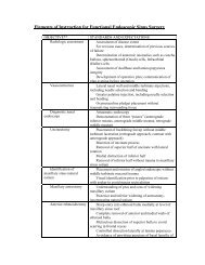
Understanding Complex Patho-Morphology - University of Utah ...
Understanding Complex Patho-Morphology - University of Utah ...
Understanding Complex Patho-Morphology - University of Utah ...

Create successful ePaper yourself
Turn your PDF publications into a flip-book with our unique Google optimized e-Paper software.
Hip Preserving Surgery:<br />
<strong>Understanding</strong> <strong>Complex</strong><br />
<strong>Patho</strong>-<strong>Morphology</strong><br />
*Christopher L. Peters MD<br />
Jill Erickson PA-C<br />
Lucas Anderson PA-C<br />
Andrew Anderson PhD<br />
Jeff Weiss PhD<br />
<strong>University</strong> <strong>of</strong> <strong>Utah</strong> Orthopaedic Center,<br />
Salt Lake City, <strong>Utah</strong><br />
Disclosures are listed in the AAOS<br />
program and website.<br />
No conflicts with this subject.
Introduction:<br />
Recent evidence suggests that abnormal hip morphology may be the<br />
primary etiology <strong>of</strong> hip osteoarthritis in young adults. Hip pathomorphology<br />
is manifested as acetabular deficiency or malorientation, or as<br />
femoral deformity and malorientation, or, most commonly, combinations<br />
<strong>of</strong> the two. Contemporary hip preservation surgical intervention has been<br />
directed at correction <strong>of</strong> these malformations and associated chondrolabral<br />
injuries and has shown promise in alleviation <strong>of</strong> hip pain and possibly<br />
retardation <strong>of</strong> osteoarthritis progression.<br />
With the increasing number <strong>of</strong> available <strong>of</strong> surgical methodologies<br />
directed at hip preservation such as surgical dislocation, osteochondroplasty,<br />
hip arthroscopy, and redirectional acetabular osteotomy, the<br />
importance <strong>of</strong> understanding the pathological process driving the painful<br />
hip has become paramount. In an effort to augment the basic information<br />
obtained from clinical examination, two-dimensional plain radiography,<br />
and magnetic resonance arthrography, we have utilized a validated 3D<br />
modeling protocol to serve as a diagnostic and surgical planning tool for<br />
hip preservation surgery. Three-dimensional modeling has helped<br />
emphasize the complex pathomorphology evident in many patients with<br />
hip dysplasia and femoroacetabular impingement and may have a future<br />
role in classification and treatment <strong>of</strong> hip maladies in young adults.<br />
Objectives:<br />
1. Describe the typical presentation <strong>of</strong> the young adult with a painful hip<br />
and corresponding case examples which illustrate common morphologic<br />
abnormalities <strong>of</strong> the femur and acetabulum.<br />
2. Using case examples, illustrate the value <strong>of</strong> a comprehensive imaging<br />
protocol to facilitate diagnosis and treatment <strong>of</strong> patients with FAI and<br />
dysplasia.<br />
3. Present our experience with 3D model development as a function <strong>of</strong>, or<br />
subset <strong>of</strong> work focused on the biomechanics <strong>of</strong> the dysplastic hip.<br />
4. Present the experience with subject-specific models including the<br />
exhibition <strong>of</strong> combined acetabula and femoral deformities.<br />
5. Outline future work including development <strong>of</strong> a 3D classification<br />
system.
Femoral Deformities<br />
20 year old male, college football player, with acetabular retroversion, positive posterior wall sign and an Os acetabuli presented with<br />
symptomatic FAI. Treatment consisted <strong>of</strong> Surgical Dislocation, rim debridement, Os excison, labral refixation and femoral head-neck<br />
osteochondroplasty.<br />
23 year old male with history <strong>of</strong> prior femoral neck fracture treated with ORIF with resultant femoral retroversion deformity and FAI. Treatment<br />
consisted <strong>of</strong> surgical dislocation, delaminated hyaline cartilage resection, rim debridement and labral refixation.<br />
14 year old female with history <strong>of</strong> Perthes disease and FAI. Treatment consisted <strong>of</strong> surgical dislocation with relative femoral neck lengthening.<br />
Acetabular cartilage was well maintained (OB1) but the labrum was detached at 12:30. Acetabulum received no treatment, femoral head-neck<br />
<strong>of</strong>fset improved with osteochondroplasty.<br />
Acetabular Deformities<br />
27 year old male presented with FAI and restricted ROM <strong>of</strong> the right hip. Radiographs revealed acetabular overcoverage and retroversion from<br />
calcified labrum. Treatment consisted <strong>of</strong> surgical dislocation, resection <strong>of</strong> calcified labrum from 8 to 4 o’clock.<br />
27 year old surfer who sustained a “hip injury” diving, presented with symptomatic FAI with acetabular retroversion, positive posterior wall sign,<br />
and an os acetabuli. CT scan and 3D model confirmed deficient posterior wall. Treatment consisted <strong>of</strong> periacetabular osteotomy with femoral<br />
head-neck osteochondroplasty, removal <strong>of</strong> os and labral repair.<br />
43 year old female presented with restricted ROM and “a labral tear”. Radiographs revealed coxa pr<strong>of</strong>unda. MRA suggested labral tear.<br />
Treatment consisted <strong>of</strong> surgical dislocation and rim resection with labral refixation from 3-9 o’clock.<br />
30 year old female presented with symptomatic classic acetabular dysplasia worse on the left. Treatment consisted <strong>of</strong> left periacetabular osteotomy.<br />
A year later she developed worsening symptoms on the right, and we performed a right PAO with femoral head-neck osteochondroplasty.<br />
She is pain-free at 4 and 5 year follow-up.
Contemporary Preoperative Planning:<br />
18 year old male previously treated for FAI <strong>of</strong> the left hip with surgical dislocation and femoral <strong>of</strong>fset improvement.<br />
The right hip exhibited acetabular retroversion with a positive posterior wall sign and visible ischial<br />
spine. 3D modeling confirmed significant acetabular posterior wall deficiency and intact hyaline cartilage.<br />
Treatment consisted <strong>of</strong> anteversion producing PAO and femoral head-neck <strong>of</strong>fset improvement. Hip pain and<br />
function is improved at 4 years follow-up. Full video <strong>of</strong> the progression and surgeries can be seen below.<br />
18 year old male with FAI and history <strong>of</strong> Perthes disease. MRA showed evidence <strong>of</strong> loose osteochondral<br />
bodies within the joint. CT arthrography and 3D modeling confirmed typical Perthes femoral deformity<br />
and osteochondral head fragmentation. Treatment consisted <strong>of</strong> relative femoral neck lengthening and<br />
grafting <strong>of</strong> the femoral head defect with osteochondral tissue resected from the head-neck junction.<br />
Acetabular hyaline cartilage delamination (OB4) was treated with resection and labral refixation. Hip<br />
pain and function is improved at 3 year follow-up.
Three-Dimensional Model Development and Validation:<br />
Overview : Preoperative surgical planning protocol (flowchart) developed in parallel with<br />
ongoing NIH grant (#RO1AR053344). Grant objective is to compare cartilage mechanics<br />
between normal and dysplastic hips using patient-specific finite element analysis (FEA)<br />
Clinic Visit,<br />
Preliminary<br />
Diagnosis<br />
1.Assess Exclusion Criteria<br />
2.Obtain Informed Consent<br />
3.Schedule CTA<br />
Patient Recruitment<br />
CTA<br />
• FAI (cam, pincer), classic dysplasia, 18-40 years old<br />
• Informed consent obtained (IRB#10983)<br />
• Excluded : Tonnis >1, pregnant women, THA candidates<br />
CT Arthrography<br />
• 15-30 ml contrast injected into capsule under fluoroscopic<br />
control (2:1 lidocaine 1% to Omnipaque 300)<br />
• With needle removed, traction applied to ensure contrast<br />
covered proximal femur essential (Figure 1)<br />
• Multi-Detector CT scan covered proximal iliac crest to trochanter<br />
and distal condyles (Figure 2)<br />
• Femur hare traction splint affixed to affected limb, ~50-80 lbs.<br />
force delivered to increase joint space; traction was a<br />
necessary precursor to obtain quality images (Figure 3)<br />
• Settings (Figure 2)<br />
– Pelvis 120 kVP, CareDose [200-400 mAs], 1 mm slice thick. w/o<br />
overlap, FOV = 250-400 mm, resolution ~0.75 x 0.75 x 1.0 mm<br />
– Femoral Condyles 80 kVP, 100 mAs, 3 mm slice thick. w/o overlap,<br />
FOV = pelvic scan<br />
Patient-<br />
Specific<br />
3D<br />
FEA<br />
Reconstruction Analyze 3D<br />
<strong>Morphology</strong><br />
Figure 1: Fluoroscope images <strong>of</strong> patient<br />
without (left) and with (right) manual traction<br />
Contrast<br />
Traction<br />
Predict Long-Term Outcome<br />
1. FE models yield contact area,<br />
peak pressure, average pressures,<br />
shear stresses<br />
2. Pre/PostOp FEA<br />
3. Develop improved classification<br />
system (FAI, Retro, Dysplasia)<br />
All Inclusive PreOp Report<br />
1. Radiological, MRA/CTA findings<br />
2. Anatomical views <strong>of</strong> 3D model<br />
3. Cartilage thickness & asphericity<br />
<strong>of</strong> femur reported as color 3D plot<br />
Figure 2: CT Scout <strong>of</strong> Study Subject<br />
Pelvic<br />
Cartilage<br />
Traction<br />
Contrast<br />
Femur<br />
Cartilage<br />
Figure 3: Coronal CTA images <strong>of</strong> patient without (left) and<br />
with (right) traction applied. Contrast clearly covers the entire<br />
proximal femur in the hip under traction, improving image<br />
quality and ability to delineate pelvic from femoral cartilage.
3D Reconstruction & Validation<br />
• Separate surfaces for cortical bone and cartilage semi-automatically reconstructed using commercial s<strong>of</strong>tware (Amira 5.2, Visage Imaging<br />
Inc.) (Figure 4)<br />
• Requires approximately 8-12 hours <strong>of</strong> segmentation time for each model (aided by undergraduate research assistants)<br />
• Surfaces smoothed and decimated to remove staircase artifact (Figure 4)<br />
• Surfaces used for preoperative surgical planning (see below)<br />
• Validation<br />
– Error between true anatomy and 3D reconstruction shown to be < 3% (1)<br />
– Cartilage >1.0 mm thick can be accurately (i.e. < 10% error) reconstructed from CTA images (2)<br />
– 2:1 dilution factor for contrast agent allows for accurate segmentation (2); not too bright / dull<br />
Amira 5.2 Console<br />
Figure 4: 3D models are generated from CT arthrography image data on a patient-specific basis using Amira 5.2. Example <strong>of</strong> process for patient with classic dysplasia.<br />
Left) Screen shot <strong>of</strong> program showing the outline for the pelvic cortex (coronal plane). Segmented splines are triangulated into a surface (middle) which is smoothed<br />
and decimated to remove digitization and CT stair-case artifacts (right).<br />
Data Analysis & Comprehensive Preoperative Surgical Report<br />
• Validated thickness algorithm (1) estimates cartilage thickness on a patient-specific basis (Figure 5)<br />
• Femur surface defining interface between bone/cartilage fit to perfect sphere using validated protocol (3) (Figure 6)<br />
• In special cases, life sized prototype made in-house using 3D printer (~$150 for femur + hemi-pelvis)<br />
• Preoperative report includes<br />
– Plain films and measurements (CEA, wall sign, cross-over sign, etc.)<br />
– CTA (degree <strong>of</strong> retroversion, lesions, labral tears, angles, etc.)<br />
– Screenshots <strong>of</strong> 3D model (full appreciation <strong>of</strong> anterior/posterior over/under-coverage)<br />
– Plot <strong>of</strong> cartilage thickness (Figure 5)<br />
– Localized asphericity <strong>of</strong> femoral head (Figure 6)<br />
Figure 5: Plot <strong>of</strong> cartilage thickness in a normal human femur (left) and<br />
acetabulum (right). The reconstruction error for cartilage has been<br />
shown to be < 10% if cartilage is at least 1.0 mm thick (2).<br />
Surface Triangulation<br />
Patient-Specific Finite Element Analysis (ongoing work)<br />
• Validated finite element analysis (FEA) protocol applied to generate patient-specific FE meshes (4)<br />
– Varying cartilage and cortical bone thickness (Figure 7)<br />
• Patient-specific kinematics acquired from motion analysis lab and applied to FE models<br />
• FE analysis yields patient-specific cartilage contact pressures, contact area, shear stresses, etc. (Figures 7, 8)<br />
– Compare normal vs. dysplastic hip joints (Figure 8) to develop a quantitative understanding <strong>of</strong> how geometrical deformities predispose<br />
the hip to early onset OA<br />
Femur<br />
Cortex Femur<br />
Cartilage<br />
2.0 mm<br />
0.8 mm<br />
7 mm<br />
0.2 mm<br />
Figure 7: Patient-specific FE mesh <strong>of</strong> a normal human hip. Left) detail <strong>of</strong> femur FE mesh showing cortical bone and cartilage.<br />
Cartilage has been modeled with varying thickness as segmented from the CT images. Middle) cortical bone is modeled using<br />
shell elements with position dependent thickness, which is also based on the CT image data. Right) magnitude and<br />
distribution <strong>of</strong> cartilage contact pressures in a normal acetabulum under simulated descending stairs.<br />
4 mm<br />
0.2 mm<br />
Smoothing,<br />
Decimati0on<br />
Rough Triangulated Surface Smoothed and Decimated Surface<br />
Cam Patient<br />
Deviation from Sphere<br />
Figure 6: Plot <strong>of</strong> asphericity <strong>of</strong> femoral head. Left) cam FAI patient. Middle) normal joint. Right) when<br />
re-scaled, normal joint continues to show very little deviation from perfect sphere. Note- positive<br />
deviations = “bone to be removed” whereas negative deviations = bone indentations. Please see<br />
ORS2009-3159 for more details.<br />
7 MPa<br />
0 MPa<br />
Normal<br />
+1.0 mm<br />
-1.0 mm<br />
Normal<br />
Dysplastic<br />
Deviation from Sphere (updated range)<br />
+0.05 mm<br />
Normal<br />
-0.05 mm<br />
Walking Stair-Climbing Descending Stairs<br />
10 MPa<br />
0 MPa<br />
Figure 8: Comparison <strong>of</strong> cartilage contact pressures between a<br />
normal (top) and dysplastic (bottom) hip joint. Pressures remained<br />
elevated in the dysplastic joint during simulated walking, stairclimbing<br />
and descending stairs (columns).
Three-dimensional Imaging and <strong>Complex</strong> <strong>Patho</strong>morphology:<br />
17 year old male with pr<strong>of</strong>ound acetabular retroversion presented with symptomatic FAI. 3D modeling<br />
clearly demonstrates posterior acetabular wall deficiency and relative lack <strong>of</strong> head-neck <strong>of</strong>fset. Treatment<br />
consisted <strong>of</strong> anteversion producing PAO with femoral head-neck <strong>of</strong>fset.<br />
24 year old male with severe bilateral classic acetabular dysplasia. 3D modeling clearly demonstrates<br />
anterior and lateral socket deficiency. Treatment consisted <strong>of</strong> PAO and femoral osteochondroplasty.<br />
35 year old male with combined cam and pincer FAI. 3D modeling demonstrates acetabular retroversion<br />
and poor femoral head-neck <strong>of</strong>fset.Treatment consisted <strong>of</strong> surgical dislocation, excision <strong>of</strong> delaminated<br />
articular cartilage from 1-3 o’clock, labral refixation and femoral <strong>of</strong>fset improvement.<br />
23 year old male with combined cam and pincer FAI. 3D modeling demonstrates acetabular retroversion with<br />
prominent Os acetabuli and posterior deficiency. Treatment consisted <strong>of</strong> periacetabular osteotomy, Os removal,<br />
labral repair and femoral head-neck osteochondroplasty as seen in the fluoroscopy pictures below.
Conclusions:<br />
1. Abnormal hip morphology may be the dominant cause <strong>of</strong> hip<br />
pain and the subsequent development <strong>of</strong> osteoarthritis <strong>of</strong> the hip<br />
in young adults.<br />
2. Hip pathomorphology may be manifested by femoral or acetabular<br />
deformity or malorientation. Most patients have combinations<br />
<strong>of</strong> the two.<br />
3. Surgical intervention should be focused on correction <strong>of</strong> the<br />
underlying pathomorphology as well as the resultant chondrolabral<br />
tissue damage.<br />
4. 3D computational modeling <strong>of</strong> the hip can facilitate proper identification<br />
<strong>of</strong> the major pathomorphologic anatomy.<br />
5. 3D computational modeling may facilitate improved understanding<br />
<strong>of</strong> FAI and dysplasia <strong>of</strong> the hip via new classification systems<br />
and verification <strong>of</strong> treatment methodologies.<br />
Acknowledgement: NIH Grant #R01AR053344<br />
References:<br />
1. Anderson, A.E., Peters, C.L., Weiss, J.A. et al. Journal <strong>of</strong> Biomechanical Engineering 127:364-373, 2005<br />
2. Anderson, A.E., Peters, C.L., Weiss, J.A. et al. Radiology, 246(1):133-41, 2008<br />
3. Anderson, A.E., Weiss, J.A. et al. Proceedings, 8th Symposium <strong>of</strong> Computational Methods in Biomechanics<br />
and Bioengineering. February, 2008<br />
4. Anderson, A.E., Peters, C.L., Weiss, J.A. et al. Journal <strong>of</strong> Biomechanical Engineering, 130(5):051008, 2008