Morning Report May 25, 2010
Morning Report May 25, 2010
Morning Report May 25, 2010

Create successful ePaper yourself
Turn your PDF publications into a flip-book with our unique Google optimized e-Paper software.
<strong>Morning</strong> <strong>Report</strong><br />
December 22, <strong>2010</strong><br />
**Welcome Applicants!**<br />
Geoffrey Smith, MD<br />
Andrew Aronson, MD<br />
Sign onto the Wireless and join<br />
Session ID #A700
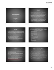
MKSAP<br />
A 55-year-old woman is evaluated for a 4-month history of progressive<br />
fatigue and pruritus. She is otherwise healthy and takes no medications.<br />
On physical examination, vital signs are normal; BMI is <strong>25</strong>. Examination<br />
reveals mild hepatomegaly and excoriations of the skin. There is no<br />
jaundice, rash, or skin eruption.<br />
Laboratory studies:<br />
• Bilirubin (total) 1.5 mg/dL (<strong>25</strong>.6 µmol/L)<br />
• Aspartate aminotransferase 60 U/L<br />
• Alanine aminotransferase 75 U/L<br />
• Alkaline phosphatase 470 U/L<br />
• Albumin 4.0 g/dL (40 g/L)<br />
• Hepatitis C virus antibody Negative<br />
• Hepatitis B surface antigen Negative<br />
Ultrasonography of the abdomen is normal with no bile duct dilatation.
Which of the following is the most appropriate<br />
next step in the diagnosis of this patient?<br />
A. α 1-Antitrypsin phenotype<br />
B. Liver biopsy<br />
C. Measurement of serum antimitochondrial<br />
antibody<br />
D. Serum protein electrophoresis
Which of the following is the most appropriate<br />
next step in the diagnosis of this patient?<br />
A. α 1-Antitrypsin phenotype<br />
B. Liver biopsy<br />
C. Measurement of serum<br />
antimitochondrial antibody<br />
D. Serum protein<br />
electrophoresis<br />
<strong>25</strong>% <strong>25</strong>% <strong>25</strong>% <strong>25</strong>%<br />
A. B. C. D.
• This patient likely has primary biliary cirrhosis, and measurement of<br />
antimitochondrial antibody is a highly sensitive and specific test for the<br />
disorder; more than 95% of patients with primary biliary cirrhosis have<br />
antimitochondrial antibodies and the false positive rate is approximately<br />
2%.<br />
• Fatigue, pruritus, an elevated alkaline phosphatase concentration, and the<br />
presence of these antibodies strongly suggest this diagnosis.<br />
• Although fatigue and pruritus used to be the most common presenting<br />
symptoms of primary biliary cirrhosis, the disease is now more widely<br />
recognized and many patients are diagnosed at earlier stages when they are<br />
asymptomatic.<br />
• If the antimitochondrial antibody is positive, liver biopsy is useful to<br />
provide evidence that would stage the disease and other liver disorders that<br />
may cause cholestasis similar to that associated with primary biliary<br />
cirrhosis.<br />
• α1-Antitrypsin deficiency would present with abnormal aminotransferase<br />
concentrations, not with a cholestatic picture.<br />
• A serum protein electrophoresis may demonstrate elevation in serum IgM,<br />
but would not be diagnostic of primary biliary cirrhosis.
34 y/o M with abdominal pain
• In USOH until yesterday.<br />
• Right upper quadrant and epigastric abdominal pain,<br />
radiating to back.<br />
• Pain constant, 10/10, began at 8pm yesterday.<br />
• Last PO intake pizza at 4:30pm yesterday. Still feels<br />
hungry, no loss of appetite.<br />
• No fevers, chills, nausea, vomiting, constipation,<br />
diarrhea, hematochezia, or melena.<br />
• Notes darker urine, but mildly decreased in quantity
Medical History<br />
• PMH: choledocholithiasis s/p cholecystectomy and ERCP, OSA<br />
not using CPAP<br />
• PSH: cholecystectomy and ERCP 2007<br />
• SH: Lives with wife. Smokes 2-3 cigarettes daily since 1995.<br />
Drinks approximately 40 oz of beer daily, but last drink was 1<br />
week PTA. Occasional marijuana, last 1 week PTA.<br />
• FH: Mother with cervical Ca; father died of unknown causes.<br />
• MEDS: None<br />
• Allergies: IV contrast, ciprofloxacin, clindamycin
Differential Diagnosis?
• Right upper quadrant<br />
– Hepatitis<br />
– Cholecystitis<br />
– Cholangitis<br />
– Pancreatitis<br />
Abdominal pain differential<br />
– Budd-Chiari syndrome<br />
– Pneumonia/empyema pleurisy<br />
– Sub-diaphragmatic abscess<br />
• Right lower quadrant<br />
– Appendicitis<br />
– Salpingitis<br />
– Ectopic pregnancy<br />
– Inguinal hernia<br />
– Nephrolithiasis<br />
– Inflammatory bowel disease<br />
– Mesenteric adenitis (yersina)<br />
• Epigastric<br />
– Peptic ulcer disease<br />
– GERD<br />
– Gastritis<br />
– Pancreatitis<br />
– Myocardial infarction<br />
– Pericarditis<br />
– Ruptured aortic aneurysm<br />
• Periumbilical<br />
– Early appendicitis<br />
– Gastroenteritis<br />
– Bowel obstruction<br />
– Ruptured aortic aneurysm<br />
• Left upper quadrant<br />
– Splenic abscess<br />
– Splenic infarct<br />
– Gastritis<br />
– Gastric ulcer<br />
– Pancreatitis<br />
• Left lower quadrant<br />
• Diverticulitis<br />
• Salpingitis<br />
• Ectopic pregnancy<br />
• Inguinal hernia<br />
• Nephrolithiasis<br />
• Irritable bowel syndrome<br />
• Inflammatory bowel disease<br />
• Diffuse Gastroenteritis<br />
• Mesenteric ischemia<br />
• Metabolic (eg, DKA, porphyria)<br />
• Malaria<br />
• Familial Mediterranean fever<br />
• Bowel obstruction<br />
• Peritonitis<br />
• Irritable bowel syndrome
Referred Pain Areas
Physical Exam<br />
• General: Obese man sitting in bed, NAD, A+Ox3<br />
• VS: Ht 172.7cm (5’8”) Wt 140kg T 36.7 P 99 R 18 BP<br />
165/109 SaO2 98% on RA<br />
• HEENT: EOMI, scleral icterus, dry MM<br />
• Neck: no lymphadenopathy, no thyromegaly<br />
• Lungs: Clear to auscultation<br />
• CV: regular, nl S1 and S2, no m/g/r, no JVD, periphery warm<br />
and well perfused, 2+ radial and d.p. pulses<br />
• Abdomen: soft, distended, RUQ tenderness (+Murphy’s<br />
sign)<br />
• GU: no CVA tenderness, no suprapubic tenderness<br />
• Neuro: CN II-XII intact, strength and sensation intact,<br />
• Skin: no rashes but several tattoos, no edema
Work up?<br />
• Which tests would you like to order?
7.5<br />
141<br />
3.7 <strong>25</strong><br />
100 10<br />
17.0<br />
48.3<br />
Laboratory Studies<br />
0.9<br />
184<br />
145<br />
Ca 9.3<br />
8.7 4.7<br />
4.9<br />
3.0/1.9<br />
534 421<br />
Lipase 39<br />
U/A: yellow, clear, s.g. 1.035, pH 6.0, Neg LE, Neg<br />
nitrites, trace Protein, Neg blood, Neg glucose,<br />
Neg ketones, Bilirubin 1+, Urobilinogen 1.0,<br />
occasional RBC, no casts, no bacteria,<br />
126<br />
Urine:<br />
Na 87<br />
Cr 111<br />
FENa 0.5%
Management at this point?<br />
• Diagnostic tests?<br />
• Additional Labs?<br />
HAV IgM NR<br />
HBV sAg NR<br />
HBV cAb IgM NR<br />
HCV Ab NR<br />
US technician not available for RUQ US
Biliary Tree
Abdominal CT w
Abdominal CT read<br />
• Probable fatty liver. Cholecystectomy clips with mild central<br />
biliary prominence. No radio-opaque foci in course of cystic<br />
duct remnant, common hepatic, or common bile duct. No<br />
focal liver lesions. Hepatic vasculature enhances normally. No<br />
other significant abnormality noted.<br />
• Impression: post cholecystectomy with mild central biliary<br />
prominence. No CT evidence of choledocholithiasis. Possible<br />
fatty liver.
Accuracy of Imaging Modalities<br />
for Detection of Biliary stones<br />
• Ultrasound<br />
– Sens 20%, Spec 83.3%<br />
• Computerized tomography<br />
– Sens 40%, Spec 91.7%<br />
• MRCP<br />
– Sens 80%, Spec 83.3%<br />
• ERCP<br />
– Sens 90%, Spec 91.7%<br />
• Endoscopic ultrasound<br />
– Sens 95%, Spec 91.7%<br />
– Moon et al., Am J Gastroenterology 2005.
Status change<br />
• ER calls to inform you patient has become<br />
drowsy/altered, spiked a fever, decreasing<br />
oxygen saturation, and is beginning to rigor.<br />
• New vitals 4h after initial exam:<br />
• T38.5 P 102 R 20 BP 141/77 Sat 92%/RA
3.3<br />
138<br />
2.7 17<br />
105 10<br />
12.2<br />
48.3<br />
N 92 L 6 M 2<br />
(+ left shift)<br />
Laboratory Studies<br />
1.0<br />
117<br />
93<br />
Ca 7.0<br />
Mg 1.0<br />
Ph 1.5<br />
INR 2.2<br />
PTT 41<br />
6.2 3.4<br />
5.7<br />
231 315<br />
Lipase 30<br />
Amylase 33<br />
Lactate 4.1<br />
274
Focused differential
Initial management?<br />
• What are we treating?
50-75% of cases
Acute Cholangitis Manifestation<br />
• Charcot triad (fever, RUQ pain, jaundice) occurs in<br />
50-75% of cases.<br />
• Reynold’s pentad (Charcot triad, hypotension,<br />
altered mental status) can occur in suppurative<br />
cholangitis.<br />
• Labs: elevated WBC with neutrophils, cholestatic<br />
liver test abnormalities, amylase can be elevated.<br />
• In the case of microabscess formation in the liver,<br />
acute hepatocyte necrosis can occur, leading to<br />
elevated aminotransferases as well.
Acute Cholangitis Microbiology<br />
• Foreign body (e.g., stone or stent) is nidus for bacterial colonization.<br />
• Bile from patients without obstruction is sterile or nearly sterile. In comparison,<br />
70% of patients with gallstones have bacteria in their bile.<br />
• Common bile duct stones - higher probability of bile culture positivity vs.<br />
gallbladder or cystic duct stones.<br />
• Causative bacteria:<br />
• GNR<br />
– Escherichia coli <strong>25</strong>-50%<br />
– Klebsiella 15-20%<br />
– Enterobacter species 5-10%<br />
• GPC<br />
– Enterococcus species 10-20%<br />
• Anaerobes<br />
– Bacteroides, Clostridia usually present as a mixed infection.<br />
– Rarely the sole infecting organisms; not clear if they play a role in acute cholangitis.<br />
– Recovery of anaerobes more common after repeated biliary tree infections or surgery.
Acute Cholangitis<br />
• What’s the next step in management of acute<br />
cholangitis?<br />
• Medical management vs. ERCP vs. surgical<br />
decompression
Management of cholangitis<br />
• Treatment: Antibiotics and Biliary drainage<br />
• 80% of patients will respond to conservative management<br />
and antibiotic therapy. Biliary drainage can then be performed<br />
on an elective basis.<br />
• In 15-20% percent of cases, cholangitis fails to settle over the<br />
first 24 hours with conservative therapy alone, requiring<br />
urgent biliary decompression.<br />
• Indications for urgent biliary decompression include:<br />
– Persistent abdominal pain<br />
– Hypotension despite adequate resuscitation<br />
– Fever greater than 39ºC (102ºF)<br />
– Mental confusion, which is a predictor of poor outcome
Patients with Endoscopic<br />
Biliary drainage:<br />
Spent less time on the<br />
ventilator*<br />
Became afebrile sooner<br />
Had shorter duration of<br />
fasting*<br />
*significant findings<br />
Surgery vs. ERCP<br />
• NEJM 1992 established non-inferiority of ERCP
Surgery vs. ERCP:<br />
Less complications and Deaths with ERCP<br />
• NEJM 1992 established non-inferiority of ERCP
Timing<br />
of<br />
ERCP
• Discussed case with GI Interventional attending on<br />
call.<br />
• Transferred to ICU for closer monitoring<br />
• Aggressive fluids, antibiotics, intubation<br />
• Defervesced with IV Zosyn, ERCP deferred until AM.<br />
• Blood Cx drawn positive for Klebsiella oxytoca<br />
• Bile fluid from ERCP positive for Enterococcus<br />
casselliflavus.
Microbiology<br />
• Klebsiella pneumoniae and oxytoca (named after 19th Century german microbiologist Edwin Klebs), are<br />
both GNR opportunistic pathogens found in the<br />
environment and mammalian mucosal surfaces,<br />
principally GI tract.<br />
• Enterococcus casseliflavus is one of the intrinsically<br />
Vancomycin-resistant enterococcus GPC that is part<br />
of the normal flora of the GI tract of mammals,<br />
occasionally causing disease.
• Impression: -<br />
Choledocholithiasis<br />
with an obstruction<br />
was found.<br />
- Two biliary stents<br />
were placed into the<br />
common hepatic<br />
duct.<br />
- Ascending cholangitis<br />
was found.<br />
ERCP
• How could he have recurrent choledocholithiasis<br />
even after his cholecystectomy?
Pathogenesis of recurrent<br />
choledocholithiasis s/p cholecystectomy<br />
• Bile-duct dysfunction<br />
– Delayed biliary clearance<br />
• Abnormal biliary motility<br />
• Post-ES papillary stenosis<br />
• Altered bile composition<br />
– Biliary cholesterol supersaturation<br />
• MDR3 missense mutations (normally secretes<br />
phosphatidylcholne.
Risk factors for Recurrent biliary stones after<br />
endoscopic sphincterotomy (EST)
Patient follow-up<br />
• Discharged to continue home IV Vanco and Zosyn<br />
• F/U ERCP:<br />
- Choledocholithiasis was found. Complete<br />
removal was accomplished by biliary<br />
sphincterotomy and lithotripsy.
Prevention<br />
• How do we prevent the next episode of<br />
cholangitis? Unclear.<br />
• Options include:<br />
1. Dismiss such patients from follow- up and encourage<br />
return only if symptoms recur.<br />
2. Periodically repeat serum liver chemistries and/or noninvasive<br />
imaging of the ducts.<br />
3. Repeat invasive ductal imaging, usually by ERCP. This<br />
alternative is rarely applied to asymptomatic patients.<br />
4. Prophylactic treatment with antibiotics, cholerrhoeic<br />
agents or stone solubilizing agents, e.g., ursodiol.
Take Home Points<br />
• Recognize ascending cholangitis<br />
• Understand how to triage these patients<br />
• Know the role of ERCP in management of<br />
cholangitis<br />
• Understand the risks for recurrent biliary<br />
stones in the absence of a gallbladder