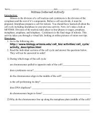
Which Cell Parts Can You See With the Microscope?
Which Cell Parts Can You See With the Microscope?
Which Cell Parts Can You See With the Microscope?

You also want an ePaper? Increase the reach of your titles
YUMPU automatically turns print PDFs into web optimized ePapers that Google loves.
Name: _________________________________________________ Date: ________________ Period: ____<br />
<strong>Which</strong> <strong>Cell</strong> <strong>Parts</strong> <strong>Can</strong> <strong>You</strong> <strong>See</strong> <strong>With</strong> <strong>the</strong> <strong>Microscope</strong>?<br />
Introduction: Living things are made of cells. All cells have parts that do certain jobs. <strong>Cell</strong>s have an outer covering called <strong>the</strong> cell (plasma)<br />
membrane. The cell membrane controls what can enter/exit a cell. The clear jellylike material inside <strong>the</strong> cell is <strong>the</strong> cytoplasm. The nucleus is <strong>the</strong><br />
control center of <strong>the</strong> cell. Plant cells have a thick outer covering called <strong>the</strong> cell wall. It is found on <strong>the</strong> outside of <strong>the</strong> cell membrane.<br />
<strong>Cell</strong> parts can be studied by making wet mounts slides. A wet mount slide is a temporary slide. It is not made to last a long time. <strong>You</strong> can make<br />
wet mount slides of living and once living materials to study cell parts.<br />
Materials:<br />
• Glass slides<br />
• Droppers<br />
• Onion skin<br />
1. Place <strong>the</strong> specimen in<br />
<strong>the</strong> center of a CLEAN<br />
slide.<br />
1. Place a drop of stain at one end of <strong>the</strong><br />
cover slip.<br />
• Cover slips<br />
• Forceps<br />
• Various prepared slides<br />
Procedures: Making a Wet Mount Using Water<br />
2. Place a drop of water<br />
on <strong>the</strong> specimen.<br />
3. Place a cover slip on <strong>the</strong><br />
slide. Put one edge of <strong>the</strong><br />
cover slip into <strong>the</strong> drop of<br />
water, and slowly dower<br />
<strong>the</strong> cover slip over <strong>the</strong><br />
specimen.<br />
Procedures: Staining a Specimen<br />
Identify <strong>Microscope</strong> <strong>Parts</strong>: Read page R8 of your textbook and label <strong>the</strong> microscope picture below.<br />
1. <strong>Which</strong> two parts do you think should be held when carrying a microscope?<br />
___________________________<br />
2. <strong>Which</strong> microscope part:<br />
a. Adjusts <strong>the</strong> amount of light that enters <strong>the</strong> microscope?<br />
_____________________<br />
b. Focuses <strong>the</strong> image under high power?<br />
_____________________<br />
c. Holds and turns <strong>the</strong> objective lenses?<br />
_____________________<br />
• <strong>Microscope</strong>s<br />
• Elodea leaf<br />
• 50 ml beaker<br />
4. Remove any air<br />
bubbles from under<br />
<strong>the</strong> cover slip by gently<br />
tapping <strong>the</strong> cover slip<br />
Filter paper<br />
5. Dry any excess<br />
water before placing<br />
<strong>the</strong> slide on <strong>the</strong><br />
microscope stage for<br />
viewing.<br />
2. Hold a piece of filter paper with forceps at <strong>the</strong><br />
o<strong>the</strong>r end of <strong>the</strong> cover slip. The stain will flow<br />
underneath <strong>the</strong> cover slip and stain <strong>the</strong> specimen.
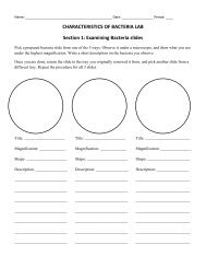
Specimen #1: Prepare a wet mount of onion cells.<br />
1. Obtain a clean slide and place a drop of iodine in <strong>the</strong> middle of <strong>the</strong> slide. If needed, rinse your slide with water and wipe dry<br />
to clean.<br />
2. Peel a thin layer of cells from <strong>the</strong> inside of an onion as seen in <strong>the</strong><br />
picture to <strong>the</strong> right. If <strong>the</strong> onion layer is not thin, peel ano<strong>the</strong>r layer.<br />
3. Place <strong>the</strong> onion peel in <strong>the</strong> drop of iodine.<br />
4. Add a cover slip over your onion.<br />
5. Examine <strong>the</strong> onion under medium and high power and using a pencil,<br />
draw your observations below.<br />
6. Be sure to find and label <strong>the</strong> <strong>Cell</strong> wall, Nucleus, Cytoplasm, and Nucleolus (optional).<br />
Onion Analysis: Best Writing Skills (complete sentences)<br />
1. Onion cells (and skin cells) are flat and seem to overlap. Explain why this arrangement is beneficial.<br />
____________________________________________________________<br />
____________________________________________________________<br />
Specimen #2: Prepare a wet mount of Elodea cells.<br />
1. Obtain a clean slide and place a drop of water in <strong>the</strong> middle of <strong>the</strong> slide. If needed, rinse your slide with water and wipe dry to<br />
clean<br />
2. Pick a leaf from <strong>the</strong> Elodea plant and place <strong>the</strong> leaf in <strong>the</strong> drop of water. Make sure <strong>the</strong> leaf is flat.<br />
3. Add a cover slip over your elodea cell.<br />
4. Examine <strong>the</strong> elodea under medium and high power and using a pencil, draw your observations below.<br />
5. Be sure to find and label <strong>the</strong> <strong>Cell</strong> wall, Chloroplast, and <strong>the</strong> Cytoplasm.
Elodea Analysis: Best Writing Skills (complete sentences)<br />
1. In what particular process do <strong>the</strong> chloroplasts function?<br />
_____________________________________________________________________<br />
_____________________________________________________________________<br />
_____________________________________________________________________<br />
2. Using <strong>the</strong> fine adjustment with high power, focus up and down on one particular cell. As you focus, some of <strong>the</strong> chloroplasts go<br />
out of view and o<strong>the</strong>rs come into view. How can you explain this?<br />
_____________________________________________________________________<br />
_____________________________________________________________________<br />
_____________________________________________________________________<br />
3. Why do you think it is important to have plants like Elodea in a balanced aquarium?<br />
_____________________________________________________________________<br />
_____________________________________________________________________<br />
_____________________________________________________________________<br />
Specimen #3: Prepare a wet mount of your cheek cells.<br />
1) Obtain a clean slide and add a drop of methylene blue in <strong>the</strong> middle of <strong>the</strong> slide. If needed, rinse your slide with water and wipe dry to<br />
clean.<br />
2) GENTLY rub <strong>the</strong> flat end of <strong>the</strong> toothpick against <strong>the</strong> inside of your cheek.<br />
3) Rub <strong>the</strong> used toothpick in <strong>the</strong> drop of methylene blue to transfer your cheek cells.<br />
4) Add a coverslip and observe under medium and high power of your microscope.<br />
5) Draw a picture of your cheek cells and label <strong>the</strong> parts you can see.<br />
Cheek Analysis: Best Writing Skills (complete sentences)<br />
1. Review your drawing of an elodea cell. What differences do you notice between an animal cell and a plant cell that can carry on<br />
photosyn<strong>the</strong>sis?<br />
____________________________________________________________<br />
____________________________________________________________<br />
____________________________________________________________<br />
Specimens 4 and 5: Examine prepared slides of cork and blood.<br />
1. Obtain a prepared slide of cork and blood from your teacher.<br />
2. Place <strong>the</strong> slides under <strong>the</strong> microscope and observe. Draw <strong>the</strong> power that best shows <strong>the</strong> cells.<br />
3. Draw and label <strong>the</strong> parts you can see on <strong>the</strong> next page.
Cork Analysis: Best Writing Skills (complete sentences)<br />
1. Knowing that cork is <strong>the</strong> remains of dead plant cells, which part (or parts) were you able to see? What is <strong>the</strong> function of this<br />
(<strong>the</strong>se) part(s)?<br />
_____________________________________________________________________<br />
_____________________________________________________________________<br />
_____________________________________________________________________<br />
Blood Analysis: Best Writing Skills (complete sentences)<br />
1. What is <strong>the</strong> function of red blood cells? How does <strong>the</strong>ir shape allow <strong>the</strong>m to do <strong>the</strong>ir job?<br />
_____________________________________________________________________<br />
_____________________________________________________________________<br />
_____________________________________________________________________<br />
General Analysis: Best Writing Skills (complete sentences)<br />
1. When first viewing an object under <strong>the</strong> microscope, explain why you should always find it under <strong>the</strong> lowest power available.<br />
_____________________________________________________________________<br />
_____________________________________________________________________<br />
_____________________________________________________________________<br />
_____________________________________________________________________<br />
2. Why do cells have different shapes?<br />
_____________________________________________________________________<br />
_____________________________________________________________________<br />
_____________________________________________________________________<br />
_____________________________________________________________________