Jules Stein Eye Institute itute
Jules Stein Eye Institute itute
Jules Stein Eye Institute itute

You also want an ePaper? Increase the reach of your titles
YUMPU automatically turns print PDFs into web optimized ePapers that Google loves.
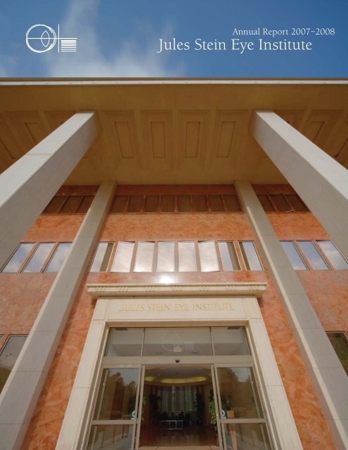
Annual Report 2007–2008<br />
<strong>Jules</strong> <strong>Stein</strong> <strong>Eye</strong> <strong>Inst<strong>itute</strong></strong>
Annual Report 2007–2008<br />
<strong>Jules</strong> <strong>Stein</strong> <strong>Eye</strong> <strong>Inst<strong>itute</strong></strong><br />
David Geffen School of Medicine at UCLA<br />
University of California, Los Angeles
ii Faculty Appendices | Endowed Professorships, Fellowships, and Other Funds<br />
<strong>Jules</strong> <strong>Stein</strong> <strong>Eye</strong> <strong>Inst<strong>itute</strong></strong><br />
Annual Report 2007–2008<br />
DIRECTOR<br />
Bartly J. Mondino, MD<br />
FACULTY ADVISOR<br />
Debora B. Farber, PhD, DPhhc<br />
MANAGING EDITOR<br />
Gloria P. Jurisic<br />
EDITORS<br />
Irene Y. Chen<br />
Tina-Marie Gauthier<br />
PHOTOGRAPHY<br />
J. Charles Martin<br />
DESIGN<br />
Ikkanda Design Group<br />
PRINTING<br />
Marina Graphic Center<br />
©2008, by the Regents of the University of California.<br />
All rights reserved.<br />
Printed on recycled paper using soy base inks<br />
This report covers the period July 1, 2007, through June 30, 2008.<br />
Requests for additional copies of the publication<br />
<strong>Jules</strong> <strong>Stein</strong> <strong>Eye</strong> <strong>Inst<strong>itute</strong></strong><br />
Annual Report 2007–2008<br />
may be sent to:<br />
Office of the Managing Editor<br />
<strong>Jules</strong> <strong>Stein</strong> <strong>Eye</strong> <strong>Inst<strong>itute</strong></strong><br />
100 <strong>Stein</strong> Plaza, UCLA<br />
Box 957000<br />
Los Angeles, California 90095–7000<br />
Phone: (310) 206-7178<br />
For more information on the <strong>Inst<strong>itute</strong></strong>, see our website: www.jsei.org<br />
The emblem of the <strong>Jules</strong> <strong>Stein</strong> <strong>Eye</strong> <strong>Inst<strong>itute</strong></strong> is adapted from the<br />
schematic eye used by Sir Isaac Newton in his classic treatise on<br />
human vision—“Opticks”—published in 1704. The horizontal lines<br />
extending from the surface of the eye represent Newton’s concept<br />
of the major colors that are in the spectrum of light.
Contents<br />
Appendices | Endowed Professorships, Fellowships, and Other Funds iii<br />
JULES STEIN v<br />
DORIS STEIN v<br />
BOARD OF TRUSTEES vi<br />
EXECUTIVE COMMITTEE vii<br />
FACILITIES viii<br />
HIGHLIGHTS 1<br />
Honors 3<br />
Research 6<br />
Education 7<br />
Philanthropy 10<br />
Thank You 12<br />
Community Outreach 13<br />
FACULTY 15<br />
PROGRAMS 63<br />
Patient Care Services 64<br />
Research and Treatment Centers 65<br />
Clinical Laboratories 72<br />
Training Programs 74<br />
APPENDICES 81<br />
Volunteer and Consulting Faculty 82<br />
Residents and Fellows 84<br />
Endowed Professorships, Fellowships, and Other Funds 86<br />
Educational Offerings 90<br />
Research Contracts and Grants 93<br />
Clinical Research Studies 106<br />
Publications of the Full-Time Faculty 111<br />
Giving Opportunities 124
iv<br />
Faculty<br />
The legacy of Dr. and Mrs. <strong>Jules</strong> <strong>Stein</strong> arises from their role<br />
in the 20th century as visionaries. Through brilliance and<br />
beneficence, they created a multitude of programs aimed<br />
specifically at one goal—preserving and restoring eyesight.<br />
They approached this task dauntlessly, integrating the<br />
worlds of business, medicine, and philanthropy in such<br />
a way as to enhance each and leave in trust the promise<br />
of limitless accomplishment in the advancement of eye<br />
research and treatment. The <strong>Jules</strong> <strong>Stein</strong> <strong>Eye</strong> <strong>Inst<strong>itute</strong></strong> was<br />
established as a result of their philanthropy.
<strong>Jules</strong> <strong>Stein</strong><br />
<strong>Jules</strong> <strong>Stein</strong> is the foremost benefactor<br />
in the world history of vision science<br />
and blindness prevention. He<br />
combined his love for music and<br />
medicine with a unique talent for<br />
analysis and organization to produce a lifetime of celebrated<br />
achievements as musician, physician, business leader,<br />
and humanitarian.<br />
Born in South Bend, Indiana, in 1896, <strong>Jules</strong> <strong>Stein</strong> received a<br />
PhB from the University of Chicago at age 18 followed by<br />
a MD degree from Rush Medical College. After completing<br />
postgraduate studies at the University of Vienna and Chicago’s<br />
Cook County Hospital, he began medical practice and was<br />
certified by the American Board of Ophthalmology.<br />
A musician from an early age, he financed his education by<br />
playing in and leading his own band. As his reputation<br />
increased, he began booking other musicians for professional<br />
engagements, and in 1924 founded Music Corporation of<br />
America (MCA). Shortly thereafter, he gave up the practice<br />
of medicine to concentrate on this enterprise. Within 10<br />
years, MCA represented most of the great name bands and<br />
corporate activities began to extend to representation of film<br />
stars, directors, writers, and musical artists. MCA entered the<br />
promising new field of television at its inception, eventually<br />
acquiring the Universal City property, Universal Pictures,<br />
and other enterprises to become pre-eminent in the<br />
entertainment industry.<br />
Throughout his phenomenally successful career, <strong>Jules</strong> <strong>Stein</strong><br />
maintained a strong interest and emotional investment in<br />
medicine, particularly his own field of ophthalmology. In the<br />
late 1950s, urged by his wife, Doris, he chose to direct his<br />
considerable talents to blindness prevention. The result<br />
was a concert of ideas and achievements that encompassed<br />
philanthropy, government, and academic medicine.<br />
By his efforts, Research to Prevent Blindness was created, now<br />
recognized as the world’s leading voluntary organization in<br />
support of studies of the eye and its diseases. <strong>Jules</strong> <strong>Stein</strong> was<br />
largely responsible for the passage of legislation to establish<br />
the National <strong>Eye</strong> <strong>Inst<strong>itute</strong></strong> as a separate entity in the National<br />
<strong>Inst<strong>itute</strong></strong>s of Health. Under his leadership, the <strong>Jules</strong> <strong>Stein</strong> <strong>Eye</strong><br />
<strong>Jules</strong> and Doris <strong>Stein</strong> v<br />
<strong>Inst<strong>itute</strong></strong> was founded as a multidisciplinary center for vision<br />
science. Since its establishment, the <strong>Inst<strong>itute</strong></strong> has become<br />
internationally identified as the focus for coordinated<br />
programs of research in the sciences related to vision,<br />
ophthalmic education, and the care of patients with eye<br />
disease. <strong>Jules</strong> <strong>Stein</strong> died in 1981, leaving a legacy of hope to<br />
the world. Through his accomplishments and philanthropy,<br />
he created ever replenishing resources for eye research and<br />
the means to preserve and restore sight for future generations.<br />
Doris <strong>Stein</strong><br />
Doris <strong>Stein</strong>’s purposeful, yet richly<br />
varied life earned the respect and<br />
affection of the many people who<br />
benefited from her humanitarianism.<br />
Inspiring partner of her husband for<br />
more than half a century, Doris <strong>Stein</strong><br />
shared with him the accomplishments of his philanthropic<br />
endeavors and guided his interests in ophthalmology,<br />
beginning with a visit to the New York Lighthouse for the<br />
Blind in the late 1950s. Deeply moved, Doris <strong>Stein</strong> urged her<br />
husband to “do something!” From that passionate beginning<br />
came a broad base of programs that catalyzed eye research.<br />
Doris <strong>Stein</strong> was a major force in this vision renaissance.<br />
She served as an officer and director of Research to Prevent<br />
Blindness, personally leading the appeal to establish more<br />
resources for investigations into eye diseases. She suggested<br />
that <strong>Jules</strong> <strong>Stein</strong> assume the principal role in the creation<br />
of an eye inst<strong>itute</strong> at UCLA, and her unflagging enthusiasm<br />
nurtured the <strong>Inst<strong>itute</strong></strong>’s development as a unique provider<br />
of every facet of vision research and patient care. Serving as<br />
Trustee, she focused special attention on <strong>Inst<strong>itute</strong></strong> initiatives<br />
to combat blindness throughout the world. She devoted her<br />
last days, until her death in 1984, to the development of an<br />
expansion and companion building for eye research. In 1989,<br />
dedication ceremonies were held for the Doris <strong>Stein</strong> <strong>Eye</strong><br />
Research Center.<br />
With grace, vision, and meaningful action, Doris <strong>Stein</strong><br />
enhanced the lives of all privileged to know her, stimulated a<br />
cascade of progress in eye research, co-founded the <strong>Inst<strong>itute</strong></strong><br />
with its boundless scientific potential, and extended the<br />
miracle of sight to untold numbers of people.
vi Faculty Board of Trustees<br />
BOARD OF TRUSTEES<br />
The <strong>Jules</strong> <strong>Stein</strong> <strong>Eye</strong> <strong>Inst<strong>itute</strong></strong> Board of Trustees was established<br />
in 1977 to ensure the <strong>Inst<strong>itute</strong></strong>’s orderly growth and<br />
development. The Board meets regularly during the year, with<br />
each Trustee providing his/her unique counsel. Collectively,<br />
their invaluable contributions have included fiscal planning<br />
Current Members<br />
Bartly J. Mondino, MD<br />
Director<br />
<strong>Jules</strong> <strong>Stein</strong> <strong>Eye</strong> <strong>Inst<strong>itute</strong></strong><br />
1994–present<br />
Ronald L. Olson, Esq.<br />
Partner<br />
Munger, Tolles, and Olson<br />
1995–present<br />
Gerald H. Oppenheimer<br />
President<br />
Gerald Oppenheimer<br />
Family Foundation<br />
President<br />
Systems Design Associates<br />
1992–present<br />
Andrea L. Rich, PhD<br />
Retired President, Chief Executive<br />
Officer and Director<br />
Los Angeles County Museum of Art<br />
Executive Vice Chancellor Emerita<br />
UCLA<br />
2007–present<br />
for the <strong>Inst<strong>itute</strong></strong>, adoption of measures to facilitate recruitment<br />
of the world’s finest vision scientists, allocation of funds for<br />
the purchase of vision research equipment, and<br />
recommendations for facilities expansion programs.<br />
Nelson C. Rising, Esq.<br />
Chairman and CEO<br />
Rising Realty Partners, LLC<br />
2004–present<br />
George A. Smith, Esq.<br />
Consultant<br />
1992–present<br />
Katrina Vanden Heuvel<br />
Publisher and Editor<br />
The Nation Magazine<br />
1984–present<br />
Casey Wasserman<br />
Chief Executive Officer<br />
The Wasserman Foundation<br />
1998–present
EXECUTIVE COMMITTEE<br />
Director, <strong>Jules</strong> <strong>Stein</strong> <strong>Eye</strong> <strong>Inst<strong>itute</strong></strong><br />
Chairman, UCLA Department of Ophthalmology<br />
Bartly J. Mondino, MD<br />
Associate Directors, <strong>Jules</strong> <strong>Stein</strong> <strong>Eye</strong> <strong>Inst<strong>itute</strong></strong><br />
Wayne L. Hubbell, PhD<br />
Gabriel H. Travis, MD<br />
<strong>Jules</strong> <strong>Stein</strong> <strong>Eye</strong> <strong>Inst<strong>itute</strong></strong> Executive Committee: (sitting from left to right) Drs. Gabriel Travis,<br />
Arthur Rosenbaum, Sherwin Isenberg, and Bartly Mondino; (standing from left to right)<br />
Mr. Jonathan Smith and Dr. Wayne Hubbell.<br />
Executive Committee vii<br />
Vice-Chairmen, UCLA Department of Ophthalmology<br />
Sherwin J. Isenberg, MD<br />
Arthur L. Rosenbaum, MD<br />
Chief Administrative Officer, <strong>Jules</strong> <strong>Stein</strong> <strong>Eye</strong> <strong>Inst<strong>itute</strong></strong><br />
Jonathan D. Smith
viii Faculty Facilities<br />
FACILITIES<br />
The facilities of the <strong>Jules</strong> <strong>Stein</strong> <strong>Eye</strong> <strong>Inst<strong>itute</strong></strong> comprise two freestanding<br />
structures of architectural note. The five-story <strong>Jules</strong><br />
<strong>Stein</strong> <strong>Eye</strong> <strong>Inst<strong>itute</strong></strong> building, occupying 110,000 square feet, is<br />
of neo-classical design. It is the original facility, constructed in<br />
1966. An expansion and companion building, the Doris <strong>Stein</strong><br />
<strong>Eye</strong> Research Center, followed in 1989. It is a four-story,<br />
red granite structure, occupying 67,000 square feet, and<br />
connecting with the <strong>Jules</strong> <strong>Stein</strong> <strong>Eye</strong> <strong>Inst<strong>itute</strong></strong> by way of a<br />
graceful portico. With support from Research to Prevent<br />
Blindness, a conference center complex was erected between<br />
the buildings as part of the Doris <strong>Stein</strong> <strong>Eye</strong> Research Center.<br />
The two facilities complement each other in design and<br />
function and together accommodate the <strong>Inst<strong>itute</strong></strong>’s outpatient<br />
treatment centers; clinical laboratories; a 30-bed ophthalmic<br />
outpatient unit; four operating rooms devoted exclusively<br />
to ophthalmic surgery; an optical dispensing facility; an<br />
ophthalmic photography unit; a pathology laboratory; basic<br />
science laboratories; a clinical research center; a library that<br />
provides dedicated study space for post doctoral vision<br />
science fellows and ophthalmology residents; and a variety<br />
of meeting facilities for lectures and conferences, including<br />
a 156-seat auditorium.<br />
The buildings reflect the considerable architectural knowledge<br />
and exquisite taste of Dr. and Mrs. <strong>Jules</strong> <strong>Stein</strong>, which is evident<br />
in the <strong>Inst<strong>itute</strong></strong>’s design, building materials, artwork and<br />
furnishings. Dr. and Mrs. <strong>Stein</strong> were committed to the belief<br />
that the special attention given to internal design created an<br />
uplifting environment for patients, visitors and staff alike.<br />
Of particular architectural note are the Reading Room,<br />
Seminar Room and Adam Room, all elegant meeting places<br />
containing antiques, original artwork and memorabilia from<br />
the <strong>Stein</strong>’s estate.<br />
Patient care suites in both facilities have been carefully<br />
planned to offer comfortable, well-appointed reception and<br />
treatment rooms, along with the most up-to-date ophthalmic<br />
equipment. These areas are enlivened by original artwork,<br />
including wall murals in the pediatric waiting rooms.<br />
The B-level lobby of the <strong>Jules</strong> <strong>Stein</strong> <strong>Eye</strong> <strong>Inst<strong>itute</strong></strong> building<br />
The <strong>Inst<strong>itute</strong></strong>’s vision research activities have benefitted<br />
enormously from the foresight of the original facility<br />
planners. A generous 20,000 square feet were devoted to<br />
basic science laboratories during the construction of the<br />
<strong>Jules</strong> <strong>Stein</strong> <strong>Eye</strong> <strong>Inst<strong>itute</strong></strong>, providing highly functional space<br />
for biochemistry, biophysics, molecular biology,<br />
electrophysiology, microsurgery, cell biology, and visual<br />
physiology research. A central infrastructure supporting<br />
all of the basic science laboratories was also incorporated<br />
into this area.<br />
Approved in 2001, and in the planning stages, is the Edie<br />
and Lew Wasserman <strong>Eye</strong> Research Center, a third building<br />
that will complete the <strong>Jules</strong> <strong>Stein</strong> <strong>Eye</strong> <strong>Inst<strong>itute</strong></strong> campus. The<br />
100,000-square-foot facility will be modeled after and situated<br />
opposite the Doris <strong>Stein</strong> <strong>Eye</strong> Research Center on <strong>Stein</strong> Plaza.<br />
Half of the space will be devoted to <strong>Inst<strong>itute</strong></strong> activities and the<br />
other half to synergistic programs of the <strong>Inst<strong>itute</strong></strong> and the<br />
David Geffen School of Medicine.<br />
Dr. <strong>Jules</strong> <strong>Stein</strong> set the standard for the <strong>Inst<strong>itute</strong></strong>’s design and<br />
function at the outset when he pronounced, “Our primary<br />
objective in building this <strong>Inst<strong>itute</strong></strong> has been to serve the needs<br />
of medical science and medical practice…At the same time, we<br />
wished to demonstrate that all these purposes can be served<br />
within an atmosphere of grace and beauty.” This standard<br />
has been maintained through the <strong>Inst<strong>itute</strong></strong>’s 42-year<br />
history and continues to be an integral component of<br />
all facilities planning.
Highlights<br />
Annual Report 2007–2008<br />
<strong>Jules</strong> <strong>Stein</strong> <strong>Eye</strong> <strong>Inst<strong>itute</strong></strong>
2 Faculty Highlights<br />
Dear Friends,<br />
I am pleased to share these highlights of the 2007–2008 academic year, which serve to<br />
strengthen our commitment to preserve sight and prevent blindness. This year we are proud<br />
to present four new faculty members, Sophie X. Deng, MD, PhD, JoAnn A. Giaconi, MD,<br />
David Sarraf, MD, and David S. Williams, PhD, who will contribute greatly to our clinical<br />
care and basic science activities. Three faculty were appointed new chair holders: Robert<br />
Alan Goldberg, MD, FACS, was appointed to the Karen and Frank Dabby Chair in<br />
Ophthalmology, Arthur L. Rosenbaum, MD, to the Brindell and Milton Gottlieb Chair<br />
in Pediatric Ophthalmology, and Steven D. Schwartz, MD, to the Ahmanson Chair<br />
in Ophthalmology.<br />
During the year, faculty members were awarded special honors from Research to Prevent<br />
Blindness, including the <strong>Jules</strong> and Doris <strong>Stein</strong> Professorship and a Career Development<br />
Award. Important research grants were awarded and renewed by the National <strong>Inst<strong>itute</strong></strong>s of<br />
Health, and other funding organizations.<br />
Philanthropic gifts to the <strong>Inst<strong>itute</strong></strong> were highlighted by the proposed establishment of two<br />
new endowed chairs by The Skirball Foundation and Ernest G. Herman, the creation of<br />
the Lydia Ann Rosenberg Fund for the Indigent Children’s Ophthalmic Care Program,<br />
and the Marie and Jerry Hornstein Family Endowed Macular Degeneration Research Fund.<br />
In addition, significant bequests were received including the Estate of Josephine M. Hewitt<br />
and the Mary E. Plummer Trust, as well as several generous contributions made in honor<br />
of friends and family.<br />
We are appreciative of these opportunities afforded to faculty and students and share the<br />
belief that we will contribute to a future full of promise.<br />
Sincerely,<br />
Bartly J. Mondino, MD<br />
Bradley R. Straatsma Professor of Ophthalmology<br />
Director, <strong>Jules</strong> <strong>Stein</strong> <strong>Eye</strong> <strong>Inst<strong>itute</strong></strong><br />
Chairman, Department of Ophthalmology, David Geffen School of Medicine at UCLA
HONORS<br />
Each year as part of their ongoing academic pursuits,<br />
faculty members achieve notable recognition derived<br />
from their accomplishments and contributions. They<br />
give invited lectures around the world; they actively<br />
participate in influential professional and community<br />
organizations; and they serve as editors and writers for<br />
a wide range of scientific journals. In some cases special<br />
honors are bestowed. Members of the faculty were<br />
honored for their contributions to ophthalmology<br />
and visual science and three new chair holders<br />
were announced.<br />
Karen and Frank Dabby Chair<br />
in Ophthalmology<br />
Robert Alan Goldberg, MD, FACS, Chief of the Orbital and<br />
Ophthalmic Plastic Surgery Division at the <strong>Jules</strong> <strong>Stein</strong> <strong>Eye</strong><br />
<strong>Inst<strong>itute</strong></strong>, has been appointed to the Karen and Frank Dabby<br />
Chair in Ophthalmology. Dr. Goldberg received his medical<br />
degree from UCLA and completed his residency and<br />
fellowship training at the <strong>Inst<strong>itute</strong></strong> before joining the faculty<br />
in 1989. He is committed to education and heavily involved<br />
in the academic administration of clinical teaching programs,<br />
directing a renowned fellowship program in orbital and<br />
ophthalmic plastic surgery and serving as Chairman of the<br />
Ophthalmology Residency Selection Committee. His<br />
surgical contributions include small-incision lateral orbital<br />
decompression, minimally invasive orbital tumor surgery,<br />
the Goldberg lacrimal stent, and non-incisional eyelid<br />
reconstruction techniques. In his clinical practice, which draws<br />
patients from across the United States and around the globe,<br />
he specializes in aesthetic and reconstructive orbital surgery,<br />
endonasal lacrimal surgery, endoscopic and small-incision<br />
facial surgery, and primary and secondary blepharoplasty.<br />
Highlights 3<br />
Brindell and Milton Gottlieb Chair in Pediatric Ophthalmology Dr. Arthur<br />
Rosenbaum (second from left), with (from left) his wife Sandra, friend and<br />
donor Mrs. Brindell Roberts Gottlieb, and daughter-in-law and son Melissa<br />
and Steve Burick.<br />
This endowment to support the activities of a distinguished<br />
faculty member in the area of orbital disease was made<br />
possible by a generous contribution from Karen and<br />
Frank Dabby.<br />
Brindell and Milton Gottlieb Chair<br />
in Pediatric Ophthalmology<br />
Arthur L. Rosenbaum, MD, appointed to the Brindell and<br />
Milton Gottlieb Chair in Pediatric Ophthalmology, has been on<br />
the UCLA faculty for 35 years, serving as Division Chief of the<br />
Pediatric Ophthalmology and Strabismus Division since 1980.<br />
He is one of the original investigators in the area of botulinum<br />
toxin injection of extraocular muscles in the treatment of<br />
strabismus and facial spastic disorders, and continues to<br />
be involved in research projects utilizing this treatment.<br />
At present, he is working on new surgical approaches to<br />
complicated strabismus problems resulting from trauma and<br />
congenital conditions. Dr. Rosenbaum has more than 200<br />
publications and co-authored a major textbook on strabismus.<br />
The recipient of several outstanding awards, including Lifetime<br />
Achievement from the American Academy of Ophthalmology<br />
and the Marshall M. Parks Medal from the Children’s <strong>Eye</strong><br />
Foundation, he is recognized for his tireless work to advance<br />
the field of pediatric ophthalmology and strabismus.<br />
Shortly before his passing in February 2006, Milton Gottlieb<br />
and his wife Brindell established the Brindell and Milton<br />
Gottlieb Chair in Pediatric Ophthalmology to support the<br />
From left, Dr. Bartly Mondino, Karen and Frank<br />
Dabby, with Dr. Robert Goldberg, Karen and Frank<br />
Dabby Chair in Ophthalmology
4 Faculty Highlights<br />
teaching and research activities of the Chief of the Pediatric<br />
Ophthalmology and Strabismus Division. As a testament to<br />
their admiration for Dr. Rosenbaum, they requested that after<br />
his retirement, the name of the chair be changed to Arthur L.<br />
Rosenbaum, MD, Chair in Pediatric Ophthalmology.<br />
The Ahmanson Chair in Ophthalmology<br />
Steven D. Schwartz, MD, Associate Professor of<br />
Ophthalmology and Chief of the Retina Division, has<br />
been appointed as the Ahmanson Chair in Ophthalmology.<br />
Dr. Schwartz received his medical degree from the University<br />
of Southern California, followed by ophthalmology residency<br />
training at UCLA's <strong>Jules</strong> <strong>Stein</strong> <strong>Eye</strong> <strong>Inst<strong>itute</strong></strong>, and a prestigious<br />
two-year fellowship at the Moorfields <strong>Eye</strong> Hospital in London,<br />
England. He joined the <strong>Inst<strong>itute</strong></strong>’s faculty in 1994 and was<br />
appointed Chief of the Retina Division in 2002. Dr. Schwartz<br />
has a strong interest in improving access to specialized<br />
ophthalmic care through telemedicine and has developed<br />
innovative screening programs for diabetic retinopathy and<br />
retinopathy of prematurity. His research contributions have<br />
helped revolutionize the way many vitreoretinal conditions,<br />
such as macular degeneration and vasoproliferative disorders,<br />
including retinopathy of prematurity and diabetic eye disease,<br />
are evaluated and treated. Recent research has contributed to<br />
innovative approaches to ocular robotic surgery.<br />
This administrative chair to support the activities of the Chief<br />
of the Retina Division was created in 2005 by The Ahmanson<br />
Foundation. The <strong>Jules</strong> <strong>Stein</strong> <strong>Eye</strong> <strong>Inst<strong>itute</strong></strong> is very grateful for<br />
Dr. Steven Schwartz, Ahmanson<br />
Chair in Ophthalmology<br />
the late Robert H. Ahmanson’s leadership and The Ahmanson<br />
Foundation’s long-standing partnership in its mission to<br />
preserve sight and prevent blindness.<br />
2007 Research to Prevent Blindness<br />
Award Grants<br />
David S. Williams, PhD, Professor of Ophthalmology and<br />
Neurobiology, is the beneficiary of<br />
the 2007 <strong>Jules</strong> and Doris <strong>Stein</strong> RPB<br />
Professorship. The <strong>Stein</strong> Professorship<br />
is the premier award of Research to<br />
Prevent Blindness, providing up to<br />
$700,000 across seven years with<br />
a possible additional $150,000 in<br />
matching funds to equip lab space.<br />
The <strong>Stein</strong> Professorship is designed<br />
to foster translational research by<br />
recruiting outstanding basic scientists to conduct clinically<br />
relevant research in a department of ophthalmology.<br />
Dr. Williams applies genetics and microscopy, including livecell<br />
imaging, to the roles of molecular motor proteins in<br />
retinal cells. He will develop translational studies on retinal<br />
degeneration and bring a cell biological approach to the study<br />
of how visual pigment is regenerated.
RPB Career Development Awards<br />
provide $200,000 across four years to<br />
outstanding young clinical and basic<br />
scientists conducting research in<br />
departments of ophthalmology.<br />
The awards provide a valuable tool to<br />
Ophthalmology Department Chairs<br />
who seek to recruit young<br />
ophthalmologists and vision scientists<br />
to eye research. In 2007, Raymond S. Douglas, MD, PhD,<br />
Assistant Professor of Ophthalmology, was one of eight<br />
researchers selected to receive the award. Dr. Douglas is<br />
investigating the autoimmune mechanisms of Graves disease<br />
in order to develop therapies to prevent the condition’s<br />
irreversible effects.<br />
Ophthalmology Rankings and Recognition<br />
“Best Ophthalmology Center in the West”<br />
The <strong>Jules</strong> <strong>Stein</strong> <strong>Eye</strong> <strong>Inst<strong>itute</strong></strong> was ranked<br />
as the best eye care center in the<br />
Western United States and<br />
number five in the nation,<br />
according to a U.S. News<br />
& World Report survey of<br />
board-certified specialists<br />
from across the country.<br />
The 19th annual guide to<br />
America’s best hospitals was<br />
Highlights 5<br />
published in the magazine’s July 21–28, 2008, edition. The<br />
<strong>Inst<strong>itute</strong></strong> continually ranks among the top ophthalmology<br />
centers in the country in this survey.<br />
Top Ophthalmology Program<br />
The <strong>Jules</strong> <strong>Stein</strong> <strong>Eye</strong> <strong>Inst<strong>itute</strong></strong> was among the top ophthalmology<br />
programs in the United States in the 2007 survey of Best<br />
Programs by Ophthalmology Times. Published on October 15,<br />
2007, the rankings were determined by polling Chairpersons<br />
and Residency Directors from Ophthalmology Departments<br />
across the nation. The <strong>Inst<strong>itute</strong></strong> ranked fourth in the Best<br />
Overall Program category for outstanding work in teaching<br />
and developing residents, educating the public about eye care,<br />
and promoting continued research among professional staff.<br />
The <strong>Inst<strong>itute</strong></strong> also received high national rankings for Best<br />
Clinical Programs, Research Programs and Residency Programs.<br />
Top Contributor of Landmark<br />
Ophthalmology Articles<br />
In a screening of Ophthalmology journals, articles authored<br />
by <strong>Jules</strong> <strong>Stein</strong> <strong>Eye</strong> <strong>Inst<strong>itute</strong></strong> faculty were ranked among the<br />
most frequently cited. The study, published in the July 2007<br />
issue of Archives of Ophthalmology, focused on articles<br />
that were published in major ophthalmology journals from<br />
1975–2006. The 100 articles with the most citations were<br />
contributed by 41 institutions, and are considered classics-landmark<br />
articles with topics that have inspired clinical and<br />
basic research in the past 30 years. The <strong>Jules</strong> <strong>Stein</strong> <strong>Eye</strong><br />
<strong>Inst<strong>itute</strong></strong> was ranked among the top five leading institutions,<br />
contributing five landmark articles.
6 Faculty Highlights<br />
RESEARCH<br />
Research is a key component of the <strong>Inst<strong>itute</strong></strong>’s academic<br />
mission, and a high priority for faculty who have often<br />
devoted their life’s work to furthering our knowledge of<br />
specific vision processes and eye diseases. Major<br />
research grants are routinely awarded to this effort each<br />
year. In 2007–2008, faculty members received important<br />
awards from both public and private organizations.<br />
Major new grants and grant renewals will enable faculty<br />
to substantially further ongoing vision science<br />
investigations that have shown promise. New clinical<br />
trials have direct application to some of the country’s<br />
most common ophthalmic problems.<br />
Stem Cell Microvesicles: Potential Tools for<br />
Retinal Regeneration<br />
A National <strong>Eye</strong> <strong>Inst<strong>itute</strong></strong> grant was awarded to Debora B.<br />
Farber, PhD, DPhhc, Karl Kirchgessner Professor of<br />
Ophthalmology, to support research on stem cell<br />
microvesicles, very small membranous vesicles released by<br />
stem cells found in their extracellular environment. Important<br />
molecular signaling occurs between progenitor/stem cells and<br />
local tissue environments or niches. Conventional dogma<br />
supports the involvement of growth factors and soluble<br />
proteins in this process. Alex Yuan, MD, PhD, and other<br />
members of Dr. Farber’s laboratory are currently investigating<br />
the ability of stem cell microvesicles to fulfill this cell-to-cell<br />
communication role and to provide cues to two different<br />
progenitor cell niches in the eye. This could result in reactivation<br />
of ciliary margin and/or retinal Müller progenitor stem cells. In<br />
addition, Dr. Farber’s group will attempt to identify and<br />
characterize microvesicles in the eye’s aqueous and vitreous<br />
humors. Delivery of stem cell microvesicles to the eye may<br />
become a new treatment for a variety of ocular disorders.<br />
Molecular Mechanism of Vitamin A Transport<br />
Hui Sun, PhD, Assistant Professor of Physiology and<br />
Ophthalmology, received a National <strong>Eye</strong> <strong>Inst<strong>itute</strong></strong> grant to<br />
study the molecular mechanism of vitamin A uptake for vision.<br />
Vitamin A is the precursor for the chromophore of<br />
photoreceptor proteins and also plays critical roles in eye<br />
development. Vitamin A deficiency is a leading cause of<br />
blindness in the world. Plasma retinol binding protein (RBP) is<br />
the principal carrier of vitamin A in the blood. Dr. Sun’s<br />
laboratory recently identified the long-sought cell-surface<br />
receptor for RBP that mediates efficient and specific transport<br />
of vitamin A into cells. Human genetics studies show that the<br />
RBP receptor is essential for the formation of the human eye.<br />
Dr. Sun’s laboratory is using a variety of techniques to study<br />
this new membrane transport system. Given the essential roles<br />
of vitamin A for both adult and developing eyes, understanding<br />
the molecular mechanism of vitamin A uptake will have a<br />
significant impact on efforts to preserve human vision.<br />
Mechanism of Visual-Pigment Regeneration in<br />
Cone Photoreceptors<br />
Gabriel H. Travis, MD, Charles Kenneth Feldman Professor of<br />
Ophthalmology and Co-Chief of the Vision Science Division,<br />
was awarded a National <strong>Eye</strong> <strong>Inst<strong>itute</strong></strong> grant to study the role<br />
of Müller cells in visual pigment regeneration. There is<br />
evidence that the Müller cell, a cell located within the retina,<br />
participates in the regeneration of visual chromophore<br />
destined for cone photoreceptors. Cones provide highresolution<br />
color vision in bright light, and are critical to<br />
normal sight in humans. Almost nothing is known about how<br />
Müller cells regenerate visual chromophore, and their relative<br />
contribution to this process. Dr. Travis’s laboratory is studying<br />
the biochemical mechanisms of visual-pigment regeneration.<br />
Light perception is mediated by visual pigments in the eye. For<br />
sustained vision, these pigments must be continuously<br />
regenerated. Since defects in pigment regeneration cause<br />
inherited blinding diseases, the project will help us<br />
understand how these genetic diseases arise.<br />
Dr. Hui Sun is studying the molecular mechanism<br />
of vitamin A uptake for vision.
Dr. Joseph Caprioli is evaluating methods to<br />
measure progressive rates of optic nerve damage<br />
in early and moderately advanced glaucoma.<br />
Evaluation of Methods to Measure Rates of<br />
Glaucomatous Optic Nerve Damage<br />
Several new clinical studies were funded during the year.<br />
One of the highlights was a substantial grant from Pfizer, Inc.,<br />
awarded to Joseph Caprioli, MD, David May II Professor of<br />
Ophthalmology, to support a clinical study of measurement<br />
and prediction of progression rates in early and moderately<br />
advanced glaucoma. Accurate assessment of optic nerve and<br />
nerve fiber layer is important to the early detection and timely<br />
treatment of glaucoma. Research is underway to develop novel<br />
structural measures of the optic nerve and nerve fiber layer,<br />
which are sensitive and specific for early and progressive<br />
glaucomatous optic nerve damage. The goals of this study<br />
include identifying clinically implementable techniques to<br />
measure the rate of progressive damage. It is unlikely that a<br />
single structural or functional technique will be best throughout<br />
the course of the disease. Different methods may need to be<br />
applied at different stages to best measure disease progression.<br />
Dr. Sophie Deng, Assistant Professor<br />
of Ophthalmology<br />
EDUCATION<br />
Highlights 7<br />
Academic education at the <strong>Jules</strong> <strong>Stein</strong> <strong>Eye</strong> <strong>Inst<strong>itute</strong></strong> is<br />
multifaceted, ranging from teaching medical students,<br />
residents, and fellows to leading national conferences. In<br />
the course of their educational duties, faculty members<br />
mentor, counsel, lecture, and demonstrate. They are<br />
responsible for hundreds of clinical and scientific<br />
publications each year, and entrusted with developing<br />
and sharing new approaches to science and medicine<br />
that will ultimately result in improved patient care. This<br />
year we are proud to introduce four new full-time faculty<br />
members, and applaud the volunteer clinical faculty who<br />
were recognized at the <strong>Inst<strong>itute</strong></strong>’s Annual Clinical and<br />
Research Seminar for their exceptional contributions to<br />
the <strong>Inst<strong>itute</strong></strong>’s training programs.<br />
New Faculty<br />
Sophie X. Deng, MD, PhD, was successfully recruited to<br />
the Cornea and Uveitis Division as Assistant Professor of<br />
Ophthalmology. Dr. Deng received a joint medical and doctor<br />
of philosophy degree through the University of Rochester’s<br />
rigorous Medical Scientist Training Program, focusing on<br />
immunology during her doctoral dissertation research.<br />
After medical school, Dr. Deng completed her residency<br />
at the Illinois <strong>Eye</strong> and Ear Infirmary at Chicago, where she<br />
conducted a study on the use of intravitreal methotrexate in<br />
the treatment of inflammatory eye diseases that won the 2005<br />
Beem Fisher Award, First Place, from the Chicago Ophthalmologic<br />
Society. She was awarded the prestigious Heed Fellowship in<br />
2005. Upon completing fellowship training in corneal and<br />
external ocular diseases and refractive surgery at the <strong>Jules</strong><br />
<strong>Stein</strong> <strong>Eye</strong> <strong>Inst<strong>itute</strong></strong>, Dr. Deng became a staff physician in the<br />
<strong>Inst<strong>itute</strong></strong>’s Cornea and Uveitis Division. In her new faculty<br />
position, she continues patient care and her research in ocular<br />
surface reconstruction using regenerative medicine.
8 Faculty Highlights<br />
Dr. JoAnn Giaconi, Assistant Clinical Professor<br />
of Ophthalmology<br />
JoAnn A. Giaconi, MD, is Assistant Clinical Professor of<br />
Ophthalmology in the Glaucoma Division. Dr. Giaconi<br />
received her medical degree from Columbia University in New<br />
York and did her residency in Ophthalmology at Stanford<br />
University Hospital. After completing a fellowship in Cornea<br />
and Refractive Surgery at the Bascom Palmer <strong>Eye</strong> <strong>Inst<strong>itute</strong></strong>,<br />
University of Miami, she came to the <strong>Jules</strong> <strong>Stein</strong> <strong>Eye</strong> <strong>Inst<strong>itute</strong></strong>,<br />
where she completed a second fellowship in Glaucoma.<br />
Dr. Giaconi provided medical and surgical care to patients<br />
in both the <strong>Inst<strong>itute</strong></strong>’s University Ophthalmology Associates<br />
and Glaucoma Division for three years prior to her faculty<br />
appointment, while concurrently teaching in the <strong>Inst<strong>itute</strong></strong>’s<br />
medical student education and residency programs at Harbor-<br />
UCLA Medical Center and the Department of Veterans Affairs<br />
Greater Los Angeles Healthcare System. As a faculty member<br />
of the Glaucoma Division, she continues her activities in<br />
patient care, teaching, and research into the effect of glaucoma<br />
surgery on the corneal endothelium.<br />
David Sarraf, MD, was appointed Associate Clinical Professor<br />
of Ophthalmology in the Retinal Disorders and Ophthalmic<br />
Genetics Division. Dr. Sarraf received his medical degree<br />
from the University of Toronto in Canada. He completed<br />
an Ophthalmology research fellowship at the <strong>Jules</strong> <strong>Stein</strong> <strong>Eye</strong><br />
<strong>Inst<strong>itute</strong></strong>, followed by Ophthalmology residency training at the<br />
University of Chicago’s Pritzker School of Medicine in Illinois<br />
and a fellowship in Medical Retina and Uveitis at Moorfields<br />
<strong>Eye</strong> Hospital in London. He returned to Los Angeles in 1998<br />
as Assistant Professor of Ophthalmology at Martin Luther<br />
King Medical Center, and as clinical staff at the <strong>Jules</strong> <strong>Stein</strong><br />
<strong>Eye</strong> <strong>Inst<strong>itute</strong></strong>. Prior to his faculty appointment, Dr. Sarraf<br />
provided medical retina services in University Ophthalmology<br />
Associates, and participated in the <strong>Inst<strong>itute</strong></strong>’s medical<br />
education and residency training programs, for which he<br />
Dr. David Williams,<br />
Professor of Ophthalmology<br />
Dr. David Sarraf, Associate Clinical Professor<br />
of Ophthalmology<br />
received the Faculty Teaching Award in 2006. He continues his<br />
patient care, research into age-related macular degeneration<br />
and diabetic retinopathy, and teaching activities as a full-time<br />
faculty member.<br />
David S. Williams, PhD, has an adjunct appointment as<br />
Professor in the Departments of Ophthalmology and<br />
Neurobiology. He was awarded the prestigious <strong>Jules</strong> and<br />
Doris <strong>Stein</strong> Research to Prevent Blindness Professorship in<br />
2007. Dr. Williams received his doctorate in neurobiology from<br />
Australian National University and completed a postdoctoral<br />
fellowship in retinal cell biology from University of California,<br />
Santa Barbara. He gained further experience as an Assistant<br />
Research Scientist at the <strong>Jules</strong> <strong>Stein</strong> <strong>Eye</strong> <strong>Inst<strong>itute</strong></strong> before joining<br />
the faculty at Indiana University. He was an Adjunct Professor<br />
in the Departments of Pharmacology and Neurosciences at the<br />
University of California, San Diego, prior to his recruitment to<br />
UCLA. Dr. Williams research interests center on cell biological<br />
studies of the retina in relation to retinal function and<br />
inherited retinal disease. His current studies focus on the role<br />
of molecular motors for the function and viability of<br />
photoreceptor and retinal pigment epithelium cells--primarily<br />
their role in protein and organelle transport within these cells.
Annual Clinical and Research Seminar<br />
The <strong>Inst<strong>itute</strong></strong>’s most prestigious academic event, the Clinical<br />
and Research Seminar, was held on May 30, 2008. Sponsored<br />
by the Department of Ophthalmology Association, it provided<br />
an opportunity for discussion of emerging vision research and<br />
a celebration of teaching and faculty volunteerism. This year’s<br />
seminar featured the Thirty-Ninth <strong>Jules</strong> <strong>Stein</strong> Lecture, the<br />
Sixth Bradley R. Straatsma Lecture and the Sixth Thomas H.<br />
Pettit Lecture.<br />
<strong>Jules</strong> <strong>Stein</strong> Lecturer<br />
Neil R. Miller, MD, FACS<br />
Professor of Ophthalmology, Neurology and Neurosurgery<br />
The Johns Hopkins University School of Medicine<br />
Bradley R. Straatsma Lecturer<br />
Catherine Bowes Rickman, PhD<br />
Professor of Ophthalmology and Cell Biology<br />
Duke University School of Medicine<br />
Thomas H. Pettit Lecturer<br />
Stuart R. Seiff, MD<br />
Professor of Ophthalmology Senate Emeritus<br />
University of California, San Francisco<br />
JSEI Clinical and Research Seminar Named<br />
Lecturers (left to right): Thomas Pettit Lecturer<br />
Dr. Stuart Seiff, Bradley Straatsma Lecturer<br />
Dr. Catherine Bowes Rickman, and <strong>Jules</strong> <strong>Stein</strong><br />
Lecturer Dr. Neil Miller<br />
Drs. Bartly Mondino (left) and Robert Goldberg (right)<br />
present Dr. Bruce Becker with the Irvine Prize.<br />
Highlights 9<br />
Volunteer and clinical faculty received awards of distinction.<br />
Bruce B. Becker, MD, received the S. Rodman Irvine<br />
Prize, which recognizes excellence among Department of<br />
Ophthalmology faculty. Senior Honor Awards were given to<br />
Andrew Henrick, MD, Morton P. Israel, MD, and Jonathan I.<br />
Macy, MD, for volunteer service to the teaching programs of<br />
UCLA and affiliated hospitals. Lynn K. Gordon, MD, PhD,<br />
and Sadiqa Stelzner, MD, received the Faculty Teaching<br />
Award for their contribution to residency education.
10 Faculty Highlights<br />
PHILANTHROPY<br />
“You make a living by what you get.<br />
You make a life by what you give.”<br />
–Sir Winston Churchill<br />
Established in 1966 through the remarkable insight<br />
and generous philanthropy of Dr. and Mrs. <strong>Jules</strong> <strong>Stein</strong>,<br />
the <strong>Jules</strong> <strong>Stein</strong> <strong>Eye</strong> <strong>Inst<strong>itute</strong></strong> continues to advance and<br />
expand its programs and facilities. Private philanthropy<br />
provides critical support for scientific innovations,<br />
exceptional education and training, and the finest,<br />
most compassionate therapeutic approaches.<br />
This year, over $8.2 million was raised to support the<br />
<strong>Inst<strong>itute</strong></strong>’s sight-saving endeavors. This commitment and<br />
dedication from more than 700 donors allows faculty to<br />
elevate the <strong>Inst<strong>itute</strong></strong> to the next level of achievement in<br />
terms of eradicating blindness and preserving vision.<br />
Jack H. Skirball Endowed Chair<br />
in Ocular Inflammatory Disease<br />
The Skirball Foundation made a $1-million pledge to establish<br />
the Jack H. Skirball Endowed Chair in Ocular Inflammatory<br />
Disease at UCLA’s <strong>Jules</strong> <strong>Stein</strong> <strong>Eye</strong> <strong>Inst<strong>itute</strong></strong>. The creation of<br />
the Skirball Chair will enable a distinguished faculty member<br />
to engage in groundbreaking investigations and training<br />
programs in the field of inflammatory eye diseases. Jack<br />
Skirball’s entire life was one of philanthropy. Since several<br />
members of his family had eyesight problems, research in this<br />
area became a priority. Beginning with the first contribution in<br />
1969, The Skirball Foundation has continued to fund vision<br />
science programs at the <strong>Jules</strong> <strong>Stein</strong> <strong>Eye</strong> <strong>Inst<strong>itute</strong></strong> and, in<br />
particular, the endeavors of the UCLA Ocular Inflammatory<br />
Disease Center.<br />
Jack Skirball<br />
Born in 1896 in Homestead, Pennsylvania, Jack H. Skirball<br />
was ordained a rabbi in 1921. He took leave of rabbinate in<br />
1933 and become a pioneer in film, first, as the manager of<br />
Educational Films Corporation and, then, as President of<br />
Skirball Productions, which was responsible for pictures such<br />
as Alfred Hitchcock’s Saboteur (1942) and Shadow of a Doubt<br />
(1943). In the 1950s, he began a third successful career, this<br />
time as a real estate developer. Through Mr. Skirball’s film<br />
career and relationship with the Music Corporation of America<br />
(MCA, Inc.), he met and became lifelong friends with Dr. <strong>Stein</strong><br />
and Mr. Wasserman. The creation of the Jack H. Skirball<br />
Endowed Chair in Ocular Inflammatory Disease will serve<br />
as a lasting legacy to this enduring and influential connection,<br />
and bring Center specialists closer to their goal of improved<br />
treatment modalities and, ultimately, a cure and methods of<br />
prevention for these blinding and painful conditions.<br />
Ernest G. Herman Chair in Ophthalmology<br />
The proposed Ernest G. Herman Chair in Ophthalmology<br />
will support a vision scientist or a clinician-investigator<br />
whose work emphasizes a significant area of ocular research.<br />
Ernest G. Herman has been helping people see for decades.<br />
At the <strong>Jules</strong> <strong>Stein</strong> <strong>Eye</strong> <strong>Inst<strong>itute</strong></strong>, he donated to an indigent<br />
care program in Pediatric Ophthalmology and made gifts<br />
for Retina research and fellowships. Over the years, his<br />
philanthropy has touched the International Council<br />
of Ophthalmology Foundation, <strong>Eye</strong>Care America, and the<br />
Association of University Professors of Ophthalmology.<br />
He made a major contribution which helped to establish<br />
the Straatsma Award for Excellence in Resident Education<br />
through the American Academy of Ophthalmology and<br />
the Association of University Professors of Ophthalmology.<br />
Mr. Ernest Herman (right)<br />
with the <strong>Jules</strong> <strong>Stein</strong> <strong>Eye</strong><br />
<strong>Inst<strong>itute</strong></strong>’s Founding Director,<br />
Dr. Bradley Straatsma
Ernest Herman, who moved from Berlin to Los Angeles as<br />
a teenager, was raised in a culture of giving. He watched his<br />
father, Berlin’s largest owner of pre-World War II residential<br />
and commercial properties, donate his first restored building<br />
back to the poor to aid Berlin’s reconstruction. Mr. Herman,<br />
with his business partner Fred Keeler, built a real estate empire<br />
that included 42 J.C. Penney department stores across the<br />
nation. After helping Dr. <strong>Jules</strong> <strong>Stein</strong> with property investments<br />
in the 1960s, Mr. Herman became a patient and lifelong friend<br />
of Bradley R. Straatsma, MD, JD, the <strong>Inst<strong>itute</strong></strong>’s founding<br />
director. The <strong>Jules</strong> <strong>Stein</strong> <strong>Eye</strong> <strong>Inst<strong>itute</strong></strong> is deeply appreciative<br />
to Mr. Herman for his broad and influential philanthropic<br />
support of ophthalmology. The Ernest G. Herman Chair is<br />
a lasting investment in the preservation of vision for children<br />
and adults.<br />
Lydia Ann Rosenberg Fund for the Indigent<br />
Children’s Ophthalmic Care Program<br />
To honor her late daughter Lydia Ann, Phyllis Rosenberg<br />
made a generous contribution to establish a fund to support<br />
the <strong>Jules</strong> <strong>Stein</strong> <strong>Eye</strong> <strong>Inst<strong>itute</strong></strong>’s Indigent Children’s Ophthalmic<br />
Care Program. This much-needed resource will provide<br />
surgical eye care and follow-up services for young patients<br />
with no medical insurance. Due to the increasing number of<br />
Americans, particularly children and their parents, who lack<br />
<strong>Jules</strong> <strong>Stein</strong> <strong>Eye</strong> <strong>Inst<strong>itute</strong></strong> Director, Dr. Bartly Mondino<br />
(left), with Phyllis Rosenberg and her son Marc.<br />
Marie and Jerry Hornstein<br />
Highlights 11<br />
health coverage, this program fulfills a growing need for<br />
ophthalmic care in our community. Phyllis and her son Marc<br />
Rosenberg heard of the <strong>Inst<strong>itute</strong></strong>’s outstanding reputation,<br />
and a program to specifically help children really appealed<br />
to them. The <strong>Jules</strong> <strong>Stein</strong> <strong>Eye</strong> <strong>Inst<strong>itute</strong></strong> is incredibly grateful for<br />
the support and friendship of Phyllis and Marc. Additional<br />
funding for indigent care is a major priority for the <strong>Inst<strong>itute</strong></strong>.<br />
The establishment of this fund is an important investment<br />
in the lives of children and a wonderful tribute to Lydia Ann.<br />
Marie and Jerry Hornstein Family Endowed<br />
Macular Degeneration Research Fund<br />
Jerry J. Hornstein and his wife Marie made a $100,000<br />
donation to establish the Marie and Jerry Hornstein Family<br />
Endowed Macular Degeneration Research Fund. This valuable<br />
resource will provide funds in perpetuity to underwrite<br />
the age-related macular degeneration studies of Steven D.<br />
Schwartz, MD, Ahmanson Professor of Ophthalmology.<br />
Both Holocast survivors, Mr. Hornstein and his wife settled<br />
independently in the United States after World War II, met,<br />
married, and ultimately opened the Security Contact Lens<br />
laboratory in Beverly Hills. Diagnosed with age-related<br />
macular degeneration, Mr. Hornstein was referred to the <strong>Jules</strong><br />
<strong>Stein</strong> <strong>Eye</strong> <strong>Inst<strong>itute</strong></strong> for treatment. Through emergency surgery,<br />
and thanks to Dr. Schwartz’s expertise, he gained peripheral<br />
vision. When the Hornsteins learned of the pioneering<br />
research on age-related macular degeneration underway at<br />
the <strong>Inst<strong>itute</strong></strong>, they were inspired to make a contribution to<br />
advance these investigations. An endowment appealed to the<br />
couple because it will serve as a lasting legacy to their family,<br />
particularly those who perished in the concentration camps.
12<br />
Faculty Highlights<br />
THANK YOU<br />
The <strong>Jules</strong> <strong>Stein</strong> <strong>Eye</strong> <strong>Inst<strong>itute</strong></strong> is grateful for the generous and steadfast<br />
support of its research, education, patient care, and outreach activities.<br />
This investment will influence Ophthalmology and related disciplines at<br />
UCLA and throughout the broader vision community. Thank you for<br />
your commitment to these important endeavors.<br />
Major Gifts over $25,000<br />
Alcon Research <strong>Inst<strong>itute</strong></strong><br />
Allergan Inc.<br />
American Geriatrics Society, Inc.<br />
American Health Assistance<br />
Foundation<br />
J. Richard and Ardis Armstrong<br />
Living Trust<br />
Bruce Ford and Anne Smith Bundy<br />
Foundation<br />
Children’s Hospital Corporation<br />
Moshe Dalman<br />
Diane and Guilford<br />
The Foundation Fighting Blindness<br />
Tom and Mary Kay Gallagher<br />
Laraine and David Gerber<br />
Brindell Roberts Gottlieb<br />
Stella and Daniel Hering<br />
Ernest G. Herman<br />
Estate of Josephine M. Hewitt<br />
Hope for Vision<br />
Marie and Jerry Hornstein<br />
Jaeb Center for Health Research<br />
The Karl Kirchgessner Foundation<br />
Irving & Estelle Levy Foundation<br />
courtesy of Dr. and Mrs. Jerome<br />
Klein<br />
Los Angeles Biomedical Research<br />
<strong>Inst<strong>itute</strong></strong><br />
MacDonald Family Foundation<br />
L and S Milken Foundation<br />
Neuro Kinetics, Inc.<br />
Gerald H. Oppenheimer Family<br />
Foundation<br />
Mary E. Plummer Family Trust<br />
Louis and Harold Price<br />
Foundation, Inc.<br />
Jeanne A. Rappaport<br />
Ritter Family Trust<br />
Phyllis and Marc Rosenberg<br />
Drs. Daljit S. and Elaine Sarkaria<br />
The Skirball Foundation<br />
Beth and David Shaw<br />
Sonin Family Trust<br />
Ruth Straatsma and Bradley R.<br />
Straatsma MD, JD<br />
The Fran and Ray Stark<br />
Foundation<br />
Research to Prevent Blindness, Inc.<br />
Vision of Children, Sam and Vivian<br />
Hardage, Co-Founders<br />
Plus numerous anonymous<br />
contributors<br />
The following individuals were<br />
honored with a tribute gift this<br />
past year:<br />
In Honor of<br />
William Ahmanson<br />
Leonard Apt, MD<br />
Richard Casey, MD<br />
Leonard and Cindy Chapman<br />
Anne Chinn<br />
Devin Freeman<br />
Marvin Meye Gladstone<br />
John Griffin, OD, MSEd<br />
Mort and Lita Heller<br />
John Hofbauer, MD<br />
Kevin Miller, MD<br />
Bartly J. Mondino, MD<br />
Edgar M. Phillips, Jr.<br />
Arthur L. Rosenbaum, MD<br />
Jennete Rosenfeld<br />
Charlotte Rubin<br />
Steven Schwartz, MD<br />
Richard L. Silver, OD<br />
Bradley Straatsma, MD, JD<br />
Marty Taback<br />
Barry Weissman, OD, PhD<br />
Rita Zide<br />
In Memory of<br />
Robert Ahmanson<br />
Mabel Chapman<br />
Seymour Fleischer<br />
Milton Gottlieb<br />
Wayne N. Graves II<br />
Cornelia Griswold<br />
Mary Hayes<br />
Carol Holoen<br />
Mary Blake Larson<br />
Nang Nou Leang<br />
Virginia E. Leyden<br />
Joseph P. Manson<br />
Laughlin B. Patrick<br />
Lydia Ann Rosenberg<br />
Haruo Yoshino
COMMUNITY OUTREACH<br />
Much of the <strong>Jules</strong> <strong>Stein</strong> <strong>Eye</strong> <strong>Inst<strong>itute</strong></strong>’s reputation springs<br />
from its innovative vision research, which translates into<br />
first-class patient care, including care for those in<br />
underserved communities. Members of the <strong>Inst<strong>itute</strong></strong>’s<br />
family: JSEI Affiliates volunteers, staff, donors, faculty,<br />
fellows, and residents, have combined their talents to<br />
provide eye care to those who would normally find it<br />
difficult to afford vision screenings, contact lenses,<br />
eyeglasses, medical eye examinations, and surgery.<br />
Indigent Children and Families Ophthalmic<br />
Care Program<br />
The <strong>Jules</strong> <strong>Stein</strong> <strong>Eye</strong> <strong>Inst<strong>itute</strong></strong>’s Indigent Children and Families<br />
Ophthalmic Care Program is a resource for children and adults<br />
with no medical insurance who require eye surgery or other<br />
specialized care in order to preserve their vision. Supported<br />
by private philanthropy, the program received an additional<br />
boost in 2003 when the Annenberg Foundation made a<br />
$1 million pledge to support its continuation and expansion.<br />
Patients are referred to the program by faculty, fellows, and<br />
residents working at the <strong>Inst<strong>itute</strong></strong>, UCLA-affiliated medical<br />
centers, UCLA Mobile <strong>Eye</strong> Clinic and Venice Family Clinic.<br />
Once the patients’ medical and financial screenings are<br />
completed, pre-operative tests are conducted, required<br />
surgeries are scheduled, and post-operative care is provided.<br />
In addition to free surgical services, the program assists<br />
parents with the purchase of pediatric contact lenses for<br />
children under the age of five who suffer from aphakia<br />
(absence of the lens of the eye) due to congenital cataract.<br />
UCLA Mobile <strong>Eye</strong> Clinic<br />
The UCLA Mobile <strong>Eye</strong> Clinic provides general eye care to<br />
approximately 4,000 adults and children annually throughout<br />
Southern California, traveling to schools, shelters, community<br />
health and senior citizen centers, health fairs, and organizations<br />
that assist homeless and low-income families. The Clinic,<br />
founded in 1975 with philanthropic support from The<br />
Karl Kirchgessner Foundation, currently uses a 39-foot bus<br />
specially equipped with eye examination equipment. The staff<br />
of trained ophthalmic personnel includes Lawrence M. Hopp,<br />
MD, Eddy Nguyen, MD, and Andrew Young, MD, as well<br />
as JSEI residents, and is led by Anne L. Coleman, MD, PhD.<br />
Vision services offered include ophthalmic examination and<br />
refraction, diagnoses of potential or existing eye disorders,<br />
treatment of some ocular diseases, and appropriate referral<br />
of patients who need additional services or surgery. <strong>Eye</strong>glass<br />
vouchers are available for children in need through the<br />
generosity of the Karl Kirchgessner Foundation, and Our Lady<br />
of Angels Cathedral provides prescription eyeglasses for adults<br />
seen on the Mobile <strong>Eye</strong> Clinic in downtown Los Angeles.<br />
JSEI Affiliates Programs—A Year in Review<br />
Since its inception, the <strong>Jules</strong> <strong>Stein</strong> <strong>Eye</strong> <strong>Inst<strong>itute</strong></strong> Affiliates have<br />
provided extensive outreach programs made possible through<br />
funding from annual membership dues and philanthropic<br />
support from program donors. Affiliates programs support<br />
vision research, vision education and vision rehabilitation for<br />
the underserved of the Los Angeles community.<br />
Preschool Vision Screening (PSVS) Program<br />
The Preschool Vision Screening Program began in 1999<br />
with support from a private donor. This program meets<br />
a growing need for early intervention for children who<br />
have undetected refractive errors or eye muscle problems.<br />
Trained lay volunteers, under the supervision of two retired<br />
optometrists, visit local preschools in Los Angeles County<br />
to screen young children for simple refractive errors and<br />
eye muscle problems. In 2008, the <strong>Inst<strong>itute</strong></strong> continued its<br />
relationship for a second year with the Santa Monica-Malibu<br />
Head Start Program, allowing volunteers to screen low-income<br />
children participating in this program that could not<br />
otherwise afford a vision exam.<br />
2007–2008 Program Statistics<br />
Number of children screened<br />
Average number screened per session<br />
Number of children referred to a<br />
physician for further examination<br />
Number of participating volunteers<br />
Highlights 13<br />
Volunteer Toshka<br />
Abrams reviews the<br />
screening symbols with<br />
a young preschool<br />
student at a recent<br />
vision screening.<br />
500<br />
30<br />
30<br />
20, including<br />
two retired<br />
optometrists
14 Faculty Highlights<br />
Vision IN-School Program<br />
Vision IN-School is a vision education program designed for<br />
fourth-to-sixth-grade students, based on curriculum provided<br />
by the National <strong>Eye</strong> <strong>Inst<strong>itute</strong></strong>. Offered to schools throughout<br />
the Greater Los Angeles area, the program began in 1996 and<br />
is a fun, interactive curriculum that covers anatomy of the eye,<br />
eye safety, and optical illusions. Volunteers visited 10 different<br />
schools this past year, presenting to 12 different classrooms.<br />
The program emphasizes eye safety and injury prevention<br />
in hopes of inspiring children to protect their precious gift<br />
of vision.<br />
2007–2008 Program Statistics<br />
Number of presentations 12<br />
Number of participating children 349<br />
Number of participating volunteers 20<br />
Shared Vision Program<br />
The Shared Vision Program collects and recycles donated<br />
eyeglasses for those in need. Most of the glasses are donated<br />
to clinic missions in Africa, Mexico, and other developing<br />
nations. Many are distributed to <strong>Jules</strong> <strong>Stein</strong> <strong>Eye</strong> <strong>Inst<strong>itute</strong></strong><br />
faculty and staff members who travel abroad to conduct<br />
specialized clinics, and some, especially pediatric frames,<br />
are utilized by the <strong>Inst<strong>itute</strong></strong>’s own Mobile <strong>Eye</strong> Clinic that<br />
conducts vision screenings in low-income areas throughout<br />
Southern California.<br />
2007–2008 Program Statistics<br />
Number of eyeglasses collected<br />
Organizations that received<br />
eyeglass donations:<br />
10,500<br />
• US Doctors for Africa<br />
• Fellowship for<br />
International Service and<br />
Health (FISH)<br />
• Lions Club International<br />
• Venice Family Clinic<br />
• Various additional<br />
International Clinic<br />
Missions<br />
JSEI Affiliates Program Volunteers, (left to<br />
right) Marcia Lloyd and Evita Cheslow,<br />
sold Make Surgery Bearable sponsorships<br />
at the fifth annual Mother’s Day<br />
“Ice Cream Social” campaign, May 2008.<br />
MagniVision<br />
MagniVision volunteers support the <strong>Inst<strong>itute</strong></strong>’s Vision<br />
Rehabilitation Center by training low-vision patients on the<br />
use of magnification devices and various new vision aids that<br />
allow them the ability to maintain their independence and<br />
improve their quality of life. Volunteers work in the clinic<br />
on a year-round basis to provide assistance to Center staff<br />
and patients. Aside from volunteer support, the JSEI Affiliates<br />
provide financial assistance to support the general needs of<br />
the Center and to purchase new assistive and magnification<br />
devices to keep the Lending Library’s inventory current.<br />
Make Surgery Bearable<br />
The Make Surgery Bearable program provides plush “Dr<br />
Teddy MD” teddy bears to each and every pediatric patient<br />
undergoing eye surgery at the <strong>Jules</strong> <strong>Stein</strong> <strong>Eye</strong> <strong>Inst<strong>itute</strong></strong>. They<br />
are small tokens but go a long way to help children feel secure<br />
during a scary time. Funds for the teddy bears are raised in<br />
a variety of ways, including the Affiliates’ annual holiday<br />
and Mother’s Day campaign drives. Sponsorships are also<br />
available year-round to honor a loved one or celebrate a<br />
special occasion.<br />
Number of teddy bears sponsored during 2007–2008 750
Faculty<br />
Annual Report 2007–2008<br />
<strong>Jules</strong> <strong>Stein</strong> <strong>Eye</strong> <strong>Inst<strong>itute</strong></strong>
16<br />
Faculty<br />
Anthony J. Aldave, MD<br />
Associate Professor of Ophthalmology<br />
Director of the Cornea Service<br />
Director of the Cornea and Refractive Surgery Fellowship<br />
Member of the <strong>Jules</strong> <strong>Stein</strong> <strong>Eye</strong> <strong>Inst<strong>itute</strong></strong><br />
Discovering the<br />
Genetic Basis of the<br />
Corneal Dystrophies<br />
The corneal genetics laboratory, under Dr. Aldave’s direction,<br />
is involved in the search for the genetic basis of inherited<br />
corneal disorders such as keratoconus, posterior polymorphous<br />
corneal dystrophy, and posterior amorphous corneal dystrophy.<br />
Additionally, the laboratory is investigating the utility of RNA<br />
interference in the management of the TGFBI dystrophies.<br />
PUBLIC SERVICE<br />
Member, American Academy of Ophthalmology Knowledge<br />
Base Development Project Cornea and External Disease Panel<br />
Member, American Academy of Ophthalmology Subspecialty<br />
Day Committee<br />
Member, American Academy of Ophthalmology Ethics<br />
Committee<br />
Associate Examiner, American Board of Ophthalmology<br />
Reviewer for many scientific journals<br />
HONORS<br />
Visiting Professor/Invited Grand Rounds Speaker at<br />
Northwestern University in Chicago, Illinois<br />
Keynote Speaker at the 66th Annual Conference of the All<br />
India Ophthalmological Society in Bangalore, India<br />
Visiting Professor at King Khaled <strong>Eye</strong> Specialist Hospital in<br />
Riyadh, Saudi Arabia<br />
Presented the Gaynelle Robertson Memorial Lectureship in<br />
Ophthalmology at the University of Texas Medical Branch in<br />
Galveston, Texas<br />
RESEARCH GRANTS<br />
NEI/NIH: Cloning the Gene for Posterior Polymorphous<br />
Corneal Dystrophy, 9/30/05–8/31/10<br />
Case Western University/NIH: A Multicenter Study to Map<br />
Genes for Fuchs Dystrophy, 2/1/06–8/31/08<br />
National Keratoconus Foundation: Identification of<br />
Differentially Expressed Genes in Keratoconus,<br />
9/1/06–8/31/07<br />
<strong>Stein</strong> Oppenheimer Endowment Award: Identification of the<br />
Genetic Basis of Keratoconus Using a Candidate Gene<br />
Approach Incorporating Gene Expression and Linkage<br />
Analysis Data, 2/13/07–2/12/08<br />
Oppenheimer Family Foundation Center For Prevention of<br />
<strong>Eye</strong> Disease Program: RNA Interference Targeting of the TGFBI<br />
Gene Transcript in Human Corneal Epithelial Cells as a<br />
Method to Inhibit Pathologic TGFBI Protein Deposition in the<br />
Corneal Dystrophies, 2/15/08–2/14/09<br />
Ophthalmic Innovations International, Inc.: To Evaluate the<br />
Safety and Effectiveness of the Phakic 6 H2 Refractive Anterior<br />
Chamber Lens, 7/3/03–7/2/07
Ischemic and<br />
Inflammatory Diseases<br />
of the Optic Nerve<br />
Dr. Arnold directs a neuro-ophthalmology research program<br />
concerned with diseases of the optic nerve. The overall goals<br />
of the program are the development of new techniques for<br />
imaging the optic nerve and its blood supply; an improved<br />
understanding and classification of ischemic and inflammatory<br />
optic nerve diseases; and the development and evaluation of<br />
new therapeutic modalities for these diseases.<br />
Dr. Arnold was a principal investigator in the National <strong>Eye</strong><br />
<strong>Inst<strong>itute</strong></strong>-sponsored clinical study of optic nerve sheath<br />
decompression surgery for nonarteritic anterior ischemic optic<br />
neuropathy, and he was on the study’s Visual Field Data Analysis<br />
Committee. He is a primary consultant for an international<br />
multicenter study of risk factors for nonarteritic anterior<br />
ischemic optic neuropathy.<br />
Ongoing additional research studies include clinical<br />
characteristics of ischemic optic neuropathy in young patients;<br />
improved differentiation of arteritic from nonarteritic anterior<br />
ischemic optic neuropathy; identification of ischemic aspects<br />
of other rare optic neuropathies, such as diabetic papillopathy,<br />
uremic optic neuropathy, and chemotherapy-induced optic<br />
neuropathy after bone marrow transplantation; and classification<br />
of unusual optic neuropathies, such as ethambutol-induced<br />
optic neuropathy and focal congenital optic nerve hypoplasia.<br />
A longitudinal study of optic nerve sheath meningiomas<br />
treated with conformal radiation is in development.<br />
Anthony C. Arnold, MD<br />
Professor of Clinical Ophthalmology<br />
Chief of the Neuro-Ophthalmology Division<br />
Director of the UCLA Optic Neuropathy Center<br />
Member of the <strong>Jules</strong> <strong>Stein</strong> <strong>Eye</strong> <strong>Inst<strong>itute</strong></strong><br />
PUBLIC SERVICE<br />
Faculty, Stanford/Bay Area Basic Science Course in<br />
Neuro-Ophthalmology<br />
Reviewer for many scientific journals<br />
HONORS<br />
President of the North American Neuro-Ophthalmology Society<br />
Director of the American Board of Ophthalmology<br />
Faculty 17<br />
Co-Director and Invited Guest Lecturer at the Symposium on<br />
Residency Education and Neuro-Ophthalmology at the World<br />
Ophthalmology Congress in Hong Kong, China<br />
Chaired the Curso de Directores de Residencias<br />
Oftalmologicas (Residency Program Directors Course) in<br />
Buenos Aires, Argentina<br />
Co-Chair at the International Neuro-Ophthalmology Society<br />
Meeting in Napa, California<br />
Invited Guest Lecturer at the International Council of<br />
Ophthalmology Program Directors Course in Florianopolis,<br />
Brazil<br />
Presented the Koplowitz Lecture at the Current Concepts of<br />
Ophthalmology Meeting at the Wilmer <strong>Eye</strong> <strong>Inst<strong>itute</strong></strong> in<br />
Baltimore, Maryland<br />
Invited Lecturer at the Saudi Ophthalmology Congress at King<br />
Fahd Cultural Center in Riyadh, Saudi Arabia<br />
Primary Guest Lecturer in Neuro-Ophthalmology at the<br />
Argentine Congress of Ophthalmology in Buenos Aires,<br />
Argentina
18<br />
Faculty<br />
Richard S. Baker, MD<br />
Associate Professor of Ophthalmology<br />
Dean of the College of Medicine, Charles Drew University<br />
of Medicine and Science<br />
Associate Dean of the David Geffen School of Medicine,<br />
UCLA<br />
Member of the <strong>Jules</strong> <strong>Stein</strong> <strong>Eye</strong> <strong>Inst<strong>itute</strong></strong><br />
Ophthalmic Epidemiology<br />
and Health Services Research<br />
Dr. Baker’s primary areas of research interest<br />
are in the fields of ophthalmic epidemiology,<br />
health services research, and health information<br />
technology including telemedicine. Current<br />
projects in ophthalmic epidemiology include<br />
statistical analysis of national and statewide<br />
databases to produce definitive populationbased<br />
estimates of the distribution and the<br />
determinants of major ophthalmic diseases<br />
and their treatments.<br />
As Director of the Charles Drew Center for<br />
Health Services Research, Dr. Baker works<br />
closely with collaborators at the <strong>Jules</strong> <strong>Stein</strong><br />
<strong>Eye</strong> <strong>Inst<strong>itute</strong></strong> and across UCLA on multiple<br />
projects related to improving access to care,<br />
optimizing quality of care, and the elimination<br />
of health disparities in diverse and<br />
underserved populations.<br />
PUBLIC SERVICE<br />
Co-Founder and Board Member, Los Angeles<br />
<strong>Eye</strong> <strong>Inst<strong>itute</strong></strong><br />
Commissioner, Blue Ribbon Commission<br />
on Los Angeles Grocery Industry and<br />
Community Health<br />
Member, South Los Angeles Health Care<br />
Leadership Roundtable<br />
Reviewer for multiple NIH and AHRQ Special<br />
Emphasis Panels<br />
Reviewer for many scientific journals<br />
HONORS<br />
Recipient of the California State Assembly<br />
Certificate of Recognition<br />
RESEARCH GRANTS<br />
Agency for Health Care Research and Quality:<br />
Drew MRISP Center for Health Services<br />
Research, 9/1/04–8/31/08<br />
National Cancer <strong>Inst<strong>itute</strong></strong>/Morehouse School<br />
of Medicine: National Black Leadership<br />
Initiative on Cancer Community Networks<br />
Program, 9/1/06–8/31/09
Molecular Biology<br />
of Vision<br />
Dr. Bhat’s laboratory conducts research in<br />
the regulation of gene expressions during<br />
differentiation and development of the<br />
vertebrate eye. This involves isolation and<br />
characterization of genes and gene products;<br />
identification of the attendant regulatory<br />
elements; and study of the regulatory controls<br />
in both in vivo and in vitro systems with<br />
manipulated gene sequences.<br />
Two areas of research are currently under<br />
investigation that will provide insight into<br />
molecular mechanisms that developmentally<br />
predispose the eye to hereditary dysfunctions<br />
such as myopia, cataracts, and retinal diseases.<br />
One area of study is the developmental and<br />
tissue-specific control of the heat shock<br />
promoter of the αB-crystallin gene and the<br />
biological function of its gene product. This<br />
involves a focus on HSF4, which Dr. Bhat<br />
recently reported to be the only heat shock<br />
transcription factor of the developing ocular<br />
lens. Another area of study is control of the<br />
growth of the eye globe with special emphasis<br />
on neurogenesis (generation of neurons) in<br />
the retina.<br />
Suraj P. Bhat, PhD<br />
Associate Professor of Ophthalmology<br />
Member of the <strong>Jules</strong> <strong>Stein</strong> <strong>Eye</strong> <strong>Inst<strong>itute</strong></strong><br />
PUBLIC SERVICE<br />
Member, National Advisory <strong>Eye</strong> Council,<br />
National <strong>Eye</strong> <strong>Inst<strong>itute</strong></strong>, National <strong>Inst<strong>itute</strong></strong>s<br />
of Health<br />
Member, University of California, Council on<br />
Research<br />
Member, Scientific Advisory Board for the <strong>Eye</strong><br />
Research <strong>Inst<strong>itute</strong></strong> of Oakland University<br />
Executive Editor, Experimental <strong>Eye</strong> Research<br />
Editor, Molecular Vision<br />
Reviewer for many scientific journals<br />
HONORS<br />
Keynote Speaker at the XXXIII Annual<br />
Conference of the Indian Society of Human<br />
Genetics and International Symposium on<br />
Genetics Revisted: The Genomics and<br />
Proteomics Advantage in Visakhapatanam, India<br />
RESEARCH GRANTS<br />
Faculty 19<br />
National <strong>Eye</strong> <strong>Inst<strong>itute</strong></strong>: Gene Expressions in<br />
Normal and Cataractous Lens, 6/1/06–5/31/11
20<br />
Faculty<br />
Dean Bok, PhD<br />
Dolly Green Professor of Ophthalmology<br />
Professor of Neurobiology<br />
Member of the <strong>Jules</strong> <strong>Stein</strong> <strong>Eye</strong> <strong>Inst<strong>itute</strong></strong><br />
Member of the Brain Research <strong>Inst<strong>itute</strong></strong><br />
Cell and Molecular<br />
Biology of the Retina<br />
Dr. Bok’s research interests involve the cell and molecular<br />
biology of the normal and diseased retina. In one research<br />
area, he is identifying and characterizing genes specific to<br />
retinal pigment epithelium (RPE) and exploring interactions<br />
that take place between RPE and retinal photoreceptors. The<br />
RPE performs a multitude of functions in the retina, including<br />
the transport of nutrients, ions, and fluid; the uptake and<br />
processing of vitamin A; and the daily removal of outer segment<br />
disc membranes that have been discarded by the photoreceptors.<br />
A second area of research involves the study of animal models<br />
for human retinitis pigmentosa and macular degeneration.<br />
Dr. Bok is using the techniques of cell and molecular biology<br />
to determine the proteins responsible for photoreceptor<br />
degeneration. One of the proteins under study in mice and<br />
humans is rds/peripherin. Because of a gene mutation, this<br />
protein is defective in a strain of mice called rds. As a result,<br />
the photoreceptors fail to form their light-sensitive organelles<br />
and eventually die. Dr. Bok and his collaborators have<br />
prevented blindness in these mice by injecting an artificial<br />
gene for rds/peripherin that performs normally. They are<br />
currently placing human rds/peripherin mutations into mice<br />
in order to study the mechanisms that cause photoreceptor<br />
death. Attempts are being made to slow the process of<br />
photoreceptor degeneration by delivery of neurotrophic factors<br />
into the retina by nonpathogenic viruses. Finally, with new<br />
information regarding the genetics of age-related macular<br />
degeneration, Dr. Bok and collaborators are studying<br />
mechanisms whereby the alternative complement pathway<br />
of the immune system contributes to this disease.<br />
PUBLIC SERVICE<br />
Member, Scientific Advisory Board: Bank of America/Giannini<br />
Foundation, the E. Matilda Ziegler Foundation for the Blind,<br />
The Karl Kirchgessner Foundation, the Macula Vision<br />
Research Foundation, Research to Prevent Blindness, and the<br />
Ruth and Milton <strong>Stein</strong>bach Fund, Inc.<br />
External Advisory Board, Center of Biomedical Research<br />
Excellence, University of Oklahoma Health Sciences Center<br />
and BRTT grant, Cole <strong>Eye</strong> <strong>Inst<strong>itute</strong></strong><br />
Editorial Board Member, International Review of Cytology<br />
Reviewer for many scientific journals<br />
HONORS<br />
Honorary Chair for the Foundation Fighting Blindness,<br />
Vision Walk<br />
RESEARCH GRANTS<br />
Macula Vision Research Foundation: Identification and<br />
Cellular Localization of Gene Products that Affect<br />
Photoreceptor Survival in Inherited Retinal Degeneration,<br />
4/1/08–3/31/09<br />
Foundation Fighting Blindness: Center Grant (Center Director,<br />
with other investigators), 7/1/05–6/30/10<br />
National <strong>Eye</strong> <strong>Inst<strong>itute</strong></strong>: Pathobiology of Inherited Retinal<br />
Degenerations, 12/1/01–11/30/07<br />
Retina Research Foundation: Paul Kayser International Award<br />
in Retina Research, 12/1/06–11/30/07<br />
Sarkaria Family Foundation: Clinical Studies of Stargardt<br />
Disease and Development of a New Mouse Model of Stargardt<br />
Disease (with other investigators), 7/1/07–6/30/11<br />
National <strong>Eye</strong> <strong>Inst<strong>itute</strong></strong>: Development of Complement<br />
Modulating Therapeutics for AMD (with other investigators),<br />
8/1/06–7/31/11<br />
National <strong>Eye</strong> <strong>Inst<strong>itute</strong></strong>: RDS Mutations; Gene Therapy for<br />
ADRP, Macular Degeneration and Pattern Dystrophy (with<br />
other investigators), 9/1/07–8/31/11
Dr. Caprioli’s long-term objective in his clinical and basic<br />
research is to identify those individuals at greatest risk for<br />
visual loss and to implement new treatment strategies to<br />
prevent blindness. Currently, the only tool that physicians<br />
have to treat glaucoma is reduction of intraocular pressure.<br />
This treatment does not prevent visual loss in a substantial<br />
proportion of patients whose damage progresses quickly.<br />
The development of effective neuroprotective avenues of<br />
treatment will be a hallmark advance to eliminate blindness<br />
from this disease.<br />
Evaluation of Methods<br />
to Measure Rates<br />
of Glaucomatous<br />
Optic Nerve Damage<br />
Accurate assessment of optic nerve and nerve fiber layer is<br />
important to the early detection and timely treatment of<br />
glaucoma. Studies are underway to develop novel structural<br />
measures of the optic nerve and nerve fiber layer, which are<br />
sensitive and specific for early and progressive glaucomatous<br />
optic nerve damage. The goals of this work include identifying<br />
clinically implementable techniques to measure the rate of<br />
progressive damage. It is unlikely that a single structural or<br />
functional technique will be best throughout the course of the<br />
disease, and that different methods will need to be applied at<br />
different stages to best measure disease progression.<br />
Molecular Mechanisms of<br />
Retinal Ganglion Cell Damage<br />
and Neuroprotective Approaches<br />
to Treatment<br />
Basic science research under Dr. Caprioli’s direction involves<br />
the mechanisms of glaucomatous optic nerve damage. The<br />
stress protein response in a glaucoma model is being intensively<br />
studied. An important goal in this research is to identify<br />
Joseph Caprioli, MD<br />
David May II Professor of Ophthalmology<br />
Chief of the Glaucoma Division<br />
Member of the <strong>Jules</strong> <strong>Stein</strong> <strong>Eye</strong> <strong>Inst<strong>itute</strong></strong><br />
neuroprotective drugs that prevent the death of retinal ganglion<br />
cells in mammalian models of glaucoma, and to apply these<br />
findings to clinical trials of human glaucoma.<br />
PUBLIC SERVICE<br />
Chair, American Academy of Ophthalmology, Practice<br />
Improvement Task Force<br />
Clinical Volunteer, Venice Family Clinic<br />
Editorial Board Member: American Journal of Ophthalmology,<br />
Investigative Ophthalmology and Visual Science, and Journal<br />
of Glaucoma<br />
Reviewer for many ophthalmic journals<br />
HONORS<br />
Presented the H. Saul Sugar Lecture in Glaucoma in Dearborn,<br />
Michigan<br />
Invited Distinguished Keynote Speaker at the State <strong>Inst<strong>itute</strong></strong> of<br />
<strong>Eye</strong> Diseases of the Russian Academy of Medical Sciences in<br />
Moscow, Russia<br />
RESEARCH GRANTS<br />
Allergan: Oral Memantine Study, 8/31/99–7/8/07<br />
Faculty 21<br />
Allergan: A 48-Month, Multicenter, Randomized, Double-<br />
Masked, Placebo-Controlled Clinical Study, 8/31/99–7/8/07<br />
Pfizer: Retrospective, Long-Term, Longitudinal Analysis of HRT<br />
Image Data in Patients with Ocular Hypertension,<br />
4/7/06–4/7/11<br />
Pfizer: Measurement and Prediction of Progression Rates in<br />
Early and Moderately Advanced Glaucoma, 9/19/07–9/30/08
22<br />
Faculty<br />
Richard Casey, MD<br />
Associate Professor of Clinical Ophthalmology<br />
Member of the <strong>Jules</strong> <strong>Stein</strong> <strong>Eye</strong> <strong>Inst<strong>itute</strong></strong><br />
Lead Physician of the Martin Luther King, Jr. Multi-Service<br />
Ambulatory Care Center<br />
Faculty of Charles Drew University of Medicine and<br />
Science<br />
Cornea External<br />
Disease and Health<br />
Services/Health Access<br />
Dr. Casey’s primary research interest involves<br />
improving access to health care in underserved<br />
communities both in the United States and<br />
throughout the world. He is working to<br />
assess the unmet needs in vision health<br />
and to develop innovative strategies to<br />
meet these deficits.<br />
PUBLIC SERVICE<br />
Co-Founder/Chairman, The Los Angeles<br />
<strong>Eye</strong> <strong>Inst<strong>itute</strong></strong><br />
President, Willowbrook Medical Center Project<br />
President, The Los Angeles Ophthalmology<br />
Medical Group
Glaucoma, Cataract,<br />
and Age-Related<br />
Macular Degeneration<br />
Dr. Coleman’s research is directed toward the<br />
diagnosis, treatment, and societal impact of<br />
glaucoma, cataracts, and age-related macular<br />
degeneration (AMD), including the study of<br />
lifestyle limitations imposed on patients with<br />
these kinds of eye diseases. Clinical projects<br />
include studies on dietary vitamin intake and<br />
open angle glaucoma, the effects of yoga on<br />
glaucoma, and glaucomatous visual field and<br />
optic nerve progression.<br />
PUBLIC SERVICE<br />
Trustee-at-Large, American Academy of<br />
Ophthalmology<br />
Assistant Secretary for Quality of Care,<br />
American Academy of Ophthalmology<br />
Planning Committee Member, National<br />
<strong>Eye</strong> Health Education Program, National<br />
Health <strong>Inst<strong>itute</strong></strong><br />
Member, University of Arizona<br />
Review Committee<br />
Executive Editor, American Journal of<br />
Ophthalmology<br />
Anne L. Coleman, MD, PhD<br />
Frances and Ray Stark Professor of Ophthalmology<br />
Professor of Epidemiology<br />
Director of the UCLA Center for <strong>Eye</strong> Epidemiology and<br />
the UCLA Mobile <strong>Eye</strong> Clinic<br />
Member of the <strong>Jules</strong> <strong>Stein</strong> <strong>Eye</strong> <strong>Inst<strong>itute</strong></strong><br />
HONORS<br />
Clinician Scientist Lecturer for the American<br />
Glaucoma Society<br />
Member of the American Ophthalmological<br />
Society<br />
RESEARCH GRANTS<br />
National <strong>Eye</strong> <strong>Inst<strong>itute</strong></strong>: Ocular Hypertension<br />
Treatment Study (OHTS), 1/1/00–12/31/08<br />
National <strong>Eye</strong> <strong>Inst<strong>itute</strong></strong>: Incidence of Late<br />
Macular Degeneration in Older Women,<br />
8/15/02–7/31/08<br />
American Health Assistance Foundation:<br />
Single Nucleotide Polymorphisms and AMD in<br />
Older Women, 4/1/07–3/31/09<br />
Friends of the Congressional Glaucoma<br />
Caucus Foundation: Student Sight Savers<br />
Program, 12/21/04–11/30/10<br />
Alcon Laboratories: Alcon Funding,<br />
12/1/07–11/31/09<br />
Faculty 23
24<br />
Faculty<br />
Joseph L. Demer, MD, PhD<br />
Leonard Apt Professor of Pediatric Ophthalmology<br />
Professor of Neurology<br />
Chief of the Comprehensive Ophthalmology Division<br />
Chair of the <strong>Eye</strong>STAR Program<br />
Director of the Ocular Motility Clinical Laboratory<br />
Member of the <strong>Jules</strong> <strong>Stein</strong> <strong>Eye</strong> <strong>Inst<strong>itute</strong></strong><br />
Motility and Vision<br />
Dr. Demer studies the role of the brain and extraocular<br />
muscles in the control of eye movements and visual<br />
perception. He is directing a National <strong>Eye</strong> <strong>Inst<strong>itute</strong></strong> research<br />
project aimed at developing an understanding of the role of<br />
orbital connective tissues in the development of binocular<br />
coordination disorders, such as strabismus, and is developing<br />
new technologies for magnetic resonance imaging of<br />
extraocular muscles and nerves. This research has contributed<br />
to the knowledge of the functional anatomy of extraocular<br />
muscles and connective tissues, allowing development of new<br />
types of surgeries. The project also involves study of an animal<br />
model of strabismus, expressing genes which cause binocular<br />
misalignment in humans.<br />
Dr. Demer is also conducting a National <strong>Eye</strong> <strong>Inst<strong>itute</strong></strong> study on<br />
magnetic resonance imaging of the extraocular muscles, which<br />
may clarify the phenotypes and mechanisms of congenital<br />
cranial dysinnervation syndromes. Patients with these<br />
syndromes have severe forms of strabismus. A project funded<br />
by Roy and Lillian Disney through Research to Prevent<br />
Blindness investigates optic nerve size, to determine if subtle<br />
optic nerve disorders are associated with amblyopia, a<br />
common cause of visual loss in children.<br />
PUBLIC SERVICE<br />
Editorial Board Member, Investigative Ophthalmology and Visual<br />
Science<br />
Editorial Board Member, Strabismus<br />
Grant Reviewer, United States Public Health Service and<br />
United States Veterans Administration<br />
Reviewer for many scientific journals<br />
HONORS<br />
Presented the Angeline Parks Lecture and was a Visiting<br />
Professor at Children’s National Medical Center in<br />
Washington, D.C.<br />
Visiting Professor at Case Western Reserve University in<br />
Cleveland, Ohio<br />
RESEARCH GRANTS<br />
National <strong>Eye</strong> <strong>Inst<strong>itute</strong></strong>: Biomechanical Analysis in Strabismus<br />
Surgery, 5/1/06–4/30/11<br />
National <strong>Eye</strong> <strong>Inst<strong>itute</strong></strong>/Children’s Hospital Boston: Genetic and<br />
Anatomic Basis of the Fibrosis Syndrome, 4/1/07–11/31/09<br />
Research to Prevent Blindness: Walt and Lilly Disney Award<br />
for Amblyopia Research, 7/1/04–6/30/09<br />
National <strong>Inst<strong>itute</strong></strong> on Deafness and Communicative Diseases:<br />
New Tests of Vestibular Function, 8/1/02–7/31/07
Limbal Stem<br />
Cell Deficiency<br />
Dr. Deng’s research is focused on improving<br />
the current treatment for patients with limbal<br />
stem cell deficiency. To achieve this goal,<br />
her laboratory takes two different approaches.<br />
The first approach is to study the<br />
microenvironment/niche of the limbal<br />
stem cells to elucidate those factors that<br />
govern the fate of limbal stem cells. Unique<br />
gene expression in the limbus using an<br />
oligonucleotide microarray technique has<br />
revealed potential molecular components<br />
of the limbal stem cell niche. The functions<br />
of the genes in the corneal epithelial cells<br />
are being investigated in human limbal cell<br />
cultures. A transgenic mouse model is being<br />
used to study the homeostasis of the corneal<br />
Sophie X. Deng, MD, PhD<br />
Assistant Professor of Ophthalmology<br />
Faculty 25<br />
Member of the <strong>Jules</strong> <strong>Stein</strong> <strong>Eye</strong> <strong>Inst<strong>itute</strong></strong><br />
Member of the UCLA Jonsson Comprehensive Cancer<br />
Center<br />
epithelial cells in normal and wound healing<br />
condition. Also under investigation is the role<br />
of Wnt signaling in the differentiation and<br />
proliferation of limbal stem cells. The second<br />
approach is to achieve patient specific therapy<br />
by regenerating autologuous limbal stem cells<br />
from various types of pluripotent stem cells<br />
through ex vivo transdifferentiation. Multiple<br />
types of pluripotent stem cells will be tested<br />
for their ability to transdifferentiate into<br />
corneal epithelial cells.
26<br />
Faculty<br />
Raymond S. Douglas, MD, PhD<br />
Assistant Professor of Ophthalmology<br />
Member of the <strong>Jules</strong> <strong>Stein</strong> <strong>Eye</strong> <strong>Inst<strong>itute</strong></strong><br />
Characterizing the<br />
Molecular Events Occurring<br />
in Graves Disease<br />
Dr. Douglas’s research laboratory is undertaking<br />
studies to help identify important therapeutic<br />
targets for modifying the clinical behavior of<br />
Graves disease and limiting the morbidity<br />
associated with it.<br />
The specific aims for this project are to identify<br />
the genes that participate in thyroid overfunction<br />
and orbital connective tissue<br />
remodeling; to characterize the proteins that<br />
are produced abnormally in the orbit and<br />
thyroid in Graves disease; to determine the<br />
signaling events that occur in Graves disease;<br />
and to identify therapeutic targets for the<br />
thyroid, glandular, and orbital manifestations<br />
of Graves disease.<br />
RESEARCH GRANTS<br />
Research to Prevent Blindness: RPB Career<br />
Development Award, 7/1/07–6/30/11<br />
National <strong>Eye</strong> <strong>Inst<strong>itute</strong></strong>: Immune Activation of<br />
Fibroblasts, 10/1/04–9/1/09<br />
Los Angeles Biomedical Research<br />
<strong>Inst<strong>itute</strong></strong>/NIH: Immune Activation of<br />
Fibroblasts, 9/1/06–8/31/07<br />
American Society of Ophthalmic Plastic and<br />
Reconstructive Surgery: ITEDS: Development<br />
of Clinical Response Index in TAO,<br />
7/1/07–6/30/09
Physiology of Neurons<br />
Dr. Fain’s primary interest is in the physiology<br />
of photoreceptors in the vertebrate eye. He is<br />
particularly interested in the biophysical<br />
nature of the electric signal and the role of<br />
calcium in its production. Such information<br />
will help investigators understand how the eye<br />
works and may provide insight into the<br />
mechanisms of retinal degeneration.<br />
Gordon L. Fain, PhD<br />
Distinguished Professor of Physiological Science,<br />
Ophthalmology, and Neuroscience<br />
Member of the <strong>Jules</strong> <strong>Stein</strong> <strong>Eye</strong> <strong>Inst<strong>itute</strong></strong><br />
RESEARCH GRANTS<br />
National <strong>Eye</strong> <strong>Inst<strong>itute</strong></strong>: Physiology of<br />
Photoreceptors, 6/1/05–5/31/09<br />
Faculty 27
28<br />
Faculty<br />
Debora B. Farber, PhD, DPhhc<br />
Karl Kirchgessner Professor of Ophthalmology<br />
Member of the <strong>Jules</strong> <strong>Stein</strong> <strong>Eye</strong> <strong>Inst<strong>itute</strong></strong><br />
Member of the Brain Research <strong>Inst<strong>itute</strong></strong><br />
Member of the Molecular Biology <strong>Inst<strong>itute</strong></strong><br />
Retinal Biochemistry,<br />
Molecular Biology, and<br />
Genetics of Retinal Degenerations<br />
Dr. Farber’s research focuses on the isolation and<br />
characterization of genes involved in inherited retinal diseases.<br />
Her team has cloned several genes encoding enzymes and<br />
proteins that play a key role in vision, including the ß-PDE<br />
gene, that leads to blindness in mice and dogs, and causes<br />
one type of autosomal recessive retinitis pigmentosa (arRP)<br />
(utilizing gene therapy, they rescued mice photoreceptors by<br />
delivering the normal gene to these cells); RP1 (responsible for<br />
a type of autosomal dominant RP); the gene causing disease in<br />
the rd7 mouse—a model for Enhanced S-Cone Syndrome; the<br />
ß-transducin gene, disrupted in the Rd4 mouse, another model<br />
of retinal disease; and the mouse homologue of the gene<br />
causing X-linked juvenile retinochisis (Xlrs1).<br />
Dr. Farber’s group is also working on the mechanisms that<br />
regulate transcription and expression of retinal genes. They<br />
found that the transcription factor SP4 controls the activity of<br />
the ß-PDE promoter and that mutation in both SP4 and ßtransducin<br />
cause digenic arRP and cone-rod dystrophy<br />
(arCRD). Other projects include the identification of cone<br />
genes (mutations in 7R and ZBED4 cause arRP and arCRD);<br />
the characterization of animal models of ocular albinism,<br />
which causes permanent visual impairment; and the study of<br />
microvesicles released by mouse embryonic stem cells that<br />
may transfer RNA or protein to other cells, in vitro.<br />
PUBLIC SERVICE<br />
Scientific Advisory Board Member: the Foundation Fighting<br />
Blindness; the Center for Vision Research, State University of<br />
New York at Syracuse; The Vision of Children Foundation; and<br />
an Advisor for The Canadian Retinitis Pigmentosa Foundation<br />
Editorial Board Member: Molecular Vision; The Open<br />
Ophthalmology Journal; The Journal of Ocular Biology, Diseases,<br />
and Informatics; and Stem Cells and Cloning Advances and<br />
Applications<br />
HONORS<br />
Recipient of “Visitante Distinguida,” Universidad Nacional de<br />
Cordoba, and Member of the National Academy of Medical<br />
Sciences of Cordoba in Republica Argentina<br />
RESEARCH GRANTS<br />
National <strong>Eye</strong> <strong>Inst<strong>itute</strong></strong>: Molecular Mechanisms in Retinal<br />
Degenerations, 7/1/06–6/30/11<br />
National <strong>Eye</strong> <strong>Inst<strong>itute</strong></strong>: Stem Cell Microvesicles: Potential Tools<br />
for Retinal Regeneration, 12/1/07–11/30/09<br />
Foundation Fighting Blindness: Center Grant (with other<br />
investigators), 7/1/05–6/30/10<br />
Vision of Children Foundation: Studies in Ocular Albinism,<br />
7/1/05–6/30/08<br />
Vision of Children Foundation: Does a Constitutively Active<br />
Gαi3 Protein Rescue the Oa1 -/- Mouse Phenotype?,<br />
4/1/08–3/31/09<br />
Hope for Vision (with Novrouz Akhmedov, PhD): Novel Locus<br />
for Retinitis Pigmentosa Associated with the Recently<br />
Identified 7R Protein, 4/1/08–3/31/09<br />
Hope for Vision: Studies on the Ability of Stem Cell<br />
Microvesicles to Activate Quiescent Ciliary Margin Stem Cells<br />
and Enhance Their Proliferation, 9/1/07–8/31/08
Glaucoma<br />
Dr. Giaconi’s primary research interests<br />
include how lifestyle impacts glaucoma, and<br />
the effect of glaucoma surgery on the cornea<br />
and vision. Using a database of women from<br />
the Multicenter Study of Osteoporotic<br />
Fractures, Dr. Giaconi is studying the<br />
association between glaucoma and the diet<br />
with Anne L. Coleman, MD, PhD. She is also<br />
enrolling patients in a clinical study to measure<br />
the effect of various glaucoma surgeries on the<br />
corneal endothelium, the layer of cells that<br />
keeps the cornea clear.<br />
JoAnn A. Giaconi, MD<br />
Assistant Clinical Professor of Ophthalmology<br />
Member of the <strong>Jules</strong> <strong>Stein</strong> <strong>Eye</strong> <strong>Inst<strong>itute</strong></strong><br />
PUBLIC SERVICE<br />
Member, Glaucoma Panel, American<br />
Academy of Ophthalmology Knowledge<br />
Base Development Project<br />
Member, Women in Ophthalmology Board<br />
of Directors<br />
Program Chair, Los Angeles Society<br />
of Ophthalmology<br />
Reviewer for many scientific journals<br />
HONORS<br />
Recipient of the American Glaucoma Society’s<br />
Clinician-Scientist Award<br />
RESEARCH GRANTS<br />
Faculty 29<br />
American Glaucoma Society: Nutritional<br />
Associations with Glaucoma/African American<br />
Women, 2/1/06–12/31/07
30<br />
Faculty<br />
Ben J. Glasgow, MD<br />
Edith and Lew Wasserman Professor of Ophthalmology<br />
Professor of Pathology and Laboratory Medicine<br />
Chief of the Ophthalmic Pathology Division<br />
Member of the <strong>Jules</strong> <strong>Stein</strong> <strong>Eye</strong> <strong>Inst<strong>itute</strong></strong><br />
Ophthalmic Pathology<br />
Dr. Glasgow’s research interests are primarily in the field of<br />
ophthalmic pathology. His major focus is the role of human<br />
lacrimal gland proteins in the protection and maintenance of<br />
the eye. His laboratory is investigating the structure-function<br />
relationship of tear lipocalin, the principal lipid carrier protein<br />
of tears. Currently, the laboratory has developed a technique<br />
called site-directed tryptophan fluorescence to probe and<br />
report information regarding molecular motion and solution<br />
structure. By studying the molecular mechanisms of tear<br />
proteins, Dr. Glasgow is seeking to learn the normal functions<br />
of tear lipocalin and its role in maintaining the health of the<br />
ocular surface and in the prevention of dry eye diseases. It is<br />
hoped that this research will lead to new treatments for dry<br />
eye and have broad application to numerous other members of<br />
this protein family that transport small, insoluble molecules<br />
through the body.<br />
PUBLIC SERVICE<br />
Member, National <strong>Inst<strong>itute</strong></strong>s of Health, Anterior <strong>Eye</strong> Diseases<br />
Study Section<br />
Reviewer for many scientific journals<br />
RESEARCH GRANTS<br />
National <strong>Eye</strong> <strong>Inst<strong>itute</strong></strong>: Proteins in Molecular Mechanisms of<br />
Tear Film Formation, 6/30/06–7/1/11
Diseases and Therapy<br />
of the <strong>Eye</strong>lid and Orbit<br />
Research into the various surgical approaches to Graves<br />
orbitopathy (thyroid eye disease) has resulted in new<br />
techniques that include less invasive small incision surgical<br />
approaches. In addition, detailed clinical information gathered<br />
from patients with Graves orbitopathy is recorded in a shared<br />
database as a way to better understand the natural history and<br />
response to treatment of this multifaceted disease, which is a<br />
cause of significant visual loss and discomfort. Dr. Goldberg<br />
and Raymond S. Douglas MD, PhD, are also investigating<br />
the underlying causes of thyroid-related orbitopathy in<br />
collaborative research with Terry J. Smith, MD, in the Division<br />
of Molecular Medicine at Harbor–UCLA Medical Center.<br />
The goal of this research is to develop better tests to monitor<br />
disease activity, as well as new treatments to address the basic<br />
cause of the disease.<br />
Research into orbital and eyelid anatomy, currently in<br />
progress, is resulting in improved techniques and approaches<br />
to deep orbital disease. Included is the use of high-resolution<br />
magnetic resonance imaging to evaluate motility problems<br />
following trauma and orbital surgery, and three-dimensional<br />
analysis of orbital anatomy. Improved understanding of eyelid<br />
and orbital anatomy is the basis for developing improved<br />
surgical techniques. Instrumentation and devices that allow<br />
less invasive surgical approaches, such as the hydrogel lacrimal<br />
stent, are being developed and studied.<br />
Ongoing research related to the reconstruction of the ocular<br />
surface in severe trauma or cicatrizing disease includes the<br />
design and evaluation of improved instrumentation and<br />
surgery techniques. It is hoped that new treatment approaches<br />
can solve problems caused when damaged eyelids, conjunctiva,<br />
and support tissues fail to provide a supportive environment<br />
for the cornea.<br />
Robert Alan Goldberg, MD<br />
Karen and Frank Dabby Professor of Ophthalmology<br />
Chief of the Orbital and Ophthalmic<br />
Plastic Surgery Division<br />
Director of the UCLA Orbital Disease Center<br />
Co-Director of the UCLA Aesthetic Center<br />
Member of the <strong>Jules</strong> <strong>Stein</strong> <strong>Eye</strong> <strong>Inst<strong>itute</strong></strong><br />
PUBLIC SERVICE<br />
Assistant Vice President Professional Education, California<br />
Academy of Ophthalmology<br />
Fellow and Member, Awards Committee, American Society of<br />
Ophthalmic Plastic and Reconstructive Surgeons<br />
Fellowship Program Director, American Academy of Cosmetic<br />
Surgery<br />
Editorial Board Member: Archives of Ophthalmology,<br />
Ophthalmic Plastic and Reconstructive Surgery, and Archives<br />
of Facial Plastic Surgery<br />
Section Editor, American Academy of Ophthalmology,<br />
O.N.E. Network<br />
HONORS<br />
Elected to the Editorial Board of the American Academy of<br />
Ophthalmology, Ophthalmic News and Education<br />
Featured Invited Speaker at the Turkish Society of<br />
Ophthalmology in Antalya, Turkey<br />
Visiting Professor and Featured Speaker at the LV Prasad <strong>Eye</strong><br />
<strong>Inst<strong>itute</strong></strong> in Hyderabad, India<br />
Presented the King Khaled Memorial Lecture at King Khaled<br />
<strong>Eye</strong> Specialist Hospital in Riyadh, Saudi Arabia<br />
Featured Invited Speaker at the Asia Pacific Ophthalmic Plastic<br />
Surgery Association Meeting in Seoul, Korea<br />
RESEARCH GRANTS<br />
Faculty 31<br />
Medicis Pharmaceuticals Corp.: A Phase III, Randomized,<br />
Placebo-Controlled, Multicenter, Double-Blind Study of<br />
Reloxin, 1/22/07–1/22/09
32<br />
Faculty<br />
Christine R. Gonzales, MD<br />
Assistant Professor of Ophthalmology<br />
Member of the <strong>Jules</strong> <strong>Stein</strong> <strong>Eye</strong> <strong>Inst<strong>itute</strong></strong><br />
Vitreoretinal Diseases<br />
Dr. Gonzales is primarily interested in retinal disease with<br />
abnormal angiogenesis such as age-related macular degeneration<br />
(AMD), diabetic retinopathy, and retinopathy of prematurity<br />
(ROP). She is the principal investigator in many clinical trials<br />
evaluating a new pharmacologic treatment for exudative<br />
macular degeneration. This treatment involves an intravitreous<br />
injection of an agent that blocks vascular endothelial growth<br />
factor (VEGF). This growth factor is known to play an<br />
important role in abnormal blood vessel growth in AMD.<br />
VEGF is also known to increase vascular permeability in<br />
patients with diabetic retinopathy and other retinal vascular<br />
diseases leading to macular edema. Dr. Gonzales is the<br />
principal investigator in other clinical trials in which an anti-<br />
VEGF agent is injected into the eye in patients with macular<br />
edema secondary to diabetic retinopathy and central retinal<br />
vein occlusion. These treatments are also being considered for<br />
ROP, in an upcoming clinical trial.<br />
RESEARCH GRANTS<br />
Opko Health: A Phase III, Randomized DM, Parallel-<br />
Assignment Study of Intravitreal Bevasiranib Sodium,<br />
3/13/08–3/14/10<br />
Ophthotech Corp.: A Phase I, Trial to Establish the Safety,<br />
Tolerability, and Pharmacokinetic Profile of Intravitreous<br />
Injections, 2/25/08–2/26/10<br />
Palmetto Retina Center: Open Label Macugen for the<br />
Treatment of Macular Edema Secondary to Branch Retinal<br />
Vein Occlusion, 11/3/06–10/2/08<br />
<strong>Eye</strong>Tech Pharmaceuticals, Inc.: Intravitreal Injections for<br />
Patients with Exudative (EOP1004) Age-Related Macular<br />
Degeneration, 10/16/01–4/1/08<br />
<strong>Eye</strong>tech Pharmaceuticals, Inc.: Macugen with Sham<br />
Photodynamic Therapy (EOP1012), 7/28/05–4/30/08<br />
<strong>Eye</strong>tech Pharmaceuticals, Inc.: A Phase IV, Open Label,<br />
Multicenter Trial of Maintenance Intravitreous Injections<br />
(EOP1023), 9/7/06–8/31/08<br />
<strong>Eye</strong>tech Pharmaceuticals, Inc.: Pegaptanib Sodium (Macugen)<br />
for Macular Edema (EOP1013), 2/3/06–2/2/10<br />
Oxigene, Inc.: Combretastin A4 Phosphate for Subfoveal<br />
Choroidal Neovascularization, 11/1/05–3/31/08<br />
NEI/JAEB Center for Health Research: The Diabetic<br />
Retinopathy Clinical Research Network, 6/1/03–12/31/09
Retinal Cell Biology<br />
and Inflammatory Diseases<br />
Dr. Gordon’s laboratory is involved in three areas of research.<br />
One project identifies that the cell line ARPE-19 uses the FAK<br />
signal transduction pathway to accomplish contraction of<br />
collagen gels, an in vitro correlate of proliferative vitreoretinopathy<br />
(PVR). PVR is observed in up to 10% of individuals following<br />
repair of retinal detachments and may lead to recurrent<br />
tractional retinal detachment and result in loss of vision.<br />
Modulation of gel contraction is accomplished by altering the<br />
expression levels of EMP2, and this modulation is mediated<br />
through a direct interaction between EMP2 and FAK, resulting<br />
in FAK activation.<br />
In a second project, Dr. Gordon’s group, collaborating with<br />
Kathleen A. Kelly, PhD, in UCLA’s Department of Pathology and<br />
Laboratory Medicine, proposes that interference with EMP2<br />
would be a potential new target for therapeutic development<br />
in prevention of chlamydial infections. Chlamydia produces<br />
trachoma, the most common cause of preventable blindness<br />
in the world. In both ocular and genitourinary tract cell lines<br />
they identified EMP2 as a new host receptor protein, which is<br />
important for chlamydial attachment and subsequent infection.<br />
They have recently developed a recombinant antibody<br />
against EMP2 that abrogates both infection and the resultant<br />
inflammatory host response.<br />
The final area of interest of Dr. Gordon’s group is the<br />
developmental role of programmed death 1 (PD-1), a molecule<br />
that is known to play an important role in immune regulation<br />
in retinal formation. Programmed cell death 1 (PD-1) has a<br />
major function as a negative regulator in the immune system.<br />
Although previous studies identified PD-1 expression in the<br />
lymphoid system, Dr. Gordon and her colleagues have recently<br />
identified its expression in neuronal cells of the retina. This<br />
observation raises the possibility of a developmental role for<br />
Lynn K. Gordon, MD, PhD<br />
Associate Professor of Ophthalmology<br />
Chief of the Section of Ophthalmology, Department of<br />
Veterans Affairs Greater Los Angeles Healthcare System<br />
Chair of the College of Applied Anatomy, David Geffen<br />
School of Medicine at UCLA<br />
Member of the <strong>Jules</strong> <strong>Stein</strong> <strong>Eye</strong> <strong>Inst<strong>itute</strong></strong><br />
PD-1 in maturation of the ganglion cell layer and retinal<br />
remodeling process as well as a possible role for PD-1 in<br />
degenerative neuronal diseases.<br />
PUBLIC SERVICE<br />
Member, Scientific Review Committee, Fight for Sight<br />
Participant, Executive Leadership Program for Women in<br />
Academic Medicine (ELAM)<br />
President, Los Angeles Society of Ophthalmology<br />
Chair, Neuro-Ophthalmology Pilot Grant Program, North<br />
American Neuro-Ophthalmology Society<br />
Councilor, American Academy of Ophthalmology,<br />
Representing Women in Ophthalmology<br />
Treasurer and Executive Board Member, Women in<br />
Ophthalmology<br />
Chair, David Geffen School of Medicine, Clinical Education<br />
Taskforce, Faculty Development/Clinical Teaching Work<br />
Group<br />
HONORS<br />
Visiting Professor at the University of Michigan Kellogg <strong>Eye</strong><br />
Center in Ann Arbor, Michigan<br />
Invited Speaker at China’s First International Symposium on<br />
Uveitis in Guangzhou, China<br />
RESEARCH GRANTS<br />
Faculty 33<br />
VA Merit Grant: EMP2, a Molecular Switch for Function of<br />
RPE2, 3/5/05–9/30/09<br />
Research to Prevent Blindness: James S. Adams Scholar Award,<br />
7/1/02–11/1/07
34<br />
Faculty<br />
Michael B. Gorin, MD, PhD<br />
Harold and Pauline Price Professor of Ophthalmology<br />
Chief of the Division of Retinal Disorders and<br />
Ophthalmic Genetics<br />
Member of the <strong>Jules</strong> <strong>Stein</strong> <strong>Eye</strong> <strong>Inst<strong>itute</strong></strong><br />
Hereditable <strong>Eye</strong> Disorders<br />
and Molecular Genetics<br />
of Age-Related Maculopathy<br />
Dr. Gorin’s primary research focus is in the field of molecular<br />
genetics of hereditable eye disorders, specifically in the<br />
complex genetics of age-related maculopathy (ARM). His<br />
research group was the first to identify specific regions of the<br />
genome that contributed to the development of age-related<br />
maculopathy in families, leading to discovery of variations in<br />
several genes that contribute to the risk of developing ARM.<br />
Dr. Gorin and other scientific collaborators investigate the<br />
molecular genetics of complex disorders such as cystoid<br />
macular edema, age-related cataracts, glaucoma, as well as<br />
monogenic disorders such as hereditary retinal degenerations,<br />
glaucoma, cataracts, and ocular syndromes. He is also<br />
pursuing studies to identify genetic variations that contribute<br />
to the severity, complications, and therapeutic responses of<br />
these conditions.<br />
A major new focus of his laboratory research will be the<br />
neurobiology of ocular pain and photophobia (sensitivity to<br />
light) using a combination of cell biology, behavioral, and<br />
molecular genetic methods. The goal of these studies is to<br />
understand the basic biology and neural pathways that<br />
contribute to photophobia so that new therapeutic strategies<br />
can be developed and tested.<br />
Clinical research efforts are also directed towards the<br />
development of methods to monitor and quantify retinal<br />
function in progressive retinal disorders (such as diabetic<br />
retinopathy, Stargardt disease, and retinitis pigmentosa) and<br />
in patients with potential ocular toxicities from systemic<br />
medications (such as Plaquenil and tamoxifen).<br />
Applied research interests include bioinformatics in clinical<br />
ophthalmic practice and public health issues pertaining to<br />
ocular disease.<br />
PUBLIC SERVICE<br />
Co-Editor, Current <strong>Eye</strong> Research<br />
Member, Association of University Professors of<br />
Ophthalmology, Consortium of Medical Education Directors<br />
Member, Special National <strong>Inst<strong>itute</strong></strong>s of Health Study Sections<br />
for the National <strong>Eye</strong> <strong>Inst<strong>itute</strong></strong>, National <strong>Inst<strong>itute</strong></strong> on Aging<br />
(Claude Pepper Grants), National Human Genome Research<br />
(GEI), Center for Inherited Disease Research<br />
Grant Reviewer, American Health Assistance Foundation<br />
Member, Data Safety Monitoring Committee for Retinitis<br />
Pigmentosa Clinical Trial<br />
Speaker, Alliance for <strong>Eye</strong> and Vision Research (AEVR)<br />
Congressional Briefing on Age-Related Macular Degeneration<br />
Reviewer for many scientific journals<br />
RESEARCH GRANTS<br />
National <strong>Eye</strong> <strong>Inst<strong>itute</strong></strong>: Genetics in Age-Related Maculopathy,<br />
4/1/07–3/31/12<br />
American Health Assistance Foundation: Linkage and<br />
Association Studies for Macular Degeneration,<br />
4/1/06–3/31/09<br />
Neuro Kinetics/NIH-NEI: Diabetic Retinopathy Diagnosis<br />
Device, 9/1/07–8/31/08<br />
Foundation Fighting Blindness: Center Grant (with other<br />
investigators), 7/1/05–6/30/10<br />
Sarkaria Family Foundation: Clinical Studies of Stargardt<br />
Disease and Development of a New Mouse Model of Stargardt<br />
Disease (with other investigators), 7/1/07–6/30/11
Retinal Surgery<br />
Dr. Gupta’s research interests are in the area of<br />
complex retinal detachments, retinal vascular<br />
diseases, novel drug delivery systems, and<br />
ocular imaging strategies.<br />
Dr. Gupta was the principal investigator of the<br />
first gene therapy clinical trial at the <strong>Jules</strong> <strong>Stein</strong><br />
<strong>Eye</strong> <strong>Inst<strong>itute</strong></strong>. His clinical trials span the<br />
development of novel surgical approaches for<br />
complex post-segment conditions to elegant<br />
drug delivery solutions for the back of the eye.<br />
He is the principal investigator in the only<br />
national trial seeking a preventive treatment<br />
for macular degeneration.<br />
As director of the Ophthalmic Ultrasound<br />
Laboratory, he has helped to pioneer novel<br />
imaging techniques for the retinal periphery.<br />
His work with the underserved at Harbor–UCLA<br />
in Torrance has led to an interest in health care<br />
delivery strategies.<br />
Anurag Gupta, MD<br />
Assistant Professor of Ophthalmology<br />
Member of the <strong>Jules</strong> <strong>Stein</strong> <strong>Eye</strong> <strong>Inst<strong>itute</strong></strong><br />
PUBLIC SERVICE<br />
Reviewer for many scientific journals<br />
RESEARCH GRANTS<br />
Faculty 35<br />
Alcon Research, Ltd.: Post Juxtascleral<br />
Administration of Anecortave Acetate v. Sham<br />
Administration for Patients with Exudative<br />
AMD, 6/1/04–5/31/10<br />
Sirna Therapeutics: Subfoveal Choroidal<br />
Neovascularization, 4/29/05–4/1/08<br />
Allergan Pharmaceutical Corp.: Standard Care<br />
v. Corticosteroid (SCORE), 8/20/04–2/19/09<br />
Gen Vec, Inc.: Intravitreal Injections in Patients<br />
with Exudative AMD, 1/31/03–12/31/09
36<br />
Faculty<br />
David Rex Hamilton, MD, FACS<br />
Assistant Professor of Ophthalmology<br />
Director of the UCLA Laser Refractive Center<br />
Member of the <strong>Jules</strong> <strong>Stein</strong> <strong>Eye</strong> <strong>Inst<strong>itute</strong></strong><br />
Corneal and Intraocular<br />
Refractive Surgery<br />
Dr. Hamilton’s research interests are in the<br />
areas of corneal biomechanics, screening for<br />
corneal ectatic disorders, and refractive<br />
surgical techniques to treat patients with<br />
complications from previous refractive surgery.<br />
He is also interested in the clinical study of<br />
intraocular lenses (IOLs) for the treatment of<br />
high myopia (Phakic IOLs) and presbyopia<br />
(multifocal and accommodating IOLs).<br />
Dr. Hamilton is actively involved in training<br />
residents and fellows in the surgical treatment<br />
of refractive errors.<br />
PUBLIC SERVICE<br />
Editorial Board Member: Ophthalmology,<br />
Journal of Refractive Surgery, and Journal of<br />
Cataract and Refractive Surgery<br />
Member, American Academy of<br />
Ophthalmology, Preferred Practice Patterns<br />
Committee for Refractive Surgery<br />
Member, American Academy of<br />
Ophthalmology, Ophthalmic News and<br />
Education Network, Refractive Surgery
Uveitis, Cornea, and<br />
External Ocular Disease<br />
Dr. Holland’s research interests involve infectious and<br />
inflammatory diseases of the eye. A major focus of current<br />
research is ocular toxoplasmosis, the most common retinal<br />
infection in the general population. This infection, caused by the<br />
parasite Toxoplasma gondii, occurs worldwide and can lead to<br />
severe vision loss. Dr. Holland is conducting epidemiological<br />
and laboratory investigations in collaboration with investigators<br />
at the National <strong>Inst<strong>itute</strong></strong>s of Health, the US Centers for Disease<br />
Control and Prevention, and other universities to understand<br />
the sources of infection, course of disease, response to treatment,<br />
and disease outcomes. Studies are being conducted to identify<br />
host and parasite factors that are related to disease severity.<br />
This information may lead to better strategies for treatment<br />
of active infections and possibly for prevention of eye<br />
involvement altogether.<br />
Since 1981, Dr. Holland has been involved in the study of HIVrelated<br />
eye disease. Studies are being performed to investigate<br />
risk factors for development of cytomegalovirus (CMV) retinitis, a<br />
blinding infection among severely immunosuppressed patients.<br />
The introduction of potent antiretroviral therapies to treat HIV<br />
infections has reduced, but not eliminated, the risk of<br />
developing CMV retinitis. Dr. Holland is participating in a large,<br />
multicenter study to investigate how the ocular manifestations<br />
of HIV disease have changed since the introduction of potent<br />
antiretroviral drugs. He is also investigating subtle changes in<br />
vision known to occur in HIV-infected individuals. These visual<br />
changes may affect an individual’s quality of life and may reflect<br />
changes in general health.<br />
In conjunction with members of the Department of Pediatrics,<br />
Dr. Holland has established a program to provide care for<br />
children with uveitis. He is studying the most effective<br />
techniques for evaluation and treatment of uveitis in this<br />
age group.<br />
Gary N. Holland, MD<br />
Vernon O. Underwood Family Professor<br />
of Ophthalmology<br />
Chief of the Cornea and Uveitis Division<br />
Director of the Ocular Inflammatory Disease Center<br />
Director of the <strong>Jules</strong> <strong>Stein</strong> <strong>Eye</strong> <strong>Inst<strong>itute</strong></strong> Clinical<br />
Research Center<br />
Member of the <strong>Jules</strong> <strong>Stein</strong> <strong>Eye</strong> <strong>Inst<strong>itute</strong></strong><br />
Dr. Holland is also investigating corneal infections,<br />
corneal transplantation in patients with glaucoma, and<br />
treatment of non-infectious uveitis in adults using various<br />
immunosuppressive drugs.<br />
PUBLIC SERVICE<br />
Associate Editor, American Journal of Ophthalmology<br />
Executive Committee Member, American Uveitis Society<br />
Steering Committee Member, Studies of the Ocular<br />
Complications of AIDS (SOCA)<br />
Steering Committee Member, Multicenter Uveitis Steroid<br />
Treatment (MUST) Trial<br />
Board of Managers Member, Fellowship Compliance<br />
Committee, Association of University Professors of<br />
Ophthalmology<br />
International Council Member, International Ocular<br />
Inflammation Society<br />
HONORS<br />
Elected to the American Ophthalmological Society<br />
RESEARCH GRANTS<br />
Faculty 37<br />
National <strong>Eye</strong> <strong>Inst<strong>itute</strong></strong>/Johns Hopkins University: Studies of<br />
the Ocular Complications of AIDS (SOCA), 8/1/05–7/31/08<br />
National <strong>Eye</strong> <strong>Inst<strong>itute</strong></strong>/Johns Hopkins University: Studies of<br />
the Ocular Complications of AIDS (LSOCA), 8/1/03–6/30/08<br />
National <strong>Eye</strong> <strong>Inst<strong>itute</strong></strong>: Multicenter Uveitis Steroid Treatment<br />
Trial (MUST), 5/1/06–4/30/09<br />
Centers for Disease Control and Prevention: Factors Related to<br />
the Severity of Ocular Toxoplasmosis, 6/1/06–12/31/07<br />
Research to Prevent Blindness: Physician-Scientist Award,<br />
1/1/03–12/31/07
38<br />
Faculty<br />
Joseph Horwitz, PhD<br />
Oppenheimer Brothers Professor of Ophthalmology<br />
Member of the <strong>Jules</strong> <strong>Stein</strong> <strong>Eye</strong> <strong>Inst<strong>itute</strong></strong><br />
Biochemistry and<br />
Biophysics of the<br />
Crystalline Lens<br />
Dr. Horwitz is conducting research on the biochemical and<br />
biophysical properties of normal and cataractous lens proteins.<br />
In his laboratory, techniques have been developed for the<br />
microdissection of single human cataractous lenses and for<br />
separating, with the aid of a microscope, opaque areas and<br />
adjacent normal areas. The cataractous and normal lens<br />
sections are then studied with the aid of a high-performance<br />
liquid chromatography system that separates the chemical<br />
substances. This work should provide valuable information<br />
about the lens proteins, and contribute directly to<br />
understanding the processes involved in the development of<br />
cataracts. In addition, Dr. Horwitz is investigating the<br />
molecular chaperone properties of the eye lens α-crystallin, a<br />
protein that plays an important role in keeping the eye lens<br />
clear during normal aging. Alpha-crystallin is also involved in<br />
age-related macular degeneration, as well as in many other<br />
neurodegenerative diseases.<br />
PUBLIC SERVICE<br />
Ad hoc Member, National <strong>Inst<strong>itute</strong></strong>s of Health, Anterior <strong>Eye</strong><br />
Disease Study Section<br />
Reviewer for many scientific journals<br />
RESEARCH GRANTS<br />
National <strong>Eye</strong> <strong>Inst<strong>itute</strong></strong>: Alpha-Crystallin & Cataractogenesis,<br />
8/1/04–7/31/09
Retinal Photoreceptor<br />
Membrane Structure<br />
and Function<br />
Dr. Hubbell’s research is focused on understanding the<br />
relationship between the molecular structure of a protein and<br />
the conformational changes that control its function. Of<br />
particular interest are membrane proteins that behave as<br />
“molecular switches,” proteins whose structures are switched<br />
to an active state by a physical or chemical signal. A primary<br />
example under study is light-activated rhodopsin, the visual<br />
pigment in photoreceptor cells of the retina. The goal is to<br />
elucidate the structure of rhodopsin, the mechanism of the<br />
molecular switch, and regulation of this switch by associated<br />
proteins, transducin and arrestin. Recently, this research has<br />
broadened to include structure/function relationships in water<br />
soluble proteins such as the lens protein α-crystallin and the<br />
family of retinoid-carrying proteins that transport vitamin A<br />
throughout photoreceptor cells.<br />
To investigate these proteins, Dr. Hubbell’s laboratory has<br />
developed the technique of site-directed spin labeling (SDSL),<br />
a novel and powerful approach to the exploration of protein<br />
structure and dynamics. By changing the genetic code, a<br />
specific attachment point in the protein is created for a<br />
nitroxide spin label probe. Analysis of the electron<br />
paramagnetic resonance (EPR) spectrum of the spin label<br />
provides information about the local environment in the<br />
protein. With a sufficiently large set of labeled proteins, global<br />
information on structure is obtained and changes in the<br />
structure during function can be followed in real time.<br />
Using SDSL, Dr. Hubbell’s laboratory, in collaboration with<br />
colleagues at the Massachusetts <strong>Inst<strong>itute</strong></strong> of Technology, has<br />
developed a topological map of rhodopsin and followed the<br />
detailed structural changes that take place upon activation by<br />
a single photon of light. Determining such molecular details is<br />
essential to understanding the underlying causes of retinal<br />
diseases such as retinitis pigmentosa.<br />
Wayne L. Hubbell, PhD<br />
<strong>Jules</strong> <strong>Stein</strong> Professor of Ophthalmology<br />
Distinguished Professor of Chemistry and Biochemistry<br />
Co-Chief of the Vision Science Division<br />
Associate Director of the <strong>Jules</strong> <strong>Stein</strong> <strong>Eye</strong> <strong>Inst<strong>itute</strong></strong><br />
PUBLIC SERVICE<br />
Member, National Academy of Sciences<br />
Member, American Academy of Arts and Sciences<br />
Chairman, Advisory Committee of the National Biomedical<br />
ESR Center, Medical College of Wisconsin<br />
Member, Advisory Committee of the Center for Very Low<br />
Frequency Imaging for In Vivo Physiology, University of<br />
Chicago<br />
Member, Advisory Committee for the Advanced ESR<br />
Technology Research Center, Cornell University<br />
HONORS<br />
Presented the Keynote Address at the EUROMAR 2007<br />
Conference in Tarragona, Spain<br />
Presented a Plenary Lecture at the Sfb-498 Conference on<br />
“Protein Cofactor Interactions in Biological Processes” in<br />
Berlin, Germany<br />
RESEARCH GRANTS<br />
National <strong>Eye</strong> <strong>Inst<strong>itute</strong></strong>: Molecular Basis of Membrane<br />
Excitation, 5/1/05–4/30/10<br />
National <strong>Eye</strong> <strong>Inst<strong>itute</strong></strong>: Core Grant for Vision Research,<br />
3/1/04–2/28/09<br />
Faculty 39
40<br />
Faculty<br />
Sherwin J. Isenberg, MD<br />
Laraine and David Gerber Professor of Pediatric<br />
Ophthalmology<br />
Professor of Pediatrics<br />
Vice-Chairman of the UCLA Department of<br />
Ophthalmology<br />
Chief of the Ophthalmology Division, Harbor–UCLA<br />
Medical Center, Torrance<br />
Member of the <strong>Jules</strong> <strong>Stein</strong> <strong>Eye</strong> <strong>Inst<strong>itute</strong></strong><br />
Pediatric Ophthalmology,<br />
Amblyopia, and<br />
Ophthalmic Pharmacology<br />
Dr. Isenberg’s research activities have concentrated on various<br />
aspects of surgical and medical diseases of children’s eyes.<br />
The goal is to decrease the frequency of blindness in children<br />
worldwide. In a series of studies of newborns, Dr. Isenberg has<br />
characterized a number of elements: the type and source of<br />
bacteria of the external eye; the evolution of iris structural<br />
changes; the development of the macula, which is the source<br />
of central vision; and pupillary responses after birth. Recent<br />
publications have characterized the production and nature of<br />
tears of infants and how the cornea develops in the first year of<br />
life. He has also reported the ocular signs in newborns whose<br />
mothers abuse cocaine, facilitating the diagnosis of newborn<br />
cocaine intoxication.<br />
In another avenue of research, povidone-iodine eye drops have<br />
been found to successfully treat bacterial conjunctivitis in a<br />
three-year, international study with the University of the<br />
Philippines. The eye drops were also found to be safer and<br />
more effective in preventing eye infections than the currently<br />
used agents. Dr. Isenberg and other investigators have now<br />
proven that the povidone-iodine eye drops can treat the<br />
number one cause of preventable pediatric blindness in the<br />
world—corneal infections due to bacteria. These studies,<br />
conducted in children and adults in India and the Philippines,<br />
should reduce the number of 400,000 children now blind<br />
from corneal infections. Another study in Kenya evaluating<br />
new treatment schedules using povidone-iodine eye drops to<br />
prevent conjunctivitis of newborns was recently published.<br />
Lastly, a new device that reports blood gases from the<br />
conjunctiva, such as oxygen and carbon dioxide, is being<br />
developed. Preliminary trials have been completed in animals<br />
and in adults undergoing cardiac bypass surgery. The hope<br />
is to eventually apply the device to the eyes of premature<br />
newborns. The continuous readout of tissue blood gas levels<br />
should enable the pediatrician to prevent damage to the baby’s<br />
brain and should keep the oxygen at an appropriate level,<br />
minimizing the possibility of blindness from retinopathy<br />
of prematurity.<br />
PUBLIC SERVICE<br />
Member, Medical Advisory Board and Board of Directors;<br />
and Research Committee Chair, Blind Children’s Center<br />
HONORS<br />
2007 Distinguished Alumnus of the Year (Ophthalmology)<br />
at Children’s Hospital National Medical Center in<br />
Washington, D.C.<br />
RESEARCH GRANTS<br />
Research to Prevent Blindness: RPB Physician Scientist Award,<br />
1/1/06–12/31/07<br />
Thrasher Research Foundation: A Clinical Trial of Povidone-<br />
Iodine for the Treatment of Fungal Corneal Ulcers,<br />
3/10/08–8/31/09<br />
National <strong>Eye</strong> <strong>Inst<strong>itute</strong></strong>/ Intelligent Optical Systems, Inc.:<br />
Conjunctival Tissue Gas Monitoring in the Animal Model,<br />
2/25/08–7/31/08
Retinal Disease and<br />
Vitreoretinal Surgery<br />
Dr. Kreiger is interested in the expanding field<br />
of vitreoretinal surgery. He has worked to<br />
clarify the indications for improved outcomes<br />
of operations on complex forms of vitreous<br />
and retinal diseases, including diabetic<br />
retinopathy, complicated rhegmatogenous<br />
retinal detachment, and ocular trauma. He<br />
has designed numerous surgical instruments<br />
and has developed a wide array of surgical<br />
techniques. He is particularly interested in<br />
the surgical incisions made in the pars plana<br />
during vitrectomy and has reported several<br />
basic science and clinical investigations<br />
that define normal healing as well as the<br />
complications that can occur when healing is<br />
abnormal. His most recent work examined the<br />
epidemiology of proliferative vitreoretinopathy,<br />
the most complex form of retinal detachment.<br />
In this work, the risk of visual loss in the fellow<br />
eye was surveyed and found to be much higher<br />
than previously suspected.<br />
Allan E. Kreiger, MD<br />
Professor of Ophthalmology Emeritus (Active Recall)<br />
Member of the <strong>Jules</strong> <strong>Stein</strong> <strong>Eye</strong> <strong>Inst<strong>itute</strong></strong><br />
PUBLIC SERVICE<br />
Reviewer for many scientific journals<br />
HONORS<br />
Presented the Taylor Smith Lecture at the 36th<br />
Aspen Retinal Detachment Society Meeting in<br />
Snowmass, Colorado<br />
Presented the Delbert Nachazel Memorial<br />
Lecture at the 27th Annual William Beaumont<br />
Hospital Conference in Royal Oak, Michigan<br />
RESEARCH GRANTS<br />
Faculty 41<br />
Foundation Fighting Blindness: Center Grant<br />
(with other investigators), 7/1/05–6/30/10
42<br />
Faculty<br />
Simon K. Law, MD, PharmD<br />
Associate Professor of Ophthalmology<br />
Chief of the Section of Ophthalmology Surgical Services,<br />
Department of Veterans Affairs, Greater Los Angeles<br />
Healthcare System<br />
Member of the <strong>Jules</strong> <strong>Stein</strong> <strong>Eye</strong> <strong>Inst<strong>itute</strong></strong><br />
Optic Disc Evaluation<br />
Dr. Law’s principal research interest focuses on the optic disc.<br />
He is working to create a computer-based internet-accessible<br />
training program for residents, fellows, general ophthalmologists,<br />
and optometrists to improve their ability to evaluate and<br />
interpret the optic disc. The course will be delivered via an<br />
Internet website to reach as many clinicians as possible.<br />
In one study, Dr. Law is evaluating the optic nerve appearance<br />
in patients with age-related macular degeneration. Its purpose<br />
is to characterize the appearance of the optic nerve at different<br />
stages of macular degeneration and evaluate for any change in<br />
appearance over time corresponding to the progression of<br />
macular degeneration. The appearance of the optic nerve will<br />
be monitored with photos and imaging studies for two years.<br />
Patients with age-related macular degeneration are invited to<br />
participate in the research and have their optic nerve evaluated<br />
by optic nerve photos and confocal imaging studies.<br />
In other studies, Dr. Law is comparing the results of two<br />
commonly used tube shunt devices for glaucoma, the Baerveldt<br />
implant and Ahmed glaucoma valve. Patients who require a<br />
tube shunt procedure to control glaucoma are randomized to<br />
receive either one of the two devices and are followed over a<br />
period of five years. He also is evaluating the difference in<br />
progression of optic neuropathy, visual field changes, IOP<br />
control, and management with a case-controlled comparison<br />
between all patients seen in his glaucoma clinic with high<br />
myopia and patients with primary open angle glaucoma.<br />
PUBLIC SERVICE<br />
Reviewer for many scientific journals<br />
RESEARCH GRANTS<br />
The American Geriatrics Society: Optic Nerve Appearance in<br />
Age-Related Macular Degeneration, 7/1/07–6/30/09<br />
Southern California Permanente Medical Group: An<br />
Evaluation of the Incidence of Glaucoma Risk Factors in<br />
Patients from a Managed Care Setting, 10/1/05–7/31/07
Ocular Inflammatory<br />
Diseases<br />
Dr. Levinson’s research interest is ocular<br />
inflammatory diseases. He is the primary<br />
investigator on international research projects<br />
in both the clinical aspects of uveitis and the<br />
immunogenetics of ocular inflammation.<br />
Current projects include a collaborative<br />
longitudinal study of a chronic inflammatory<br />
disease, birdshot chorioretinopathy, with<br />
investigators in France. The study focuses on<br />
the interrelationship of disease factors, as well<br />
as the course of disease and response to<br />
treatment. Dr. Levinson is also studying the<br />
genetic components of the several forms of<br />
ocular inflammation in collaboration with<br />
investigators in the United States, France,<br />
Mexico, and Japan.<br />
Ralph D. Levinson, MD<br />
Associate Professor of Ophthalmology<br />
Member of the <strong>Jules</strong> <strong>Stein</strong> <strong>Eye</strong> <strong>Inst<strong>itute</strong></strong><br />
PUBLIC SERVICE<br />
Reviewer for many scientific journals<br />
RESEARCH GRANTS<br />
Faculty 43<br />
MacDonald Family Foundation: Studies in<br />
Immunogenetics of Ocular Inflammatory<br />
Disease, 5/1/02–6/30/09
44<br />
Faculty<br />
Kevin M. Miller, MD<br />
Kolokotrones Professor of Ophthalmology<br />
Member of the <strong>Jules</strong> <strong>Stein</strong> <strong>Eye</strong> <strong>Inst<strong>itute</strong></strong><br />
Cataract and Refractive Surgery<br />
Dr. Miller’s research interests are in cataract and refractive<br />
surgery, intraocular lenses, ophthalmic optics, ophthalmic<br />
devices, and surgical outcomes. He performs much of his<br />
research in collaboration with Michael D. Olson, OD, PhD,<br />
with residents and fellows at the <strong>Inst<strong>itute</strong></strong>, and medical<br />
students at UCLA.<br />
Dr. Miller’s clinical practice is focused primarily on refractive<br />
cataract surgery. He developed an Astigmatism Management<br />
Service for treating corneal astigmatism at the time of cataract<br />
surgery that optimizes uncorrected visual acuity following<br />
surgery. This service has been adopted by leading surgeons<br />
throughout the United States.<br />
The need for cataract surgery is expected to reach epidemic<br />
proportions in the United States in the next 10 to 20 years<br />
as baby boomers reach the age of cataract development.<br />
Dr. Miller is actively working to establish a Comprehensive<br />
Cataract Center at UCLA, seeking gifts of all sizes to endow<br />
and fund facilities, research projects, equipment purchases,<br />
and fellowships.<br />
Dr. Miller is also working with Physical Optics Corporation to<br />
develop an adaptive optics eye-tracking scanning laser<br />
ophthalmoscope or TASLO. The 2 µ theoretical transverse<br />
resolution of the new instrument should allow visualization of<br />
retinal details not previously seen by other imaging<br />
techniques.<br />
The iris is the colored portion at the front of the eye that<br />
contains the pupil. Traumatic aniridia is a condition in which<br />
all or part of the iris is damaged in an eye injury.<br />
Dr. Miller obtained an individual device exemption from the<br />
FDA to study Morcher GmbH implants, and he is an investigator<br />
for Ophtec USA in the clinical trial of the 311 iris reconstruction<br />
lens. These artificial iris implants have a positive impact on<br />
the lives of many unfortunate patients who suffer from severe<br />
eye trauma.<br />
PUBLIC SERVICE<br />
Chairman, American Academy of Ophthalmology, Anterior<br />
Segment COMPASS Committee<br />
Course Faculty Member, American Academy of<br />
Ophthalmology Maintenance of Certification<br />
Member, American Academy of Ophthalmology, Preferred<br />
Practice Patterns Committee<br />
Member, American Society of Cataract and Refractive Surgery<br />
Cataract Clinical Committee<br />
Editorial Board Member, Comprehensive Ophthalmology Update<br />
HONORS<br />
Inducted into the International Intraocular Implant Club<br />
Recipient of the Senior Honor Award from the American<br />
Academy of Ophthalmology<br />
RESEARCH GRANTS<br />
Ophtec, USA: Device Study, 7/14/04–7/15/09<br />
Hoya Corp.: UV Absorbing Acrylic Posterior, 11/1/04–6/30/08
Cornea and External<br />
Ocular Diseases and<br />
Immunological Disorders<br />
Dr. Mondino’s research activity is focused on<br />
cornea-external ocular diseases, with particular<br />
emphasis on immunological disorders. He is<br />
studying the role of lymphocytes and the<br />
complement system in the immunopathogenesis<br />
of anterior segment diseases. In addition, a<br />
model of staphylococcal hypersensitivity<br />
lesions of the cornea is being explored as well<br />
as the immune response to staphylococcal<br />
endophthalmitis. Other research interests<br />
include corneal dystrophies, peripheral corneal<br />
ulcers, bullous diseases of the skin and<br />
mucous membranes, collagen shields, and<br />
contact lens-related corneal ulcers.<br />
Bartly J. Mondino, MD<br />
Bradley R. Straatsma Professor of Ophthalmology<br />
Chairman of the UCLA Department of Ophthalmology<br />
Director of the <strong>Jules</strong> <strong>Stein</strong> <strong>Eye</strong> <strong>Inst<strong>itute</strong></strong><br />
Member of the UCLA Brain Research <strong>Inst<strong>itute</strong></strong><br />
PUBLIC SERVICE<br />
Board of Directors Member, Braille <strong>Inst<strong>itute</strong></strong><br />
Board of Trustees Member, Association of<br />
University Professors of Ophthalmology<br />
Executive Vice-President, Association of<br />
University Professors of Ophthalmology<br />
Editorial Board Member, Ophthalmic Surgery,<br />
Lasers and Imaging<br />
Editor, Association of University Professors of<br />
Ophthalmology, News & Views<br />
Board of Directors Member, National Alliance<br />
for <strong>Eye</strong> and Vision Research<br />
Board of Directors Member, Alliance for <strong>Eye</strong><br />
and Vision Research<br />
RESEARCH GRANTS<br />
Faculty 45<br />
Research to Prevent Blindness: Departmental<br />
Unrestricted Grant Award, 1/1/07–12/31/08
46<br />
Faculty<br />
Steven Nusinowitz, PhD<br />
Assistant Professor of Ophthalmology<br />
Co-Director of the Visual Physiology Laboratory<br />
Member of the <strong>Jules</strong> <strong>Stein</strong> <strong>Eye</strong> <strong>Inst<strong>itute</strong></strong><br />
Mechanisms of<br />
Retinal Degeneration<br />
Dr. Nusinowitz’s primary research interest is focused on<br />
understanding the cellular contributions to non-invasive<br />
measures of visual function and defining the sites and<br />
mechanisms of disease action in inherited retinal and visual<br />
pathway disorders. His main approach to gaining an<br />
understanding of the site and underlying mechanism of<br />
disease action in humans is to study the patterns of<br />
electrophysiological and psychophysical responses obtained<br />
from mice in which the disruption of different cells or<br />
pathways in the visual system are specifically targeted by<br />
genetic manipulation. By comparing the patterns of responses<br />
in human disease with the patterns of responses from rodents<br />
with targeted cellular disruption, Dr. Nusinowitz is able to test<br />
hypotheses about the underlying pathophysiology in human<br />
disease, to provide a mechanism for the development of novel<br />
non-invasive diagnostic tools, to develop tools for better<br />
diagnosis of clinical disease, and to determine the efficacy<br />
of a variety of treatment strategies.<br />
PUBLIC SERVICE<br />
Editorial Board Member, Current <strong>Eye</strong> Research<br />
Ad hoc Editorial Board Member, Investigative Ophthalmology<br />
and Visual Science<br />
Advisor, Mutant Mouse Resource at the Jackson Laboratories<br />
Scientific Advisory Board Member, SYTERA, Inc. and SIRION<br />
Pharmaceuticals<br />
Director, JSEI Electrophysiology Reading Center (ERC)<br />
RESEARCH GRANTS<br />
The Karl Kirchgessner Foundation: Unrestricted Grant,<br />
12/1/04–6/30/08<br />
The Vision of Children: Photoreceptor and Retinal Pigment<br />
Epithelium, 4/30/06–6/30/08<br />
Foundation Fighting Blindness: Center Grant (with other<br />
investigators), 7/1/05–6/30/10<br />
<strong>Stein</strong> Oppenheimer Award, 4/29/06–4/30/08<br />
Sirion Therapeutics: A Phase II Study of the Safety and Efficacy<br />
of Fenretinide, 9/1/07–8/31/09<br />
Drs. Daljit S. and Elaine Sarkaria Fund: Clinical Studies of<br />
Stargardt Disease and Development of a New Mouse Model of<br />
Stargardt Disease (Principal Investigator, with other<br />
investigators), 7/1/07–6/30/11
Biochemistry and Molecular<br />
Biology of Retinal Ganglion<br />
Cells; Mechanism of Retinal<br />
Ganglion Cell Death<br />
in Glaucoma<br />
Dr. Piri’s research is aimed toward understanding<br />
the molecular mechanisms leading to retinal<br />
ganglion cell (RGC) death in glaucoma. It has<br />
been established that RGCs die by apoptosis in<br />
glaucoma, but the exact pathway from death<br />
stimulus to cell death is not understood. As an<br />
initial step in identifying potential factors<br />
responsible for RGC apoptosis, Dr. Piri’s<br />
laboratory is analyzing gene expression patterns<br />
that are altered in glaucomatous retinas using<br />
DNA microarrays and proteomic technologies,<br />
as well as conventional molecular biology,<br />
biochemistry, and genetic methods.<br />
Another area of interest is the identification of<br />
new genes that are expressed exclusively or<br />
preferentially in RGCs. Currently, Dr. Piri is<br />
investigating the roles of several RGC-expressed<br />
novel genes in RGC differentiation and<br />
Natik I. Piri, PhD<br />
Assistant Professor of Ophthalmology<br />
Member of the <strong>Jules</strong> <strong>Stein</strong> <strong>Eye</strong> <strong>Inst<strong>itute</strong></strong><br />
metabolism, and their possible involvement in<br />
glaucomatous or other optic neuropathies.<br />
Gene and protein expression studies may lead<br />
to a better understanding of the regulatory<br />
events involved in RGC apoptosis, and provide<br />
molecular targets for the development of new<br />
therapeutic agents with neuroprotective effects<br />
in order to prevent or delay the loss of ganglion<br />
cells in glaucoma.<br />
PUBLIC SERVICE<br />
Reviewer for many scientific journals<br />
Faculty 47
48<br />
Faculty<br />
Arthur L. Rosenbaum, MD<br />
Brindell and Milton Gottlieb Professor of<br />
Pediatric Ophthalmology<br />
Chief of the Pediatric Ophthalmology and<br />
Strabismus Division<br />
Vice-Chairman of the UCLA Department<br />
of Ophthalmology<br />
Member of the <strong>Jules</strong> <strong>Stein</strong> <strong>Eye</strong> <strong>Inst<strong>itute</strong></strong><br />
Pediatric Ophthalmology,<br />
Strabismus, Retinal Disease,<br />
and Ophthalmic Surgery<br />
Dr. Rosenbaum’s research emphasis is in the field of<br />
strabismus (misalignment of the eyes). He is one of the<br />
original investigators in the area of botulinum toxin injection<br />
of extraocular muscles in the treatment of strabismus and<br />
facial spastic disorder. He continues to be involved in research<br />
projects utilizing this treatment following surgical<br />
overcorrection and undercorrection in an attempt to avoid<br />
reoperation; and for sixth nerve palsy.<br />
Dr. Rosenbaum is presently working on new surgical<br />
approaches to complicated strabismus problems resulting<br />
from trauma and congenital problems, where most of the<br />
ocular muscles are not functioning correctly. He has recently<br />
completed studies on strabismus complications following<br />
cosmetic blepharoplasty and on the use of adjustable suture<br />
strabismus surgery in children. Dr. Rosenbaum has recently<br />
co-authored a major textbook on strabismus.<br />
HONORS<br />
Presented the Von Noorden Lecture at Baylor University in<br />
Houston, Texas<br />
RESEARCH GRANTS<br />
National <strong>Eye</strong> <strong>Inst<strong>itute</strong></strong>: Lateral Rectus Reanimation Following<br />
Sixth Nerve Palsy, 9/1/04–8/31/08<br />
Research to Prevent Blindness: Physician-Scientist Award,<br />
1/1/04–12/31/08
Age-Related<br />
Macular Degeneration<br />
and Diabetic Retinopathy<br />
Dr. Sarraf’s research activity has focused on two major<br />
diseases: age-related macular degeneration and diabetic<br />
retinopathy.<br />
Dr. Sarraf has studied complications of the revolutionary new<br />
anti-VEGF therapies. He has determined the incidence of<br />
retinal pigment epithelial tears associated with intraocular<br />
anti-VEGF therapy and has determined the imaging (including<br />
fluorescein angiography and optical coherence tomography)<br />
risk factors for development of this event, allowing retinologists<br />
a better ability to predict this complication and better inform<br />
their patients. Future studies will aim to better classify this<br />
complication and to develop therapies to decrease the<br />
incidence of this visually debilitating event.<br />
Having studied the racial presentation of diabetic retinopathy,<br />
Dr. Sarraf determined that the clinical phenotype of diabetic<br />
retinopathy differs between the African American and<br />
Hispanic populations.<br />
Dr. Sarraf has also identified novel diseases and signature<br />
imaging characteristics for various conditions, including<br />
crystalline retinopathy and solar retinopathy.<br />
David Sarraf, MD<br />
Associate Clinical Professor of Ophthalmology<br />
Member of the <strong>Jules</strong> <strong>Stein</strong> <strong>Eye</strong> <strong>Inst<strong>itute</strong></strong><br />
PUBLIC SERVICE<br />
Director, Annual JSEI and DEI Comprehensive Ophthalmology<br />
Review Course<br />
Director, Lasers in Ophthalmology Course<br />
Co-Director, Los Angeles Imaging Conference for Retinal<br />
Specialists<br />
Interspecialty Committee Member, American Academy of<br />
Ophthalmology<br />
Reviewer for many scientific journals<br />
HONORS<br />
Faculty 49<br />
Recipient of the Achievement Award and the Secretariat Award<br />
from the American Academy of Ophthalmology
50<br />
Faculty<br />
Steven D. Schwartz, MD<br />
Ahmanson Professor of Ophthalmology<br />
Chief of the Retina Division<br />
Director of the UCLA Diabetic <strong>Eye</strong> Disease<br />
and Retinal Vascular Center<br />
Co-Director of the Macula Center<br />
Associate Professor of Ophthalmology<br />
Member of the <strong>Jules</strong> <strong>Stein</strong> <strong>Eye</strong> <strong>Inst<strong>itute</strong></strong><br />
Retinal Diseases<br />
Dr. Schwartz’s research interests involve retinal diseases, with<br />
particular emphasis on vasoproliferative diseases, such as<br />
retinopathy of prematurity and diabetic eye disease; and<br />
degenerative diseases like macular degeneration. His basic<br />
research includes novel medical device technologies such as<br />
scanning lasers, imaging technologies, and surgical equipment<br />
including surgical robotics, with particular emphasis centered<br />
on diagnostic and treatment applications. Dr. Schwartz’s<br />
clinical research focuses on clinical trials where novel<br />
pharmacotherapeutic agents are studied to discover treatments<br />
for both wet and dry age-related macular degeneration,<br />
retinopathy of prematurity (ROP), and diabetic retinopathy.<br />
Dr. Schwartz also has a strong interest in improving both the<br />
quality of and access to specialized ophthalmology care<br />
through innovative teleophthalmological approaches to<br />
screening for eye diseases, specifically diabetic retinopathy<br />
and ROP. Currently, a collaborative program with the Gonda<br />
Diabetes Center and the Venice Family Clinic is underway, in<br />
which screening for diabetic retinopathy is conducted with a<br />
nonmydriatic camera (a camera that does not require dilation<br />
of the eyes) as part of each patient’s regular diabetes treatment.<br />
Results are telecommunicated to specialists at the <strong>Jules</strong> <strong>Stein</strong><br />
<strong>Eye</strong> <strong>Inst<strong>itute</strong></strong> for interpretation and follow-up.<br />
A number of novel imaging devices are now being studied to<br />
determine if early detection of retinal problems can lead to<br />
better outcomes for individuals and for patient populations.<br />
PUBLIC SERVICE<br />
Board Member, American Society of Retinal Specialists<br />
Program Committee Member, Association for Research in<br />
Vision and Ophthalmology<br />
Diabetic <strong>Eye</strong> Disease Screening, Venice Family Clinic<br />
RESEARCH GRANTS<br />
Research to Prevent Blindness: Mrs. Merrill Park Award,<br />
6/15/01–6/30/08<br />
Alcon Research, Ltd.: Post Juxtascleral Administration of<br />
Anecortave Acetate v. Sham Administration in Patients with<br />
Exudative AMD, 6/1/04–5/31/10<br />
Sirna Therapeutics: Subfoveal Choroidal Neovascularization,<br />
4/29/05–4/1/08<br />
Allergan Sales, LLC: Dexamethasone Posterior Segment Drug<br />
Delivery System, 8/16/04–1/31/09<br />
Allergan Sales, LLC: DEX PS DDS Applicator System in the<br />
Treatment of Patients with Diabetic Macular Edema,<br />
8/25/05–12/31/09<br />
Chiltern Int.: A Multicenter, Randomized, Placebo-Controlled,<br />
Double-Masked, Parallel Group, Dose Ranging Clinical Trial,<br />
4/1/07–3/31/09<br />
Emmes Corp.: Age Related <strong>Eye</strong> Disease Study II,<br />
1/1/06–12/31/08<br />
Genentech, Inc.: Ranibizumab with CNV, 3/1/06–2/28/09<br />
Genentech, Inc.: Ranibizumab Injection, 10/5/07–8/31/12<br />
Lowy Medical Research <strong>Inst<strong>itute</strong></strong>/NEI: Macular Telangiectasia,<br />
9/1/05–8/31/10<br />
NEI/JAEB Center for Health Research: The Diabetic<br />
Retinopathy Clinical Research Network, 6/1/03–12/31/09<br />
University of Pennsylvania: Comparison of AMD Treatment<br />
Trials (Coordinating Center), 9/1/07–7/31/08
Molecular Mechanism<br />
of Vitamin A Transport;<br />
Macular Degeneration<br />
Dr. Sun’s laboratory studies mechanisms of macular<br />
degeneration and vitamin A transport for vision. Macular<br />
degeneration is a leading cause of blindness in the developed<br />
world and vitamin A deficiency is the leading cause of blindness<br />
in the third world. Vitamin A is essential for vision because<br />
it is the precursor for the chromophore of photoreceptor<br />
proteins and also plays critical roles in eye development.<br />
Plasma retinol binding protein (RBP) is the principal carrier<br />
of vitamin A in the blood. Despite an overwhelming amount<br />
of evidence accumulated during the past three decades<br />
supporting the existence of a cell-surface receptor for RBP,<br />
it has eluded identification.<br />
Using a novel biochemical strategy, Dr. Sun’s laboratory<br />
identified the RBP receptor as a multi-transmembrane protein<br />
of previously unknown function. Expression of this protein<br />
is highly enriched in blood-organ barriers such as the retinal<br />
pigment epithelium (RPE). The RBP receptor specifically<br />
binds to RBP and facilitates the release of vitamin A from<br />
the vitamin A/RBP complex and the transport of vitamin A<br />
into the cell. In this process, it simultaneously functions as<br />
a membrane receptor and a membrane transporter. The<br />
RBP/RBP receptor system represents a rare example in<br />
eukaryotic cells of a small molecule delivery system that<br />
involves an extracellular carrier protein but does not depend<br />
on endocytosis.<br />
Recent human genetic studies found that the RBP receptor<br />
is essential for the formation of human eyes, consistent with<br />
the critical role of vitamin A in eye development. Dr. Sun’s<br />
laboratory is using a variety of techniques to study this new<br />
membrane transport system. To elucidate the etiology of<br />
macular degeneration, his group is studying both the<br />
Hui Sun, PhD<br />
Assistant Professor of Physiology and Ophthalmology<br />
Member of the <strong>Jules</strong> <strong>Stein</strong> <strong>Eye</strong> <strong>Inst<strong>itute</strong></strong><br />
pathogenic and protective mechanisms of this puzzling<br />
disease. The long-term goal of these studies is to reveal the<br />
fundamental causes of the disease so that more effective and<br />
efficient therapies can be developed.<br />
PUBLIC SERVICE<br />
Ad hoc reviewer, National <strong>Inst<strong>itute</strong></strong>s of Health, National <strong>Eye</strong><br />
<strong>Inst<strong>itute</strong></strong><br />
Ad hoc reviewer, Medical Research Council, U.K.<br />
Reviewer for many scientific journals<br />
HONORS<br />
Ellison Medical Foundation Scholar<br />
RESEARCH GRANTS<br />
Faculty 51<br />
National <strong>Eye</strong> <strong>Inst<strong>itute</strong></strong>: Molecular Mechanism of Vitamin A<br />
Uptake for Vision, 9/30/07–8/31/12<br />
Karl Kirchgessner Foundation: Research Award,<br />
3/1/07–2/28/08<br />
Ellison Medical Foundation: New Scholar Award,<br />
9/1/06–8/31/10<br />
Foundation Fighting Blindness: Research Grant,<br />
5/1/07–4/30/10<br />
E. Matilda Ziegler Foundation, 12/1/04–11/30/07
52<br />
Faculty<br />
Gabriel H. Travis, MD<br />
Charles Kenneth Feldman Professor of Ophthalmology<br />
Associate Director of the <strong>Jules</strong> <strong>Stein</strong> <strong>Eye</strong> <strong>Inst<strong>itute</strong></strong><br />
Co-Chief of the Vision Science Division<br />
Biochemistry of<br />
Vertebrate Photoreceptors<br />
and Mechanisms of<br />
Retinal Degeneration<br />
Dr. Travis’s research group uses biochemical and genetic<br />
approaches to study the function of photoreceptor cells. Vision<br />
in vertebrates is mediated by two types of light-sensitive cells,<br />
rods, and cones. The light-sensitive structure of rods and cones<br />
is the outer segment, comprising a stack of flattened disks<br />
filled with opsin visual-pigment. Mice with the spontaneous<br />
mutation, retinal degeneration slow or rds, completely lack<br />
outer segments. Dr. Travis originally cloned the gene for rds<br />
and helped to show that in some families, mutations in the<br />
corresponding human gene are responsible for an inherited<br />
blinding disease called retinitis pigmentosa. One project in his<br />
laboratory is to define the role of rds in the folding of outersegment<br />
discs. A second project is to study the function of a<br />
newly identified transporter protein called ABCR. Mutations in<br />
the human ABCR gene are responsible for Stargardt disease,<br />
the most common form of inherited juvenile macular<br />
degeneration. Dr. Travis’s group generated mice with a<br />
mutation in this gene. Biochemical analysis of the phenotype<br />
in abcr-mutant mice led them to the molecular cause of<br />
Stargardt disease. This understanding suggested a<br />
pharmacological strategy to reverse the biochemical defect in<br />
abcr-mutant mice. Testing of this strategy is underway with<br />
encouraging preliminary results. Another project ongoing in<br />
his laboratory concerns the mechanism of visual-pigment<br />
regeneration in cone photoreceptors. Despite the importance<br />
of cones, little is known about how visual pigments are<br />
replenished to permit sustained vision under daylight<br />
conditions. Recent results from his group point to the<br />
existence of a new pathway for regenerating visual pigments in<br />
cones. The Travis group is currently working to purify and<br />
clone the enzymes that define this new biochemical pathway.<br />
PUBLIC SERVICE<br />
Scientific Advisory Panel Member, The Karl Kirchgessner<br />
Foundation Vision Science Program<br />
Reviewer for many scientific journals<br />
RESEARCH GRANTS<br />
Research to Prevent Blindness: <strong>Stein</strong> Professorship Award,<br />
7/1/01–6/30/08<br />
National <strong>Eye</strong> <strong>Inst<strong>itute</strong></strong>: The Role of Muller Cells in Visual<br />
Pigment Regeneration, 3/1/08–2/28/13<br />
National <strong>Eye</strong> <strong>Inst<strong>itute</strong></strong>: Biochemical and Genetic Analysis of the<br />
Visual Cycle, 9/9/05–7/31/10<br />
Bruce Ford and Anne Smith Bundy Foundation Grant,<br />
7/1/03–8/15/08<br />
National <strong>Eye</strong> <strong>Inst<strong>itute</strong></strong>: Vision Science Training Grant,<br />
9/30/05–9/29/10<br />
Macula Vision Research Foundation: A2E Accumulation in the<br />
Macular Degenerations: Pathogenic Significance &<br />
Implications for Treatment, 7/31/07–6/30/10<br />
Foundation Fighting Blindness: Center Grant (with other<br />
investigators), 7/1/05–6/30/10
Corneal Contact<br />
Lenses and Corneal<br />
Oxygen Transport<br />
Dr. Weissman continues to study the optics<br />
and physiological tolerance of contact lens<br />
systems. He has specified a model that predicts<br />
optical changes that are induced when a<br />
hydrogel (soft) contact lens “wraps” onto a<br />
human cornea. This model has shown its<br />
applications in lens design. Dr. Weissman<br />
investigates the severe complications<br />
occasionally encountered with contact lens<br />
wear, such as neovascularization, abrasion, and<br />
corneal infection. He is interested in systems<br />
for oxygen supply to the corneas of contact<br />
lens wearers, and in the ability of contact<br />
lenses and emerging ophthalmic devices to<br />
transmit oxygen. He recently published a<br />
model that predicts the tear layer oxygen<br />
under different contact lens designs.<br />
Keratoconus, a corneal disease treated with<br />
contact lenses, is another area of interest. Dr.<br />
Weissman was the principal investigator for<br />
the UCLA center of the National <strong>Eye</strong> <strong>Inst<strong>itute</strong></strong>sponsored<br />
Collaborative Longitudinal<br />
Evaluation of Keratoconus (CLEK) study.<br />
Barry A. Weissman, OD, PhD<br />
Professor of Ophthalmology<br />
Member of the <strong>Jules</strong> <strong>Stein</strong> <strong>Eye</strong> <strong>Inst<strong>itute</strong></strong><br />
PUBLIC SERVICE<br />
Faculty 53<br />
Education Committee Member and Board<br />
Member, Los Angeles County Optometric<br />
Society
54<br />
Faculty<br />
David S. Williams, PhD<br />
Professor of Ophthalmology and Professor<br />
of Neurobiology<br />
Member of the <strong>Jules</strong> <strong>Stein</strong> <strong>Eye</strong> <strong>Inst<strong>itute</strong></strong><br />
Cell Biology of<br />
the Retina and<br />
Inherited Retinal Disease<br />
Dr. Williams’s laboratory focuses on the cell biology of<br />
photoreceptor and RPE cells. His group is especially interested<br />
in the proteins that underlie Usher syndrome, and one area<br />
of his research involves gene therapy experiments aimed at<br />
preventing the blindness that ensues from Usher syndrome<br />
type 1B. Past studies have elucidated transport roles for the<br />
Usher 1B protein, myosin VIIa, in the retina. Recently, a proofof-principle<br />
study was published, showing that virus-based<br />
delivery of the myosin VIIa gene to the retina could correct<br />
defects in a mouse model of Usher 1B. In more basic studies,<br />
his laboratory is investigating how proteins and organelles are<br />
moved around within the photoreceptor and RPE cells.<br />
PUBLIC SERVICE<br />
Scientific Advisory Board, Foundation Fighting Blindness<br />
Executive Board Member, Sustainability Council of New<br />
Zealand<br />
Invited Panel Member, United Nations FAO Workshop on<br />
Safety of Genetic Engineering<br />
Founding Committee Member, International Society for<br />
Ocular Cell Biology<br />
Organizing Committee, First International Symposium on<br />
Usher Syndrome and Related Disorders<br />
Associate Editor, Visual Neuroscience<br />
Reviewer for many scientific journals<br />
HONORS<br />
Presented the Centre of Excellence in Vision Science Plenary<br />
Lecture at the AOVSM Meeting in Canberra, Australia<br />
Recipient of a Research to Prevent Blindness, <strong>Jules</strong> and Doris<br />
<strong>Stein</strong> Professorship<br />
RESEARCH GRANTS<br />
National <strong>Eye</strong> <strong>Inst<strong>itute</strong></strong>: Retinal Cell Biology of Usher 1 Proteins,<br />
12/1/07–11/30/08<br />
National Neurovision Research <strong>Inst<strong>itute</strong></strong>/FFB: Development of<br />
Lentivirus-Based Usher 1B Gene Therapy, 10/1/07–6/30/08<br />
Research to Prevent Blindness: Doris and <strong>Jules</strong> <strong>Stein</strong><br />
Professorship, 1/1/08–12/31/12
Development and<br />
Disease Therapy<br />
of the Retina<br />
Dr. Yang is interested in the molecular and<br />
cellular mechanisms underlying retinal<br />
development and disease. Her research efforts<br />
are directed toward understanding how retinal<br />
progenitor (precursor) cells become different<br />
types of mature retinal neurons during<br />
formation of the retina. To achieve these goals,<br />
her laboratory uses a variety of molecular and<br />
cellular approaches to study genes involved in<br />
cell-to-cell communication and neuronal<br />
differentiation. As important research tools<br />
Dr. Yang utilizes special laboratory-based<br />
viruses to mediate gene transfer and advanced<br />
transgenic technologies. In addition, Dr. Yang’s<br />
laboratory is developing viral vector based<br />
gene therapy and stem cell based cell therapy<br />
for a variety of retinal degenerative diseases.<br />
Her research will enhance researchers’<br />
capabilities to manipulate retinal progenitor<br />
and stem cells, thereby contributing to the<br />
effort to combat retinal degenerative diseases.<br />
Xian-Jie Yang, PhD<br />
Associate Professor of Ophthalmology<br />
Member of the <strong>Jules</strong> <strong>Stein</strong> <strong>Eye</strong> <strong>Inst<strong>itute</strong></strong><br />
RESEARCH GRANTS<br />
Faculty 55<br />
National <strong>Eye</strong> <strong>Inst<strong>itute</strong></strong>: Cytokine Signal<br />
Transduction in Retinal Development,<br />
9/30/05–7/31/09<br />
Foundation Fighting Blindness/UC San Diego:<br />
Development of Lentivirus-Based Usher 1B<br />
Gene Therapy, 7/1/06–4/29/08<br />
Research to Prevent Blindness: Dolly Green<br />
Scholar Award, 7/1/03–6/30/08<br />
Foundation Fighting Blindness: Center Grant<br />
(with other investigators), 7/1/05–6/30/10
56<br />
Faculty<br />
Tara A. Young, MD, PhD<br />
Assistant Professor of Ophthalmology<br />
Director of the Ophthalmic Oncology Center<br />
Member of the <strong>Jules</strong> <strong>Stein</strong> <strong>Eye</strong> <strong>Inst<strong>itute</strong></strong><br />
Metastatic Ocular Melanoma<br />
The Ophthalmic Oncology Center, under Dr. Young’s<br />
direction, is researching molecular markers in ocular<br />
melanoma to provide prognostic information to patients and<br />
to better understand metastatic disease. This information may<br />
be important to establish better treatments for this cancer.<br />
New modalities are being investigated to predict, detect, and<br />
ultimately treat choroidal melanoma metastasis.<br />
Surgical Approaches<br />
to Vitreoretinal Disease<br />
and Cancer<br />
Dr. Young is one of a very few physicians who is a vitreoretinal<br />
surgeon and an ophthalmic oncologist. She manages the<br />
spectrum of vitreoretinal disease in addition to the surgical<br />
management of ocular melanoma, allowing patients to<br />
maximally benefit from a wide range of surgical prognostic<br />
and therapeutic expertise she brings to the <strong>Jules</strong> <strong>Stein</strong><br />
<strong>Eye</strong> <strong>Inst<strong>itute</strong></strong>.<br />
PUBLIC SERVICE<br />
Member, Macula Society<br />
Member, Retina Society<br />
Member, American Academy of Ophthalmology<br />
Member, International Congress of Ophthalmic Oncology<br />
Member, Association for Research and Vision in<br />
Ophthalmology<br />
Fellow, Royal College of Physicians and Surgeons of Canada,<br />
Ophthalmology<br />
Reviewer for many scientific journals<br />
RESEARCH GRANTS<br />
Council on Research: Faculty Research Grant, 7/1/07–6/30/08<br />
American Association of Cancer Research: Career<br />
Development Award, 7/1/08–6/30/10
INSTITUTE MEMBERS BASED AT OTHER SITES<br />
James W. Bisley, PhD<br />
Assistant Professor of Neurobiology<br />
Vision and Visual Perception<br />
Dr. Bisley’s research revolves around understanding the neural<br />
mechanisms underlying the cognitive processing of visual<br />
information. These cognitive processes include visual<br />
perception, visual memory and visual attention. Recent work<br />
has focused on how the responses of neurons in posterior<br />
parietal cortex are involved in the allocation of visual attention<br />
and how they drive eye movements.<br />
Nicholas C. Brecha, PhD<br />
Professor of Neurobiology and Medicine<br />
Vice Chair of the Department of Neurobiology<br />
Member of the <strong>Jules</strong> <strong>Stein</strong> <strong>Eye</strong> <strong>Inst<strong>itute</strong></strong><br />
Member of the Brain Research <strong>Inst<strong>itute</strong></strong><br />
Member of CURE: Center for Digestive Diseases<br />
Functional Organization of the<br />
Mammalian Retina<br />
Dr. Brecha’s research focuses on the elucidation of the cellular<br />
and neurochemical organization of the outer and inner retina.<br />
Morphological studies have defined cell types and classes, and<br />
neurochemical studies have investigated the modulatory action<br />
of neurotransmitters and neuroactive peptides. Experimental<br />
work has led to the formulation of a model to investigate the<br />
functional role of neuropeptides in the retina and provide<br />
evidence for the current hypothesis that neuropeptides are<br />
modulators of retinal neurons and circuitry. Other experimental<br />
work has suggested a vesicular mechanism underlies transmitter<br />
release from horizontal cells in the mammalian retina. These<br />
investigations are fundamental steps in establishing the<br />
retina’s functional organization and provide the basis for<br />
understanding the pathophysiology of retinal dysfunction.<br />
Michael Danciger, PhD<br />
Research Ophthalmologist<br />
Professor of Biology at Loyola-Marymount University,<br />
Los Angeles<br />
Member of the <strong>Jules</strong> <strong>Stein</strong> <strong>Eye</strong> <strong>Inst<strong>itute</strong></strong><br />
Genetic Factors Influencing<br />
Retinal Degenerations<br />
Faculty 57<br />
The focus of Dr. Danciger’s research is twofold: Identify genetic<br />
factors (especially those that are protective) that influence or<br />
modify the course of retinal degenerations as a result of light<br />
exposure or inherited mutations; and identify the genetic<br />
factors that contribute to age-related retinal degeneration. It is<br />
hoped that this research will open pathways of study leading<br />
to treatments that will prevent retinal degenerations or<br />
decrease their severity.<br />
Dario L. Ringach, PhD<br />
Professor of Neurobiology and Psychology<br />
Member of the <strong>Jules</strong> <strong>Stein</strong> <strong>Eye</strong> <strong>Inst<strong>itute</strong></strong><br />
Visual Perception, <strong>Eye</strong> Movement and<br />
Sensorimotor Integration<br />
Dr. Ringach’s research focuses on the relationship between eye<br />
movements and visual perception, as well as how motor<br />
planning and execution, such as reaching, grasping and<br />
navigating, is influenced by visual information. Using virtual<br />
reality environments, he and his colleagues are studying how<br />
human subjects interact with visual stimuli that can be<br />
controlled in real time depending on the individual’s eye,<br />
head, and limb movements. They are examining how visual<br />
feedback is used to guide motor actions as well as how the<br />
brain adapts to novel situations, such as simulated increases in<br />
the total feedback delay.
58<br />
Faculty<br />
Terry J. Smith, MD<br />
Professor of Medicine<br />
Milly and Steve Liu Scholar<br />
Chief of the Division of Molecular Medicine at Harbor-–UCLA<br />
Medical Center<br />
Member of the <strong>Jules</strong> <strong>Stein</strong> <strong>Eye</strong> <strong>Inst<strong>itute</strong></strong><br />
Orbital Connective Tissue and its Involvement<br />
in Graves Disease<br />
Dr. Smith’s research involves the molecular pathogenesis of<br />
thyroid-associated ophthalmopathy (TAO). He and his<br />
colleagues have been characterizing the unique phenotype of<br />
human orbital fibroblasts because those cells appear to be<br />
intimately involved in TAO. In particular, Dr. Smith has<br />
applied a wide array of molecular techniques to identify<br />
specific fibroblast genes and their products, which he believes<br />
lead to the dramatic inflammation and tissue remodeling that<br />
occurs in the orbit in TAO. The relationship between the orbit<br />
and thyroid gland is uncertain and why the two tissues should<br />
share involvement in Graves disease is unknown at this time.<br />
One feature common to both, is the infiltration of activated T<br />
lymphocytes. Dr. Smith has found that both orbital fibroblasts<br />
and thyrocytes, when activated, participate in the trafficking of<br />
T lymphocytes to diseased tissues. He and his colleagues have<br />
discovered that Graves disease-specific antibodies bind to and<br />
activate the insulin-like growth factor receptor. In so doing,<br />
powerful T lymphocyte chemoattractant molecules are<br />
expressed. His research group is currently studying<br />
intracellular signaling pathways utilized in this gene inductive<br />
process.<br />
Guido A. Zampighi, PhD<br />
Professor of Neurobiology<br />
Member of the <strong>Jules</strong> <strong>Stein</strong> <strong>Eye</strong> <strong>Inst<strong>itute</strong></strong><br />
Intercellular Junctions and Communication<br />
A fundamental property of cells organized in tissues is their<br />
ability to communicate with each other via highly specialized<br />
areas of plasma membrane contact, called junctions. Dr.<br />
Zampighi is studying the gap junction and the chemical<br />
synapse, two specialized junctions that mediate electrical and<br />
chemical transmission, respectively. The gap junction is<br />
composed of specialized channels containing small<br />
hydrophilic pores that span the two plasma membranes and<br />
the intervening extracellular space (the cell-to-cell channel).<br />
He is investigating the high resolution structure of the<br />
channels as well as the mechanisms of assembly, insertion,<br />
and retrieval of the connexin proteins which form them. The<br />
chemical synapse is composed of a presynaptic neuron filled<br />
with small diameter vesicles that contain high concentration of<br />
neurotransmitters and a postsynaptic neuron with receptors.<br />
He is studying the mechanisms by which the synaptic vesicles<br />
attach (“dock”) to the presynaptic membrane. He uses<br />
structural (electron microscopy), biochemical, and<br />
electrophysiological techniques in an attempt to identify,<br />
purify, and characterize the channels and transporters of both<br />
junctions in an effort to further understand the mechanisms<br />
mediating cell communication.
PROFESSIONAL RESEARCH SERIES<br />
Novrouz Akhmedov, PhD<br />
Assistant Research Ophthalmologist<br />
Molecular Biology of the Retina<br />
Dr. Akhmedov’s primary research objectives include the<br />
identification of differentially expressed and novel retinal<br />
genes, the evaluation of their function in the maintenance of<br />
the retina, and their possible involvement in human hereditary<br />
retinal degenerative diseases. His initial studies on the 7R<br />
(recently isolated) gene revealed that a mutation in its sequence<br />
is associated with autosomal recessive retinitis pigmentosa. He<br />
showed that 7R is a membrane-associated protein and detected<br />
its juxtanuclear localization in the ER/Golgi apparatus region<br />
of transfected HEK 293 cells. Dr. Akhemdov is currently<br />
investigating how the 7R protein, in conjunction with other<br />
partner proteins, exerts an effect on the structural and<br />
functional organization of the Golgi apparatus.<br />
Christian Altenbach, PhD<br />
Research Ophthalmologist<br />
Structure and Function of Rhodopsin<br />
The membrane protein rhodopsin is a critical first step in<br />
visual transduction, converting light energy into a chemical<br />
form in the photoreceptor cell of the eye. To understand this<br />
process on a detailed, molecular level, Dr. Altenbach is using<br />
site-directed spin labeling (SDSL) and electron paramagnetic<br />
resonance spectroscopy (EPR) to study the structure of<br />
rhodopsin in the absence of light, as well as the changes in<br />
structure caused by light. This research will add to the<br />
scientific base for hereditary eye diseases.<br />
Qingling Huang MD<br />
Assistant Research Ophthalmologist<br />
Structure and Function of Alpha-crystallin<br />
Dr. Huang’s research is focused on understanding the function<br />
and structure of alpha-crystallin. Alpha-crystallin is one of the<br />
major structure proteins of the eye lens. This protein is a<br />
member of the small heat-shock proteins and is capable of<br />
protecting other proteins from denaturation and aggregations.<br />
This protein is very important because it is over-expressed in<br />
many neurological diseases.<br />
Minghao Jin, PhD<br />
Associate Research Ophthalmologist<br />
Faculty 59<br />
Molecular Biology and Biochemistry of the<br />
Visual Cycle and Retinal Degeneration<br />
Dr. Jin’s research focuses on the molecular and biochemical<br />
mechanisms that regulate the retinoid visual cycle. His major<br />
activities include cloning and characterization of novel or<br />
unidentified genes involved in the visual cycle. With his<br />
colleagues, he has identified the Rpe65 gene, and demonstrated<br />
that the disease-causing mutations in the Rpe65 gene abolish<br />
the isomerase activity. Loss of Rpe65 isomerase activity has<br />
been linked to the early onset blinding disease known as<br />
Leber’s congenital amaurosis. Dr. Jin is also investigating the<br />
function of interphotoreceptor retinoid-binding protein (IRBP),<br />
a major soluble protein component of the interphotoreceptor<br />
matrix in the visual cycle and retinal degeneration. Using mice<br />
that lack IRBP, he recently found that it plays an essential role<br />
for cone vision and cone survival. Understanding the detailed<br />
function of IRBP at the molecular level may provide valuable<br />
knowledge for the development of a novel therapeutic<br />
intervention for photoreceptor degeneration.<br />
Jacky M.K. Kwong, PhD<br />
Assistant Research Ophthalmologist<br />
Degeneration of Retinal Ganglion Cells<br />
and Neuroprotection<br />
Dr. Kwong’s research goal is to understand the mechanisms<br />
of glaucomatous optic nerve damage and to identify novel<br />
neuroprotective therapies for glaucoma. He is studying the<br />
response and the cell death pathway of retinal ganglion cells in<br />
experimental animal models related to optic nerve injury and<br />
glaucoma. He is also applying pharmacologic techniques to<br />
evaluate therapies that enhance endogenous neuroprotective<br />
responses against glaucomatous, excitotoxic and axonal damage<br />
to nerve cells, and utilizing multi-disciplinary methods to<br />
understand the protective mechanisms.
60<br />
Faculty<br />
Michael D. Olson, OD, PhD<br />
Associate Research Ophthalmologist<br />
Comprehensive Ophthalmology<br />
Dr. Olson’s research activities focus on the visual and surgical<br />
outcomes following cataract surgery and the surgical correction<br />
of refractive errors. He is currently studying the Hoya YA-60BB<br />
intraocular lens, which absorbs UV and Blue Light for improved<br />
protection of the retina, and the Ophtec Reconstruction Lens<br />
for the treatment of partial or complete aniridia. With Kevin M.<br />
Miller, MD, he obtained a humanitarian device exemption from<br />
the U.S. Food and Drug Administration for Morcher GmbH<br />
aniridia implants to treat eyes with congenital and acquired<br />
iris defects. This investigation has recently been expanded<br />
to include additional Morcher devices, and patients with<br />
compromised corneas. Other research includes surgical and<br />
visual performance outcomes of functionally monocular<br />
patients, thermal imaging comparison of phacoemulsification<br />
instruments, efficacy and safety of minus power intraocular<br />
lens (IOL) implantation in highly myopic eyes having cataract<br />
surgery, and the evaluation of Pentacam vs. standard corneal<br />
topography for IOL power calculation following<br />
keratorefractive surgery.<br />
Maria Carolina Ortube, MD<br />
Assistant Research Ophthalmologist<br />
Ocular Genetics<br />
Dr. Ortube is currently an investigator in four clinical and<br />
translational research projects related to ocular genetics. The<br />
Genetics of Age-Related Maculopathy (GARM) study focuses<br />
on the genetic and environmental risks that contribute to agerelated<br />
maculopathy (ARM). Findings may contribute to the<br />
development of therapeutic and preventive strategies for ARM<br />
and to the identification of risk factors for developing this<br />
disease. The genetics of inherited eye disorders protocol and<br />
the Stargardt studies provide for the clinical characterization of<br />
affected individuals and at-risk family members, using state-ofthe<br />
art imaging and functional technologies in conjunction<br />
with molecular genetic testing to identify causative genes and<br />
mutations. A major goal of the Stargardt Study is to establish<br />
the most sensitive and reliable methods for assessing disease<br />
severity and progression for future clinical trials. Dr. Ortube is<br />
also investigating a simple, noninvasive, rapid method for<br />
widespread screening of diabetics in order to identify those<br />
who may require medical attention and/or therapy for diabetic<br />
retinopathy. The study monitors the responses of the pupil to<br />
light as a method for detecting regional losses of function of<br />
the retina.<br />
Roxana A. Radu, PhD<br />
Assistant Research Ophthalmologist<br />
Retinoids Metabolism in the <strong>Eye</strong> and Underlying<br />
Mechanisms of Macular Degeneration<br />
Dr. Radu’s research focuses on the visual cycle and underlying<br />
biochemical and molecular mechanisms of macular<br />
degeneration. Several research groups have observed that<br />
the gene for complement factor H (CFH), a component of the<br />
innate immune system, is a strong susceptibility locus for AMD.<br />
However, the mechanism by which dysfunction of CFH causes<br />
AMD is not known. Using the abca4 null mutant mouse, a well<br />
established animal model for lipofuscin-based maculopathies,<br />
Dr. Radu will test the hypothesis that abnormal metabolism of<br />
vitamin A and its derivatives could lead to overt activation of<br />
the complement system. She will also generate two new mouse<br />
models for AMD to investigate the biochemical and molecular<br />
mechanisms used by the retinal pigment epithelium to deal<br />
with abnormal build-up of vitamin A-based toxic compounds<br />
such as A2E. These complex mouse genetic models will<br />
advance understanding of the relationship between lipofuscin<br />
accumulation, complement activation, and photoreceptor<br />
degeneration in AMD, and will be a valuable tool for<br />
developing new treatments for this disease.<br />
Jun-ru Tian, MD, PhD<br />
Associate Research Ophthalmologist<br />
Interaction Between Vestibulo-Ocular Reflex<br />
(VOR) and Saccadic <strong>Eye</strong> Movements During<br />
<strong>Eye</strong>-Head Coordination<br />
Dr. Tian is investigating the mechanism of abnormal gaze reflexes<br />
caused by cerebellar dysfunction, inner ear disturbances, and<br />
aging. These impairments can affect eye and head coordination.<br />
Specifically, she is studying the interaction of vestibular-ocular<br />
reflex or VOR (involuntary rotation of the eyes in the opposite<br />
direction from head rotation) and saccadic eye movements<br />
that normally occur to shift the gaze promptly from one cued<br />
target location to another. Investigation of this interaction may<br />
provide insight into the neural reflex system that coordinates<br />
eye and head movement, leading to the development of<br />
synergistic strategies for compensation.
PROFESSIONAL CLINICAL SERIES<br />
John D. Bartlett, MD<br />
Clinical Instructor of Ophthalmology<br />
Cataract Surgery<br />
Dr. Bartlett provides clinical supervision to resident physicians<br />
at the University Ophthalmology Associates and teaches<br />
medical students during their ophthalmology surgical subspecialties<br />
clinical rotation.<br />
Melissa W. Chun, OD<br />
Associate Clinical Professor of Ophthalmology<br />
Director of the UCLA Vision Rehabilitation Center<br />
Vision Rehabilitation<br />
Dr. Chun’s research interest is in the area of vision rehabilitation<br />
for patients with low vision. She is currently conducting<br />
research on the effect of internet access training on quality of<br />
life of patients with age-related macular degeneration. She is<br />
also participating in a multi-center pilot study to evaluate the<br />
effectiveness of visual rehabilitation services and how to<br />
improve methods of vision rehabilitation care.<br />
Jean-Pierre Hubschman, MD<br />
Clinical Instructor of Ophthalmology<br />
Advanced Vitreoretinal Surgical<br />
Interventions and Robotics<br />
Dr. Hubschman’s clinical research focuses on the development<br />
and evaluation of new vitreoretinal surgical techniques and<br />
robotics for ophthalmic surgery. Automated surgery utilizing<br />
robotics promises to increase surgical precision and accuracy,<br />
and improve access to medical care. His recent publications<br />
include research papers as well as a book chapter about the<br />
feasibility of robotic surgery in ophthalmology.<br />
Tanuj Nakra, MD<br />
Clinical Instructor of Ophthalmology<br />
Faculty 61<br />
Diseases and Therapy of the <strong>Eye</strong>lid and Orbit<br />
Dr. Nakra is participating in several clinical research projects<br />
related to diseases and therapy of the eyelid and orbit. His<br />
investigation of Graves disease has led to the description of<br />
the lateral displacement of the globe in Graves disease, and<br />
the clinical and surgical implications of this finding. He is<br />
quantifying patterns of facial asymmetry and their predictive<br />
relationship with pathologic eyelid positions. Other clinical<br />
projects underway include novel methods of post-cancer<br />
reconstruction and the extended uses of flurouracil in treating<br />
periocular lesions. This past year, Dr. Nakra authored the<br />
Ophthalmic Plastic Surgery chapter in the textbook “Risk<br />
Prevention in Ophthalmology.”<br />
Susan S. Ransome, MD<br />
Clinical Instructor of Ophthalmology<br />
Cytomegalovirus (CMV) Retinitis<br />
Dr. Ransome is participating in several clinical research studies<br />
involving HIV-infected patients, some of whom have AIDS and<br />
cytomegalovirus (CMV) retinitis. In one study, investigators are<br />
following patients over time to see what types of eye problems<br />
develop in HIV-infected individuals in the era of potent<br />
antiretroviral therapies.<br />
Meryl L. Shapiro-Tuchin, MD<br />
Assistant Clinical Professor of Ophthalmology<br />
Director of the Ophthalmology Inpatient Consultation Service<br />
Comprehensive Ophthalmology<br />
Dr. Shapiro-Tuchin provides clinical supervision to resident<br />
physicians while they are attending patients at University<br />
Ophthalmology Associates clinics. She also functions as<br />
Director of the Ophthalmology Inpatient Consultation Service,<br />
assisting resident physicians in their evaluation of inpatients<br />
admitted to the David Geffen School of Medicine at UCLA.<br />
She provides clinical instruction to medical students during<br />
their rotation in Ophthalmology.
62<br />
Faculty<br />
Angelo Tsirbas, MD<br />
Clinical Instructor of Ophthalmology<br />
Orbital Diseases and Ophthalmic<br />
Plastic Surgery<br />
As part of a multidisciplinary team of researchers, Dr. Tsirbas<br />
is studying the mechanisms of thyroid orbitopathy and is<br />
involved in the development of new minimally invasive<br />
surgical approaches for the treatment of complicated orbital<br />
lesions. He is currently working on robotic applications of eye<br />
surgery and endoscopic orbital approaches. Dr. Tsirbas is also<br />
actively involved in the evaluation of new minimally invasive<br />
aesthetic procedures for facial rejuvenation.<br />
Federico G. Velez, MD<br />
Clinical Instructor of Ophthalmology<br />
New Approaches to Strabismus<br />
As part of a multidisciplinary team of researchers, Dr. Velez is<br />
studying the mechanisms of congenital and acquired forms of<br />
strabismus and is involved in the development of new surgical<br />
approaches for the treatment of complicated forms of ocular<br />
motor deficiencies. He has participated in the development of<br />
guidelines for preoperative assessment and surgical<br />
approaches to patients with convergent (esotropia), divergent<br />
(extropia), and vertical forms of strabismus, and has<br />
developed new techniques to treat pediatric patients with<br />
eyelid abnormalities and cataracts.<br />
EMERITUS FACULTY<br />
Leonard Apt, MD<br />
Professor of Ophthalmology Emeritus (Active Recall)<br />
Founding Chief of the Division of Pediatric Ophthalmology<br />
and Strabismus<br />
Member of the <strong>Jules</strong> <strong>Stein</strong> <strong>Eye</strong> <strong>Inst<strong>itute</strong></strong><br />
Michael O. Hall, PhD<br />
Professor of Ophthalmology Emeritus (Active Recall)<br />
Founding Member of the <strong>Jules</strong> <strong>Stein</strong> <strong>Eye</strong> <strong>Inst<strong>itute</strong></strong><br />
Robert S. Hepler, MD<br />
Professor of Ophthalmology Emeritus (Active Recall)<br />
Founding Chief of the Neuro-Ophthalmology Division<br />
Member of the <strong>Jules</strong> <strong>Stein</strong> <strong>Eye</strong> <strong>Inst<strong>itute</strong></strong><br />
Bradley R. Straatsma, MD, JD<br />
Professor of Ophthalmology Emeritus (Active Recall)<br />
Founding Chairman of the Department of Ophthalmology<br />
Founding Director of the <strong>Jules</strong> <strong>Stein</strong> <strong>Eye</strong> <strong>Inst<strong>itute</strong></strong><br />
Richard W. Young, PhD<br />
Professor of Neurobiology Emeritus<br />
Member of the <strong>Jules</strong> <strong>Stein</strong> <strong>Eye</strong> <strong>Inst<strong>itute</strong></strong><br />
LECTURERS<br />
Kathleen L. Boldy, V. MD<br />
Lecturer in Ophthalmology<br />
Samuel M. Genensky, PhD<br />
Lecturer in Ophthalmology
Programs<br />
Annual Report 2007–2008<br />
<strong>Jules</strong> <strong>Stein</strong> <strong>Eye</strong> <strong>Inst<strong>itute</strong></strong>
64 Faculty Programs | Patient Care Services<br />
PATIENT CARE SERVICES<br />
The <strong>Inst<strong>itute</strong></strong>’s program of care for patients encompasses the<br />
full range of eye diseases. Nationally and internationally<br />
renowned faculty, along with highly skilled clinical fellows<br />
and physician residents, provide integrated consultation<br />
and treatment, including new diagnostic and therapeutic<br />
procedures that have been made available through recent<br />
scientific advances. <strong>Inst<strong>itute</strong></strong> ophthalmologists are supported<br />
by optometrists, orthoptists, technicians, and nurses. Care is<br />
delivered in distinctive subspecialty treatment centers, service<br />
areas, and clinical laboratories, as well as in specially equipped<br />
ophthalmic surgical suites and a dedicated inpatient unit.<br />
Faculty Consultation Service<br />
<strong>Inst<strong>itute</strong></strong> faculty provide direct consultation and treatment,<br />
including surgery, to patients through the Ophthalmology<br />
Faculty Consultation Service. This service offers both referring<br />
physicians and patients a valuable and unique resource for<br />
special eye problems. Faculty have extensive, advanced<br />
training in ophthalmic subspecialties, which enables them to<br />
offer emerging therapies.<br />
University Ophthalmology Associates<br />
Comprehensive and subspecialty eye care in all medical and<br />
surgical areas of ophthalmology is offered through University<br />
Ophthalmology Associates (UOA). Dr. John D. Bartlett<br />
leads UOA as Medical Director. Ophthalmologists, who are<br />
members of the UCLA Medical Group, staff the practice.<br />
Inpatient Services<br />
Inpatient ophthalmology services are provided within the<br />
medical center to both pediatric and adult patients. The<br />
Ophthalmology Inpatient Consultation Service, operating<br />
24 hours a day through the Ronald Reagan UCLA Medical<br />
Center, provides consultation and treatment to patients<br />
admitted to the hospital’s medical and surgical inpatient<br />
services. Led by Dr. Meryl L. Shapiro-Tuchin, the<br />
consultation team consists of physician resident staff.<br />
Subspecialty coverage from faculty is called upon as needed.<br />
Surgical Services<br />
Ophthalmic surgery of all types, from cataract extraction to<br />
removal of ocular and orbital tumors, is performed in the<br />
<strong>Inst<strong>itute</strong></strong>’s dedicated operating rooms. Additionally, a number<br />
of laser surgeries, such as laser-assisted in situ keratomileusis<br />
(LASIK), and certain aesthetic and retinal laser surgeries and<br />
injection procedures, are performed in designated outpatient<br />
procedure rooms. Faculty perform surgical procedures<br />
according to their specific ophthalmic specialty. They are joined<br />
by other medical specialists, including anesthesiologists, nurses,<br />
and technicians, to ensure the highest quality pre- and postoperative<br />
care.<br />
UCLA Mobile <strong>Eye</strong> Clinic<br />
The UCLA Mobile <strong>Eye</strong> Clinic, a 39-foot-long bus specially<br />
outfitted with eye examination equipment, was donated to the<br />
<strong>Jules</strong> <strong>Stein</strong> <strong>Eye</strong> <strong>Inst<strong>itute</strong></strong> in 1996 by The Karl Kirchgessner<br />
Foundation, Barbara Mott McCarthy, and the Burton C.<br />
Bettingen Corporation. This is the third bus to be donated<br />
since the program’s founding by the Uncle Claude Fund in<br />
1975. The Mobile <strong>Eye</strong> Clinic’s staff of trained ophthalmic<br />
personnel, led by Dr. Anne L. Coleman, includes Drs.<br />
Lawrence M. Hopp, Eddy Nguyen, and Andrew Young. They<br />
provide general eye care to over 4,000 adults and children<br />
annually throughout Southern California, traveling to schools,<br />
shelters, community health and senior citizen centers, health<br />
fairs, and organizations that assist homeless and low-income<br />
families. Vision services offered include ophthalmic examination<br />
and refraction, diagnosis of potential or existing eye disorders,<br />
treatment of some ocular diseases, and appropriate referral of<br />
patients who need additional services.<br />
<strong>Eye</strong> and Tissue Bank and Donor <strong>Eye</strong> Program<br />
The Donor <strong>Eye</strong> Program, under the direction of Dr. Anthony J.<br />
Aldave, was established at the <strong>Jules</strong> <strong>Stein</strong> <strong>Eye</strong> <strong>Inst<strong>itute</strong></strong> to<br />
inform the public of the need for eye tissue and to retrieve<br />
donated eyes. The program endeavors to increase the<br />
availability of eye tissue for corneal transplantation and for<br />
scientific study of underlying causes of various eye diseases.<br />
Potential donors are recruited through patient contact by<br />
ophthalmology faculty and by a donor information brochure<br />
containing all of the documents necessary for donating to the<br />
UCLA <strong>Eye</strong> and Tissue Bank in accordance with the Uniform<br />
Anatomical Gift Act. The UCLA <strong>Eye</strong> and Tissue Bank has<br />
established a strong relationship with the Doheny <strong>Eye</strong> Bank to<br />
ensure coordinated efforts.
Dr. Gary Holland, Chief of the Cornea and Uveitis<br />
Division, examines a young patient.<br />
In the two decades that the <strong>Eye</strong> and Tissue Bank has been in<br />
operation, the waiting time for corneal transplantation has<br />
decreased from several months to less than two weeks, not<br />
only at the <strong>Jules</strong> <strong>Stein</strong> <strong>Eye</strong> <strong>Inst<strong>itute</strong></strong>, but also at the UCLAaffiliated<br />
hospitals where this surgical procedure is performed.<br />
For emergency cases, tissue is available within 24 hours.<br />
SUMMARY OF PATIENT CARE STATISTICS<br />
2006–2007 2007–2008<br />
Faculty Consultation Service<br />
Patient visits 63,146 63,058<br />
University Ophthalmology Associates<br />
Patient visits 19,653 20,819<br />
Inpatient Consultation Service<br />
Patient evaluations 331 335<br />
Clinical Laboratories<br />
Procedures 24,699 25,699<br />
Surgery Services<br />
Number of procedures 7,462 9,204<br />
Mobile <strong>Eye</strong> Clinic<br />
Number of patients seen 3,792 3,655<br />
Ocular abnormalities 38% 40%<br />
Number of trips 165 179<br />
Programs | Research and Treatment Centers 65<br />
RESEARCH AND TREATMENT CENTERS<br />
The Ophthalmology Treatment Centers provide subspecialty<br />
care from faculty physicians who are actively involved in<br />
related research, enabling emerging and experimental<br />
treatment options to be developed for a gamut of eye<br />
disorders. In addition to comprehensive treatment, the centers<br />
provide both patients and physicians expert diagnostic and<br />
consultation services for diseases that are difficult to identify<br />
and treat. Ophthalmology faculty work closely with other<br />
specialists, both within the <strong>Jules</strong> <strong>Stein</strong> <strong>Eye</strong> <strong>Inst<strong>itute</strong></strong> and other<br />
UCLA clinical departments, to create a multidisciplinary team<br />
customized for each patient’s unique medical needs. Services<br />
provided by the centers may be inpatient or outpatient in<br />
nature, draw upon the surgical skills of ophthalmology faculty<br />
when necessary, and be coordinated with patients’ nonvision<br />
treatment needs when appropriate.<br />
Aesthetic Center<br />
The Aesthetic Center, under the direction of Dr. Robert Alan<br />
Goldberg, provides services to patients who are interested in<br />
enhancing their appearance through aesthetic surgery.<br />
Established in 1998, the Center has rapidly earned a<br />
reputation for high quality, individualized care delivered by<br />
surgeons trained in both ophthalmic and plastic surgery.<br />
Surgical services offered in the Aesthetic Center include laserassisted<br />
upper and lower eyelid blepharoplasty, endoscopic<br />
forehead lifting, liposuction, lifting of the neck and face, and<br />
dermal filler and other cosmetic injections to smooth facial<br />
lines. Center physicians have pioneered surgical techniques to<br />
improve the normal function and appearance of the eyes and<br />
face, and often receive referrals for correction of complications<br />
from previous plastic surgery.<br />
A major goal of the Center is to educate patients on what they<br />
might achieve with aesthetic surgery. In conjunction with indepth<br />
consultation, videotapes and digital photography are<br />
used to facilitate discussion. A comfortable, private suite<br />
provides a confidential setting for pre- and post-operative care,<br />
and procedures are performed in dedicated surgical facilities. A<br />
range of post-surgical nursing options are available, from home<br />
care to the <strong>Inst<strong>itute</strong></strong>’s ophthalmic inpatient unit.<br />
Aesthetic Center faculty conduct clinical research that focuses<br />
on improving understanding of skin processes, such as aging<br />
and healing, and on developing new techniques and<br />
substances for aesthetic surgery.
66 Faculty Programs | Research and Treatment Centers<br />
UCLA Center for <strong>Eye</strong> Epidemiology<br />
The UCLA Center for <strong>Eye</strong> Epidemiology, under the direction<br />
of Dr. Anne L. Coleman, was established in 1998 to promote<br />
interdisciplinary investigations into blinding diseases of public<br />
health importance and is supported by private donations<br />
including an endowment established by The Ahmanson<br />
Foundation. Colleagues with expertise in epidemiology,<br />
biostatistics, health policy, public health, and international<br />
health collaborate with ophthalmologists around the world to<br />
advance knowledge related to the causes and prevention of<br />
specific eye diseases. It is a coordinating body for expanding<br />
and sharing information about vision health. The Center<br />
maintains and improves vision health through public health<br />
research and intervention.<br />
The pursuits and goals of CEE further its mission:<br />
• Study the eye and its function and health in the broader<br />
context of the individual, the community, and society<br />
• Incorporate the prevention, treatment, and rehabilitation of<br />
blinding eye diseases into its programs<br />
• Promote vibrant communication among scientists,<br />
professionals, research groups, students, and advocates for<br />
vision health<br />
• Develop and support experts from several disciplines<br />
• Develop a vision healthcare message for the public<br />
• Disseminate findings to researchers, educators, and the<br />
broader community<br />
• Increase general awareness of vision health issues by<br />
individuals and populations<br />
Center to Prevent Childhood Blindness<br />
The Center to Prevent Childhood Blindness, under the<br />
direction of Dr. Sherwin J. Isenberg, is committed to reducing<br />
pediatric blindness. Currently 1.5 million children worldwide<br />
are blind, primarily due to premature births, genetic disorders,<br />
and infections. UCLA physicians and basic scientists,<br />
including Drs. Leonard Apt, Gary N. Holland, Steven<br />
Nusinowitz, and Irwin Weiss, have joined forces to conduct<br />
collaborative research, education, and patient care programs<br />
designed to increase awareness and help treat pediatric<br />
blindness. A significant emphasis of the Center is on a<br />
noninvasive method of measuring blood gases from the<br />
surface of the eye, which may be critical in preventing<br />
retinopathy of prematurity, a leading cause of blindness in<br />
premature newborns. Another emphasis is to develop<br />
inexpensive ocular antibiotics for underdeveloped areas to<br />
treat corneal infections that now blind more than 400,000<br />
children worldwide.<br />
The Center has completed a study showing that a very<br />
inexpensive antiseptic solution can treat corneal ulcers in<br />
children. For a few pennies a day, the number one preventable<br />
cause of childhood blindness can be treated. This will hopefully<br />
prevent thousands of children from going blind, especially in<br />
underdeveloped areas. Additional private support will enable<br />
center physicians and basic scientists to develop new medical<br />
and surgical treatment options for boys and girls worldwide.<br />
Clinical Research Center<br />
The <strong>Jules</strong> <strong>Stein</strong> <strong>Eye</strong> <strong>Inst<strong>itute</strong></strong>’s Clinical Research Center<br />
functions under the direction of Dr. Gary N. Holland, with<br />
Co-Directors Drs. Joseph Caprioli, Steven D. Schwartz, and<br />
Ralph D. Levinson. Established in 1998, the Center provides<br />
core support to faculty members conducting patient-based<br />
research studies. This support involves vital, behind-the-scenes<br />
activities that facilitate the clinical research process. Center<br />
staff liaise with grant agencies and government regulatory<br />
bodies, assist with the preparation of grant applications,<br />
participate in the design and management of clinical studies,<br />
and perform data collection and analysis functions.<br />
<strong>Inst<strong>itute</strong></strong> faculty are currently conducting more than 30 clinical<br />
research studies (listed in the Appendices). Patients can<br />
volunteer to participate in studies that contribute to a better<br />
understanding of ocular disorders or that evaluate new,<br />
potentially better treatments for various diseases of the eye.<br />
Contact Lens Center<br />
The Contact Lens Center, under the direction of Dr. Barry A.<br />
Weissman, was created through a reorganization of the contact<br />
lens service in 2002 to provide patients with an expanded<br />
treatment program and facilities. The Center serves patients<br />
with all ophthalmic diagnoses that can be treated with contact<br />
lenses, including nearsightedness and farsightedness, regular<br />
and irregular astigmatism, and presbyopia. The Center also<br />
treats patients who have had eye diseases that are only optically<br />
or therapeutically approached with contact lenses (e.g.,<br />
aphakia, keratoconus, post-corneal transplants, corneal<br />
trauma, and infection).<br />
The Center is one of several across the nation that participated<br />
in the landmark CLEK (Collaborative Longitudinal Evaluation<br />
of Keratoconus) Study, sponsored by the National <strong>Eye</strong> <strong>Inst<strong>itute</strong></strong>.<br />
Other research conducted by faculty at the Center includes<br />
contact lens wear complications, such as neovascularization,<br />
abrasion and corneal infection; and systems of oxygen supply<br />
to the corneas of contact lens wearers.
Diabetic <strong>Eye</strong> Disease and<br />
Retinal Vascular Center<br />
Under the direction of Dr. Steven D. Schwartz, the Diabetic<br />
<strong>Eye</strong> Disease and Retinal Vascular Center provides diabetic<br />
patients with medical consultation and treatment services<br />
ranging from basic vision screening to the management of<br />
severe eye disease. Established almost a decade ago, the Center<br />
has since contributed significantly to the understanding,<br />
treatment, and prevention of diabetic eye disease. Currently,<br />
the focus is on innovation in technologies and techniques that<br />
will expand the standard of treatment, such as<br />
• new lasers and unique strategies for employing existing lasers,<br />
• refinement of microsurgical techniques specific to diabetic<br />
eye diseases, and<br />
• non-traditional treatment approaches.<br />
The Center’s treatment philosophy is based upon the<br />
systemic nature of diabetes and stresses viewing the patient<br />
as a whole. Ophthalmologists take into consideration<br />
complications and requirements of diabetes that lie outside<br />
the field of ophthalmology, and a diabetes patient coordinator<br />
provides education and psychosocial support to reinforce<br />
disease prevention, treatment applications, and lifestyle<br />
changes. Treatment interventions include laser and ophthalmic<br />
surgery. Recognizing the special care needed for diabetics<br />
in any ophthalmic surgical situation, Center physicians<br />
perform all eye surgeries for diabetics, including those<br />
specific to the disease, as well as vitrectomy, cataract<br />
surgery, and retinal reattachment.<br />
Programs | Research and Treatment Centers 67<br />
Genetic counselor Ariadna<br />
Martinez discusses the option of<br />
genetic testing with a patient and<br />
her daughter.<br />
<strong>Eye</strong> Trauma and Emergency Center<br />
The <strong>Eye</strong> Trauma and Emergency Center, under the direction<br />
of Dr. Robert Alan Goldberg, provides immediate response<br />
to ophthalmic emergencies through an eye trauma team<br />
available 24 hours a day for consultative, medical, and surgical<br />
care involving both primary and secondary ocular repairs.<br />
Ophthalmic emergency care has been provided by the UCLA<br />
Department of Ophthalmology since its inception. In 1980,<br />
the <strong>Eye</strong> Trauma and Emergency Center was formally<br />
established to encompass all levels of ocular trauma within<br />
the UCLA hospital system, including support to affiliated<br />
institutions. The Center offers complete evaluation and<br />
treatment of the traumatically injured eye, including<br />
vitreoretinal and/or orbital and ophthalmic plastic surgery,<br />
anterior segment surgery, and medical followup.<br />
Patients are commonly referred to the Center for such ocular<br />
traumas as<br />
• ruptured globe,<br />
• intraocular foreign bodies,<br />
• acute orbital hypertension,<br />
• retinal detachment,<br />
• chemical burns of the cornea and conjunctiva, and<br />
• acute vitreous hemorrhage.<br />
Primary surgical repairs are performed immediately for new<br />
trauma while secondary repairs are usually scheduled. Less<br />
than half of patients experiencing ocular trauma require<br />
immediate surgery and, in many cases, treatment involves<br />
medical follow-up alone.
68 Faculty Programs | Research and Treatment Centers<br />
Gerald Oppenheimer Family Foundation<br />
Center for the Prevention of <strong>Eye</strong> Disease<br />
Established with a generous pledge from Gail and Gerald H.<br />
Oppenheimer, the Gerald Oppenheimer Family Foundation<br />
Center for the Prevention of <strong>Eye</strong> Disease is committed to the<br />
discovery of agents and methods to prevent ophthalmic<br />
diseases. Areas of study include genetic and environmental<br />
factors that may cause eye disease, and pharmacologic and<br />
natural agents to prevent eye disease. The latter includes<br />
lifestyle modification, nutrition, vitamins, herbs, acupuncture,<br />
and massage. The following projects were funded by the<br />
Gerald Oppenheimer Family Foundation Center for the<br />
Prevention of <strong>Eye</strong> Disease in 2007–2008:<br />
Novel Locus for Retinitis Pigmentosa Associated with the<br />
Recently Identified 7R Protein<br />
Novrouz Akhmedov, PhD<br />
Assistant Research Ophthalmologist<br />
Debora B. Farber, PhD, DPhhc<br />
Karl Kirchgessner Professor of Ophthalmology<br />
RNA Interference Targeting of the TGFBI Gene Transcript<br />
in Human Corneal Epithelial Cells as a Method to Inhibit<br />
Pathologic TGFBI Protein Deposition in the Corneal Dystrophies<br />
Anthony J. Aldave, MD<br />
Associate Professor of Ophthalmology<br />
The Influence of Posterior Parietal Cortex on Visual Processing<br />
James W. Bisley, PhD<br />
Assistant Professor of Neurobiology<br />
Characterization of an Avirulent Mutant of Toxoplasma Gondii,<br />
the Causative Agent of Ocular Toxoplasmosis<br />
Peter J. Bradley, PhD<br />
Assistant Professor of Microbiology, Immunology, and<br />
Molecular Genetics<br />
Dietary Vitamin Intake and Open Angle Glaucoma:<br />
A Case-Control Study<br />
Lucy Shen, MD<br />
Fourth-year Resident of Ophthalmology<br />
Anne L. Coleman, MD, PhD<br />
Frances and Ray Stark Professor of Ophthalmology<br />
Professor of Epidemiology<br />
Dr. Tara Young, Director of the <strong>Inst<strong>itute</strong></strong>’s<br />
Ophthalmic Oncology Center, prepares for surgery.<br />
Screen for Small Molecules that slow Dominant Optic Atrophy<br />
Alexander M. van der Bliek, PhD<br />
Associate Professor of Biological Chemistry<br />
In addition to these projects, the Gerald Oppenheimer Family<br />
Foundation Center for the Prevention of <strong>Eye</strong> Disease also<br />
directed funding to support the <strong>Inst<strong>itute</strong></strong>’s Indigent Children<br />
and Families Ophthalmic Care Program, which provides<br />
much-needed ophthalmic medical care to economically<br />
disadvantaged children and adults.<br />
Laser Refractive Center<br />
The Laser Refractive Center is under the direction of Dr. David<br />
Rex Hamilton. Founded in 1991, the Center utilizes the skills<br />
of faculty specializing in refractive corneal surgery, including<br />
clinical and research applications of new laser technology.<br />
The Center is one of a few in the United States to pioneer<br />
investigations into laser eye surgery.<br />
The Laser Refractive Center provides patients with all standard<br />
refractive procedures and offers emerging therapies as they<br />
become available. The most common forms of refractive vision<br />
correction are:<br />
• Custom LASIK (laser in situ keratomileusis)<br />
• Custom LASEK (laser epithelial keratomileusis)<br />
• Presbyopia-correcting intraocular lens implant<br />
• Custom PRK (photorefractive keratectomy)<br />
• AK (astigmatic keratotomy)<br />
• CK (conductive keratoplasty)<br />
• Phakic intraocular lenses<br />
• Intracorneal ring implant
Patients referred to the Laser Refractive Center undergo a<br />
complete ocular examination, including advanced corneal<br />
topographic maps to analyze corneal shape and identify<br />
conditions that may interfere with surgical correction.<br />
Candidates for laser surgery receive intensive education so that<br />
they understand the benefits, risks, and alternatives to surgery.<br />
Clinical trials for new refractive devices and techniques are<br />
open to patients on an ongoing basis. These include studies<br />
for the treatment of nearsightedness, farsightedness, and<br />
presbyopia that are not available in community-based laser<br />
refractive centers.<br />
Macular Disease Center<br />
The Macular Disease Center, under the direction of Drs.<br />
Steven D. Schwartz and Christine R. Gonzales, was created<br />
in 1994 in response to the growing, national incidence of<br />
macular degeneration.<br />
The thrust of the Center is threefold:<br />
• provide high-quality patient care, utilizing standard and<br />
developing treatments;<br />
• offer associated rehabilitation services, such as low-vision<br />
aids and counseling, in order to enhance quality of life for<br />
patients; and<br />
• promote collaborative research between clinicians and basic<br />
science researchers into the cause of macular disease.<br />
Patients with the atrophic or dry form of macular disease are<br />
evaluated and their cases followed at the Macular Disease<br />
Comprehensive Cataract Center<br />
specialist, Dr. Kevin Miller, holds<br />
an educational forum about new<br />
treatments for cataracts.<br />
Programs | Research and Treatment Centers 69<br />
Center in conjunction with services offered by the Vision<br />
Rehabilitation Center. Patients with the exudative or wet form<br />
of macular disease receive an immediate angiogram to identify<br />
lesions. Treatment options for wet macular disease include<br />
participation in emerging therapies and clinical studies that<br />
are matched to the patient’s disease status. A patient coordinator<br />
is available to answer questions and provide information on an<br />
ongoing basis.<br />
Ocular Inflammatory Disease Center<br />
The Ocular Inflammatory Disease Center, under the direction<br />
of Dr. Gary N. Holland, was established in 1985 to provide<br />
patients with a broad spectrum of coordinated care that bridges<br />
specialties both within the <strong>Inst<strong>itute</strong></strong> and outside the field of<br />
ophthalmology. The Center has a long history of participating<br />
in clinical studies and drug therapy investigations that have<br />
furthered the understanding and treatment of inflammatory<br />
eye diseases.<br />
Center faculty were the first to describe cytomegalovirus (CMV)<br />
retinitis as an ophthalmic manifestation of AIDS. Their ongoing<br />
investigations have made the Ocular Inflammatory Disease<br />
Center a premier site of expertise for AIDS-related ophthalmic<br />
disease in the country. Additionally, the Center has worldrenowned<br />
scientists and clinicians working on the research and<br />
treatment of uveitis. Other common inflammatory eye diseases<br />
treated in the Center are corneal ulcers, endophthalmitis, and<br />
autoimmune diseases of the cornea and ocular surface.
70 Faculty Programs | Research and Treatment Centers<br />
Due to the frequency with which ocular inflammatory diseases<br />
are associated with systemic diseases, the Ocular Inflammatory<br />
Disease Center physicians routinely collaborate with nonophthalmologists.<br />
During each evaluation, a determination is<br />
made regarding additional services or specialties that may be<br />
necessary as part of a patient’s care. The Center offers a<br />
Corneal Ulcer Service and an HIV Ocular Disease Service<br />
staffed by clinical coordinators to facilitate the integration of<br />
care with other medical specialties. If indicated, diagnostic<br />
testing ranging from cultures to biopsies to special ultrasound<br />
biomicroscopic examinations; complex medical treatments<br />
including immunosuppression and investigational drugs; and<br />
surgical intervention such as corneal, glaucoma, and<br />
retinovitreous procedures are also available.<br />
Ophthalmic Oncology Center<br />
The Ophthalmic Oncology Center, under the direction of<br />
Dr. Tara A. Young, is renowned for its expertise in teaching,<br />
research, and clinical management of adult posterior segment<br />
tumors. Established in the early 1980s by the founding<br />
director of the <strong>Jules</strong> <strong>Stein</strong> <strong>Eye</strong> <strong>Inst<strong>itute</strong></strong>, Dr. Bradley R.<br />
Straatsma, the Center is internationally recognized as one of<br />
the leading centers for the diagnosis and management of<br />
ocular melanoma. The Center serves as a hub for national,<br />
long-term studies investigating the progression and outcome<br />
of ocular melanoma and its treatment. It played an important<br />
role in the Collaborative Ocular Melanoma Study (COMS), a<br />
multicenter, randomized study sponsored by the National <strong>Eye</strong><br />
<strong>Inst<strong>itute</strong></strong>, to evaluate the effectiveness of standard treatment<br />
options for this cancer.<br />
Basic science research leading to molecular discoveries in<br />
cancer metastasis is the central goal of the <strong>Jules</strong> <strong>Stein</strong> <strong>Eye</strong><br />
<strong>Inst<strong>itute</strong></strong> Ocular Melanoma Laboratory. Patients may<br />
participate directly in this research by contributing tumor<br />
material obtained via needle biopsy at the time of their cancer<br />
surgery. Imaging research to detect melanoma metastasis,<br />
cancer immunotherapy studies for melanoma metastasis with<br />
partners at the UCLA Jonsson Comprehensive Cancer Center,<br />
and health psychology studies are among the clinical research<br />
studies in which patients may participate.<br />
Patients diagnosed with ocular melanoma may be offered a<br />
highly sophisticated treatment plan coordinated by UCLA<br />
radiation physicists and radiation oncologists. With several<br />
decades of experience in radioactive plaque therapy for ocular<br />
melanoma, successful treatment of the primary eye tumor is<br />
the central clinical goal.<br />
Optic Neuropathy Center<br />
Under the direction of Dr. Anthony C. Arnold, the Optic<br />
Neuropathy Center provides multidisciplinary consultation,<br />
diagnosis, and treatment for patients with complex diseases<br />
involving the optic nerve. Established in 1991, the Center<br />
incorporates specialized facilities and equipment for diagnostic<br />
testing and offers consultation from neuro-ophthalmologists,<br />
orbital surgeons, neurologists, neuroradiologists, and<br />
neurosurgeons. The professional and technical expertise<br />
available to patients, many of whom are referred for definitive<br />
diagnosis, is unparalleled.<br />
Patients referred to the Optic Neuropathy Center receive<br />
advanced diagnostic testing of the orbit and optic nerve to<br />
assist in evaluation. Extensive analysis of diagnostic data,<br />
together with information from the patient’s medical history<br />
and comprehensive physical examination, enables Center<br />
physicians to determine the nature of the disease and devise a<br />
treatment plan. Treatment may be medical or surgical<br />
depending on the nature of the referral and the patient’s<br />
disease status. As part of the Center’s clinical research effort,<br />
physicians are refining disease classifications along with<br />
diagnostic and treatment approaches to further the field of<br />
optic neuropathy and improve options for patients with these<br />
complex disorders.<br />
Orbital Disease Center<br />
The Orbital Disease Center, under the direction of Dr. Robert<br />
Alan Goldberg, brings multidisciplinary expertise to the<br />
treatment and study of orbital diseases arising from trauma,<br />
cancer, inflammation, and infection. The Center was founded<br />
in 1991, building upon an established, international reputation<br />
in the treatment of orbital diseases. Care is organized<br />
around a team of experts in ophthalmology, neuroradiology,<br />
neurosurgery, head and neck surgery, radiation oncology, and<br />
craniofacial surgery, who bring to the treatment of orbital<br />
diseases a depth of knowledge and experience not available<br />
elsewhere in Southern California.<br />
Both medical and surgical management are employed in the<br />
Center; however, surgery is the primary treatment approach.<br />
Modern ophthalmic operating rooms within the <strong>Inst<strong>itute</strong></strong><br />
provide advanced instrumentation for ophthalmic surgery,<br />
oculoplastic surgery, orbital bony reconstruction, and orbital<br />
microsurgery. The treatment team performs procedures that<br />
are not usually available in the community, including orbital<br />
decompression microsurgery for orbital apical tumors, optic<br />
canal decompression, and bony reconstruction to address<br />
traumatic or congenital defects.
The Orbital Disease Center has an active program on Graves<br />
disease. New surgical techniques are evaluated for patients<br />
with Graves disease and basic science research is carried out to<br />
further understand the disease.<br />
Vision Rehabilitation Center<br />
The Vision Rehabilitation Center is under the direction of<br />
Dr. Melissa W. Chun, with Dr. Steven D. Schwartz as Medical<br />
Advisor. The Center was established to provide rehabilitation<br />
for patients with low vision and maximize their visual function<br />
and quality of life. Low vision is defined as best corrected<br />
vision of 20/70 or worse in the better eye. Though less<br />
restrictive than legal blindness (20/200 or worse) or total<br />
blindness, low vision can nevertheless limit daily life. Patients<br />
with low vision have a wide range of eye diseases, including<br />
age-related macular degeneration, diabetic retinopathy,<br />
glaucoma, corneal disease, and retinitis pigmentosa.<br />
Programs | Research and Treatment Centers 71<br />
The Vision Rehabilitation Center provides assistance in the<br />
form of patient education and training, including reading and<br />
computer training, as well as consultation on a wide array of<br />
technologically advanced devices that can help patients adapt<br />
to their visual restrictions. Center services are customized for<br />
each patient’s individual needs, and may include instruction<br />
on simple techniques that optimize lighting and contrast,<br />
such as pouring coffee into a white mug, to instruction on<br />
sophisticated devices like a computer system that scans<br />
written materials and reads it back in a synthesized voice.<br />
One unique feature is a special “lending library” of low vision<br />
devices that enables patients to try them at home or in the<br />
office prior to purchase.<br />
From left, Drs. Robert Goldberg,<br />
Raymond Douglas and Terry<br />
Smith conduct research on<br />
Graves disease.
72 Faculty Programs | Clinical Laboratories<br />
CLINICAL LABORATORIES<br />
The Ophthalmology Clinical Laboratories provide precise<br />
measurements, photographs, and quantitative studies of the<br />
eye and the visual system. Quantitative information of this<br />
type enhances patient care by increasing the accuracy of<br />
diagnosis and enlarging the parameters employed to assess<br />
the clinical course and effectiveness of treatment. Additionally,<br />
the clinical laboratories expand the scope of treatment<br />
alternatives, promote clinical research, and generally<br />
augment the effectiveness of ophthalmic disease management.<br />
The laboratories are available to all ophthalmologists in<br />
the community.<br />
Glaucoma Photography Laboratory<br />
The Glaucoma Photography Laboratory, under the direction of<br />
Dr. Joseph Caprioli, takes a series of specialized photographs<br />
for new and follow-up glaucoma patients to assist the<br />
ophthalmologist in the management of patients with this<br />
disease. All of the photographic modalities capture varying<br />
aspects of the optic nerve fiber layer, a critical component in<br />
predicting, diagnosing, and monitoring glaucoma. Additional<br />
abnormalities associated with glaucoma and other eye diseases<br />
can also be measured and visually represented.<br />
The GDX Nerve Fiber Analyzer utilizes polarized light in place<br />
of dilation to measure the thickness of the nerve fiber layer.<br />
This test is particularly useful in diagnosing new glaucoma.<br />
Heidleberg retinal tomography (HRT), using confocal laser<br />
light, measures additional parameters of the optic nerve and<br />
provides more information on the nerve fiber layer. Optical<br />
coherence tomography (OCT) utilizes reflected light to<br />
measure the nerve fiber layer as well as to measure macular<br />
holes as a staging procedure for surgical repair. An ophthalmic<br />
fundus camera photographs the optic nerve in stereo. The<br />
Laboratory is conducting clinical studies to evaluate the<br />
effectiveness of each photographic modality in terms of<br />
predictive accuracy and early detection of glaucoma.<br />
Ocular Motility Clinical and<br />
Basic Science Laboratory<br />
The Ocular Motility Clinical and Basic Science Laboratory,<br />
under the direction of Dr. Joseph L. Demer, records and<br />
quantitatively analyzes eye movement abnormalities resulting<br />
from ocular and neurological disorders, such as ocular<br />
myasthenia gravis. Four types of tests are performed. Electrooculography<br />
(placing electrodes around the eye) evaluates<br />
nerve muscle palsies and lost or slipped eye muscles. The Hess<br />
test utilizes specialized eye charts and lenses to assist in the<br />
diagnosis of a number of problems, including double vision.<br />
Magnetic scleral search coil techniques are utilized in clinical<br />
research studies to detect fine movements not evident through<br />
normal visual examination. Another test involves the visual<br />
recording of eye movement using a video camera.<br />
Basic science research is an integral component of the<br />
Laboratory. Using donor tissue, investigators further their<br />
understanding of eye movement as well as diseases of the eye,<br />
brain, and muscles, and related tissues of the inner ear. The<br />
research goals are twofold: To advance etiological theories of<br />
disease; and to develop new tests and new applications of<br />
existing tests that will aid in the diagnosis of eye diseases.<br />
Ophthalmic Photography<br />
Clinical Laboratory<br />
The Ophthalmic Photography Clinical Laboratory, under the<br />
direction of Dr. Tara A. Young, provides a wide array of<br />
photographic techniques important in patient care, research,<br />
and teaching. The primary purpose of ophthalmic<br />
photography in patient care is to record the present state of<br />
the eye, and in cases of abnormality, to establish a baseline and<br />
monitor the patient’s condition over time. Patient care services<br />
include photographic documentation of anterior segment<br />
diseases involving corneal problems like growths, infection,<br />
and trauma; photographs of ocular motility to record<br />
abnormalities in eye movement; fundus photography, which<br />
captures pictures of the retina; and diagnostic testing using<br />
fluorescein and indocyanine green angiography, which records<br />
the dynamics of blood flow in the eye. The Laboratory also<br />
supports the research and teaching activities of the <strong>Jules</strong> <strong>Stein</strong><br />
<strong>Eye</strong> <strong>Inst<strong>itute</strong></strong> by preparing and duplicating graphic materials<br />
for presentation and publication.<br />
Ophthalmic Ultrasonography<br />
Clinical Laboratory<br />
The Ophthalmic Ultrasonography Clinical Laboratory,<br />
directed by Drs. Anurag Gupta and Ralph D. Levinson,<br />
performs clinical examinations that are useful in diagnosing<br />
both ocular and orbital eye diseases. Patients are referred for<br />
ocular ultrasonic examination when internal structures of the<br />
eye cannot be seen directly or to differentiate known or<br />
suspected ocular pathology.
Diagnostic examinations include standardized A-scan, B-scan,<br />
and biomicroscopy. Standardized A-scan is useful in tissue<br />
differentiation and is commonly employed to diagnose ocular<br />
and orbital tumors, including choroidal melanoma. B-scan<br />
provides location and contour information and is particularly<br />
useful in differentiating vitreous membranes from retinal<br />
detachment. Ultrasound biomicroscopy provides exquisitely<br />
detailed, high-resolution views of the anterior segment of the<br />
eye and is a critical tool for the evaluation of ocular pathology,<br />
especially in opaque corneas. Biometry and lens calculation<br />
examinations are also performed in the Laboratory. Biometry<br />
measures the axial eye length, anterior chamber depth, and<br />
lens thickness; lens calculations are performed to determine<br />
the power of the lens implant for cataract patients.<br />
Ophthalmology Diagnostic Laboratory<br />
The Ophthalmology Diagnostic Laboratory, under the<br />
direction of Dr. Joseph Caprioli, offers four quantitative tests,<br />
including measurement of vision acuity and field of vision. The<br />
potential acuity meter (PAM) and the laser interferometer<br />
measure potential vision acuity, usually preparatory to cataract<br />
surgery for patients with complicating eye diseases such as<br />
macular degeneration. Patients with little potential visual<br />
acuity are often not candidates for surgery. The Goldmann<br />
perimeter utilizes manual perimetry to measure the field of<br />
vision (including peripheral vision). Patients with retinal<br />
degenerations are commonly referred for this type of test.<br />
Lastly, the endothelial cell count uses a high-powered<br />
microscope and video camera to photograph the inner layer of<br />
the cornea and corneal thickness. Patients with a low cell<br />
count do not have a healthy cornea and consequently may not<br />
be good candidates for surgery due to difficulty in healing.<br />
Programs | Clinical Laboratories 73<br />
Dr. Steven Nusinowitz (standing), assisted<br />
by Staff Research Assistant Ms. Lisa<br />
Blasco, prepares a patient for testing in the<br />
<strong>Inst<strong>itute</strong></strong>’s Visual Physiology Laboratory.<br />
Perimetry Laboratory<br />
The Perimetry Laboratory, under the direction of Dr. Joseph<br />
Caprioli, performs visual field examinations that determine<br />
the sensitivity of central and peripheral vision. Examinations<br />
are conducted with advanced Humphrey automated perimetry<br />
equipment. Testing detects visual field deficits associated with<br />
certain kinds of eye diseases such as glaucoma, retinal disorders,<br />
and neuro-ophthalmic conditions. Utilizing pinpoints of light<br />
around a perimetry bowl, the test evaluates different areas of the<br />
field of vision. Test results are computerized and compared to<br />
a range of normal values by age group. Patterns of diminished<br />
fields of vision are related to specific eye diseases. Perimetry<br />
testing is employed for diagnostic purposes and to monitor<br />
visual field sensitivity over time, especially for glaucoma patients.<br />
Both standard and shortwave automated techniques are<br />
available, in addition to frequency-doubling perimetry and<br />
motion-detection perimetry.<br />
Visual Physiology Clinical Laboratory<br />
The Visual Physiology Clinical Laboratory, under the<br />
direction of Drs. Michael B. Gorin and Steven Nusinowitz,<br />
quantitatively evaluates the function of the retina and visual<br />
pathways. Patients are referred for functional testing to<br />
confirm a specific diagnosis or, in cases where the<br />
etiology is unknown, to rule out alternative possibilities.<br />
Electrophysiological tests, including the electroretinogram<br />
(ERG), electro-oculogram (EOG), and visually evoked<br />
potential test (VEP), record electrical signals from different<br />
layers of the visual system, much like an electrocardiogram<br />
(ECG) records electrical signals of the heart. Psychophysical<br />
tests, such as reading an eye chart, require the participation<br />
of the patient in specific tasks to evaluate visual functions like<br />
color blindness and contrast sensitivity. In many cases, both<br />
electrophysical and psychophysical tests are performed<br />
together in order to obtain the optimum amount of<br />
information for diagnosis.
74 Faculty Programs | Training Programs<br />
TRAINING PROGRAMS<br />
The <strong>Jules</strong> <strong>Stein</strong> <strong>Eye</strong> <strong>Inst<strong>itute</strong></strong> and the UCLA Department of<br />
Ophthalmology jointly provide comprehensive training in<br />
ophthalmology and vision science to medical students,<br />
residents, and clinical and research fellows. Integrated, multifaceted<br />
programming offers a stimulating environment for<br />
learning and for meeting degree and certificate requirements.<br />
A strong commitment to teaching by <strong>Inst<strong>itute</strong></strong> faculty assures<br />
that ophthalmic education is maintained as a priority amidst<br />
the imperatives of patient care and research.<br />
The training programs at the <strong>Inst<strong>itute</strong></strong> encompass the gamut<br />
of ophthalmic and vision science education, representing<br />
every level of training and incorporating a full range of subjects<br />
in the study of the eye. The residency program is rated one<br />
of the top in the country. A large patient population with<br />
diverse vision problems, including many that require surgical<br />
intervention, offers innumerable training opportunities for both<br />
residents and clinical fellows. The availability of more than<br />
15 research laboratories ensures a wide choice of vision science<br />
projects for all trainees. Pre- and post-doctoral research fellows<br />
particularly benefit from the wealth of new and unfolding<br />
research generated by vision scientists at the <strong>Inst<strong>itute</strong></strong>.<br />
UCLA Medical Student Program<br />
Each academic year, the Department of Ophthalmology and<br />
the <strong>Inst<strong>itute</strong></strong> extend instruction to UCLA medical students in<br />
their second, third, and fourth years of enrollment. Through<br />
lectures, demonstrations, discussions, and clinical practice, the<br />
students are afforded numerous training opportunities from<br />
which to gain knowledge and experience in ophthalmology.<br />
All second-year medical students participate in a four-day<br />
program that encompasses the ophthalmology portion of<br />
Fundamentals of Clinical Medicine. The program consists of<br />
lectures, demonstrations, instructions on ophthalmic historytaking,<br />
and techniques for examination of the eye and related<br />
structures of the visual system. Students in small groups are<br />
assigned to clinical instructors for practical and personalized<br />
training in examination procedures and in the use of<br />
ophthalmic instruments. In this way, each student is exposed<br />
to major ophthalmic diseases, significant ophthalmic findings,<br />
and actual examination experience.<br />
Third-year medical students complete a one-week rotation in<br />
ophthalmology. Instruction includes lectures, reading,<br />
computer-assisted learning, discussion, demonstrations, and<br />
patient examinations. The basic schedule for this rotation<br />
provides instruction in clinical ophthalmology and participation<br />
Director of the UCLA Ophthalmology Residency Program,<br />
Dr. Anthony Arnold (left), with third-year residents,<br />
Drs. Barbara Yates and Allen Hu.<br />
in departmental seminars and conferences. Students observe<br />
and participate in patient care and experience a concentrated<br />
period of basic instruction in ophthalmology.<br />
The fourth-year medical student program is made up of various<br />
elective programs, including the Advanced Clinical Clerkship<br />
in Ophthalmology. Elective courses provide intensive exposure<br />
to clinical ophthalmology and basic visual sciences.<br />
UCLA Ophthalmology Residency Program<br />
The Department of Ophthalmology conducts an accredited<br />
three-year residency program for 21 residents; seven residents<br />
begin training each July. The full breadth of ophthalmology<br />
training is offered, including experience in general<br />
ophthalmology and in the following ophthalmic subspecialties:<br />
cornea-external ocular disease, glaucoma, neuro-ophthalmology,<br />
ophthalmic anesthesiology, orbital and ophthalmic plastic<br />
surgery, pediatric ophthalmology and strabismus, retinal<br />
disease, ophthalmic genetics, ocular inflammatory disease,<br />
ophthalmic oncology, and vision science.<br />
Training incorporates the resources of several major medical<br />
centers in Los Angeles: UCLA <strong>Jules</strong> <strong>Stein</strong> <strong>Eye</strong> <strong>Inst<strong>itute</strong></strong>;<br />
Harbor–UCLA Medical Center; Olive View–UCLA Medical<br />
Center; and the Veterans Affairs Greater Los Angeles<br />
Healthcare System at West Los Angeles and Sepulveda. Every<br />
resident has exposure to each medical center during the<br />
course of training, thereby ensuring clinical experience with a<br />
wide range of problems and patient populations. Certification<br />
by the American Board of Ophthalmology is a natural objective<br />
of the program.
Clinical Rotations<br />
Clinical rotations at the <strong>Jules</strong> <strong>Stein</strong> <strong>Eye</strong> <strong>Inst<strong>itute</strong></strong> include<br />
both general ophthalmology and subspecialties. In general,<br />
ophthalmology residents work as a team, handling clinics,<br />
emergencies (including overnight in-house call), and walk-in<br />
patients. They also serve as an ophthalmology consult service<br />
for inpatients in the Ronald Reagan UCLA Medical Center.<br />
Residents assigned to subspecialty service rotations are<br />
provided with intensive exposure to the various divisions<br />
within the Department, working closely with faculty members<br />
in a private practice environment. At the UCLA-affiliated<br />
medical centers, residents work in teams that provide both<br />
general and subspecialty patient care.<br />
During the first year of training, residents are given a firm<br />
clinical foundation with an emphasis on general<br />
ophthalmology. They develop skills in refraction, diagnostic<br />
evaluations, and the medical management of ophthalmic<br />
problems. They also begin their surgery training and take an<br />
intensive rotation in ophthalmic pathology under the direction<br />
of a full-time ophthalmic pathologist. In the second year,<br />
residents begin in-depth training in all of the ophthalmic<br />
subspecialties. During the third year, they assume greater<br />
responsibilities for medical and surgical patient care and<br />
participate in supervision and teaching of medical students<br />
and first- and second-year ophthalmology residents. They also<br />
engage in administrative duties at the various medical centers.<br />
Didactic Education<br />
Residents receive didactic education in the classroom on an<br />
ongoing basis. Once each week, all residents attend a<br />
Programs | Training Programs 75<br />
mandatory half-day program that includes faculty lectures<br />
that, over the course of the three-year program, cover each of<br />
the required subjects in the American Academy of<br />
Ophthalmology and Clinical Sciences Course. These lectures<br />
are followed by Grand Rounds, consisting of presentation and<br />
discussion of specific patient cases, and faculty lectures on<br />
clinical topics related to ophthalmic subspecialties.<br />
Throughout the week, clinical conferences in ophthalmic<br />
subspecialties are held, where problems are presented and<br />
discussed. These conferences sometimes consist of visiting<br />
faculty lectures. Residents attend according to their rotations<br />
and interests. First-year residents also meet weekly with faculty<br />
members in a course designed specifically for their needs as<br />
new trainees. There are additional opportunities for residents<br />
to participate in any of the extensive continuing education<br />
programs conducted by the department and the <strong>Inst<strong>itute</strong></strong>. All<br />
residents attend at least one annual meeting of the American<br />
Academy of Ophthalmology in their second or third year of<br />
the program.<br />
Surgery Training<br />
Residents begin to perform surgery in their first year of<br />
training and continue to operate throughout their residencies.<br />
Surgical cases are assigned commensurate with level of<br />
training and experience. First-year residents begin in the<br />
<strong>Inst<strong>itute</strong></strong>’s Microsurgery Laboratory, learning basic techniques.<br />
The Laboratory is available to residents throughout their<br />
training. Residents first assist on selected surgical cases;<br />
by the end of their residencies, they are performing<br />
procedures independently.<br />
First-year residents are (from left)<br />
Drs. Gina Yoo, Vicki Chan, Alex<br />
Yuan, Seongmu Lee, Monica Ralli, Le<br />
Yu, and Heather Chang.
76 Faculty Programs | Training Programs<br />
Research<br />
An understanding of and an appreciation for research are<br />
major prerequisites for assimilating future developments in<br />
ophthalmology. Accordingly, ophthalmic research is an<br />
integral component of residency training. Residents are<br />
expected to undertake independent investigation or to<br />
participate in an ongoing clinical or basic science research<br />
project in ophthalmology. They present the results of their<br />
work at the <strong>Jules</strong> <strong>Stein</strong> <strong>Eye</strong> <strong>Inst<strong>itute</strong></strong> Clinical and Research<br />
Seminar during their second and third years of residency.<br />
They are also encouraged to report their studies at regional<br />
and national meetings and publish their results in scientific<br />
journals. Residents with special clinical or research interests<br />
have an opportunity to use elective time to increase their<br />
exposure to a particular area of ophthalmology. This time can<br />
be spent with full-time or volunteer faculty at UCLA or at<br />
other institutions.<br />
<strong>Eye</strong>STAR Program<br />
The <strong>Eye</strong>STAR (Specialty Training and Advanced Research)<br />
Program offers vision science training combined with an<br />
ophthalmology residency. Appointees complete a residency<br />
program leading to certification in ophthalmology, as well as<br />
laboratory research experience leading to a doctorate or<br />
postdoctoral training in the event that the trainee has a<br />
doctorate. <strong>Eye</strong>STAR trainees work under the guidance of a<br />
faculty advisory panel representing the trainee’s clinical and<br />
research interests.<br />
This unique program is especially geared to physicians<br />
committed to academic careers in ophthalmology, combining<br />
basic science with clinical practice in a six-year curriculum.<br />
Trainees select their faculty mentors and laboratories or<br />
research groups from a wide range of participants throughout<br />
the David Geffen School of Medicine at UCLA, College of<br />
Letters and Sciences, School of Public Health, Clinical Scholars<br />
Program, and RAND Graduate School.<br />
Second-year residents are (standing from<br />
left) Drs. Allen Chiang, Barbara Yates,<br />
Ahmad Mansury, Allen Hu, (seated from<br />
left), Pradeep Prasad, Tanvi Shah, Tania<br />
Tai, and Sumit Shah.<br />
The curriculum is a blend of graduate courses, laboratory<br />
research, and clinical training. The trainee is expected to<br />
commit three years to the residency program in ophthalmology<br />
and conduct vision science research for three years. Each<br />
trainee’s educational course is individualized depending on<br />
his or her background, interests, and needs. The goal is to<br />
generate leaders in ophthalmology who are investigators as<br />
well as practitioners—physicians who are as comfortable at the<br />
laboratory bench as in the examining or operating rooms.<br />
UCLA Ophthalmology and Vision Science<br />
Fellowship Programs<br />
The <strong>Jules</strong> <strong>Stein</strong> <strong>Eye</strong> <strong>Inst<strong>itute</strong></strong> offers stipends to enable<br />
particularly well-qualified persons to receive and contribute to<br />
training and research at the predoctoral and postdoctoral<br />
levels. Fellows usually concentrate on specific areas of clinical<br />
ophthalmology or vision science.<br />
Clinical fellowship training combines outpatient, inpatient,<br />
and surgical experience in an ophthalmic subspecialty. The<br />
fellow assumes increasing responsibility for patient care,<br />
under the supervision of faculty members responsible for the<br />
program. In addition to receiving instruction from faculty, the<br />
fellow instructs medical students and residents. Research is<br />
considered an important aspect of specialty training and a<br />
major prerequisite for assimilating future developments in<br />
ophthalmology. Clinical fellows are expected to undertake<br />
independent investigation or to participate in one of the
Third-year residents are (standing from<br />
left) Drs. Lucy Shen, Jacob Khoubian,<br />
Karen Shih, Stacy Pineles (seated from<br />
left) Lauren Eckstein, Yvonne Ou, and<br />
Marc Shomer.<br />
ongoing research projects in a basic or clinical field related to<br />
their specialty. Time is allotted appropriately for this experience,<br />
and its value is enhanced by careful supervision, availability of<br />
laboratory facilities, and access to technical assistance.<br />
Vision science fellowship training is laboratory based and<br />
offers both predoctoral and postdoctoral opportunities to<br />
trainees in specific areas of vision science, or over a wide range<br />
of topics. Trainees work under the supervision of <strong>Inst<strong>itute</strong></strong><br />
faculty members who are engaged in basic science research<br />
and have active laboratories. They may work on individual<br />
projects or contribute to faculty projects. The scope and nature<br />
of each vision science fellowship is developed by the trainee<br />
and his/her faculty mentor.<br />
Clinical and vision science fellowships offered by the <strong>Inst<strong>itute</strong></strong><br />
are described below.<br />
Fellowship in Comprehensive Ophthalmology<br />
The Comprehensive Ophthalmology Division offers a<br />
one-year fellowship under the direction of Dr. Kevin M.<br />
Miller. The fellowship prepares the graduates of residency<br />
training programs for careers in academic comprehensive<br />
ophthalmology, emphasizing the latest techniques in<br />
cataract surgery and combined cataract-refractive surgery.<br />
The learning experience is augmented by participation in<br />
the Annual Clinical and Research Seminar and Grand<br />
Rounds, and through interaction with members of the<br />
volunteer clinical faculty.<br />
Programs | Training Programs 77<br />
Clinical experience is gained by working under the program<br />
director in the comprehensive ophthalmology consultation<br />
suite and University Ophthalmology Associates. Surgical<br />
experience is gained by assisting the faculty preceptor,<br />
performing independent and supervised surgery, and<br />
supervising residents. Although cataract surgery accounts<br />
for the majority of surgery performed by the service, the<br />
fellow gains experience in many areas, including anterior<br />
segment surgery, laser refractive surgery, strabismus surgery,<br />
glaucoma filtration surgery, oculoplastics, and some retinal<br />
laser procedures.<br />
Teaching is an integral part of the fellowship experience.<br />
The fellow is expected to be an instructor in an annual<br />
anterior segment surgery course, the lens and cataract basic<br />
science course, journal club, and other courses offered by the<br />
Comprehensive Ophthalmology Division. The fellow also is<br />
expected to instruct medical students and speak at community<br />
hospitals in the southern California area through arrangements<br />
with the Ronald Reagan UCLA Medical Center Visiting Speakers<br />
Program. The fellow presents cases at Grand Rounds and<br />
participates as an instructor or lecturer at courses offered<br />
during the annual American Society of Cataract and Refractive<br />
Surgery and American Academy of Ophthalmology meetings.<br />
The fellow is expected to undertake several clinical research<br />
projects during the year. A microsurgery laboratory is available<br />
for surgical experiments. Collaboration with members of the<br />
vision science faculty can be arranged for projects of mutual<br />
interest. Research findings are submitted for presentation at<br />
national meetings and significant findings are submitted for<br />
publication. The fellow is required to present the results of one<br />
study at the <strong>Jules</strong> <strong>Stein</strong> <strong>Eye</strong> <strong>Inst<strong>itute</strong></strong>’s Clinical and Research<br />
Day and Post-ARVO Seminar.
78 Faculty Programs | Training Programs<br />
Fellowship in Contact Lens Practice<br />
The one-year fellowship in contact lens, under the direction<br />
of Drs. Barry A. Weissman and Melissa W. Chun, offers<br />
optometrists and ophthalmologists advanced study in<br />
accordance with the program’s mission to<br />
• provide state-of-the-art clinical training in contact lens care,<br />
• foster scientific investigation of related topics in the study of<br />
contact lenses and corneal physiology,<br />
• prepare individuals to deliver excellence in clinical care,<br />
• encourage individuals in careers in academic or industrial<br />
settings, and<br />
• promote professional relations between optometry<br />
and ophthalmology.<br />
The contact lens fellow participates in patient care in the<br />
<strong>Jules</strong> <strong>Stein</strong> <strong>Eye</strong> <strong>Inst<strong>itute</strong></strong> Contact Lens Center. Working with<br />
optometrists, ophthalmology residents, and ophthalmology<br />
fellows of various subspecialties provides excellent training<br />
opportunities for routine and specialized contact lens and<br />
comprehensive ophthalmology services in a multidisciplinary<br />
setting. Specialized services include complicated contact lens<br />
fittings for all types of astigmatism, aphakia (both adult and<br />
pediatric), presbyopia, post surgical corneas (including<br />
corneal grafts and keratorefractive surgery), irregular corneas<br />
secondary to trauma, and diseased corneas (including<br />
keratoconus and healed herpetic keratitis).<br />
The fellow is encouraged to participate in ongoing research in<br />
contact lens care and to initiate personal research activities<br />
related to patient care and/or laboratory study. In this way, the<br />
fellow becomes versed in current scientific thought related to a<br />
variety of contact lens topics, such as immunology and<br />
microbiology of contact lens wear, contact lens optics, and<br />
oxygen delivery through contact lens materials. More than one<br />
day per week is allocated to research activities.<br />
Fellowship in Cornea-External Ocular Diseases<br />
and Refractive Surgery<br />
Under the direction of faculty members Drs. Anthony J.<br />
Aldave, Bartly J. Mondino, Gary N. Holland, Sophie X.<br />
Deng, Barry A. Weissman, and David Rex Hamilton, in the<br />
Cornea and Uveitis Division, one-year fellowships are offered<br />
in the study of diseases of the cornea, external eye, anterior<br />
segment, and refractive surgery. Each faculty member has an<br />
area of specialization, creating a broad clinical and laboratory<br />
training experience for applicants who are preparing for an<br />
academic career in ophthalmology. Clinical experience<br />
consists of participation in the cornea faculty practices,<br />
including surgery, and assisting and/or directing the care<br />
of emergency cornea cases at the <strong>Jules</strong> <strong>Stein</strong> <strong>Eye</strong> <strong>Inst<strong>itute</strong></strong>.<br />
Fellows work in the microsurgical laboratory at the <strong>Inst<strong>itute</strong></strong>,<br />
developing their microsurgical skills, and assist in teaching<br />
these skills to ophthalmology residents. Under the direction of<br />
faculty, they also perform primary surgical procedures in the<br />
UCLA Laser Refractive Center. In addition to in-depth training<br />
at the <strong>Jules</strong> <strong>Stein</strong> <strong>Eye</strong> <strong>Inst<strong>itute</strong></strong>, fellows supervise patient care in<br />
the cornea clinics at the West Los Angeles Veterans Affairs<br />
Healthcare Center and at Harbor–UCLA Medical Center.<br />
Experience and knowledge concerning contact lens fitting,<br />
contact lens management and related aspects of corneal<br />
physiology are also obtained.<br />
Research may be clinically oriented or geared to basic science.<br />
Faculty are actively engaged in research at the <strong>Inst<strong>itute</strong></strong> and<br />
collaborate with fellows on worthwhile projects of mutual<br />
interest. Fellows typically complete an original clinical or<br />
laboratory research project, and frequently co-author a book<br />
chapter or review during their training.<br />
Fellowship in Glaucoma<br />
Under the direction of Drs. Joseph Caprioli, Anne L.<br />
Coleman, JoAnn A. Giaconi, and Simon K. Law, the one- or<br />
two-year glaucoma fellowship provides clinical and laboratory<br />
experience in glaucoma diagnosis and management for<br />
physicians who are preparing for an academic career in<br />
ophthalmology. Clinical experience is gained by working with<br />
the faculty advisor in the consultation suite examining patients<br />
and participating in clinical and surgical management. Fellows<br />
assist in glaucoma surgical cases and perform surgery under<br />
the guidance of the program director. They work in the<br />
glaucoma microsurgical laboratory, participate in microsurgery<br />
courses for resident and practicing physicians, assist in the<br />
glaucoma clinic, and develop expertise in the various<br />
diagnostic techniques used in glaucoma treatment through<br />
preceptor-type relationships with faculty.<br />
Fellows participate in glaucoma teaching performed at the<br />
<strong>Jules</strong> <strong>Stein</strong> <strong>Eye</strong> <strong>Inst<strong>itute</strong></strong> and affiliated institutions, present<br />
cases at teaching rounds, and prepare presentations for<br />
regularly scheduled glaucoma conferences. They participate in<br />
teaching of staff, students, and residents through practical and<br />
didactic presentations, and improve their techniques of<br />
examination and interpretation of ancillary tests.<br />
Fellows undertake at least one research project that can be<br />
accomplished during the fellowship period. This project, which<br />
may be a clinical study or an applied research project in the<br />
laboratory, is performed in cooperation with the faculty advisor.
Use of the excellent facilities in eye pathology is encouraged.<br />
Vision scientists, including biochemists, physiologists,<br />
pathologists, anatomists, and clinical ophthalmologists,<br />
are amenable to collaborating on worthwhile projects.<br />
Fellowship in Neuro-Ophthalmology<br />
The one-year fellowship in neuro-ophthalmology, under<br />
the direction of Dr. Anthony C. Arnold, involves a close<br />
preceptor-preceptee relationship, participation in teaching<br />
rounds, and work in the private consultation suite. The fellow<br />
is encouraged to develop rapport with members of the neurology<br />
and neurosurgery services at UCLA, and to become aware of<br />
neuro-ophthalmologic problems elsewhere in the hospital.<br />
The David Geffen School of Medicine at UCLA maintains major<br />
clinical and research programs in neurology, neurosurgery,<br />
and neuroradiology.<br />
The fellow attends the weekly Neurology and Neurosurgery<br />
Grand Rounds, takes an active part in seeing relevant inpatient<br />
consultations throughout the medical center, and assists<br />
in selected surgical procedures of interest to neuroophthalmologists.<br />
Attendance at the weekly neuroradiology<br />
teaching conferences is encouraged. Participation in clinical<br />
research, such as studies of eye movement disorders and<br />
disturbances of visual pathways, is expected. Time is allotted<br />
for scientific reading and for research activities.<br />
Fellowship in Orbital and Ophthalmic<br />
Plastic Surgery<br />
Fellowships in orbital and ophthalmic plastic surgery, under<br />
the overall supervision of Drs. Henry I. Baylis, Raymond S.<br />
Douglas, Robert Alan Goldberg, Jonathan Hoenig, and<br />
Norman Shorr, provide special training for physicians who<br />
have completed ophthalmology residency training and are<br />
Programs | Training Programs 79<br />
interested in specializing in orbital and adnexal disorders, and<br />
in aesthetic and reconstructive orbitofacial surgery. The Orbital<br />
and Ophthalmic Plastic Surgery Service conjoins efforts of the<br />
neuro-ophthalmology, neuroradiology, neurosurgery, and<br />
ocular pathology units in diagnosis and treatment of disorders<br />
affecting the optic nerve, orbit, and adjacent tissues (paranasal<br />
sinuses and intracranial tissues) that affect vision and ocular<br />
motility. The fellowship program is approved by the American<br />
Society of Ophthalmic Plastic and Reconstructive Surgery and<br />
the American Academy of Cosmetic Surgery.<br />
The fellowship program includes ophthalmic plastic surgery<br />
outpatient consultation, inpatient activities, and surgical<br />
procedures at the <strong>Jules</strong> <strong>Stein</strong> <strong>Eye</strong> <strong>Inst<strong>itute</strong></strong> and affiliated hospitals.<br />
Fellows participate extensively in the research activities of the<br />
Orbital and Ophthalmic Plastic Surgery Division. In addition<br />
to publishing results of original research in peer-reviewed<br />
scientific journals, fellows complete a formal thesis that<br />
partially satisfies the membership requirements of the American<br />
Society of Ophthalmic Plastic and Reconstructive Surgery.<br />
As part of the <strong>Inst<strong>itute</strong></strong>’s orbital and ophthalmic plastic surgery<br />
continuing education program, several surgical and dissection<br />
workshops, including a seminar on eyelid and orbit surgery,<br />
are held each year. The highlight of these workshops is the<br />
one-on-one teaching between faculty members and participants.<br />
Fellows are an integral part of these courses.<br />
Fellowship in Pediatric Ophthalmology<br />
and Strabismus<br />
Fellowships in pediatric ophthalmology and strabismus<br />
provide in-depth clinical and laboratory experience for<br />
individuals preparing for academic careers in ophthalmology.<br />
The division offers one-year fellowships, under the direction<br />
of Drs. Arthur L. Rosenbaum, Sherwin J. Isenberg, and<br />
Joseph L. Demer for U.S. licensed physicians as well as<br />
ophthalmologists from abroad.<br />
Clinical experience for each fellowship consists of supervised<br />
participation in the ophthalmic care of pediatric patients seen<br />
at the <strong>Jules</strong> <strong>Stein</strong> <strong>Eye</strong> <strong>Inst<strong>itute</strong></strong>, Harbor–UCLA Medical Center,<br />
and Olive View–UCLA Medical Center. Specific activities<br />
Ophthalmology fellows are (standing from left) Drs. Ryan Taban, Eric Lee, Scott<br />
Oliver, Ronald Mancini, Peter Kappel (seated from left) My Hanh Nguyen,<br />
Adrienne Ruth, Noa Ela-Dalman, Atiporn Thuangtong, and Tina Rutar.
80 Faculty Programs | Training Programs<br />
include participation in University Ophthalmology Associates,<br />
the nursery and neonatal intensive care units, ophthalmic<br />
plastic and reconstructive surgery, and the pediatric retinal<br />
service. Other activities in pediatric ophthalmology include<br />
experience in the private consultation suites and participation<br />
in pediatric cases that are handled through other services.<br />
Fellows may collaborate with vision scientists, including<br />
biochemists, physiologists, pathologists, and anatomists, on<br />
research projects of mutual interest.<br />
Fellowship in Vitreoretinal Diseases and Surgery<br />
The fellowship in vitreoretinal diseases and surgery is designed<br />
to provide an opportunity for appropriate clinical training and<br />
for clinical or vision science research related to retinal disease,<br />
over a two-year period. Special training includes the<br />
prevention, diagnosis, and treatment of retinal, choroidal,<br />
vitreous, and related ocular diseases. Under the direction of<br />
Drs. Steven D. Schwartz, Christine R. Gonzales, Michael B.<br />
Gorin, Allan E. Kreiger, Anurag Gupta, Jean-Pierre<br />
Hubschman, David Sarraf, Tara A. Young, and Bradley R.<br />
Straatsma, the fellowship consists of the following major<br />
components:<br />
• diabetic retinopathy,<br />
• diseases of the macula and retina,<br />
• fluorescein angiography and retinal photography,<br />
• hereditary retinal degenerations,<br />
• ocular trauma,<br />
• ophthalmic oncology,<br />
• rhegmatogenous retinal disease and vitreoretinal surgery, and<br />
• ultrasonography.<br />
Fellowships in Vision Science<br />
Predoctoral and postdoctoral fellowships in vision science are<br />
offered to individuals who have an interest in specific research<br />
areas being pursued by <strong>Inst<strong>itute</strong></strong> faculty in highly specialized<br />
laboratory environments. These fellowships are supported<br />
either by individual funds available to <strong>Inst<strong>itute</strong></strong> professors<br />
or as part of a special program offered under the auspices<br />
of a National <strong>Eye</strong> <strong>Inst<strong>itute</strong></strong> Vision Science Training Grant.<br />
Predoctoral fellows are required to take a defined program<br />
of core courses and become rapidly acquainted with the<br />
fundamentals of vision research. They carry out their eye-<br />
related research and obtain their doctorates in about six<br />
years, receiving training in the methods of neurobiology,<br />
neurosciences, pharmacology, chemistry, biochemistry,<br />
molecular biology, molecular genetics, cell biology,<br />
biomechanics, or visual physiology, according to their<br />
individual needs and interests. Fellows are required to<br />
present their research at informal and formal seminars, and<br />
are encouraged to participate in national and international<br />
meetings and to write and publish scientific papers. They gain<br />
a broad background in the vision sciences by interacting with<br />
members of adjacent laboratories and collaborating with<br />
faculty members other than their own preceptors.<br />
Postdoctoral research fellowships are offered for one to three<br />
years. Each one is unique with research programs established<br />
according to mutual agreement between trainees and mentors.<br />
Research areas for postdoctoral fellows include molecular<br />
biology, genetics, biophysics, biomechanics, cell biology, eye<br />
development, and biochemistry. Upon completion of their<br />
fellowships, trainees usually pursue careers in academia<br />
or industry.<br />
International Fellowship and Exchange Program<br />
To promote and encourage research and education interaction<br />
with ophthalmology institutions throughout the world, the<br />
<strong>Jules</strong> <strong>Stein</strong> <strong>Eye</strong> <strong>Inst<strong>itute</strong></strong> offers an International Ophthalmology<br />
Fellowship and Exchange Program consisting of one- to twoyear<br />
fellowships under the supervision of specific <strong>Inst<strong>itute</strong></strong><br />
faculty. Candidates for these international fellowships are<br />
nominated by prestigious institutions outside the United<br />
States and often hold academic positions within their own<br />
countries. Fellows participate in the clinical and research<br />
activities of ophthalmic-subspecialties according to their<br />
training needs.
Appendices<br />
Annual Report 2007–2008<br />
<strong>Jules</strong> <strong>Stein</strong> <strong>Eye</strong> <strong>Inst<strong>itute</strong></strong>
82 Faculty Appendices | Volunteer and Consulting Faculty<br />
VOLUNTEER AND CONSULTING FACULTY<br />
VOLUNTEER FACULTY<br />
IN OPHTHALMOLOGY<br />
Clinical Professor<br />
of Ophthalmology<br />
Henry I. Baylis, MD<br />
Founding Chief of the<br />
Orbital and Ophthalmic Plastic<br />
Surgery Division<br />
Bruce B. Becker, MD<br />
Michael S. Berlin, MD<br />
Norman E. Byer, MD<br />
William P. Chen, MD<br />
Glenn O. Dayton, MD<br />
Paul D. Deiter, MD<br />
Donald E. Dickerson, MD<br />
Richard Elander, MD<br />
Leland M. Garrison, MD<br />
John D. Hofbauer, MD<br />
Kenneth J. Hoffer, MD<br />
C. Richard Hulquist, MD<br />
Barry M. Kerman, MD<br />
Roger A. Kohn, MD<br />
Howard R. Krauss, MD<br />
Benjamin C. Kwan, MD<br />
Jeremy E. Levenson, MD<br />
Ezra Maguen, MD<br />
Samuel Masket, MD<br />
Albert T. Milauskas, MD<br />
Irvin S. Pilger, MD<br />
George B. Primbs, MD<br />
Yaron S. Rabinowitz, MD<br />
Teresa O. Rosales, MD<br />
Robert J. Schechter, MD<br />
Stephen S. Seiff, MD<br />
Alan L. Shabo, MD<br />
Norman Shorr, MD<br />
Robert M. Sinskey, MD<br />
Sherwin H. Sloan, MD<br />
Roger W. Sorenson, MD<br />
Howard H. Stone, MD<br />
Associate Clinical Professor<br />
of Ophthalmology<br />
Charles R. Barnes, MD<br />
Gerald J. Barron, MD<br />
Arnold L. Barton, MD<br />
Louis Bernstein, MD<br />
W. Benton Boone, MD<br />
Andrew E. Choy, MD<br />
Melissa W. Chun, OD<br />
Peter J. Cornell, MD<br />
Bernard S. Davidorf, MD<br />
Uday Devgan, MD<br />
Paul B. Donzis, MD<br />
Robert E. Engstrom, Jr., MD<br />
Donald S. Fong, MD, MPH<br />
Donald I. Goldstein, MD<br />
Michael J. Groth, MD<br />
Thomas A. Hanscom, MD<br />
Andrew Henrick, MD<br />
Edwin P. Hill, MD<br />
Eugene F. Hoffman, Jr., MD<br />
David F. Kamin, MD<br />
Stanley M. Kopelow, MD<br />
Joseph N. Lambert, MD<br />
Jonathan I. Macy, MD<br />
Robert K. Maloney, MD<br />
M. Gene Matzkin, MD<br />
Joan E. McFarland, MD<br />
James W. McKinzie, MD<br />
Alan L. Norton, MD<br />
Leon G. Partamian, MD<br />
John F. Paschal, MD<br />
Gene J. Pawlowski, MD<br />
Sidney W. Penn, MD<br />
Michael Reynard, MD<br />
David S. Robbin, MD<br />
David E. Savar, MD<br />
Timothy V. Scott, MD<br />
James F. Sharp, MD<br />
Albert Sheffer, MD<br />
James D. Shuler, MD<br />
Yossi Sidikaro, MD, PhD<br />
Matthew Sloan, MD<br />
Ronald J. Smith, MD<br />
Alfred Solish, MD, MS<br />
William C. Stivelman, MD<br />
Hector L. Sulit, MD<br />
Kamal A. Zakka, MD<br />
Assistant Clinical Professor<br />
of Ophthalmology<br />
David H. Aizuss, MD<br />
Malvin B. Anders, MD<br />
Richard K. Apt, MD<br />
Reginald G. Ariyasu, MD, PhD<br />
Arthur A. Astorino, MD<br />
Mark A. Baskin, MD<br />
Arthur Benjamin, MD<br />
Katherine L. Bergwerk, MD<br />
Betsy E. Blechman, MD<br />
Cynthia A. Boxrud, MD<br />
Amarpreet S. Brar, MD<br />
Harvey A. Brown, MD<br />
Almira W. Cann, MD, PhD<br />
Arnett Carraby, MD<br />
Andrew M. Chang, MD<br />
Thomas B-H. Choi, MD<br />
Milton W. Chu, MD<br />
Robert A. Clark, MD<br />
Charles A. Cooper, MD<br />
Yadavinder P. Dang, MD<br />
Jonathan M. Davidorf, MD<br />
John L. Davidson, MD<br />
Sanford S. Davidson, MD<br />
Louise Cooley Davis, MD<br />
Farid Eghbali, OD<br />
Troy R. Elander, MD<br />
Naomi L. Ellenhorn, MD<br />
Calvin T. Eng, MD<br />
Joseph M. Faust, MD<br />
Doreen T. Fazio, MD<br />
Sanford G. Feldman, MD<br />
David R. Fett, MD<br />
Laura E. Fox, MD<br />
Ron P. Gallemore, MD<br />
George H. Garcia, MD<br />
Kathryn M. Gardner, MD<br />
Leslie C. Garland, MD<br />
W. James Gealy, Jr., MD<br />
Lawrence H. Green, MD<br />
Man M. Singh Hayreh, MD<br />
Matthew L. Hecht, MD<br />
Jonathan A. Hoenig, MD<br />
Jeffrey Hong, MD<br />
Morton P. Israel, MD<br />
Steven J. Jacobson, MD<br />
Véronique H. Jotterand, MD<br />
J. David Karlin, MD<br />
David S. Katzin, MD<br />
James F. Kleckner, MD<br />
Jerome R. Klein, MD<br />
Craig H. Kliger, MD<br />
Howard E. Lazerson, MD<br />
Brian L. Lee, MD<br />
Steven Leibowitz, MD<br />
Robert T. Lin, MD<br />
Joanne E. Low, MD<br />
Bryant J. Lum, MD<br />
Michael C. Lynch, MD<br />
M. Polly McKinstry, MD<br />
Ashish M. Mehta, MD<br />
George L. Miller, MD<br />
Kenneth J. Miller, MD<br />
David R. Milstein, MD<br />
Ronald L. Morton, MD<br />
Lee T. Nordan, MD<br />
Roger L. Novack, MD, PhD<br />
James H. Peace, MD<br />
Gilbert S. Perlman, MD<br />
Cheryl J. Powell, MD<br />
John R. Privett, MD<br />
Firas Rahhal, MD<br />
George M. Rajacich, MD<br />
Jay J. Richlin, MD<br />
Laurence N. Roer, MD<br />
Gerald S. Sanders, MD<br />
Barry S. Seibel, MD<br />
Meryl Shapiro-Tuchin, MD<br />
David M. Shultz, MD
Eliot B. Siegel, MD<br />
Lance M. Siegel, MD<br />
John D. Slaney, MD<br />
Robert J. Smyth-Medina, MD<br />
Kenneth O. Sparks, MD<br />
Kenneth D. <strong>Stein</strong>sapir, MD<br />
Sadiqa Stelzner, MD<br />
Robert C. Tarter, MD<br />
Debra G. Tennen, MD<br />
Teddy Y. Tong, MD<br />
Sterling M. Trenberth, MD<br />
Robert C. Tudor, MD<br />
Henry E. Ullman, MD<br />
Nancy S. Wang, MD<br />
Tay J. Weinman, MD<br />
Irwin S. Weiss, MD<br />
Sidney J. Weiss, MD<br />
Scott Whitcup, MD<br />
David L. Williams, MD<br />
Jeffrey V. Winston, MD<br />
David L. Wirta, MD<br />
Barry J. Wolstan, MD<br />
Wilson C. Wu, MD, PhD<br />
Michael C. Yang, MD<br />
Richard H. Yook, MD<br />
Peter D. Zeegen, MD<br />
Clinical Instructor<br />
in Ophthalmology<br />
John D. Bartlett, MD<br />
J. Kevin Belville, MD<br />
Eduardo Besser, MD<br />
Maria Braun, MD<br />
Neil D. Brourman, MD<br />
Stephen S. Bylsma, MD<br />
Joseph H. Chang, MD<br />
John J. Darin, MD<br />
Paul J. Dougherty, MD<br />
Daniel Ebroon, MD<br />
Brad S. Elkins, MD<br />
Appendices | Volunteer and Consulting Faculty 83<br />
Satvinder Gujral, MD<br />
Lawrence M. Hopp, MD, MS<br />
John A. Hovanesian, MD<br />
Aarchan Joshi, MD<br />
Anisha J. Judge, MD<br />
Rajesh Khanna, MD<br />
Julie A. King, MD<br />
Mark H. Kramar, MD<br />
Daniel Krivoy, MD<br />
Laurie C. McCall, MD<br />
David Paikal, MD<br />
Alpa A.S. Patel, MD<br />
Jayantkumar Patel, MD<br />
Susan Ransome, MD<br />
Steven H. Rauchman, MD<br />
Robert A. Schwarcz, MD<br />
Kayar Shah, MD<br />
Mark Silverberg, MD<br />
Sharon N. Spooner-Dailey, MD<br />
Dana P. Tannenbaum, MD<br />
William L. Trotter, MD<br />
Mathew Wang, MD<br />
Peter H. Win, MD<br />
Patrick Yeh, MD<br />
CONSULTING MEMBERS OF THE<br />
JULES STEIN EYE INSTITUTE<br />
Robert W. Baloh, MD<br />
Professor of Neurology and<br />
Surgery (Head and Neck)<br />
Ferdinand V. Coroniti, PhD<br />
Professor, Department of Physics<br />
and Astronomy<br />
David Eisenberg, DPhil<br />
Investigator, Howard<br />
Hughes Medical <strong>Inst<strong>itute</strong></strong>;<br />
Director, UCLA-DOE <strong>Inst<strong>itute</strong></strong><br />
for Genomics and Proteomics;<br />
Volunteer clinical faculty members (from left)<br />
Drs. Andrew Henrick, Morton Israel, and<br />
Jonathan Macy received the 2008 Senior<br />
Honor Award for providing more than 25<br />
years of outstanding service to the UCLA<br />
Department of Ophthalmology.<br />
Professor, Departments of<br />
Chemistry & Biochemistry and<br />
Biological Chemistry Molecular<br />
Biology <strong>Inst<strong>itute</strong></strong><br />
Alan M. Fogelman, MD<br />
Castera Professor and Executive<br />
Chair, Department of Medicine<br />
Alan D. Grinnell, PhD<br />
Professor of Physiology and<br />
Physiological Science;<br />
Director, Jerry Lewis<br />
Neuromuscular Research Center;<br />
Director, Ahmanson Laboratory<br />
of Neurobiology<br />
Vicente Honrubia, MD, DMSc<br />
Professor Emeritus of Surgery,<br />
Division of Head and Neck<br />
Surgery; Director, Victor<br />
Goodhill Ear Center<br />
Sherman M. Mellinkoff, MD<br />
Professor Emeritus of Medicine;<br />
Former Dean, UCLA School<br />
of Medicine<br />
C. Kumar Patel, PhD<br />
Professor, Department of Physics<br />
and Astronomy<br />
Leonard H. Rome, PhD<br />
Senior Associate Dean for<br />
Research; Professor of<br />
Biological Chemistry<br />
Peter C. Whybrow, MD<br />
Judson Braun Professor and<br />
Executive Chair, Department of<br />
Psychiatry and Biobehavioral<br />
Sciences; Director,<br />
Neuropsychiatric <strong>Inst<strong>itute</strong></strong>;<br />
Physician in Chief,<br />
Neuropsychiatric Hospital
84 Faculty Appendices | Residents and Fellows<br />
Dr. Lynn Gordon (left) presented third-year resident<br />
Dr. Karen Shih with the Ophthalmic Surgery Award for<br />
demonstrating outstanding surgical skills during the<br />
residency training program.<br />
Retina ophthalmology fellow Dr. Scott Oliver (center) received<br />
the Fellow Teaching Award from third-year residents (from left)<br />
Drs. Stacy Pineles, Yvonne Ou, Karen Shih, Jacob Khoubian,<br />
Lucy Shen, and Marc Shomer.<br />
RESIDENTS AND FELLOWS<br />
RESIDENTS<br />
Third-Year Residents, 2005–2008<br />
Lauren A. Eckstein, MD, PhD<br />
F. Jacob Khoubian, MD<br />
Yvonne Ou, MD<br />
Stacy L. Pineles, MD<br />
Lucy Q. Shen, MD<br />
Karen Shih, MD<br />
Marc H. Shomer, MD, PhD<br />
Second-Year Residents, 2006–2009<br />
Allen Chiang, MD<br />
Allen Hu, MD<br />
Ahmad M. Mansury, MD<br />
Pradeep S. Prasad, MD<br />
Sumit P. Shah, MD<br />
Tanvi M. Shah, MD<br />
Tania Tai, MD<br />
Barbara S. Yates, MD<br />
First-Year Residents, 2007–2010<br />
Vicki K. Chan, MD<br />
Heather S. Chang, MD<br />
Seongmu Lee, MD<br />
Monica Ralli, MD<br />
Gina L. Yoo, MD<br />
Le Yu, MD<br />
EYESTAR TRAINEES<br />
Alex Yuan, MD, PhD<br />
Shaheen P. Karim, MD<br />
Helen Lee, MD<br />
CLINICAL FELLOWS<br />
Corneal and External Ocular<br />
Diseases and Refractive Surgery<br />
My Hanh T. Nguyen, MD<br />
Jared R. Younger, MD, MPH<br />
Glaucoma<br />
Priya V. Desai, MD<br />
Ryan Taban, MD<br />
Orbital and Ophthalmic<br />
Plastic Surgery<br />
Ronald Mancini, MD<br />
Mehryar (Ray) Taban, MD<br />
Pediatric Ophthalmology<br />
and Strabismus<br />
Tina Rutar, MD<br />
Adrienne Ruth, MD<br />
Uveitis and Inflammatory<br />
<strong>Eye</strong> Disease<br />
Peter J. Kappel, MD<br />
Vitreoretinal Diseases and Surgery<br />
Robert Coffee, MD<br />
Eric S. Lee, MD<br />
Scott C. Oliver, MD<br />
Shantan Reddy, MD<br />
Specialized Clinical Fellow<br />
Marisa Chung, OD (Contact Lens)
INTERNATIONAL FELLOWS<br />
Comprehensive Ophthalmology<br />
Qiong-yan Tang, MD<br />
Daye, China<br />
Cornea–External Ocular Disease<br />
Kamal Khairidzan, MD<br />
Kuala Lumpur, Malaysia<br />
Glaucoma<br />
Jaehong Ahn, MD<br />
Seoul, South Korea<br />
Yasunari Munemasa, MD, PhD<br />
Kawasaki, Japan<br />
Orbital and Ophthalmic<br />
Plastic Surgery<br />
Benjamin Burt, MD<br />
Melbourne, Australia<br />
Pediatric Ophthalmology<br />
Noa Ela-Dalman, MD<br />
Tel-Aviv, Israel<br />
Atiporn Thuangtong, MD<br />
Bangkok, Thailand<br />
Vitreoretinal Diseases<br />
J.P. Hubschman, MD<br />
Marseille, France<br />
International pediatrics fellow Dr. Noa Ela-<br />
Dalman (center) won the 2008 American<br />
Association for Pediatric Ophthalmology and<br />
Strabismus Research Award. The awardwinning<br />
project was co-authored by Drs.<br />
Arthur Rosenbaum (left), Joseph Demer<br />
(right), and Federico Velez (not shown).<br />
POSTDOCTORAL RESEARCH<br />
FELLOWS<br />
Tanya Diemer, PhD<br />
Zhen Hua Ding, PhD<br />
Oluwatoyin Fafowara, MD<br />
Mark Fleissner, PhD<br />
Rajendra Kumar Gangalum, PhD<br />
Louise Hughes, PhD<br />
Catherine Hwang, MD<br />
Zhe Jing, PhD<br />
Riki Kawaguchi, PhD<br />
Joanna Kaylor, PhD<br />
Miyeon Kim, PhD<br />
Srinvas Koduru, PhD<br />
Vanda Lopes, PhD<br />
Shawn Morales, PhD<br />
Kun Do Rhee, PhD<br />
Kiyo Sakagami, PhD<br />
Veena Theendakara, PhD<br />
Amy Tien, PhD<br />
Depti Trivedi, PhD<br />
Ned Van Eps, PhD<br />
Jiamei Yu, PhD<br />
Quan Yuan, PhD<br />
Appendices | Residents and Fellows 85<br />
PREDOCTORAL RESEARCH<br />
FELLOWS<br />
Tammy Beran<br />
Michael Bridges<br />
Ling Chen, MD<br />
Robert Kent Fanter<br />
Yi-Wen (Evan) Hsieh<br />
Gergana Kodjebacheva<br />
Carlos Lopez<br />
Sheryll Mangahas<br />
John McCoy<br />
Shawn Morales<br />
Anita Narasimhan<br />
Caroline Sham<br />
Dora Toledo Warshaviak<br />
Jang (Lawrence) Yoo<br />
Alejandra Young
86 Faculty Appendices | Endowed Professorships, Fellowships, and Other Funds<br />
ENDOWED PROFESSORSHIPS, FELLOWSHIPS, AND OTHER FUNDS<br />
ENDOWED PROFESSORSHIPS<br />
Ahmanson Chair in Ophthalmology<br />
Established in 2005 by The<br />
Ahmanson Foundation, as an<br />
administrative chair for the<br />
Retina Division Chief to further<br />
research, education, and clinical<br />
care programs<br />
Steven D. Schwartz, MD<br />
2007–Present<br />
Leonard Apt Chair<br />
in Pediatric Ophthalmology<br />
Established in 2004 by Dr. Leonard<br />
Apt, Professor Emeritus of<br />
Ophthalmology and Founding<br />
Director of the Division of Pediatric<br />
Ophthalmology and Strabismus,<br />
with a gift from the trust of Frederic<br />
G. Rappaport, Dr. Apt’s nephew<br />
Joseph L. Demer, MD, PhD<br />
2005–Present<br />
Karen and Frank Dabby Endowed<br />
Chair in Ophthalmology<br />
Established in 2007 as a term<br />
chair to support the activities of a<br />
distinguished faculty member in<br />
the area of orbital disease<br />
Robert Alan Goldberg, MD<br />
2008–Present<br />
Charles Kenneth Feldman Chair<br />
in Ophthalmology<br />
Established in 1982 in memory of<br />
Charles Kenneth Feldman, an<br />
entertainment industry executive<br />
Robert D. Yee, MD<br />
Professor 1984–1987<br />
Hillel Lewis, MD<br />
Scholar 1989–1993<br />
Gabriel H.Travis, MD<br />
2001–Present<br />
Laraine and David Gerber Chair<br />
in Ophthalmology<br />
Established in 1998 as a term chair<br />
by Mr. and Mrs. Gerber and<br />
converted to a permanent<br />
appointment chair in 2007 with an<br />
additional pledge<br />
Joseph L. Demer, MD, PhD<br />
2000–2004<br />
Sherwin J. Isenberg, MD<br />
2004–Present<br />
Brindell and Milton Gottlieb Chair<br />
in Pediatric Ophthalmology<br />
Established in 2005 as an<br />
administrative chair for the Division<br />
of Pediatric Ophthalmology and<br />
Strabismus, in honor of Dr. Arthur<br />
L. Rosenbaum, and will change to<br />
the Arthur L. Rosenbaum, MD,<br />
Chair in Pediatric Ophthalmology,<br />
upon his retirement from UCLA<br />
Arthur L. Rosenbaum, MD<br />
2008–Present<br />
Dolly Green Chair<br />
in Ophthalmology<br />
Established in 1980 by Dorothy<br />
(Dolly) Green<br />
Dean Bok, PhD<br />
1984–Present<br />
Ernest G. Herman Endowed Chair<br />
in Ophthalmology<br />
Initiated in 2007 by Mr. Ernest G.<br />
Herman to support a visionscientist<br />
or a clinician-investigator<br />
Karl Kirchgessner Foundation Chair<br />
in Vision Science<br />
Established in 2002 as a term chair<br />
by a colleague of Dr. <strong>Jules</strong> <strong>Stein</strong> to<br />
promote basic science research<br />
initiatives of the Department<br />
of Ophthalmology<br />
Debora B. Farber, PhD, DPhhc<br />
2002–Present<br />
Kolokotrones Chair<br />
in Ophthalmology<br />
Established in 2004 by Wendy and<br />
Theo Kolokotrones to support the<br />
teaching and research of a cataract<br />
surgeon and scientist<br />
Kevin M. Miller, MD<br />
2005–Present<br />
Grace and Walter Lantz Endowed<br />
Chair in Ophthalmology<br />
Established in 1991 as a term chair<br />
by Mr. and Mrs. Lantz<br />
J. Bronwyn Bateman, MD<br />
Grace and Walter Lantz Scholar<br />
1993–1995<br />
Sherwin J. Isenberg, MD<br />
Professor, 1996–2004<br />
Joseph L. Demer, MD, PhD<br />
Professor, 2004–2005<br />
David May II Chair<br />
in Ophthalmology<br />
Established in 1998 as a term chair<br />
by the family of David May II, a<br />
founding member of the <strong>Inst<strong>itute</strong></strong>’s<br />
Board of Trustees, to perpetuate, in<br />
memoriam, Mr. May’s association<br />
with the <strong>Jules</strong> <strong>Stein</strong> <strong>Eye</strong> <strong>Inst<strong>itute</strong></strong>,<br />
and converted to a permanent chair<br />
with an additional pledge from the<br />
Wilbur May Foundation<br />
Gary N. Holland, MD<br />
1999–2004<br />
Joseph Caprioli, MD<br />
2004–Present<br />
Oppenheimer Brothers Chair<br />
in Ophthalmology<br />
Established in 2002 as a term<br />
chair by the Oppenheimer<br />
Brothers Foundation<br />
Joseph Horwitz, PhD<br />
2003–Present
Harold and Pauline Price Chair<br />
in Ophthalmology<br />
Established in 2000 by the Louis<br />
and Harold Price Foundation and<br />
converted to a permanent<br />
appointment chair in 2006 with an<br />
additional pledge<br />
Michael B. Gorin, MD, PhD<br />
2006–Present<br />
Jerome and Joan Snyder Chair<br />
in Ophthalmology<br />
Established in 2007 to support<br />
the activities of a distinguished<br />
faculty member who directs<br />
the Ophthalmology Residency<br />
Program, ensuring that UCLA’s<br />
accredited program continues to<br />
offer rigorous and comprehensive<br />
instruction for individuals of the<br />
highest caliber<br />
Frances and Ray Stark Chair<br />
in Ophthalmology<br />
Established in 1992 as a term<br />
chair by the Fran and Ray Stark<br />
Foundation and converted to a<br />
permanent appointment position<br />
in 2007 with an additional<br />
commitment<br />
Joseph Caprioli, MD<br />
1997–2004<br />
Anne L. Coleman, MD, PhD<br />
2004–Present<br />
<strong>Jules</strong> <strong>Stein</strong> Chair in Ophthalmology<br />
Established in 1982 as a memorial<br />
tribute to Dr. <strong>Jules</strong> <strong>Stein</strong> by his<br />
many friends, with the leadership<br />
of Samuel Goldwyn, Jr.<br />
Wayne L. Hubbell, PhD<br />
1983–Present<br />
Jack H. Skirball Endowed Chair<br />
in Ocular Inflammatory Disease<br />
Initiated in 2007 by the Skirball<br />
Foundation in honor of Jack H.<br />
Skirball’s longstanding friendship<br />
with Dr <strong>Jules</strong> <strong>Stein</strong> and<br />
Lew Wasserman<br />
Appendices | Endowed Professorships, Fellowships, and Other Funds 87<br />
Bradley R. Straatsma, MD, Chair<br />
in Ophthalmology<br />
Established in 1994 to honor<br />
Bradley R. Straatsma, MD,<br />
Founding Director of the<br />
<strong>Jules</strong> <strong>Stein</strong> <strong>Eye</strong> <strong>Inst<strong>itute</strong></strong><br />
Bartly J. Mondino, MD<br />
2000–Present<br />
Vernon O. Underwood Family Chair<br />
in Ophthalmology<br />
Established in 1995 as a term chair<br />
by Adrienne Underwood in<br />
memory of her late husband,<br />
Vernon O. Underwood<br />
Gary N. Holland, MD<br />
2004–Present<br />
Edith and Lew Wasserman Chair<br />
in Ophthalmology<br />
Established in 1977 by Edie and<br />
Lew Wasserman to honor<br />
Dr. <strong>Jules</strong> <strong>Stein</strong><br />
Manfred Spitznas, MD<br />
1979–1981<br />
Bartly J. Mondino, MD<br />
Scholar 1984–1988;<br />
Professor 1988–2000<br />
Ben J. Glasgow, MD<br />
2003–Present<br />
ENDOWED FELLOWSHIPS<br />
Rosalind W. Alcott Fellowship<br />
Established in 1978 for the training<br />
of outstanding postdoctoral fellows<br />
Leonard Apt Fellowship<br />
Established in 2002 by Leonard<br />
Apt, MD, Founding Chief of the<br />
Pediatric Ophthalmology and<br />
Strabismus Division, to support<br />
outstanding clinical fellows in the<br />
field of pediatric ophthalmology<br />
and strabismus<br />
Tina Rutar, MD<br />
2007–2008<br />
The Thelma and William Brand<br />
Director’s Fund<br />
Established in 2004 with a trust<br />
from William F. Brand to benefit<br />
worthy students at the <strong>Jules</strong> <strong>Stein</strong><br />
<strong>Eye</strong> <strong>Inst<strong>itute</strong></strong><br />
Shantan Reddy, MD<br />
2007–2008<br />
Steven and Nancy Cooperman<br />
Fellowship Fund<br />
To support eye research and<br />
education, with emphasis on<br />
clinical ophthalmology<br />
Jared Younger, MD<br />
2007–2008<br />
Klara Spinks Fleming<br />
Fellowship Fund<br />
Established in 1985 for the support<br />
of cataract research<br />
Frances Howard Goldwyn<br />
Fellowship<br />
Established in 1977 by Samuel<br />
Goldwyn, Jr., with gifts from Mrs.<br />
Goldwyn’s estate and Dr. and Mrs.<br />
<strong>Jules</strong> <strong>Stein</strong><br />
Mehryar Taban, MD<br />
2007–2008<br />
Elsa and Louis Kelton Fellowship<br />
Endowed in 1982 to support<br />
postdoctoral research and training<br />
Tina Rutar, MD<br />
2007–2008<br />
Bert Levy Research<br />
Fellowship Fund<br />
Established in 1995 to enhance the<br />
educational opportunities of vision<br />
science scholars and advance<br />
research in neuro-ophthalmology<br />
David May II Fellowship Fund<br />
To support advanced study and<br />
research in ophthalmology and<br />
vision science<br />
Ronald Mancini, MD<br />
2006–2008
88 Faculty Appendices | Endowed Professorships, Fellowships, and Other Funds<br />
Abe Meyer Memorial<br />
Fellowship Fund<br />
Established in 1969 to support<br />
clinical fellows at the <strong>Inst<strong>itute</strong></strong><br />
Eric S. Lee, MD<br />
2006–2008<br />
Scott C. Oliver, MD<br />
2006–2008<br />
Shantan Reddy, MD<br />
2007–2008<br />
Adelaide <strong>Stein</strong> Miller<br />
Research Fellowship<br />
Established in 1977 as a tribute to<br />
Dr. <strong>Jules</strong> <strong>Stein</strong>’s sister<br />
My Hanh Nguyen, MD<br />
2007–2008<br />
Harold and Pauline Price<br />
Fellowship<br />
To support research and education<br />
in ophthalmology and vision care<br />
Frederic G. Rappaport Fellowship<br />
in Retina/Oncology<br />
Established in 2004 by Mrs. Jeanne<br />
A. Rappaport, as a memorial to her<br />
son, Frederic<br />
Scott C. Oliver, MD<br />
2007–2008<br />
Ann C. Rosenfield Fund<br />
Established in 2000 to support<br />
the Division of Orbital and<br />
Ophthalmic Surgery’s International<br />
Fellowship Program<br />
Dr. Jack Rubin Memorial Fellowship<br />
To support postdoctoral fellows<br />
Sanford and Erna Schulhofer<br />
Fellowship Fund<br />
To support postdoctoral research<br />
and training in vision science<br />
Lee and Mae Sherman<br />
Fellowship Fund<br />
Established in 1971 to support<br />
postdoctoral fellows<br />
Priya Desai, MD<br />
2007–2008<br />
<strong>Jules</strong> <strong>Stein</strong> Research Fellowship<br />
Established in 1982 to honor<br />
the memory of Charles<br />
Kenneth Feldman<br />
Robert Coffee, MD<br />
2007–2008<br />
Adrienne Ruth, MD<br />
2007–2008<br />
Vernon O. Underwood Family<br />
Fellowship Fund<br />
Established in 1993 to support<br />
clinical fellows<br />
Ryan Taban, MD<br />
2007–2008<br />
ENDOWMENTS FOR<br />
RESEARCH, EDUCATION,<br />
AND PATIENT CARE<br />
The Annenberg Foundation Fund<br />
Established in 2003 for follow-up<br />
treatment of patients who have<br />
benefited from The Annenberg<br />
Foundation’s direct-service<br />
programs for children and adults<br />
J. Richard Armstrong and Ardis<br />
Armstrong Fund<br />
Established in 2007 from a<br />
generous gift from the J. Richard<br />
Armstrong and Ardis Armstrong<br />
Revocable Living Trust to support<br />
degenerative eye diseases<br />
Elsie B. Ballantyne Regents Fund<br />
For educational and patient<br />
care projects within the<br />
Glaucoma Division<br />
Elsie B. Ballantyne UCLA<br />
Foundation Fund<br />
Established in 1971 for research<br />
related to glaucoma<br />
Virginia Burns Oppenheimer<br />
Endowment Fund<br />
Established in 1998 with memorial<br />
gifts as a tribute to Virginia Burns<br />
Oppenheimer for interior<br />
maintenance projects<br />
Card Family Research Fund<br />
Established in 1998 for vision<br />
research with an emphasis on<br />
corneal disease<br />
Edward and Hannah Carter Fund<br />
Established in 1990 for continuing<br />
medical education<br />
Anthony Eannelli Fund<br />
Established in 1998 with a bequest<br />
from the estate of Anthony Eannelli<br />
for research into the treatment and<br />
cure of macular degeneration<br />
Katherine L. Gardner<br />
Research Fund<br />
Established in 1984 for<br />
vision research<br />
Emma B. Gillespie Fund<br />
Established in 1968 for the<br />
development and enrichment of<br />
teaching, research, and patient<br />
care programs<br />
Audrey Hayden-Gradle Trust<br />
Established in 1994 with an<br />
unrestricted trust<br />
Marie and Jerry Hornstein Family<br />
Endowed Macular Degeneration<br />
Research Fund<br />
Established in 2007 by Mr. and<br />
Mrs. Hornstein to support agerelated<br />
macular degeneration<br />
under the direction of Steven D.<br />
Schwartz, MD
Michael Huffington Ophthalmology<br />
Scholarship Fund<br />
Established in 1994 for educational<br />
activities within the Retina Division<br />
Stella F. Joseph Fund<br />
Established in 1982 for the cataract<br />
research of Bradley R. Straatsma, MD<br />
JSEI Maintenance Fund<br />
For special maintenance of the<br />
exterior marble and other unique<br />
characteristics of the <strong>Jules</strong> <strong>Stein</strong> <strong>Eye</strong><br />
<strong>Inst<strong>itute</strong></strong> buildings<br />
Herman King Fund<br />
Established in 1993 with a bequest<br />
from Herman King to support agerelated<br />
cataract research under the<br />
direction of Joseph Horwitz, PhD<br />
The Karl Kirchgessner Foundation<br />
Ophthalmology Endowment Fund<br />
Established in 1984 for promising<br />
areas of vision science research<br />
Sara Kolb Memorial Fund<br />
Established in 1984 for research in<br />
pediatric ophthalmology<br />
John and Theiline McCone<br />
Macular Disease Research Fund<br />
Established in 1989 for macular<br />
disease research<br />
Memorial Library Funds<br />
In honor of General and Mrs. H.L.<br />
Oppenheimer, Jerome T. Pearlman,<br />
and Susan <strong>Stein</strong> Shiva<br />
Chesley Jack Mills Trust<br />
Established in 1990 for vision<br />
research with special emphasis on<br />
glaucoma associated with corneal<br />
disease and/or uveitis<br />
Patricia Pearl Morrison<br />
Research Fund<br />
Established in 1982 for the<br />
investigation of retinal structure<br />
and disease<br />
Appendices | Endowed Professorships, Fellowships, and Other Funds 89<br />
Gerald Oppenheimer Family<br />
Foundation Center for the<br />
Prevention of <strong>Eye</strong> Disease<br />
Endowment Fund<br />
Mr. and Mrs. Oppenheimer<br />
established this endowment in<br />
2007 to support the Center’s<br />
mission to discover agents and<br />
methods to prevent ophthalmic<br />
diseases and disorders<br />
Emily G. Plumb Estate and Trust<br />
Established in 2003 to support<br />
research for the prevention and<br />
cure of blindness<br />
Harold and Pauline Price Retina<br />
Research Fund<br />
Established in 2000 by the Louis<br />
and Harold Price Foundation for<br />
retina research under the direction<br />
of Steven D. Schwartz, MD<br />
Herb Ritts, Jr. Memorial<br />
Vision Fund<br />
Established in 2004 by Herb’s<br />
family and friends to provide<br />
monies to support AIDS-related<br />
vision care, research, and education<br />
Albert Sarnoff Endowed<br />
Cataract Fund<br />
Established in 2008 to support<br />
cataract research and education<br />
programs under the direction of<br />
Kevin M. Miller, MD<br />
Richard B. Shapiro Vision Fund<br />
Established by Mr. Shapiro’s friends<br />
and family, and endowed in 2006,<br />
will underwrite investigations in<br />
uveitis and its complications, such<br />
as glaucoma<br />
The Skirball Foundation Fund<br />
Established in 1990 for research,<br />
education, and patient care in the<br />
Ocular Inflammatory Disease Center<br />
Arthur Spitzer Fund<br />
Established with a gift annuity in<br />
1995 by Arthur Spitzer for<br />
unrestricted support<br />
Raymond and Ruth Stotter Vision<br />
Science Research Fund<br />
Established in 1990 for vision<br />
science research<br />
Bradley R. Straatsma<br />
Research Fund<br />
Established in 1988 for research<br />
related to ophthalmology under the<br />
direction of Bradley R. Straatsma, MD<br />
Barbara P. Taylor Fund<br />
For the vision science program<br />
UCLA Center for <strong>Eye</strong> Epidemiology<br />
Established in 1997 by The<br />
Ahmanson Foundation to support<br />
research and clinical studies to<br />
further knowledge of the<br />
development, treatment, and<br />
prevention of eye disease<br />
Paul J. Vicari Endowed Cataract<br />
Research Fund<br />
Established in 2006 by the Resnick<br />
Family Foundation to honor Paul J.<br />
Vicari, will support cataract<br />
research and educational activities<br />
currently under the direction of<br />
Kevin M. Miller, MD, Professor of<br />
Clinical Ophthalmology at UCLA<br />
Uncle Claude Fund<br />
Established in 1972 for vision care<br />
services of needy children and<br />
adults through the UCLA Mobile<br />
<strong>Eye</strong> Clinic<br />
Anne H. West Estate Fund<br />
Established in 1987 for medical<br />
research in eye diseases and<br />
disorders and for related<br />
equipment and supplies<br />
Daniel B. Whipple Fund<br />
Established in 1982 for the study of<br />
the transplantation of eye tissue
90 Faculty Appendices | Educational Offerings<br />
EDUCATIONAL OFFERINGS<br />
OPHTHALMOLOGY AND VISION<br />
SCIENCE TRAINING PROGRAMS<br />
Thirteenth Annual Vision<br />
Science Conference<br />
September 7–9, 2007<br />
Sponsored jointly by the <strong>Jules</strong> <strong>Stein</strong><br />
<strong>Eye</strong> <strong>Inst<strong>itute</strong></strong> and the National <strong>Eye</strong><br />
<strong>Inst<strong>itute</strong></strong>, the Annual Vision Science<br />
Conference brings together preand<br />
postdoctoral research fellows<br />
and faculty from the <strong>Jules</strong> <strong>Stein</strong> <strong>Eye</strong><br />
<strong>Inst<strong>itute</strong></strong> to discuss a wide range of<br />
topics in vision science research.<br />
Ophthalmology Basic and Clinical<br />
Science Course<br />
Course Chairman<br />
Bartly J. Mondino, MD<br />
This course is a major segment<br />
of the education program for<br />
ophthalmology residents, as well as<br />
a review course for ophthalmologists.<br />
Sections are presented each year<br />
in a rotation designed to provide<br />
complete review of all sections<br />
in a three-year period. First-year<br />
residents participate in a more<br />
intensive curriculum in order to<br />
obtain a comprehensive foundation<br />
of ophthalmologic knowledge.<br />
In 2007–2008, the following course<br />
components were offered:<br />
Fundamentals and Principles<br />
of Ophthalmology<br />
September 5–October 24, 2007<br />
Joseph L. Demer, MD, PhD<br />
Section Chairman<br />
Optics Refraction and<br />
Contact Lenses<br />
October 31–January 2, 2008<br />
Barry A. Weissman, OD, PhD<br />
Section Chairman<br />
Ocular Inflammation and Uveitis<br />
January 9–March 12, 2008<br />
Gary N. Holland, MD<br />
Section Chairman<br />
Neuro-Ophthalmology<br />
March 19–June 11, 2008<br />
Anthony C. Arnold, MD<br />
Section Chairman<br />
Ophthalmology Clinical<br />
Conferences<br />
Coordinators<br />
Anthony C. Arnold, MD<br />
Gary N. Holland, MD<br />
The Clinical Conferences are<br />
offered in conjunction with the<br />
regular weekly Ophthalmology<br />
Basic and Clinical Science Course.<br />
These conferences are accompanied<br />
by lectures in the Ophthalmology<br />
Specialty Service Program, in which<br />
all faculty members of the <strong>Inst<strong>itute</strong></strong><br />
participate on a rotating basis, and<br />
the Invited Lecture Series, which<br />
features guest speakers from other<br />
UCLA Departments, from across<br />
the United States, and from abroad.<br />
The Clinical Conferences review<br />
patient care activities of the UCLA<br />
Department of Ophthalmology,<br />
present general topics in ophthalmic<br />
science, and promote discussion<br />
of relevant aspects of ophthalmic<br />
pathology and pharmacy.<br />
Vision Science Seminar Series<br />
Coordinator<br />
Suraj P. Bhat, PhD<br />
This seminar series, conducted<br />
throughout the academic year,<br />
allows faculty within the <strong>Jules</strong> <strong>Stein</strong><br />
<strong>Eye</strong> <strong>Inst<strong>itute</strong></strong> to present their<br />
research to other members of the<br />
<strong>Inst<strong>itute</strong></strong>, thereby fostering the<br />
exchange of knowledge and<br />
cooperation. In addition, the series<br />
frequently includes presentations<br />
by eminent visitors to the<br />
UCLA campus.<br />
Phacoemulsification Course<br />
September 9, 2007<br />
Course Director<br />
Kevin M. Miller, MD<br />
This course is a key component of<br />
the residency training program, as<br />
well as a resource for practicing<br />
ophthalmologists. Both classroom<br />
and laboratory instruction are<br />
offered, covering in detail the<br />
procedural and anatomical<br />
components of modern sutureless<br />
phacoemulsification.<br />
Study Groups<br />
Focusing on specific topics in<br />
clinical ophthalmology, study<br />
groups meet regularly under the<br />
leadership of faculty members who<br />
are acknowledged specialists in<br />
their respective fields. The study<br />
groups are an integral part of the<br />
residency and clinical fellowship<br />
training programs and serve as an<br />
informal resource for practicing<br />
ophthalmologists in the community.<br />
HIV-Related <strong>Eye</strong> Disease<br />
Study Group<br />
At monthly conferences, Gary N.<br />
Holland, MD, and Susan S.<br />
Ransome, MD, meet with infectious<br />
disease specialists from the UCLA<br />
Center for AIDS Research and<br />
Education (CARE) to discuss new<br />
developments in the management<br />
of cytomegalovirus (CMV) retinitis<br />
and other issues related to the<br />
ophthalmic manifestations of<br />
AIDS. Interested clinical and<br />
research professionals are invited<br />
to the meetings.
Fluorescein Angiography Conference<br />
Arranged by the clinical fellows in<br />
vitreoretinal studies, this conference<br />
convenes periodically to review<br />
current angiograms representing<br />
disease entities, unusual<br />
abnormalities, and controversial<br />
interpretations of angiographic<br />
findings. Steven D. Schwartz, MD,<br />
and other members of the Retina<br />
Division supervise the conference.<br />
Glaucoma Weekly Conference<br />
This conference is designed to<br />
teach residents and fellows a<br />
basic understanding of the<br />
pathophysiology and clinical<br />
care of glaucoma. Faculty, fellows<br />
and residents all participate in<br />
case and subject presentations and<br />
discussions. The conferences are<br />
coordinated by Joseph Caprioli, MD.<br />
Oculoplastics Conference<br />
This conference meets bimonthly<br />
and includes full-time and volunteer<br />
clinical faculty and visitors from the<br />
community who meet to discuss<br />
oculoplastics and orbital cases<br />
presented by fellows and residents.<br />
The coordinator for the year was<br />
Robert Alan Goldberg, MD.<br />
Ophthalmic Pathology Conference<br />
Faculty and residents meet daily to<br />
review pathological findings from<br />
current ophthalmology cases.<br />
The coordinator for the year was<br />
Ben J. Glasgow, MD.<br />
Pediatric Ophthalmology and<br />
Strabismus Conference<br />
At monthly meetings rotating<br />
among Harbor-UCLA Medical<br />
Center, Olive View-UCLA Medical<br />
Center, and the <strong>Jules</strong> <strong>Stein</strong> <strong>Eye</strong><br />
<strong>Inst<strong>itute</strong></strong>, difficult pediatric<br />
ophthalmology and strabismus<br />
cases are presented and discussed.<br />
These conferences were<br />
coordinated for the year by Sherwin<br />
J. Isenberg, MD, at Harbor-UCLA<br />
Medical Center; Federico Velez,<br />
MD, at Olive View-UCLA Medical<br />
Center; and Joseph L. Demer, MD,<br />
PhD, and Arthur L. Rosenbaum,<br />
MD, at the <strong>Jules</strong> <strong>Stein</strong> <strong>Eye</strong> <strong>Inst<strong>itute</strong></strong>.<br />
Neuro-Ophthalmology Conference<br />
This conference meets bimonthly<br />
and includes full-time and<br />
volunteer clinical faculty and<br />
visitors from the community who<br />
meet to discuss neuroophthalmology<br />
cases presented by<br />
clinical fellows and residents. The<br />
coordinator for the year was<br />
Anthony C. Arnold, MD.<br />
CONTINUING EDUCATION<br />
PROGRAMS<br />
Aesthetic <strong>Eye</strong>lid and Orbitofacial<br />
Surgery Course<br />
July 6–7, 2007<br />
Robert Axelrod Memorial Lecturer<br />
Jeffrey Green, MD<br />
New York, New York<br />
Special Featured Lecturer<br />
Val Lambros, MD<br />
Newport Beach, California<br />
The Aesthetic <strong>Eye</strong>lid and Orbitofacial<br />
Surgery course combines live<br />
surgical demonstrations, cadaver<br />
dissection and didactic lectures.<br />
It draws ophthalmologists from<br />
around the world to learn about<br />
the latest advances in aesthetic<br />
and reconstructive surgery of the<br />
eyelids and face, many of which<br />
have been pioneered at the<br />
<strong>Jules</strong> <strong>Stein</strong> <strong>Eye</strong> <strong>Inst<strong>itute</strong></strong>.<br />
Appendices | Educational Offerings 91<br />
Comprehensive Ophthalmology<br />
Review Course<br />
February 28–March 2, 2008<br />
Course Director<br />
David Sarraf, MD<br />
The <strong>Jules</strong> <strong>Stein</strong> <strong>Eye</strong> <strong>Inst<strong>itute</strong></strong> and<br />
the Doheny <strong>Eye</strong> <strong>Inst<strong>itute</strong></strong> teamed<br />
up this year to present the<br />
Third Annual Comprehensive<br />
Ophthalmology Review Course.<br />
The collaborative effort was<br />
developed to serve ophthalmology<br />
training programs in Southern<br />
California. The program<br />
concentrated on the epidemiology,<br />
clinical presentation, diagnosis<br />
and management of<br />
ophthalmological disease.<br />
Clinical <strong>Eye</strong> Care Update<br />
May 4, 2008<br />
Course Directors<br />
Steven Bae, OD<br />
Jule Lamm, OD<br />
Barry A. Weissman, OD, PhD<br />
The <strong>Jules</strong> <strong>Stein</strong> <strong>Eye</strong> <strong>Inst<strong>itute</strong></strong>, the<br />
Los Angeles County Optometric<br />
Society and the Garrick Optometric<br />
Welfare Clinic organized an<br />
accredited program that served the<br />
dual purpose of honoring Lesley L.<br />
Walls, OD, MD, DOS, President<br />
and Professor of the Southern<br />
California College of Optometry,<br />
and increasing awareness of the<br />
Optometric Clinician-Scientist<br />
Endowed Chair Campaign. This<br />
endowment will provide significant<br />
resources in perpetuity to enable<br />
the optometric and ophthalmic<br />
professions to collaborate on<br />
promising and pioneering<br />
investigations to uncover new<br />
treatment options that will greatly<br />
benefit patients.
92 Faculty Appendices | Educational Offerings<br />
JSEI Clinical and Research Seminar<br />
May 30, 2008<br />
Coordinators<br />
Anthony C. Arnold, MD<br />
Robert Alan Goldberg, MD<br />
Gary N. Holland, MD<br />
Bartly J. Mondino, MD<br />
Xian-Jie Yang, PhD<br />
The JSEI Clinical and Research<br />
Seminar is geared to physicians and<br />
basic scientists. It is an intensive<br />
course in which UCLA and guest<br />
faculty present current concepts<br />
and recent advances in<br />
ophthalmology. The curriculum<br />
emphasizes practical scientific<br />
material and incorporates a detailed<br />
syllabus. The <strong>Jules</strong> <strong>Stein</strong> Lecture<br />
series was initiated in 1970 to be<br />
held in conjunction with the<br />
<strong>Inst<strong>itute</strong></strong>’s annual seminar. In 2002,<br />
the Bradley R. Straatsma and<br />
Thomas H. Pettit Lecture series<br />
were initiated. The invited lectures<br />
commemorate the contributions of<br />
Drs. <strong>Jules</strong> <strong>Stein</strong>, Bradley Straatsma<br />
and Thomas Pettit to ophthalmic<br />
science at UCLA and throughout<br />
the United States. The lectures are<br />
the academic highlights of the year.<br />
Over 60 basic scientists and clinical<br />
researchers attended the Thirteenth Annual<br />
Vision Science Conference in Lake Arrowhead,<br />
California, on September 7–9, 2007.<br />
39th <strong>Jules</strong> <strong>Stein</strong> Lecturer<br />
Neil R Miller, MD<br />
Frank B. Walsh Professor of Neuro-<br />
Ophthalmology<br />
Johns Hopkins Medical Institutions<br />
6th Bradley R. Straatsma Lecturer<br />
Catherine Bowes Rickman, PhD<br />
Associate Professor of<br />
Ophthalmology and Cell Biology<br />
Duke University Medical Center<br />
6th Thomas H. Pettit Lecturer<br />
Stuart R. Seiff, MD<br />
Professor of Ophthalmology<br />
Director, Ophthalmic Plastic and<br />
Reconstructive Surgery<br />
Chief of Ophthalmology, San<br />
Francisco General Hospital<br />
LASERS in Ophthalmology: A<br />
Review and Update<br />
June 14, 2008<br />
Course Director<br />
David Sarraf, MD<br />
This half-day seminar reviews<br />
indications for laser applications in<br />
the various subspecialties of<br />
ophthalmology. Landmark studies<br />
and trials validating indications are<br />
discussed. Course faculty provide<br />
an update on new laser systems as<br />
well as new applications of old<br />
systems. Discussion includes the<br />
mechanics or optics of the laser<br />
system.
RESEARCH CONTRACTS AND GRANTS<br />
Appendices | Research Contracts and Grants 93<br />
FISCAL YEAR 2007–2008 AWARD YEAR TOTAL TOTAL AWARD<br />
Vision Science Grants<br />
Anthony J. Aldave, MD — 1 $10,000<br />
Identification of Differentially Expressed Genes in Keratoconus<br />
National Keratoconus Foundation<br />
Duration: 9/1/06–8/31/07<br />
Anthony J. Aldave, MD $166,239 $831,195<br />
Cloning the Gene for Posterior Polymorphous Corneal Dystrophy<br />
National <strong>Eye</strong> <strong>Inst<strong>itute</strong></strong><br />
Duration: 9/30/05–8/31/10<br />
Anthony J. Aldave, MD — 1 $30,000<br />
Identification of the Genetic Basis of Keratoconus<br />
Using a Candidate Gene Approach Incorporating<br />
Gene Expression and Linkage Analysis Data<br />
<strong>Stein</strong> Oppenheimer Endowment Award<br />
Duration: 2/13/07–2/12/08<br />
Anthony J. Aldave, MD $30,000 $30,000<br />
RNA Interference Targeting of the TGFBI Gene<br />
Transcript in Human Corneal Epithelial Cells<br />
Oppenheimer Family Foundation Center<br />
Duration: 2/15/08–2/14/09<br />
Suraj P. Bhat, PhD $300,006 $1,629,995<br />
Gene Expressions in Normal and Cataractous Lens<br />
National <strong>Eye</strong> <strong>Inst<strong>itute</strong></strong><br />
Duration: 6/1/06–5/31/11<br />
Dean Bok, PhD $50,000 $50,000<br />
Identification and Cellular Localization of Gene Products<br />
that Affect Photoreceptor Survival in Inherited Retinal Degeneration<br />
Macula Vision Research Foundation<br />
Duration: 4/1/08–3/31/09<br />
Dean Bok, PhD — 1 $45,000<br />
Paul Kayser International Award in Retina Research<br />
Retina Research Foundation<br />
Duration: 12/1/06–11/30/07<br />
Dean Bok, PhD $316,175 2 $1,929,977<br />
Pathobiology of Inherited Retinal Degenerations<br />
National <strong>Eye</strong> <strong>Inst<strong>itute</strong></strong><br />
Duration: 12/1/01–11/30/07<br />
1 Total amount of grant awarded in previous year(s)
94 Faculty Appendices | Research Contracts and Grants<br />
FISCAL YEAR 2007–2008 AWARD YEAR TOTAL TOTAL AWARD<br />
Dean Bok, PhD $375,949 $1,105,878<br />
w/ Debora B. Farber, PhD, DPhhc, Michael B. Gorin, MD, PhD,<br />
Allan Kreiger, MD, Steven Nusinowitz, PhD,<br />
Gabriel H. Travis, MD, Xian-Jie Yang, PhD<br />
Foundation Fighting Blindness Center Grant<br />
Foundation Fighting Blindness<br />
Duration: 7/1/05–6/30/10<br />
Dean Bok, PhD $125,000 2 $675,000<br />
w/ other investigators<br />
Development of Complement Modulating Therapeutics for AMD<br />
National <strong>Eye</strong> <strong>Inst<strong>itute</strong></strong><br />
Duration: 8/1/06–7/31/11<br />
Dean Bok, PhD $70,581 2 $282,324<br />
w/ other investigators<br />
RDS Mutations; Gene Therapy for ADRP,<br />
Macular Degeneration and Pattern Dystrophy<br />
National <strong>Eye</strong> <strong>Inst<strong>itute</strong></strong><br />
Duration: 9/1/07–8/31/11<br />
Anne L. Coleman, MD, PhD $100,000 $100,000<br />
Single Nucleotide Polymorphisms and AMD in Older Women<br />
American Health Assistance Foundation<br />
Duration: 4/1/07–3/31/09<br />
Anne L. Coleman, MD, PhD $4,500 $21,849<br />
Student Sight Savers Program<br />
Friends of the Congressional Glaucoma Caucus Foundation<br />
Duration: 12/21/04–11/30/10<br />
Anne L. Coleman, MD, PhD $120,966 $575,941<br />
Ocular Hypertension Treatment Study (OHTS)<br />
NEI/Charles R. Drew University<br />
Duration: 1/1/00–12/31/08<br />
Anne L. Coleman, MD, PhD — 1 $2,122,754<br />
Incidence of Late Macular Degeneration in Older Women<br />
National <strong>Eye</strong> <strong>Inst<strong>itute</strong></strong><br />
Duration: 8/15/02–7/31/08<br />
Anne L. Coleman, MD, PhD — 1 $67,054<br />
Alcon Funding<br />
Alcon Laboratories<br />
Duration: 12/1/07–11/31/09<br />
1Total amount of grant awarded in previous year(s)<br />
2Grant administered outside of the <strong>Jules</strong> <strong>Stein</strong> <strong>Eye</strong> <strong>Inst<strong>itute</strong></strong>
Appendices | Research Contracts and Grants 95<br />
FISCAL YEAR 2007–2008 AWARD YEAR TOTAL TOTAL AWARD<br />
Joseph L. Demer, MD, PhD $336,543 $1,716,985<br />
Biomechanical Analysis in Strabismus Surgery<br />
National <strong>Eye</strong> <strong>Inst<strong>itute</strong></strong><br />
Duration: 5/1/06–4/30/11<br />
Joseph L. Demer, MD, PhD $270,449 $1,934,345<br />
New Tests of Vestibular Function<br />
National <strong>Inst<strong>itute</strong></strong> on Deafness and Communicative Diseases<br />
Duration: 8/1/02–7/31/07<br />
Joseph L. Demer, MD, PhD — 1 $75,000<br />
Walt and Lilly Disney Award for Amblyopia Research Award<br />
Research to Prevent Blindness<br />
Duration: 7/1/04–6/30/09<br />
Joseph L. Demer, MD, PhD $25,753 $49,980<br />
Genetic and Anatomic Basis of the Fibrosis Syndrome<br />
NEI/Boston Children's Hospital<br />
Duration: 4/1/07–11/31/09<br />
Raymond S. Douglas, MD, PhD $232,491 $985,588<br />
Immune Activation of Fibroblasts<br />
National <strong>Eye</strong> <strong>Inst<strong>itute</strong></strong><br />
Duration: 10/1/04–9/1/09<br />
Raymond S. Douglas, MD, PhD $50,000 $200,000<br />
RPB Career Development Award<br />
Research to Prevent Blindness<br />
Duration: 7/1/07–6/30/11<br />
Raymond S. Douglas, MD, PhD — 1 $31,570<br />
Immune Activation of Fibroblasts<br />
Los Angeles BioMedical Research <strong>Inst<strong>itute</strong></strong>/NIH<br />
Duration: 9/1/06–8/31/07<br />
Raymond S. Douglas, MD, PhD $25,000 $25,000<br />
ITEDS: Development of Clinical Response Index in TAO<br />
American Society of Ophthalmic Plastic and Reconstructive Surgery<br />
Duration: 7/1/07–6/30/09<br />
Noa Ela-Dalman, MD $7,500 $15,000<br />
Optic Nerve Blood Flow and Retinal Nerve Fiber Layer<br />
Thickness in Patients with Unilateral Amblyopia<br />
Blind Children's Center<br />
Duration: 7/1/06–12/31/07<br />
Gordon L. Fain, PhD $281,264 $843,792<br />
Physiology of Photoreceptors<br />
National <strong>Eye</strong> <strong>Inst<strong>itute</strong></strong><br />
Duration: 6/1/05–5/31/09
96 Faculty Appendices | Research Contracts and Grants<br />
FISCAL YEAR 2007–2008 AWARD YEAR TOTAL TOTAL AWARD<br />
Debora B. Farber, PhD, DPhhc $242,750 $1,221,000<br />
Molecular Mechanisms in Retinal Degenerations<br />
National <strong>Eye</strong> <strong>Inst<strong>itute</strong></strong><br />
Duration: 7/1/06–6/30/11<br />
Debora B. Farber, PhD, DPhhc $50,000 $50,000<br />
Studies in Ocular Albinism<br />
Vision of Children<br />
Duration: 7/1/05–6/30/08<br />
Debora B. Farber, PhD, DPhhc — 1 $35,000<br />
Studies in Microvesicles to Activate Quiescent Ciliary<br />
Margin Stem Cells and Enhance Their Proliferation<br />
Hope for Vision<br />
Duration: 9/1/07–8/31/08<br />
Debora B. Farber, PhD, DPhhc $12,500 $50,000<br />
Constitutively Active Gai3 Protein Rescue the Oa1-/-phenotype<br />
Vision of Children<br />
Duration: 4/1/08–3/31/09<br />
Debora B. Farber, PhD, DPhhc $152,000 $277,000<br />
Stem Cell Microvesicles: Potential Tools for Retinal Regeneration<br />
National <strong>Eye</strong> <strong>Inst<strong>itute</strong></strong><br />
Duration: 12/1/07–11/30/09<br />
JoAnn A. Giaconi, MD — 1 $40,000<br />
Nutritional Associations w/Glaucoma/African American Women<br />
American Glaucoma Society<br />
Duration: 2/1/06–12/31/07<br />
Ben J. Glasgow, MD $250,000 $1,250,000<br />
Proteins in Molecular Mechanisms of Tear Film Formation<br />
National <strong>Eye</strong> <strong>Inst<strong>itute</strong></strong><br />
Duration: 6/30/06–7/1/11<br />
Lynn K. Gordon, MD, PhD — 1 $50,000<br />
James S. Adams Scholar Award<br />
Research to Prevent Blindness<br />
Duration: 7/1/02–11/01/07<br />
Lynn K. Gordon, MD, PhD $150,000 $600,000<br />
EMP2, a Molecular Switch for Function of RPE2<br />
VA Merit Grant<br />
Duration: 3/5/05–9/30/09<br />
1 Total amount of grant awarded in previous year(s)
Appendices | Research Contracts and Grants 97<br />
FISCAL YEAR 2007–2008 AWARD YEAR TOTAL TOTAL AWARD<br />
Michael B. Gorin, MD, PhD $878,663 $4,691,120<br />
Genetics in Age-Related Maculopathy<br />
National <strong>Eye</strong> <strong>Inst<strong>itute</strong></strong><br />
Duration: 4/1/07–3/31/12<br />
Michael B. Gorin, MD, PhD $131,250 $150,000<br />
Linkage and Association Studies for Macular Degeneration<br />
American Health Assistance Foundation<br />
Duration: 4/1/06–3/31/09<br />
Michael B. Gorin, MD, PhD $34,500 $34,500<br />
Diabetic Retinopathy Diagnosis Device<br />
Neuro Kinetics/NIH-NEI<br />
Duration: 9/1/07–8/31/08<br />
Gary N. Holland, MD — 1 $55,000<br />
Physician-Scientist Award<br />
Research to Prevent Blindness<br />
Duration: 1/1/03–12/31/07<br />
Gary N. Holland, MD $229,450 $575,634<br />
Studies of Ocular Complications of AIDS (SOCA)<br />
National <strong>Eye</strong> <strong>Inst<strong>itute</strong></strong><br />
Duration: 8/1/05–7/31/08<br />
Gary N. Holland, MD $62,460 $361,022<br />
Studies of Ocular Complications of AIDS (LSOCA)<br />
National <strong>Eye</strong> <strong>Inst<strong>itute</strong></strong><br />
Duration: 8/1/03–6/30/08<br />
Gary N. Holland, MD $78,585 $391,060<br />
Multicenter Uveitis Steroid Treatment (MUST) Trial<br />
National <strong>Eye</strong> <strong>Inst<strong>itute</strong></strong><br />
Duration: 5/1/06–4/30/09<br />
Gary N. Holland, MD $10,000 $29,000<br />
Factors Related to the Severity of Ocular Toxoplasmosis<br />
Centers for Disease Control and Prevention<br />
Duration: 6/1/06–12/31/07<br />
Joseph Horwitz, PhD $329,065 $1,645,325<br />
Alpha-Crystallin and Cataractogenesis<br />
National <strong>Eye</strong> <strong>Inst<strong>itute</strong></strong><br />
Duration: 8/1/04–7/31/09<br />
Wayne L. Hubbell, PhD $354,025 $1,692,149<br />
Molecular Basis of Membrane Excitation<br />
National <strong>Eye</strong> <strong>Inst<strong>itute</strong></strong><br />
Duration: 5/1/05–4/30/10
98 Faculty Appendices | Research Contracts and Grants<br />
FISCAL YEAR 2007–2008 AWARD YEAR TOTAL TOTAL AWARD<br />
Wayne L. Hubbell, PhD $321,308 $1,212,780<br />
Core Grant for Vision Research<br />
National <strong>Eye</strong> <strong>Inst<strong>itute</strong></strong><br />
Duration: 3/1/04–2/28/09<br />
Sherwin J. Isenberg, MD — 1 $55,000<br />
RPB Physician-Scientist Award<br />
Research To Prevent Blindness<br />
Duration: 1/1/06–12/31/07<br />
Sherwin J. Isenberg, MD $72,784 2 $254,219<br />
A Clinical Trial of Povidone-Iodine for the Treatment of<br />
Fungal Corneal Ulcers<br />
Thrasher Research Foundation<br />
Duration: 3/10/08–8/31/09<br />
Sherwin J. Isenberg, MD $10,096 2 $139,995<br />
Conjunctival Tissue Gas Monitoring in the Animal Model<br />
Intelligent Optical Systems/NEI<br />
Duration: 2/25/08–7/31/08<br />
Ralph D. Levinson, MD $25,000 $350,000<br />
Studies in Immunogenetics of Ocular Inflammatory Disease<br />
MacDonald Family Foundation<br />
Duration: 5/1/02–6/30/09<br />
Bartly J. Mondino, MD $110,000 $110,000<br />
Departmental Grant Award<br />
Research to Prevent Blindness<br />
Duration: 1/1/07–12/31/08<br />
Steven Nusinowitz, PhD — 1 $50,000<br />
Kirchgessner Foundation Research Award<br />
Kirchgessner Foundation<br />
Duration: 12/1/04–6/30/08<br />
Steven Nusinowitz, PhD $25,500 $51,542<br />
Photoreceptor and Retinal Pigment Epithelium (RPE)<br />
The Vision of Children<br />
Duration: 4/3/06–6/30/08<br />
Steven Nusinowitz, PhD $212,000 $1,025,000<br />
w/Dean Bok, PhD, Michael B. Gorin, MD, PhD<br />
Clinical Studies of Stargardt Disease and Development<br />
of a New Mouse Model of Stargardt Disease<br />
Drs. Daljit S. and Elaine Sarkaria Fund<br />
Duration: 7/1/07–6/30/11<br />
1Total amount of grant awarded in previous year(s)<br />
2Grant administered outside of the <strong>Jules</strong> <strong>Stein</strong> <strong>Eye</strong> <strong>Inst<strong>itute</strong></strong>
Appendices | Research Contracts and Grants 99<br />
FISCAL YEAR 2007–2008 AWARD YEAR TOTAL TOTAL AWARD<br />
Steven Nusinowitz, PhD — 1 $16,449<br />
<strong>Stein</strong> Oppenheimer Award<br />
Duration: 4/29/06–4/30/08<br />
Roxanna A. Radu, PhD $50,000 $100,000<br />
Analysis of A2E Degradation and Complement Activation<br />
American Health Assistance Foundation<br />
Duration: 4/1/08–3/31/10<br />
Arthur L. Rosenbaum, MD — 1 $55,000<br />
Physician-Scientist Award<br />
Research to Prevent Blindness<br />
Duration: 1/1/07–12/31/08<br />
Arthur L. Rosenbaum, MD — 1 $300,000<br />
Lateral Rectus Reanimation Following Sixth Nerve Palsy<br />
National <strong>Eye</strong> <strong>Inst<strong>itute</strong></strong><br />
Duration: 9/1/04–8/31/08<br />
Steven D. Schwartz, MD — 1 $10,000<br />
Mrs. Merrill Park Award<br />
Research to Prevent Blindness<br />
Duration: 6/15/07–6/30/08<br />
Hui Sun, PhD $50,000 $50,000<br />
Research Award<br />
Karl Kirchgessner Foundation<br />
Duration: 3/1/07–2/28/08<br />
Hui Sun, PhD $225,000 $1,125,000<br />
Molecular Mechanism of Vitamin A Uptake for Vision<br />
National <strong>Eye</strong> <strong>Inst<strong>itute</strong></strong><br />
Duration: 9/30/07–8/31/12<br />
Hui Sun, PhD $46,000 $184,000<br />
New Scholar Award<br />
Ellison Medical Foundation<br />
Duration: 9/1/06–8/31/10<br />
Hui Sun, PhD $87,785 $263,355<br />
Research Grant<br />
Foundation Fighting Blindness<br />
Duration: 5/1/07–4/30/10<br />
Hui Sun, PhD — 1 $210,000<br />
E. Matilda Ziegler Foundation<br />
Duration: 12/1/04–11/30/07
100 Faculty Appendices | Research Contracts and Grants<br />
FISCAL YEAR 2007–2008 AWARD YEAR TOTAL TOTAL AWARD<br />
Gabriel H. Travis, MD $100,000 $575,000<br />
<strong>Stein</strong> Professorship Award<br />
Research to Prevent Blindness<br />
Duration: 7/1/01–6/30/08<br />
Gabriel H. Travis, MD $250,000 $1,250,000<br />
The Role of Muller Cells in Visual Pigment Regeneration<br />
National <strong>Eye</strong> <strong>Inst<strong>itute</strong></strong><br />
Duration: 3/1/08–2/28/13<br />
Gabriel H. Travis, MD $305,275 $1,526,453<br />
Vision Science Training Grant<br />
National <strong>Eye</strong> <strong>Inst<strong>itute</strong></strong><br />
Duration: 9/30/05–9/29/10<br />
Gabriel H. Travis, MD $100,000 $500,000<br />
Bruce Ford and Anne Smith Bundy Foundation Grant<br />
Bruce Ford and Anne Smith Bundy Foundation<br />
Duration: 7/1/03–8/15/08<br />
Gabriel H. Travis, MD $242,758 $1,222,399<br />
Biochemical and Genetic Analysis of the Visual Cycle<br />
National <strong>Eye</strong> <strong>Inst<strong>itute</strong></strong><br />
Duration: 9/9/05–7/31/10<br />
Gabriel H. Travis, MD $80,000 $240,000<br />
A2E Accumulation in the Macular Degenerations:<br />
Pathogenic Significance and Implications for Treatment<br />
Macula Vision Research Foundation<br />
Duration: 7/31/07–6/30/10<br />
David S. Williams, PhD $100,000 $500,000<br />
Doris and <strong>Jules</strong> <strong>Stein</strong> Research to Prevent Blindness Professorship<br />
Research to Prevent Blindness<br />
Duration: 1/1/08–12/31/12<br />
David S. Williams, PhD $237,903 $237,903<br />
Retinal Cell Biology of Usher 1 Proteins<br />
National <strong>Eye</strong> <strong>Inst<strong>itute</strong></strong><br />
Duration: 12/1/07–11/30/08<br />
David S. Williams, PhD $200,200 $200,200<br />
Development of Lentivirus-Based Usher 1B Gene Therapy<br />
National Neurovision Research <strong>Inst<strong>itute</strong></strong><br />
Foundation Fighting Blindness<br />
Duration: 10/1/07–6/30/08<br />
1 Total amount of grant awarded in previous year(s)
Appendices | Research Contracts and Grants 101<br />
FISCAL YEAR 2007–2008 AWARD YEAR TOTAL TOTAL AWARD<br />
Xian-Jie Yang, PhD $242,758 $979,641<br />
Cytokine Signal Transduction in Retinal Development<br />
National <strong>Eye</strong> <strong>Inst<strong>itute</strong></strong><br />
Duration: 9/30/05–7/31/09<br />
Xian-Jie Yang, PhD — 1 $70,000<br />
Dolly Green Scholar Award<br />
Research to Prevent Blindness<br />
Duration: 7/1/03–6/30/08<br />
Xian-Jie Yang, PhD $31,308 $93,923<br />
Development of Lentivirus-Based Usher 1B Gene Therapy<br />
Foundation Fighting Blindness/UC San Diego<br />
Duration: 7/1/06–4/29/08<br />
Tara A. Young, MD, PhD — 1 $10,000<br />
Faculty Research Grant<br />
Council on Research<br />
Duration: 7/1/07–6/30/08<br />
Tara A. Young, MD, PhD $50,000 $100,000<br />
AACR-FNAB Career Development Award for Translational Cancer Research<br />
American Association for Cancer Research<br />
Duration: 7/1/08–6/30/10<br />
Postdoctoral Fellow Grants<br />
Novrouz Akhmedov, PhD — 1 $50,000<br />
Debora B. Farber, PhD, DPhhc (Mentor)<br />
Studies on the 7R Protein that is Associated with a Novel<br />
Lucus for Retinitis Pigmentosa<br />
Hope for Vision<br />
Duration: 4/1/08–3/31/09<br />
Alex Yuan, MD, PhD $100,144 $200,144<br />
Debora B. Farber, PhD, DPhhc (Mentor)<br />
Embryonic Stem Cell Microvesicles: A New Approach to RNA Transfer<br />
The Vision of Children<br />
Duration: 6/1/07–5/31/09<br />
Alex Yuan, MD, PhD — 1 $30,000<br />
Oppenheimer Family Foundation<br />
Prevent <strong>Eye</strong> Disease<br />
Duration: 2/1/07–1/31/09
102 Faculty Appendices | Research Contracts and Grants<br />
FISCAL YEAR 2007–2008 AWARD YEAR TOTAL TOTAL AWARD<br />
Clinical Trials<br />
Anthony J. Aldave, MD — 1 $82,212<br />
To Evaluate the Safety and Effectiveness<br />
of the Phakic 6 H2 Refractive Anterior Chamber Lens<br />
Ophthalmic Innovations Int.<br />
Duration: 7/3/03–7/2/07<br />
Anthony J. Aldave, MD $5,469 $11,288<br />
A Multicenter Study to Map Genes for Fuchs Dystrophy<br />
Case Western University/NIH<br />
Duration: 2/1/06–8/31/08<br />
Joseph Caprioli, MD — 1 $101,942<br />
Retrospective, Long-Term, Longitudinal Analysis of HRT Image<br />
Data in Patients with Ocular Hypertension<br />
Pfizer, Inc.<br />
Duration: 4/7/06–4/7/11<br />
Joseph Caprioli, MD $6,153 $320,695<br />
A 48-Month, Multicenter, Randomized, Double-Masked,<br />
Placebo-Controlled Clinical Study<br />
Allergan, Inc.<br />
Duration: 8/31/99–7/8/07<br />
Joseph Caprioli, MD $293,523 $293,523<br />
Measurement and Prediction of Progression Rates in<br />
Early and Moderately Advanced Glaucoma<br />
Pfizer, Inc.<br />
Duration: 9/19/07–9/30/08<br />
Joseph Caprioli, MD — 1 $472,836<br />
Oral Memantine Study<br />
Allergan, Inc.<br />
Duration: 8/31/99–7/8/07<br />
Robert Alan Goldberg, MD $7,540 $74,603<br />
A Phase III, Randomized, Placebo-Controlled, Multicenter,<br />
Double-Blind Study of Reloxin<br />
Medicis Pharmaceuticals Corp.<br />
Duration: 1/22/07–1/22/09<br />
Christine R. Gonzales, MD — 1 $121,425<br />
A Phase III, Randomized DM, Parallel-Assignment<br />
Study of Intravitreal Bevasiranib Sodium<br />
Opko Health<br />
Duration: 3/13/08–3/14/10<br />
1 Total amount of grant awarded in previous year(s)
Appendices | Research Contracts and Grants 103<br />
FISCAL YEAR 2007–2008 AWARD YEAR TOTAL TOTAL AWARD<br />
Christine R. Gonzales, MD $12,067 $50,985<br />
A Phase I, Trial to Establish the Safety, Tolerability, and<br />
Pharmacokinetic Profile of Intravitreous Injections<br />
Ophthotech Corp.<br />
Duration: 2/25/08–2/26/10<br />
Christine R. Gonzales, MD $3,050 $32,950<br />
Open Label Macugen for the Treatment of Macular<br />
Edema Secondary to Branch Retinal Vein Occlusion<br />
Palmetto Retina Center<br />
Duration: 11/3/06–10/2/08<br />
Christine R. Gonzales, MD $8,192 $516,580<br />
Intravitreal Injections for Patients with Exudative (EOP1004)<br />
Age-Related Macular Degeneration<br />
<strong>Eye</strong>Tech Pharmaceuticals, Inc.<br />
Duration: 10/16/01–4/1/08<br />
Christine R. Gonzales, MD $9,174 $229,129<br />
Macugen with Sham Photodynamic Therapy (EOP1012)<br />
<strong>Eye</strong>tech Pharmaceuticals, Inc.<br />
Duration: 7/28/05–4/30/08<br />
Christine R. Gonzales, MD $97,906 $141,153<br />
A Phase IV, Open Label, Multicenter Trial of<br />
Maintenance Intravitreous Injections (EOP1023)<br />
<strong>Eye</strong>tech Pharmaceuticals, Inc.<br />
Duration: 9/7/06–8/31/08<br />
Christine R. Gonzales, MD $208,461 $208,461<br />
Pegaptanib Sodium (Macugen) for Macular Edema (EOP1013)<br />
<strong>Eye</strong>tech Pharmaceuticals, Inc.<br />
Duration: 2/3/06–2/2/10<br />
Christine R. Gonzales, MD $1,750 $4,362<br />
Combretastin A4 Phosphate for<br />
Subfoveal Choroidal Neovascularization<br />
Oxigene, Inc.<br />
Duration: 11/1/05–3/31/08<br />
Anurag Gupta, MD $76,602 $242,948<br />
Intravitreal Injections in Patients with Exudative AMD<br />
Gen Vec, Inc.<br />
Duration: 1/31/03–12/31/09
104 Faculty Appendices | Research Contracts and Grants<br />
FISCAL YEAR 2007–2008 AWARD YEAR TOTAL TOTAL AWARD<br />
Anurag Gupta, MD and Steven D. Schwartz, MD $24,209 $105,471<br />
Post Juxtascleral Administration of Anecortave Acetate<br />
v. Sham Administration in Patients with Exudative AMD<br />
Alcon Research, LTD<br />
Duration: 6/1/04–5/31/10<br />
Anurag Gupta, MD $16,358 $82,541<br />
Standard Care v. Corticosteroid (SCORE)<br />
Allergan Pharmaceutical Corp.<br />
Duration: 8/20/04–2/19/09<br />
Anurag Gupta, MD and Steven D. Schwartz, MD $34,394 $152,948<br />
Subfoveal Choroidal Neovascularization<br />
Sirna Therapeutics, Inc.<br />
Duration: 4/29/05–4/1/08<br />
Simon K. Law, MD, PharmD — 1 $34,304<br />
An Evaluation of the Incidence of Glaucoma Risk Factors<br />
in Patients from a Managed Care Setting<br />
Southern California Permanente Medical Group<br />
Duration: 10/1/05–7/31/07<br />
Simon K. Law, MD, PharmD $37,500 $37,500<br />
Optic Nerve Appearance in Age-Related Macular Degeneration<br />
The American Geriatrics Society<br />
Duration: 7/1/07–6/30/09<br />
Kevin M. Miller, MD N/A N/A<br />
Device Study<br />
Ophtec, USA<br />
Duration: 7/14/04–7/15/09<br />
Kevin M. Miller, MD — 1 $45,977<br />
UV Absorbing Acrylic Posterior<br />
Hoya Corp.<br />
Duration: 11/1/04–06/30/08<br />
Steven Nusinowitz, PhD $20,000 $21,500<br />
A Phase II Study of the Safety and Efficacy of Fenretinide<br />
Sirion Therapeutics, Inc.<br />
Duration: 9/1/07–8/31/09<br />
Steven D. Schwartz, MD — 1 $297,712<br />
Dexamethasone Posterior Segment Drug Delivery System<br />
Allergan Sales, LLC<br />
Duration: 8/16/04–1/31/09<br />
1 Total amount of grant awarded in previous year(s)
Appendices | Research Contracts and Grants 105<br />
FISCAL YEAR 2007–2008 AWARD YEAR TOTAL TOTAL AWARD<br />
Steven D. Schwartz, MD $45,475 $171,916<br />
DEX PS DDS Applicator System in the Treatment of Patients<br />
with Diabetic Macular Edema<br />
Allergan Sales, LLC<br />
Duration: 8/25/05–12/31/09<br />
Steven D. Schwartz, MD $13,162 $82,337<br />
A Multicenter, Randomized, Placebo-Controlled, Double-Masked,<br />
Parallel Group, Dose Ranging Clinical Trial<br />
Chiltern Int.<br />
Duration: 4/1/07–3/31/09<br />
Steven D. Schwartz, MD $24,961 $24,961<br />
Age Related <strong>Eye</strong> Disease Study II<br />
Emmes Corp.<br />
Duration: 1/1/06–12/31/08<br />
Steven D. Schwartz, MD $17,792 $181,112<br />
Ranibizumab with (CNV) - FVF3689g<br />
Genetech, Inc.<br />
Duration: 3/1/06–2/28/09<br />
Steven D. Schwartz, MD $3,413 $3,413<br />
Ranibizumab Injection - FVF4168g<br />
Genetech, Inc.<br />
Duration: 10/5/07–8/31/12<br />
Steven D. Schwartz, MD — 1 $175,334<br />
Macular Telangiectasia<br />
Lowy Medical Research <strong>Inst<strong>itute</strong></strong>/NEI<br />
Duration: 9/1/05–8/31/10<br />
Steven D. Schwartz, MD and Christine R. Gonzales, MD — 1 $23,059<br />
The Diabetic Retinopathy Clinical Research Network<br />
NEI/JAEB Center for Health Research<br />
Duration: 6/1/03–12/31/09<br />
Steven D. Schwartz, MD $74,300 $142,200<br />
Coordinating Center<br />
Comparison of AMD Treatment Trials<br />
University of Pennsylvania<br />
Duration: 9/1/07–7/31/08
106 Faculty Appendices | Clinical Research Studies<br />
CLINICAL RESEARCH STUDIES<br />
CORNEA AND EXTERNAL EYE<br />
A Study of the Genetic Basis<br />
of Posterior Polymorphous<br />
Corneal Dystrophy<br />
The <strong>Inst<strong>itute</strong></strong> is participating in a<br />
study funded by the National <strong>Eye</strong><br />
<strong>Inst<strong>itute</strong></strong> to identify the gene(s)<br />
responsible for posterior<br />
polymorphous dystrophy, an<br />
inherited corneal endothelial<br />
disorder that may result in<br />
irreversible corneal swelling and<br />
loss of vision. Investigator:<br />
Anthony J. Aldave, MD<br />
Identifying Novel Genes for Fuchs’<br />
Endothelial Corneal Dystrophy<br />
The <strong>Inst<strong>itute</strong></strong> is participating in a<br />
multi-center study funded by the<br />
National <strong>Eye</strong> <strong>Inst<strong>itute</strong></strong> to identify<br />
the gene(s) responsible for Fuchs’<br />
corneal endothelial dystrophy, an<br />
inherited corneal endothelial<br />
disorder that may result in<br />
irreversible corneal swelling and<br />
loss of vision. Investigators:<br />
Anthony J. Aldave, MD; Bartly J.<br />
Mondino, MD; and Gary N.<br />
Holland, MD<br />
EYE INFECTIONS AND<br />
INFLAMMATIONS<br />
Multicenter Uveitis<br />
Steroid Treatment<br />
The study compares two currently<br />
available treatments for uveitis.<br />
Systemic treatment utilizing<br />
medications (corticosteroids or<br />
immunosuppressive drugs) taken<br />
orally, by injection, or by<br />
intravenous infusion will be<br />
compared to treatment with an<br />
intraocular implant containing<br />
corticosteroid that is placed<br />
surgically. Neither is experimental.<br />
Both treatment approaches are<br />
known to be effective for treating<br />
uveitis, but have different potential<br />
adverse events. Investigators: Gary<br />
N. Holland, MD; Ralph D.<br />
Levinson, MD; Susan S. Ransome,<br />
MD; and David Sarraf, MD<br />
Longitudinal Studies of the Ocular<br />
Complications of AIDS (LSOCA)<br />
“LSOCA” is a multicenter, NIHsupported<br />
epidemiological study<br />
designed to investigate the nature<br />
of HIV-related eye diseases since<br />
the introduction of potent antiretroviral<br />
therapies. Nearly 2,000<br />
people are being followed nationwide.<br />
Investigators: Gary N.<br />
Holland, MD; and Susan S.<br />
Ransome, MD<br />
Relation Between KIR Genes &<br />
CMV Retinitis<br />
<strong>Inst<strong>itute</strong></strong> faculty are studying why<br />
some people with AIDS develop<br />
CMV retinitis, an infection of the<br />
retina, while others do not. People<br />
are at risk for CMV retinitis when<br />
the number of circulating CD4+ Tlymphocytes<br />
drops substantially<br />
and there are a high number of HIV<br />
particles in the blood. This study is<br />
designed to determine whether KIR<br />
genes, which control natural killer<br />
(NK) cell activities and other<br />
immune functions, differ between<br />
HIV-infected individuals who<br />
develop CMV retinitis and those<br />
who do not, despite similar risk<br />
factors otherwise. Investigators:<br />
Gary N. Holland, MD; and Ralph D.<br />
Levinson, MD<br />
Factors Related to the Severity of<br />
Ocular Toxoplasmosis<br />
Toxoplasmosis is a common<br />
parasitic disease that can cause a<br />
vision-threatening infection of the<br />
retina. Severity varies from<br />
asymptomatic lesions to extensive<br />
destruction of the retina with<br />
blindness. Individuals with and<br />
without ocular toxoplasmosis are<br />
being evaluated with a blood test to<br />
determine whether (1) people can<br />
have a genetic predisposition to<br />
severe disease when infected with<br />
the parasite, or (2) there is a<br />
particular strain of parasite that<br />
causes more severe disease than<br />
others. Investigators: Gary N.<br />
Holland, MD; and Ralph D.<br />
Levinson, MD<br />
GENERAL OPHTHALMOLOGY<br />
Retinopathy in Adult Patients with<br />
Congenital Heart Disease<br />
The purpose of this study is to<br />
investigate whether adult patients<br />
with congenital heart disease have<br />
retinal blood vessel abnormalities.<br />
The patients are divided into two<br />
groups: (1) patients with coarctatio<br />
of the aorta, and (2) patients with<br />
cyanotic disease. This study is<br />
being conducted in cooperation<br />
with the Adult Congenital Heart<br />
Disease Center at UCLA.<br />
Investigators: Steven D. Schwartz,<br />
MD; and Eric S. Lee, MD<br />
GLAUCOMA AND OPTIC NERVE<br />
Clinical Measurements of the Optic<br />
Nerve in Glaucoma<br />
Accurate assessment of optic nerve<br />
and nerve fiber layer is important to<br />
the early detection and timely<br />
treatment of glaucoma. Studies are<br />
underway to develop novel<br />
structural measures of the optic<br />
nerve and nerve fiber layer, which<br />
are sensitive and specific for early<br />
and progressive, glaucomatous<br />
optic nerve damage. Investigators:<br />
Joseph Caprioli, MD; Anne L.<br />
Coleman, MD, PhD; and Simon K.<br />
Law, MD, PharmD<br />
Optic Nerve Appearance in Age-<br />
Related Macular Degeneration<br />
To evaluate the relationship<br />
between macular degeneration and<br />
optic nerve change, digital imaging<br />
technology and photography are<br />
used to assess the structural<br />
appearance of the optic nerve in<br />
patients with age-related macular<br />
degeneration. Investigator: Simon<br />
K. Law, MD, PharmD
Ahmed Valve Implant vs. Baerveldt<br />
Implant in Glaucoma<br />
Tube shunt devices for glaucoma<br />
have received little comparison.<br />
This study compares the long-term<br />
efficacy and safety of the two most<br />
commonly used glaucoma tube<br />
shunt surgical devices in clinical<br />
settings. Investigators: Joseph<br />
Caprioli, MD; Anne L. Coleman,<br />
MD, PhD; and Simon K.<br />
Law, MD, PharmD<br />
LENS AND CATARACT<br />
Ophtec Iris Reconstruction Lens<br />
The <strong>Inst<strong>itute</strong></strong> is participating in a<br />
multicenter clinical investigation<br />
designed to evaluate the safety and<br />
effectiveness of the Ophtec Model<br />
311 Iris Reconstruction Lens for the<br />
treatment of visual disturbances<br />
(glare, halos, and photophobia)<br />
related to the absence of part or the<br />
entire human iris. This study is<br />
designed to determine the level of<br />
reduction in visual disturbances<br />
and improvement in uncorrected<br />
and best spectacle corrected visual<br />
acuity associated with Model 311<br />
lens implantation. The implant is<br />
designed for patients who are<br />
aniridic and either aphakic or in<br />
need of cataract surgery.<br />
Investigators: Kevin M. Miller, MD;<br />
and Michael D. Olson, OD, PhD<br />
Morcher Iris Diaphragm<br />
The <strong>Inst<strong>itute</strong></strong> has obtained a<br />
compassionate use Individual<br />
Device Exemption from the U.S.<br />
Food and Drug Administration to<br />
use the Morcher Iris Diaphragm<br />
implant in patients with partial or<br />
complete aniridia, and to evaluate<br />
its preliminary effectiveness. The<br />
implant is designed to limit the<br />
amount of light coming into the eye,<br />
like a natural iris, reducing symptoms<br />
of light sensitivity and glare.<br />
Investigators: Kevin M. Miller, MD;<br />
and Michael D. Olson, OD, PhD<br />
Pentacam for Intraocular<br />
Lens Calculation Following<br />
Refractive Surgery<br />
Refractive surgery is a procedure<br />
that changes the corneal curvature<br />
to correct or eliminate a patient’s<br />
refractive error. This change to the<br />
corneal curvature significantly<br />
confounds the intraocular lens<br />
implant power calculation. A new<br />
corneal topography instrument, the<br />
Pentacam, has been developed that<br />
measures both the front and back<br />
of the cornea. This new instrument<br />
may have the capability to enable<br />
us to more accurately calculate the<br />
original corneal curvatures, thus<br />
improving the accuracy of the lens<br />
implant power calculation.<br />
Investigators: Kevin M. Miller, MD;<br />
and Qiong-Yan Tang, MD<br />
MACULA, RETINA,<br />
AND VITREOUS<br />
Dexamethasone Injections<br />
in the Treatment of Diabetic<br />
Macular Edema<br />
Retina faculty are participating in a<br />
study to assess the safety and<br />
efficacy of 70 mg and 350 mg<br />
dexamethasone posterior segment<br />
drug delivery system in the<br />
treatment of patients with diabetic<br />
macular edema. The three-year<br />
multicenter study compares 350<br />
mg and 70 mg dexamethasone<br />
injections (slow release) to placebo<br />
injections for diabetic macular<br />
edema. Investigators: Steven D.<br />
Schwartz, MD; Christine R.<br />
Gonzales, MD; Allan E. Kreiger,<br />
MD; Tara A. Young, MD, PhD; Eric<br />
S. Lee, MD; Scott C. Oliver, MD;<br />
Robert E. Coffee, MD; Shantan<br />
Reddy, MD; and Anurag Gupta, MD<br />
Clinical Characterization, Genetic<br />
Testing, and Visual Function in<br />
Patients with Stargardt Disease<br />
The <strong>Inst<strong>itute</strong></strong> is doing a<br />
comprehensive analysis of visual<br />
function in patients diagnosed with<br />
Appendices | Clinical Research Studies 107<br />
Stargardt disease, an early onset<br />
form of macular degeneration<br />
caused by a number of mutations<br />
in the abcr gene. Investigators<br />
perform molecular genetic testing<br />
to confirm the Stargardt diagnosis<br />
and better understand the diversity<br />
of the condition, and obtain<br />
baseline measures of visual function<br />
with a battery of established clinical<br />
electrodiagnostic and behavioral<br />
tests. Physiological parameters are<br />
correlated with specific genetic<br />
mutations to establish which<br />
diagnostic tests can provide the<br />
most reliable measures of retinal<br />
dysfunction and retinal cell death<br />
(atrophy). Investigators: Michael B.<br />
Gorin, MD, PhD; Steven Nusinowitz,<br />
PhD; Debora B. Farber, PhD,<br />
DPhhc; Maria Carolina Ortube, MD;<br />
and Ariadna Martinez, MS CGC<br />
Understanding the Genetics of<br />
Inherited <strong>Eye</strong> Disorders<br />
The <strong>Inst<strong>itute</strong></strong> is participating in a<br />
study to search for the gene(s) that<br />
are responsible for inherited<br />
disorders that are either specific to<br />
the eye or that have eye findings as<br />
part of the medical condition. This<br />
study provides for the clinical<br />
characterization of affected<br />
individuals and at-risk family<br />
members, in conjunction with<br />
molecular genetic testing, to<br />
identify the causative genes and<br />
mutations. Study investigators also<br />
examine variants in modifier genes<br />
(genes that modify the expression<br />
of a second gene) that may affect<br />
the severity or progression of these<br />
conditions. Ocular conditions that<br />
affect the development and initial<br />
function of the eye and visual<br />
system, as well as those that lead to<br />
later degenerative changes that<br />
compromise vision or the health of<br />
the eyes are included in these<br />
studies. Investigators: Anthony J.<br />
Aldave, MD; and Michael B. Gorin,<br />
MD, PhD
108 Faculty Appendices | Clinical Research Studies<br />
Evaluating the Use of<br />
Microplasmin before Undergoing<br />
Vitrectomy Surgery<br />
Retina faculty are evaluating<br />
whether the injection of<br />
Microplasmin, an experimental<br />
drug, will induce a total Posterior<br />
Vitreous Detachment (PVD) –<br />
loosening the connection between<br />
the vitreous (the jelly-like substance<br />
in the center of the eye) and the<br />
retina (the part of the eye<br />
responsible for vision).<br />
Microplasmin injected into animal<br />
eyes has been shown to help loosen<br />
the connection between the<br />
vitreous and the retina, called a<br />
detachment. Detachment of the<br />
vitreous from the retina may<br />
improve certain retinal conditions,<br />
such as diabetic retinopathy or<br />
diabetic macular edema.<br />
Investigators: Steven D. Schwartz,<br />
MD; Christine R. Gonzales, MD;<br />
Allan E. Kreiger, MD; Tara A. Young,<br />
MD, PhD; Eric S. Lee, MD; Scott C.<br />
Oliver, MD; Jean Pierre<br />
Hubschman, MD; and Anurag<br />
Gupta, MD<br />
Effects of Lucentis ® on<br />
Autofluorescence Imaging<br />
The purpose of this study is to<br />
evaluate the effect of Lucentis®<br />
injections on autofluorescence<br />
imaging in patients with wet agerelated<br />
macular degeneration.<br />
Investigators: Jean Pierre<br />
Hubschman, MD; Steven D.<br />
Schwartz, MD; and Robert E.<br />
Coffee, MD<br />
Effects of Lucentis ® Injections on<br />
the Quality of Binocular Function<br />
<strong>Inst<strong>itute</strong></strong> investigators are evaluating<br />
the effect of Lucentis® injections on<br />
the quality of binocular vision<br />
(contrast sensitivity and visual<br />
depth perception) in patients with<br />
wet age-related macular<br />
degeneration. Investigators: Jean<br />
Pierre Hubschman, MD; Sherwin J.<br />
Isenberg, MD; and Steven D.<br />
Schwartz, MD<br />
Comparison of 25 and 23 Gauge<br />
Vitrectomy Sutureless Techniques<br />
using Ultrasound Biomicroscopy<br />
Members of the Retina Division are<br />
collecting additional data using<br />
ultrasound biomicroscopy to<br />
evaluate and compare differences in<br />
the healing process of the eye<br />
following vitrectomy surgery with<br />
different sutureless techniques.<br />
Investigators: Jean Pierre<br />
Hubschman, MD; and Steven D.<br />
Schwartz, MD<br />
Genetics of Age-related<br />
Maculopathy (GARM)<br />
The <strong>Inst<strong>itute</strong></strong> is participating in a<br />
multicenter study with University<br />
of Pittsburgh to understand the<br />
genetic and environmental risks<br />
that contribute to age-related<br />
maculopathy (ARM). The study is<br />
designed to identify genes that alter<br />
susceptibility to ARM and determine<br />
the extent to which variants in these<br />
genes and other factors affect one’s<br />
risk of developing the condition.<br />
Study candidates include patients<br />
with macular degeneration and their<br />
children (more than 49 years old),<br />
as well as the spouses or partners<br />
of these children, and additional<br />
family members of ARM patients.<br />
Investigators: Michael B. Gorin,<br />
MD, PhD; Maria Carolina Ortube,<br />
MD; Robert Ferrell, PhD; Daniel<br />
Weeks, PhD; and Yvette Conley, PhD<br />
Rapid, Non-Invasive, Regional<br />
Functional Imaging of the Retina<br />
<strong>Inst<strong>itute</strong></strong> investigators are<br />
participating in a study funded by<br />
the National <strong>Inst<strong>itute</strong></strong>s of Health to<br />
monitor the responses of the pupil<br />
to light as a method for detecting<br />
regional losses of function of the<br />
retina. The ultimate goal of this<br />
research is to develop a simple,<br />
noninvasive, rapid method for<br />
widespread screening of diabetics<br />
in order to identify those who may<br />
require medical attention and/or<br />
therapy for diabetic retinopathy.<br />
The basic device used for monitoring<br />
the eye positions and pupil changes<br />
uses invisible levels of light to<br />
measure the eye. The instrument<br />
was constructed by Neuro Kinetics<br />
Inc., a company that has licensed<br />
this technology from the University<br />
of Pittsburgh, which holds the<br />
patent on the design and use of this<br />
instrument with Dr. Gorin listed as<br />
the primary inventor. (Dr. Gorin has<br />
a financial interest in this research.)<br />
Investigators: Michael B. Gorin,<br />
MD, PhD; Alex Kiderman, PhD; and<br />
Maria Carolina Ortube, MD<br />
Comparison of Methods to Detect<br />
Early Retinal Changes Associated<br />
with Hydroxychloroquine<br />
(Plaquenil) Exposure<br />
Hydroxychloroquine (Plaquenil) is<br />
an oral medication used in the<br />
treatment of several chronic<br />
autoimmune diseases including<br />
systemic lupus erythematosus,<br />
rheumatoid arthritis, and Sjögren<br />
syndrome. Because Plaquenil has<br />
been found to cause retinal damage<br />
in a small number of individuals,<br />
screening for Plaquenil-related<br />
retinal toxicity is considered by<br />
many to be an important part of<br />
patient management. Study<br />
investigators will review existing<br />
data from individuals who had<br />
testing done at the <strong>Inst<strong>itute</strong></strong>’s Visual<br />
Physiology Laboratory to determine<br />
which tests have a higher degree of<br />
sensitivity to detect early retinal<br />
changes that might reflect a toxic<br />
response to Plaquenil therapy.<br />
Investigators: Michael B. Gorin,<br />
MD, PhD; Steven Nusinowitz, PhD;<br />
Maria Carolina Ortube, MD; and<br />
Ariadna Martinez, MS CGC
Vitamin Supplementation as<br />
Treatment for Dry Age-Related<br />
Macular Degeneration<br />
This study explores the effects of<br />
oral supplementation of high doses<br />
of lutein and zeaxanthin and/or<br />
omega-3 long chain polyunsaturated<br />
fatty acids, called DHA and EPA, on<br />
the development of age-related<br />
macular degeneration (AMD) and<br />
vision loss. The goal of the optional<br />
sub-study is to evaluate the effects<br />
of eliminating beta-carotene and/or<br />
reducing zinc from the original Age-<br />
Related <strong>Eye</strong> Disease Study formula<br />
on the progression and development<br />
of AMD. Investigators: Steven D.<br />
Schwartz, MD; Christine R.<br />
Gonzales, MD; Allan E. Kreiger,<br />
MD; David Sarraf, MD; Tara A.<br />
Young, MD, PhD; Eric S. Lee, MD;<br />
Scott C. Oliver, MD; Jean Pierre<br />
Hubschman, MD; Michael B. Gorin,<br />
MD, PhD; Robert E. Coffee, MD;<br />
Shantan Reddy, MD; and Anurag<br />
Gupta, MD<br />
Effects of Fenretinide on the<br />
Treatment of Age-Related<br />
Macular Degeneration<br />
The Retina Division is investigating<br />
the safety and effectiveness of<br />
fenretinide as a treatment for<br />
people who suffer from age-related<br />
macular degeneration (AMD). AMD<br />
progression is caused by the buildup<br />
of lipofuscin, a chemical found<br />
naturally in the eye. Fenretinide has<br />
been shown to reduce the amount<br />
of lipofuscin in the eye. Fenretinide<br />
is an investigational drug and has<br />
not been approved by the U.S.<br />
Food and Drug Administration<br />
(FDA). Investigators: Steven D.<br />
Schwartz, MD; Christine R.<br />
Gonzales, MD; Allan E. Kreiger,<br />
MD; Eric S. Lee, MD; Scott C.<br />
Oliver, MD; Robert E. Coffee, MD;<br />
Shantan Reddy, MD; Alejandra<br />
Young; and Anurag Gupta, MD<br />
Appendices | Endowed Professorships, Fellowships, and Other Funds 109<br />
A Natural History Study of<br />
Macular Telangiectasia<br />
This is a natural history study of<br />
macular telangiectasia. Investigators<br />
are collecting data about this rare<br />
retinal disease with the hope of<br />
developing both a better knowledge<br />
of macular telangiectasia as well as<br />
a treatment. Investigators: Steven D.<br />
Schwartz, MD; Christine R.<br />
Gonzales, MD; Allan E. Kreiger,<br />
MD; Tara A. Young, MD, PhD;<br />
Michael B. Gorin, MD, PhD; David<br />
Sarraf, MD; Jean-Pierre<br />
Hubschman, MD; Scott C. Oliver,<br />
MD; Eric S. Lee, MD; Robert E.<br />
Coffee, MD; Shantan Reddy, MD;<br />
and Anurag Gupta, MD<br />
Evaluating the Effectiveness and<br />
Safety of E10030 <strong>Eye</strong> Injections for<br />
Treatment of Wet Macular<br />
Degeneration<br />
This is a 12-week study to test a<br />
new experimental drug E10030,<br />
alone and in combination with<br />
Lucentis, in patients who have wet<br />
macular degeneration. One<br />
injection of E10030, or E10030 in<br />
combination with Lucentis, is<br />
administered followed by Lucentis<br />
every 4 weeks. There are nine study<br />
visits total, each requiring various<br />
tests including photographs of the<br />
eye, vision tests, blood work and<br />
ECGs. Investigators: Christine R.<br />
Gonzales, MD; Allan E. Kreiger,<br />
MD; David Sarraf, MD; Tara A.<br />
Young, MD, PhD; Eric S. Lee, MD;<br />
Scott C. Oliver, MD; Jean Pierre<br />
Hubschman, MD; Robert E. Coffee,<br />
MD; and Shantan Reddy, MD<br />
OCULAR MELANOMA<br />
Molecular and Cytogenetic Studies<br />
of Ocular Melanoma<br />
The goal of this research is to study<br />
ocular melanoma tumor tissue, and<br />
identify key molecular and genetic<br />
features that could help predict<br />
those patients who may be at high<br />
risk for metastasis. A sample of<br />
tumor tissue will be removed at the<br />
time of radioactive plaque<br />
placement surgery or tumor<br />
resection and used for molecular<br />
and genetic testing. Patients will be<br />
informed of the results and,<br />
depending on the outcome, will<br />
have increased monitoring to detect<br />
metastasis at the earliest possible<br />
stage and the opportunity to<br />
participate in clinical trials of<br />
experimental treatments that might<br />
not normally be offered to patients<br />
with ocular melanoma.<br />
Investigators: Tara A. Young, MD,<br />
PhD; Lynn K. Gordon, MD, PhD;<br />
and Bradley R. Straatsma, MD, JD<br />
PET/CT Imaging for Early Detection<br />
of Ocular Melanoma<br />
Subjects with ocular melanoma<br />
undergo a series of combined<br />
position emission tomography<br />
(PET)/CT scans. Results are<br />
studied to evaluate the use of this<br />
new imaging procedure compared<br />
to CT scanning alone. This<br />
information may be useful in<br />
detecting metastasis (spread of<br />
tumors) at an early stage. The<br />
research may ultimately provide<br />
new knowledge that will be used to<br />
develop better ways of monitoring<br />
for tumor spread and allow for<br />
early treatment if metastasis is<br />
found. Investigators: Tara A. Young,<br />
MD, PhD; and Bradley R. Straatsma,<br />
MD, JD
110 Faculty Appendices | Clinical Research Studies<br />
Optical Coherence Tomography of<br />
Regional Abnormalities Associated<br />
with Choroidal Nevus, Choroidal<br />
Melanoma and Choroidal<br />
Melanoma Treated with<br />
Iodine-125 Brachytherapy<br />
Optical coherence therapy (OCT)<br />
imaging is performed during<br />
regularly scheduled visits on<br />
patients with choroidal nevus,<br />
choroidal melanoma, and choroidal<br />
melanoma treated with iodine-125<br />
brachytherapy. The purpose of this<br />
study is threefold: to study the<br />
structure and function of the retina<br />
overlying the tumor and the<br />
macula, to evaluate the effects of<br />
radiation on the retina, and to<br />
compare OCT imaging to other<br />
imaging procedures. Investigators:<br />
Melissa W. Chun, OD; Jennie<br />
Kageyama, OD; Bradley R.<br />
Straatsma, MD, JD; Tara A. Young,<br />
MD, PhD; and Scott C. Oliver, MD<br />
ORBITAL AND OPHTHALMIC<br />
PLASTIC SURGERY<br />
Hydrogel Lacrimal Stent Study<br />
Faculty in the Orbital and<br />
Ophthalmic Plastic Surgery<br />
Division are evaluating the use of<br />
the Hydrogel Lacrimal Stent in<br />
dacryocystorhinostomy (DCR)<br />
surgery. DCR surgery creates an<br />
ostium or drainage hole between<br />
the tear duct and the nose,<br />
bypassing obstructed tear ducts. A<br />
stent is inserted following surgery<br />
to maintain the ostium. The new<br />
lacrimal stent made of Hydrogel, a<br />
medical plastic that can absorb<br />
more than 90% of its weight in<br />
water, absorbs fluid from<br />
surrounding tissue to expand to a<br />
diameter of approximately 1/5 inch<br />
in a spherical fashion. This fluidabsorbing<br />
property allows the stent<br />
to be inserted small and expand<br />
after insertion, thus minimizing<br />
scarring within the nasal cavity.<br />
Investigators: Robert Alan<br />
Goldberg, MD; and Raymond S.<br />
Douglas, MD, PhD<br />
Thyroid-Related Orbitopathy<br />
In this research, cells from the<br />
orbital tissue of patients with<br />
Graves’ disease, removed as part of<br />
surgery, are harvested and grown in<br />
the laboratory. Molecular biologic<br />
features of the disease identified in<br />
these cells are correlated with<br />
clinical parameters of the disease. It<br />
is hoped that this research will lead<br />
to better therapies and more<br />
specific tests to determine the<br />
effectiveness of therapies.<br />
Investigators: Robert Alan<br />
Goldberg, MD; Terry J. Smith, MD;<br />
and Raymond S. Douglas, MD, PhD<br />
PEDIATRICS AND STRABISMUS<br />
Biomechanical Analysis in<br />
Strabismus Surgery<br />
Now in its second decade of<br />
support from the National <strong>Eye</strong><br />
<strong>Inst<strong>itute</strong></strong>, this study aims to develop<br />
new diagnostic tests and computer<br />
models that will lead to<br />
improvements in strabismus<br />
surgery. Tests of binocular<br />
alignment and eye movements, as<br />
well as magnetic resonance imaging<br />
(MRI) of the extraocular muscles,<br />
are performed in the <strong>Inst<strong>itute</strong></strong>’s<br />
Clinical and Basic Science Ocular<br />
Motility Laboratory before and after<br />
strabismus surgery. Selected<br />
patients undergo molecular genetic<br />
studies of the extraocular muscles<br />
and orbital connective tissues.<br />
Results are correlated with state-ofthe-art<br />
anatomic studies done in<br />
the laboratory and with<br />
comparative anatomic studies in<br />
volunteers who do not have<br />
strabismus. Investigator: Joseph L.<br />
Demer, MD, PhD<br />
Genetic and Anatomic Basis of the<br />
Fibrosis Syndrome<br />
The long-term goal of this National<br />
<strong>Eye</strong> <strong>Inst<strong>itute</strong></strong>-sponsored project is to<br />
determine the cause of congenital<br />
fibrosis of the extraocular muscles,<br />
a rare, inherited condition resulting<br />
in strabismus and drooping eyelids.<br />
A collaborative investigation is being<br />
conducted with investigators from<br />
Children’s Hospital in Boston.<br />
Nerve versus muscular causes of<br />
this syndrome are being studied in<br />
individual families around the<br />
country and linked through<br />
molecular genetics testing of blood<br />
samples to the causal genes.<br />
Investigator: Joseph L. Demer,<br />
MD, PhD<br />
Optic Nerve in Amblyopia<br />
Amblyopia is a major cause of<br />
childhood visual loss. With the<br />
support of a Research to Prevent<br />
Blindness Roy and Lillian Disney<br />
Award for Amblyopia Research,<br />
Dr. Demer’s laboratory uses high<br />
resolution, surface-coil magnetic<br />
resonance imaging (MRI) to study<br />
optic nerve size in amblyopia. This<br />
study tests the theory that the optic<br />
nerve is smaller than normal in<br />
amblyopia and that optic nerve size<br />
may be a limiting factor in<br />
restoration of vision by amblyopia<br />
treatment. Investigator: Joseph L.<br />
Demer, MD, PhD
PUBLICATIONS OF THE FULL-TIME FACULTY<br />
Anthony J. Aldave, MD<br />
Aldave AJ. Classification of the<br />
corneal dystrophies. Arch<br />
Ophthalmol. 2008 May;126(5):747.<br />
Aldave AJ, Yellore VS, Sonmez B,<br />
Bourla N, Salem AK, Khan MA,<br />
Rayner SA, Glasgow BJ. A novel<br />
variant of combined granular-lattice<br />
corneal dystrophy associated with<br />
the Met619Lys mutation in the<br />
TGFBI gene. Arch Ophthalmol.<br />
2008 Mar;126(3):371-7.<br />
Yellore VS, Sonmez B, Rayner SA,<br />
Aldave AJ. A late-onset unilateral<br />
variant of lattice corneal dystrophy<br />
not associated with a TGFBI<br />
mutation. Br J Ophthalmol. 2008<br />
Mar;92(3):426-7.<br />
Aldave AJ, Yellore VS, Yu F, Bourla<br />
N, Sonmez B, Salem AK, Rayner SA,<br />
Sampat KM, Krafchak CM, Richards<br />
JE. Posterior polymorphous corneal<br />
dystrophy is associated with TCF8<br />
gene mutations and abdominal<br />
hernia. Am J Med Genet A. 2007<br />
Nov 1;143(21):2549-56.<br />
Yellore VS, Khan MA, Bourla N,<br />
Rayner SA, Chen MC, Sonmez B,<br />
Momi RS, Sampat KM, Gorin MB,<br />
Aldave AJ. Identification of<br />
mutations in UBIAD1 following<br />
exclusion of coding mutations in<br />
the chromosome 1p36 locus for<br />
Schnyder crystalline corneal<br />
dystrophy. Mol Vis. 2007 Sep<br />
24;13:1777-82.<br />
Aldave AJ, Bourla N, Yellore VS,<br />
Rayner SA, Khan MA, Salem AK,<br />
Sonmez B. Keratoconus is not<br />
associated with mutations in<br />
COL8A1 and COL8A2. Cornea.<br />
2007 Sep;26(8):963-5.<br />
Appendices | Publications of the Full-Time Faculty 111<br />
Yellore VS, Sonmez B, Chen MC,<br />
Rayner SA, Thonar EJ, Aldave AJ.<br />
An unusual presentation of macular<br />
corneal dystrophy associated with<br />
uniparental isodisomy and a novel<br />
Leu173Pro mutation. Ophthalmic<br />
Genet. 2007 Sep;28(3):169-74.<br />
Aldave AJ, Nguyen A. Ocular surface<br />
toxicity associated with topical<br />
interferon alpha-2b. Br J<br />
Ophthalmol. 2007 Aug;91(8):<br />
1087-8.<br />
Aldave AJ, Yellore VS, Bourla N,<br />
Momi RS, Khan MA, Salem AK,<br />
Rayner SA, Glasgow BJ, Kurtz I.<br />
Autosomal recessive CHED<br />
associated with novel compound<br />
heterozygous mutations in<br />
SLC4A11. Cornea. 2007<br />
Aug;26(7):896-900.<br />
Weissman BA, Aldave AJ, Mondino<br />
BJ. Contact lens related corneal<br />
infection. In Player U, Foster CS<br />
(eds). Uveitis and Immunological<br />
Disorders II. Springer, Berlin. 2007.<br />
Anthony C. Arnold, MD<br />
Pineles SL, Arnold AC. Giant cell<br />
arteritis. Int Ophthalmol Clin. 2007<br />
Fall;47(4):105-19, x. Review.<br />
Ela-Dalman N, Arnold AC, Chang<br />
LK, Velez FG, Lasky JL 3rd.<br />
Abducens nerve ocular<br />
neuromyotonia following non-sellar<br />
or parasellar tumors. Strabismus.<br />
2007 Jul-Sep;15(3):149-51.<br />
Dean Bok, PhD<br />
Radu RA, Yuan Q, Hu J, Peng JH,<br />
Lloyd M, Nusinowitz S, Bok D,<br />
Travis GH. Vitamin A<br />
supplementation accelerates<br />
Lipofuscin accumulation in the<br />
retinal pigment epithelium of a<br />
mouse model for ABCA4-mediated<br />
inherited retinal dystrophies. Invest<br />
Ophthalmol Vis Sci. 2008 May 30.<br />
Radu RA, Hu J, Peng J, Bok D, Mata<br />
NL, Travis GH. RGR-opsin mediates<br />
light-dependent translocation of alltrans-retinyl<br />
esters for synthesis of<br />
visual chromophore in retinal<br />
pigment epithelial cells. J Biol<br />
Chem. 2008 May 12.<br />
Brill E, Malanson KM, Radu RA,<br />
Boukharov NV, Wang Z, Chung HY,<br />
Lloyd MB, Bok D, Travis GH, Obin<br />
M, Lem J. A novel form of<br />
transducin-dependent retinal<br />
degeneration: accelerated retinal<br />
degeneration in the absence of rod<br />
transducin. Invest Ophthalmol Vis<br />
Sci. 2007 Dec;48(12):5445-53.<br />
Ruiz A, Ghyselinck NB, Mata N,<br />
Nusinowitz S, Lloyd M, Dennefeld<br />
C, Chambon P, Bok D. Somatic<br />
ablation of the Lrat gene in the<br />
mouse retinal pigment epithelium<br />
drastically reduces its retinoid<br />
storage. Invest Ophthalmol Vis Sci.<br />
2007 Dec;48(12):5377-87.<br />
Mukherjee PK, Marcheselli VL,<br />
Barreiro S, Hu J, Bok D, Bazan NG.<br />
Neurotrophins enhance retinal<br />
pigment epithelial cell survival<br />
through neuroprotectin D1<br />
signaling. Proc Natl Acad Sci U S A.<br />
2007 Aug 7;104(32):13152-7. Epub<br />
2007 Aug 1.<br />
Joseph Caprioli, MD<br />
Kim SH, Munemasa Y, Kwong JM,<br />
Ahn JH, Mareninov S, Gordon LK,<br />
Caprioli J, Piri N. Activation of<br />
autophagy in retinal ganglion cells.<br />
J Neurosci Res. 2008 Jun 2.<br />
Nouri-Mahdavi K, Nikkhou K,<br />
Hoffman DC, Law SK, Caprioli J.<br />
Detection of early glaucoma with<br />
optical coherence tomography<br />
(StratusOCT). J Glaucoma. 2008<br />
Apr-May;17(3):183-8.
112 Faculty Appendices | Publications of the Full-Time Faculty<br />
Munemasa Y, Kim SH, Ahn JH,<br />
Kwong JM, Caprioli J, Piri N.<br />
Thioredoxins 1 and 2 are<br />
neuroprotective in retinal ganglion<br />
cells after optic nerve transection<br />
and oxidative stress. Invest<br />
Ophthalmol Vis Sci. 2008 Apr 25.<br />
Law SK, Song BJ, Yu F, Kurbanyan<br />
K, Yang TA, Caprioli J. Hemorrhagic<br />
complications from glaucoma<br />
surgery in patients on<br />
anticoagulation therapy or<br />
antiplatelet therapy. Am J<br />
Ophthalmol. 2008 Apr;145(4):736-<br />
746. Epub 2008 Feb 6.<br />
Caprioli J. The importance of rates<br />
in glaucoma. Am J Ophthalmol.<br />
2008 Feb;145(2):191-2.<br />
Caprioli J. and Piri N.<br />
Neuroprotection in glaucoma. In<br />
Yorio T, Clark A, Wax M (eds).<br />
Ocular Therapeutics: <strong>Eye</strong> on New<br />
Discoveries. Academic Press. New<br />
York, 2008, pp 423-443.<br />
Taban RT, Caprioli J. The<br />
importance of lowering intraocular<br />
pressure. Medscape Ophthalmology,<br />
Medscape.com, WebMD, 2008.<br />
Ahn J, Piri N, Caprioli J, Munemasa<br />
Y, Kim SH, Kwong JM. Expression<br />
of heat shock transcription factors<br />
and heat shock protein 72 in rat<br />
retina after intravitreal injection of<br />
low dose N-methyl-D-aspartate.<br />
Neurosci Lett. 2008 Mar<br />
5;433(1):11-6. Epub 2007 Dec 25.<br />
Caprioli J, Coleman AL. Intraocular<br />
pressure fluctuation a risk factor<br />
for visual field progression at low<br />
intraocular pressures in the<br />
Advanced Glaucoma Intervention<br />
Study. Ophthalmology. 2007 Dec 13.<br />
Caprioli J, Garway-Heath DF;<br />
International Glaucoma Think<br />
Tank. A critical reevaluation of<br />
current glaucoma management:<br />
International Glaucoma Think<br />
Tank, July 27-29, 2006, Taormina,<br />
Sicily. Ophthalmology. 2007<br />
Nov;114(11 Suppl):S1-41.<br />
Souza C, Tran DH, Loman J, Law<br />
SK, Coleman AL, Caprioli J. Longterm<br />
outcomes of Ahmed glaucoma<br />
valve implantation in refractory<br />
glaucomas. Am J Ophthalmol. 2007<br />
Dec;144(6):893-900. Epub 2007<br />
Oct 4.<br />
Badalà F, Nouri-Mahdavi K, Raoof<br />
DA, Leeprechanon N, Law SK,<br />
Caprioli J. Optic disk and nerve<br />
fiber layer imaging to detect<br />
glaucoma. Am J Ophthalmol. 2007<br />
Nov;144(5):724-32. Epub 2007<br />
Sep 14.<br />
Nouri-Mahdavi K, Hoffman D, Ralli<br />
M, Caprioli J. Comparison of<br />
methods to predict visual field<br />
progression in glaucoma. Arch<br />
Ophthalmol. 2007<br />
Sep;125(9):1176-81.<br />
Caprioli J. Intraocular pressure<br />
fluctuation: an independent risk<br />
factor for glaucoma? Arch<br />
Ophthalmol. 2007<br />
Aug;125(8):1124-5.<br />
Law SK, Nguyen AM, Coleman AL,<br />
Caprioli J. Severe loss of central<br />
vision in patients with advanced<br />
glaucoma undergoing<br />
trabeculectomy. Arch Ophthalmol.<br />
2007 Aug;125(8):1044-50.<br />
Nouri-Mahdavi K, Hoffman DC,<br />
Ralli M, Caprioli J. Prediction of<br />
visual field progression in<br />
glaucoma: comparison of methods.<br />
Arch Ophthalmol, 125:<br />
1176-1181, 2007.<br />
Caprioli J. Preoperative evaluation<br />
and diagnostic approach. In<br />
Glaucoma; Shaaraway, Sherwood,<br />
Hitchings, and Crowston (eds).<br />
Cameos written for chapter by<br />
Zeyen et al. Elsevier. 2007.<br />
Anne L. Coleman, MD, PhD<br />
Roizen A, Ela-Dalman N, Velez FG,<br />
Coleman AL, Rosenbaum AL.<br />
Surgical treatment of strabismus<br />
secondary to glaucoma drainage<br />
device. Arch Ophthalmol. 2008<br />
Apr;126(4):480-6.<br />
Coleman AL, Stone KL,<br />
Kodjebacheva G, Yu F, Pedula KL,<br />
Ensrud KE, Cauley JA, Hochberg<br />
MC, Topouzis F, Badala F,<br />
Mangione CM; Study of<br />
Osteoporotic Fractures Research<br />
Group. Glaucoma risk and the<br />
consumption of fruits and<br />
vegetables among older women in<br />
the study of osteoporotic fractures.<br />
Am J Ophthalmol. 2008<br />
Jun;145(6):1081-9. Epub 2008<br />
Mar 20.<br />
Seitzman RL, Mahajan VB,<br />
Mangione C, Cauley JA, Ensrud KE,<br />
Stone KL, Cummings SR, Hochberg<br />
MC, Hillier TA, Sinsheimer JS, Yu F,<br />
Coleman AL; Study of Osteoporotic<br />
Fractures Research Group. Estrogen<br />
receptor alpha and matrix<br />
metalloproteinase 2<br />
polymorphisms and age-related<br />
maculopathy in older women. Am J<br />
Epidemiol. 2008 May<br />
15;167(10):1217-25. Epub 2008<br />
Mar 20.<br />
Giaconi JA, Coleman AL. Evidencebased<br />
medicine in glaucoma. Focal<br />
Points: Clinical Modules for<br />
Ophthalmologists. 2008 Mar<br />
volume XXVI, number 3.
Wilson MR, Lee PP, Weinreb RN,<br />
Lee BL, Singh K; Glaucoma<br />
Modified RAND-like Methodology<br />
Group. A panel assessment of<br />
glaucoma management:<br />
modification of existing RAND-like<br />
methodology for consensus in<br />
ophthalmology. Part I:<br />
Methodology and design. Am J<br />
Ophthalmol 2008 Mar; 145:570-4.<br />
Singh K, Lee BL, Wilson MR;<br />
Glaucoma Modified RAND-like<br />
Methodology Group. An assessment<br />
of glaucoma management:<br />
modification of existing RAND-like<br />
methodology for consensus in<br />
ophthalmology. Part II: Results and<br />
interpretation. Am J Ophthalmol<br />
2008 Mar; 145:575-81.<br />
Coleman AL, Mansberger SL,<br />
Wilson MR. Epidemiology of<br />
Primary Open Angle Glaucoma. In<br />
Albert DM and Miller JW (eds).<br />
Albert & Jakobiec’s Principles and<br />
Practice of Ophthalmology.<br />
Elsevier, Third edition, 2008.<br />
Caprioli J, Coleman AL. Intraocular<br />
Pressure Fluctuation A Risk Factor<br />
for Visual Field Progression at Low<br />
Intraocular Pressures in the<br />
Advanced Glaucoma Intervention<br />
Study. Ophthalmology. 2007 Dec 13.<br />
Coleman AL. Sources of binocular<br />
suprathreshold visual field loss in a<br />
cohort of older women being<br />
followed for risk of falls (an<br />
American Ophthalmological Society<br />
thesis). Trans Am Ophthalmol Soc.<br />
2007 Dec;105:312-29.<br />
Topouzis F, Coleman AL, Harris A,<br />
Koskosas A, Founti P, Gong G, Yu F,<br />
Anastasopoulos E, Pappas T,<br />
Wilson MR. Factors associated with<br />
undiagnosed open-angle glaucoma:<br />
the Thessaloniki <strong>Eye</strong> Study. Am J<br />
Ophthalmol. 2008 Feb;145(2):327-<br />
335. Epub 2007 Nov 28.<br />
Souza C, Tran DH, Loman J, Law<br />
SK, Coleman AL, Caprioli J. Longterm<br />
outcomes of Ahmed glaucoma<br />
valve implantation in refractory<br />
glaucomas. Am J Ophthalmol. 2007<br />
Dec;144(6):893-900. Epub 2007<br />
Oct 4.<br />
Topouzis F, Wilson MR, Harris A,<br />
Anastasopoulos E, Yu F, Mavroudis<br />
L, Pappas T, Koskosas A, Coleman<br />
AL. Prevalence of open-angle<br />
glaucoma in Greece: the<br />
Thessaloniki <strong>Eye</strong> Study. Am J<br />
Ophthalmol. 2007 Oct;144(4):<br />
511-9.<br />
Mavroudis L, Harris A, Topouzis F,<br />
Wilson MR, Yu F, Anastasopoulos<br />
E, Koskosas A, Siesky B, Pappas T,<br />
Founti P, Coleman AL.<br />
Reproducibility of pixel-by-pixel<br />
analysis of Heidelberg retinal<br />
flowmetry images: the Thessaloniki<br />
<strong>Eye</strong> Study. Acta Ophthalmol. 2008<br />
Feb;86(1):81-6. Epub 2007 Sep 10.<br />
Coleman AL. Gathering evidence<br />
from claims data: caveats.<br />
Ophthalmology. 2007<br />
Sep;114(9):1597-8.<br />
Law SK, Nguyen AM, Coleman AL,<br />
Caprioli J. Severe loss of central<br />
vision in patients with advanced<br />
glaucoma undergoing<br />
trabeculectomy. Arch Ophthalmol.<br />
2007 Aug;125(8):1044-50.<br />
Jung S, Coleman A, Weintraub NT.<br />
Vision screening in the elderly. J<br />
Am Med Dir Assoc. 2007 Jul;8:<br />
355-62.<br />
Coleman AL, Yu F. <strong>Eye</strong>-related<br />
medicare costs for patients with<br />
age-related macular degeneration<br />
from 1995 to 1999.<br />
Ophthalmology. 2008<br />
Jan;115(1):18-25. Epub 2007<br />
Jun 18.<br />
Appendices | Publications of the Full-Time Faculty 113<br />
Coleman AL, Giaconi JA. Aqueous<br />
shunts. In Choplin and Lundy<br />
(eds). Atlas of Glaucoma, 2nd<br />
edition, Martin Dunitz, Ltd., 2007.<br />
Coleman AL, Pasquale L, Girkin C,<br />
Bourne R, Iwase A. Epidemiology of<br />
intraocular pressure. In Weinreb,<br />
RN and Brandt J (eds). Intraocular<br />
pressure: Consensus Series. Kugler<br />
Publications, 2007.<br />
Joseph L. Demer, MD, PhD<br />
Dumars S, Andrews C, Chan WM,<br />
Engle EC, Demer JL. Magnetic<br />
resonance imaging of the<br />
endophenotype of a novel familial<br />
Möbius-like syndrome. J AAPOS.<br />
2008 May 1.<br />
Kono R, Okanobu H, Ohtsuki H,<br />
Demer JL. Displacement of the<br />
rectus muscle pulleys simulating<br />
superior oblique palsy. Jpn J<br />
Ophthalmol. 2008 Jan-<br />
Feb;52(1):36-43. Epub 2008<br />
Mar 28.<br />
Demer JL. More respect for<br />
connective tissues. J AAPOS. 2008<br />
Feb;12(1):5-6.<br />
Demer, JL. Imaging and strabismus.<br />
In Albert DM, Miller JW, Azar DT,<br />
and Blodi BA, Albert and Jacobiec’s<br />
Principles and Practice of<br />
Ophthalmology, 3rd ed.<br />
Philadelphia: Saunders, 2008 Jan,<br />
pp. 4325-4332.<br />
Crane BT, Tian J, Ishiyama A,<br />
Demer JL. Unilateral<br />
deafferentation and eye position<br />
misdirect the initial vestibulo-ocular<br />
reflex: a model-based study. Invest<br />
Ophthalmol Vis Sci. 2007<br />
Dec;48(12):5512-22.
114 Faculty Appendices | Publications of the Full-Time Faculty<br />
Demer JL, Clark RA, Lim KH, Engle<br />
EC. Magnetic resonance imaging of<br />
innervational and extraocular<br />
muscle abnormalities in Duaneradial<br />
ray syndrome. Invest<br />
Ophthalmol Vis Sci. 2007<br />
Dec;48(12):5505-11.<br />
Goldberg RA, Mancini R, Demer JL.<br />
The transcaruncular approach:<br />
surgical anatomy and technique.<br />
Arch Facial Plast Surg. 2007 Nov-<br />
Dec;9(6):443-7.<br />
Lim KH, Poukens V, Demer JL.<br />
Fascicular specialization in human<br />
and monkey rectus muscles:<br />
evidence for anatomic<br />
independence of global and orbital<br />
layers. Invest Ophthalmol Vis Sci.<br />
2007 Jul;48(7):3089-97.<br />
Tian JR, Crane BT, Ishiyama A,<br />
Demer JL. Three dimensional<br />
kinematics of rapid compensatory<br />
eye movements in humans with<br />
unilateral vestibular<br />
deafferentation. Exp Brain Res.<br />
2007 Sep;182(2):143-55. Epub<br />
2007 Jun 5.<br />
Demer, JL. <strong>Eye</strong> movements and<br />
positions. In: Jaeger EA and Tasman<br />
PR (eds). Clinical Ophthalmology.<br />
Philadelphia: Lippincott Williams<br />
and Wilkins, 2007, vol. 1, ch. 2.<br />
Sophie X. Deng, MD, PhD<br />
Asbell PA, Colby KA, Deng S,<br />
McDonnell P, Meisler DM, Raizman<br />
MB, Sheppard JD Jr, Sahm DF.<br />
Ocular TRUST: nationwide<br />
antimicrobial susceptibility patterns<br />
in ocular isolates. Am J<br />
Ophthalmol. 2008 Jun;145(6):951-<br />
958. Epub 2008 Mar 28.<br />
Raymond S. Douglas, MD, PhD<br />
Bhatnagar A, Eckstein LA, Douglas<br />
RS, Goldberg RA. Congenital<br />
lacrimal sac fistula: intraoperative<br />
visualization by polyvinyl siloxane<br />
cast. Ophthal Plast Reconstr Surg.<br />
2008 Mar-Apr;24(2):158-60.<br />
Goldberg RA, Samimi DB, Tsirbas<br />
A, Douglas RS. The hydrogel<br />
lacrimal stent for<br />
dacryocystorhinostomy:<br />
preliminary experience. Ophthal<br />
Plast Reconstr Surg. 2008 Mar-<br />
Apr;24(2):85-9.<br />
Goldberg RA, Lee S, Jayasundera T,<br />
Tsirbas A, Douglas RS, McCann JD.<br />
Treatment of lower eyelid retraction<br />
by expansion of the lower eyelid<br />
with hyaluronic Acid gel. Ophthal<br />
Plast Reconstr Surg. 2007 Sep-<br />
Oct;23(5):343-8.<br />
Zoumalan CI, Cockerham KP,<br />
Turbin RE, Volpe NJ, Kazim M,<br />
Douglas RS, Feldon SE. Efficacy of<br />
corticosteroids and external beam<br />
radiation in the management of<br />
moderate to severe thyroid eye<br />
disease. J Neuroophthalmol. 2007<br />
Sep;27(3):205-14.<br />
Smith TJ, Douglas RS.<br />
Pathophysiology of Graves’<br />
Orbitopathy. In Albert and Miller;<br />
Albert and Jakobiec’s Principles of<br />
Ophthalmology 3rd edition.<br />
Elsevier Press, 2007.<br />
Douglas RS, Smith TJ. Thyroid<br />
orbitopathy: new immunologic<br />
concepts and future implications.<br />
In Essentials of Ophthalmology.<br />
Springer, 2007.<br />
Gordon L. Fain, PhD<br />
Woodruff ML, Janisch KM,<br />
Peshenko IV, Dizhoor AM, Tsang<br />
SH, Fain GL. Modulation of<br />
phosphodiesterase6 turnoff during<br />
background illumination in mouse<br />
rod photoreceptors. J Neurosci.<br />
2008 Feb 27;28(9):2064-74.<br />
Woodruff ML, Olshevskaya EV,<br />
Savchenko AB, Peshenko IV, Barrett<br />
R, Bush RA, Sieving PA, Fain GL,<br />
Dizhoor AM. Constitutive<br />
excitation by Gly90Asp rhodopsin<br />
rescues rods from degeneration<br />
caused by elevated production of<br />
cGMP in the dark. J Neurosci. 2007<br />
Aug 15;27(33):8805-15.<br />
Debora B. Farber, PhD, DPhhc<br />
Young A, Powelson EB, Whitney IE,<br />
Raven MA, Nusinowitz S, Jiang M,<br />
Birnbaumer L, Reese BE, Farber<br />
DB. Involvement of OA1, an<br />
intracellular GPCR, and G{alpha}i3,<br />
its binding protein, in melanosomal<br />
biogenesis and optic pathway<br />
formation. Invest Ophthalmol Vis<br />
Sci. 2008 Mar 31.<br />
Saghizadeh M, Akhmedov NB,<br />
Farber DB. Identification and<br />
characterization of genes expressed<br />
in cone photoreceptors. Adv Exp<br />
Med Biol. 2008;613:235-44.<br />
JoAnn A. Giaconi, MD<br />
Ramirez AM, Chaya CJ, Gordon LK,<br />
Giaconi JA. A comparison of<br />
semiautomated versus manual<br />
Goldmann kinetic perimetry in<br />
patients with visually significant<br />
glaucoma. J Glaucoma. 2008<br />
Mar;17(2):111-7.<br />
Giaconi JA, Coleman AL. Evidencebased<br />
medicine in glaucoma. Focal<br />
Points: Clinical Modules for<br />
Ophthalmologists. 2008 Mar<br />
volume XXVI, number 3.<br />
Kymionis GD, Bouzoukis D,<br />
Diakonis V, Tsiklis N, Gkenos E,<br />
Pallikaris AI, Giaconi JA, Yoo SH.<br />
Long-term results of thin corneas<br />
after refractive laser surgery. Am J<br />
Ophthalmol. 2007 Aug;144(2):181-<br />
185. Epub 2007 May 29.
Coleman AL, Giaconi JA. Aqueous<br />
shunts. In Choplin and Lundy<br />
(eds). Atlas of Glaucoma, 2nd<br />
edition. Martin Dunitz, Ltd., 2007.<br />
Ben J. Glasgow, MD<br />
Aldave AJ, Yellore VS, Sonmez B,<br />
Bourla N, Salem AK, Khan MA,<br />
Rayner SA, Glasgow BJ. A novel<br />
variant of combined granular-lattice<br />
corneal dystrophy associated with<br />
the Met619Lys mutation in the<br />
TGFBI gene. Arch Ophthalmol.<br />
2008 Mar;126(3):371-7.<br />
Tai TY, Glasgow BJ. Illuminating<br />
the invisible specimen: Descemet<br />
membrane endothelial keratoplasty.<br />
Arch Pathol Lab Med. 2008<br />
Mar;132(3):322.<br />
Yusifov TN, Abduragimov AR,<br />
Narsinh K, Gasymov OK, Glasgow<br />
BJ. Tear lipocalin is the major<br />
endonuclease in tears. Mol Vis.<br />
2008 Jan 29;14:180-8.<br />
Gasymov OK, Abduragimov AR,<br />
Glasgow BJ. Ligand binding site of<br />
tear lipocalin: contribution of a<br />
trigonal cluster of charged residues<br />
probed by 8-anilino-1naphthalenesulfonic<br />
acid.<br />
Biochemistry. 2008 Feb<br />
5;47(5):1414-24. Epub 2008 Jan 8.<br />
Young TA, Burgess BL, Rao NP,<br />
Glasgow BJ, Straatsma BR.<br />
Transscleral fine-needle aspiration<br />
biopsy of macular choroidal<br />
melanoma. Am J Ophthalmol. 2008<br />
Feb;145(2):297-302. Epub 2007<br />
Dec 11.<br />
Korn BS, Glasgow BJ, Kikkawa DO.<br />
Epiphora as a presenting sign of<br />
angioleiomyoma of the lacrimal sac.<br />
Ophthal Plast Reconstr Surg. 2007<br />
Nov-Dec;23(6):490-2.<br />
Gasymov OK, Abduragimov AR,<br />
Glasgow BJ. Characterization of<br />
fluorescence of ANS-tear lipocalin<br />
complex: evidence for multiplebinding<br />
modes. Photochem<br />
Photobiol. 2007 Nov-<br />
Dec;83(6):1405-14.<br />
Gasymov OK, Abduragimov AR,<br />
Glasgow BJ. Site-directed circular<br />
dichroism of proteins: 1Lb bands of<br />
Trp resolve position-specific<br />
features in tear lipocalin. Anal<br />
Biochem. 2008 Mar 15;374(2):386-<br />
95. Epub 2007 Nov 6.<br />
Gasymov OK, Abduragimov AR,<br />
Glasgow BJ. Evidence for internal<br />
and external binding sites on<br />
human tear lipocalin. Arch Biochem<br />
Biophys. 2007 Dec 1;468(1):15-21.<br />
Epub 2007 Sep 21.<br />
Aldave AJ, Yellore VS, Bourla N,<br />
Momi RS, Khan MA, Salem AK,<br />
Rayner SA, Glasgow BJ, Kurtz I.<br />
Autosomal recessive CHED<br />
associated with novel compound<br />
heterozygous mutations in<br />
SLC4A11. Cornea. 2007<br />
Aug;26(7):896-900.<br />
Gasymov OK, Abduragimov AR,<br />
Merschak P, Redl B, Glasgow BJ.<br />
Oligomeric state of lipocalin-1<br />
(LCN1) by multiangle laser light<br />
scattering and fluorescence<br />
anisotropy decay. Biochim Biophys<br />
Acta. 2007 Oct;1774(10):1307-15.<br />
Epub 2007 Aug 14.<br />
Robert Alan Goldberg, MD<br />
Bhatnagar A, Eckstein LA, Douglas<br />
RS, Goldberg RA. Congenital<br />
lacrimal sac fistula: intraoperative<br />
visualization by polyvinyl siloxane<br />
cast. Ophthal Plast Reconstr Surg.<br />
2008 Mar-Apr;24(2):158-60.<br />
Appendices | Publications of the Full-Time Faculty 115<br />
Goldberg RA, Samimi DB, Tsirbas<br />
A, Douglas RS. The hydrogel<br />
lacrimal stent for<br />
dacryocystorhinostomy:<br />
preliminary experience. Ophthal<br />
Plast Reconstr Surg. 2008 Mar-<br />
Apr;24(2):85-9.<br />
Goldberg RA, Mancini R, Nakra T,<br />
Shorr N. A stepladder approach for<br />
persistent lower eyelid retraction<br />
after blepharoplasty. Saudi Journal<br />
Ophthalmology. 2008;22:19-27.<br />
Goldberg RA. The orbital and<br />
ophthalmic plastic surgery theme<br />
issue. Arch Ophthalmol. 2007<br />
Dec;125(12):1708-9.<br />
Goldberg RA, Mancini R, Demer JL.<br />
The transcaruncular approach:<br />
surgical anatomy and technique.<br />
Arch Facial Plast Surg. 2007 Nov-<br />
Dec;9(6):443-7.<br />
Ben Simon GJ, Lee S, Schwarcz RM,<br />
McCann JD, Goldberg RA. Muller's<br />
muscle-conjunctival resection for<br />
correction of upper eyelid ptosis:<br />
relationship between<br />
phenylephrine testing and the<br />
amount of tissue resected with final<br />
eyelid position. Arch Facial Plast<br />
Surg. 2007 Nov-Dec;9(6):413-7.<br />
Goldberg RA. The orbital and<br />
ophthalmic plastic surgery theme<br />
issue. Arch Facial Plast Surg. 2007<br />
Nov-Dec;9(6):384.<br />
Goldberg RA, Lee S, Jayasundera T,<br />
Tsirbas A, Douglas RS, McCann JD.<br />
Treatment of lower eyelid retraction<br />
by expansion of the lower eyelid<br />
with hyaluronic Acid gel. Ophthal<br />
Plast Reconstr Surg. 2007 Sep-<br />
Oct;23(5):343-8.<br />
Lee S, Selva D, Huilgol SC,<br />
Goldberg RA, Leibovitch I.<br />
Pharmacological treatments for<br />
basal cell carcinoma. Drugs.<br />
2007;67(6):915-34. Review.
116 Faculty Appendices | Publications of the Full-Time Faculty<br />
Goldberg RA. Lower blepharoplasty<br />
with fat repositioning, Fagien<br />
Putterman Cosmetic Oculoplastic<br />
Surgery, Fourth Edition. Saunders,<br />
Philadelphia, PA, 2007.<br />
Christine R. Gonzales, MD<br />
Gonzales CR, Singh S, Yu F,<br />
Kreiger AE, Gupta A, Schwartz SD.<br />
Pediatric rhegmatogenous retinal<br />
detachment: clinical features and<br />
surgical outcomes. Retina. 2008<br />
Jun;28(6):847-852.<br />
Vinekar A, Trese MT, Capone A Jr;<br />
Photographic Screening for<br />
Retinopathy of Prematurity<br />
(PHOTO-ROP) Cooperative Group.<br />
Evolution of retinal detachment in<br />
posterior retinopathy of<br />
prematurity: impact on treatment<br />
approach. Am J Ophthalmol. 2008<br />
Mar;145(3):548-555. Epub 2008<br />
Jan 22.<br />
Telander DG, Renno RZ, Velez FG,<br />
Gonzales, CR. Traumatic macular<br />
hole in shaken baby syndrome<br />
successfully treated with pars<br />
plicata vitrectomy and gas<br />
tamponade. Retinal Cases & Brief<br />
Reports, 2008; 2: 53-54.<br />
Bourla D, Gonzales CR, Schwartz<br />
SD. Systemic risk factors for the<br />
progression of laser treated<br />
retinopathy of prematurity to stage<br />
4 or 5 disease. Retina, 2008;<br />
supplement.<br />
Jean-François E, Low JY, Gonzales<br />
CR, Sarraf D. Sjögren-larsson<br />
syndrome and crystalline<br />
maculopathy associated with a<br />
novel mutation. Arch Ophthalmol.<br />
2007 Nov;125(11):1582-3.<br />
Kapamajian M, Gonzales CR, Gupta<br />
A, Schwartz SD. Suprachoroidal<br />
hemorrhage as an intraoperative<br />
complication of 25-gauge pars<br />
plana vitrectomy. Semin<br />
Ophthalmol. 2007 Jul-<br />
Sep;22(3):197-9.<br />
Win PH, Gonzales CR, Young T,<br />
Yates B, Sarraf D. Syphilitic scleritis<br />
and choroidal malignant melanoma<br />
of the same eye. Semin<br />
Ophthalmol. 2007 Jul-<br />
Sep;22(3):193-5.<br />
Hubschman JP, Gonzales CR,<br />
Bourla DH, Gupta A, Schwartz SD.<br />
Combined 25- and 23-gauge<br />
surgery: a new sutureless<br />
vitrectomy technique. Ophthalmic<br />
Surg Lasers Imaging. 2007 Jul-<br />
Aug;38(4):345-8.<br />
Diabetic Retinopathy Clinical<br />
Research Network (DRCR).<br />
Relationship between optical<br />
coherence tomography–measured<br />
central retinal thickness and visual<br />
acuity in diabetic macular edema.<br />
Ophthalmology. 2007; 114:525-536.<br />
Purohit AR, Gonzales CR.Vitreous<br />
hemorrhage: a discussion of<br />
etiologies, controversies, current<br />
and future therapeutics. Review of<br />
Ophthalmology. 2007; 2:249-254.<br />
Khwarg JW, Bourla D, Gonzales CR,<br />
Schwartz SD. Familial exudative<br />
vitreoretinopathy and macular hole<br />
exhibited in same individual.<br />
Seminars in Ophthalmology. 2007;<br />
22:85-6.<br />
Lynn K. Gordon, MD, PhD<br />
Kim SH, Munemasa Y, Kwong JM,<br />
Ahn JH, Mareninov S, Gordon LK,<br />
Caprioli J, Piri N. Activation of<br />
autophagy in retinal ganglion cells.<br />
J Neurosci Res. 2008 Jun 2.<br />
Morales SA, Mareninov S, Wadehra<br />
M, Zhang L, Goodglick L, Braun J,<br />
Gordon LK. Epithelial Membrane<br />
Protein 2 (EMP2) controls collagen<br />
gel contraction in ARPE-19 cells by<br />
modulating FAK activation. Invest<br />
Ophthalmol Vis Sci. 2008 May 9.<br />
Wadehra M, Mainigi M, Morales SA,<br />
Rao RG, Gordon LK, Williams CJ,<br />
Braun J. Steroid hormone<br />
regulation of EMP2 expression and<br />
localization in the endometrium.<br />
Reprod Biol Endocrinol. 2008<br />
Apr 9;6:15.<br />
Ramirez AM, Chaya CJ, Gordon LK,<br />
Giaconi JA. A comparison of<br />
semiautomated versus manual<br />
Goldmann kinetic perimetry in<br />
patients with visually significant<br />
glaucoma. J Glaucoma. 2008<br />
Mar;17(2):111-7.<br />
Shimazaki K, Jirawuthiworavong<br />
GV, Heckenlively JR, Gordon LK.<br />
Frequency of anti-retinal antibodies<br />
in normal human serum. J<br />
Neuroophthalmol. 2008<br />
Mar;28(1):5-11.<br />
Shimazaki K, Jirawuthiworavong<br />
GV, Nguyen EV, Awazu M,<br />
Levinson RD, Gordon LK.<br />
Tubulointerstitial nephritis and<br />
uveitis syndrome: a case with an<br />
autoimmune reactivity against<br />
retinal and renal antigens. Ocul<br />
Immunol Inflamm. 2008 Jan-<br />
Feb;16(1):51-3.<br />
Gordon LK, Monnet D, Holland<br />
GN, Brézin AP, Yu F, Levinson RD.<br />
Longitudinal cohort study of<br />
patients with birdshot<br />
chorioretinopathy. IV. Visual field<br />
results at baseline. Am J<br />
Ophthalmol. 2007 Dec;144(6):829-<br />
837. Epub 2007 Oct 15.<br />
Chang V, McCurdy D, Gordon LK.<br />
Etanercept associated optic<br />
neuropathy. Clin Experiment<br />
Ophthalmol. 2007 Sep-<br />
Oct;35(7):680-2. Review.<br />
Morales SA, Mareninov S, Prasad P,<br />
Wadehra M, Braun J, Gordon LK.<br />
Collagen gel contraction by ARPE-<br />
19 cells is mediated by a FAK-Src<br />
dependent pathway. Exp <strong>Eye</strong> Res.<br />
2007 Dec;85(6):790-8. Epub 2007<br />
Aug 29.
Forbes A, Wadehra M, Mareninov S,<br />
Morales S, Shimazaki K, Gordon<br />
LK, Braun J. The tetraspan protein<br />
EMP2 regulates expression of<br />
caveolin-1. J Biol Chem. 2007 Sep<br />
7;282(36):26542-51. Epub 2007<br />
Jul 3.<br />
Shimazaki K, Wadehra M, Forbes A,<br />
Chan AM, Goodglick L, Kelly KA,<br />
Braun J, Gordon LK. Epithelial<br />
membrane protein 2 modulates<br />
infectivity of Chlamydia muridarum<br />
(MoPn). Microbes Infect. 2007<br />
Jul;9(8):1003-10. Epub 2007<br />
Apr 18.<br />
Michael B. Gorin, MD, PhD<br />
Jakobsdottir J, Conley YP, Weeks<br />
DE, Ferrell RE, Gorin MB. C2 and<br />
CFB genes in age-related<br />
maculopathy and joint action with<br />
CFH and LOC387715 genes. PLoS<br />
ONE. 2008 May 21;3(5):e2199.<br />
Thiels E, Hoffman EK, Gorin MB.<br />
A reliable behavioral assay for the<br />
assessment of sustained<br />
photophobia in mice. Curr <strong>Eye</strong> Res.<br />
2008 May;33(5&6):483-491.<br />
Young TA, Burgess BL, Rao NP,<br />
Gorin MB, Straatsma BR. Highdensity<br />
genome array is superior to<br />
fluorescence in-situ hybridization<br />
analysis of monosomy 3 in<br />
choroidal melanoma fine needle<br />
aspiration biopsy. Mol Vis. 2007<br />
Dec 21;13:2328-33.<br />
Yellore VS, Khan MA, Bourla N,<br />
Rayner SA, Chen MC, Sonmez B,<br />
Momi RS, Sampat KM, Gorin MB,<br />
Aldave AJ. Identification of<br />
mutations in UBIAD1 following<br />
exclusion of coding mutations in<br />
the chromosome 1p36 locus for<br />
Schnyder crystalline corneal<br />
dystrophy. Mol Vis. 2007 Sep<br />
24;13:1777-82.<br />
Anurag Gupta, MD<br />
Gonzales CR, Singh S, Yu F, Kreiger<br />
AE, Gupta A, Schwartz SD. Pediatric<br />
rhegmatogenous retinal<br />
detachment: clinical features and<br />
surgical outcomes. Retina. 2008<br />
Jun;28(6):847-852.<br />
Hubschman JP, Gupta A, Bourla<br />
DH, Culjat M, Yu F, Schwartz SD.<br />
20-, 23-, and 25-gauge vitreous<br />
cutters: performance and<br />
characteristics evaluation. Retina.<br />
2008 Feb;28(2):249-57.<br />
Holland GN, Vaudaux JD,<br />
Shiramizu KM, Yu F, Goldenberg<br />
DT, Gupta A, Carlson M, Read RW,<br />
Novack RD, Kuppermann BD;<br />
Southern California HIV/<strong>Eye</strong><br />
Consortium. Characteristics of<br />
untreated AIDS-related<br />
cytomegalovirus retinitis. II.<br />
Findings in the era of highly active<br />
antiretroviral therapy (1997 to<br />
2000). Am J Ophthalmol. 2008<br />
Jan;145(1):12-22.<br />
Bourla DH, Hubschman JP, Culjat<br />
M, Tsirbas A, Gupta A, Schwartz<br />
SD. Feasibility study of intraocular<br />
robotic surgery with the da Vinci<br />
surgical system. Retina. 2008<br />
Jan;28(1):154-8.<br />
Kapamajian M, Gonzales CR, Gupta<br />
A, Schwartz SD. Suprachoroidal<br />
hemorrhage as an intraoperative<br />
complication of 25-gauge pars<br />
plana vitrectomy. Semin<br />
Ophthalmol. 2007 Jul-<br />
Sep;22(3):197-9.<br />
Hubschman JP, Gonzales CR,<br />
Bourla DH, Gupta A, Schwartz SD.<br />
Combined 25- and 23-gauge<br />
surgery: a new sutureless<br />
vitrectomy technique. Ophthalmic<br />
Surg Lasers Imaging. 2007 Jul-<br />
Aug;38(4):345-8.<br />
Appendices | Publications of the Full-Time Faculty 117<br />
David Rex Hamilton, MD, FACS<br />
Samimi DB, Hamilton DR.<br />
Recalcitrant epithelial ingrowth in<br />
patients with compromised eyelid<br />
function. J Refract Surg. 2008<br />
May;24(5):544-6.<br />
Sargsyan AE, Dulchavsky AG,<br />
Adams J, Melton S, Hamilton DR,<br />
Dulchavsky SA. Ultrasound<br />
detection of simulated intra-ocular<br />
foreign bodies by minimally trained<br />
personnel. Aviat Space Environ<br />
Med. 2008 Jan;79(1):58-61.<br />
Gary N. Holland, MD<br />
Levinson RD, Du Z, Luo L, Holland<br />
GN, Rao NA, Reed EF, Rajalingam<br />
R. KIR and HLA gene combinations<br />
in Vogt-Koyanagi-Harada disease.<br />
Hum Immunol. 2008 Jun;69(6):<br />
349-53. Epub 2008 May 9.<br />
Holland GN, Crespi CM, Ten Damvan<br />
Loon N, Charonis AC, Yu F,<br />
Bosch-Driessen LH, Rothova A.<br />
Analysis of recurrence patterns<br />
associated with toxoplasmic<br />
retinochoroiditis. Am J<br />
Ophthalmol. 2008<br />
Jun;145(6):1007-1013. Epub 2008<br />
Mar 17.<br />
Levinson RD, Du Z, Luo L, Monnet<br />
D, Tabary T, Brezin AP, Zhao L,<br />
Gjertson DW, Holland GN, Reed<br />
EF, Cohen JH, Rajalingam R.<br />
Combination of KIR and HLA gene<br />
variants augments the risk of<br />
developing birdshot<br />
chorioretinopathy in HLA-A*29positive<br />
individuals. Genes Immun.<br />
2008 Apr;9(3):249-58. Epub 2008<br />
Mar 13.<br />
Holland GN. AIDS and<br />
ophthalmology: the first quarter<br />
century. Am J Ophthalmol. 2008<br />
Mar;145(3):397-408. Review.
118 Faculty Appendices | Publications of the Full-Time Faculty<br />
Freeman WR, Van Natta ML, Jabs<br />
D, Sample PA, Sadun AA, Thorne J,<br />
Shah KH, Holland GN; SOCA<br />
Research Group. Vision function in<br />
HIV-infected individuals without<br />
retinitis: report of the Studies of<br />
Ocular Complications of AIDS<br />
Research Group. Am J Ophthalmol.<br />
2008 Mar;145(3):453-462. Epub<br />
2008 Jan 11.<br />
Holland GN, Vaudaux JD, Jeng SM,<br />
Yu F, Goldenberg DT, Folz IC,<br />
Cumberland WG, McCannel CA,<br />
Helm CJ, Hardy WD; UCLA CMV<br />
Retinitis Study Group.<br />
Characteristics of untreated AIDSrelated<br />
cytomegalovirus retinitis. I.<br />
Findings before the era of highly<br />
active antiretroviral therapy (1988<br />
to 1994). Am J Ophthalmol. 2008<br />
Jan;145(1):5-11.<br />
Holland GN, Vaudaux JD,<br />
Shiramizu KM, Yu F, Goldenberg<br />
DT, Gupta A, Carlson M, Read RW,<br />
Novack RD, Kuppermann BD;<br />
Southern California HIV/<strong>Eye</strong><br />
Consortium. Characteristics of<br />
untreated AIDS-related<br />
cytomegalovirus retinitis. II.<br />
Findings in the era of highly active<br />
antiretroviral therapy (1997 to<br />
2000). Am J Ophthalmol. 2008<br />
Jan;145(1):12-22.<br />
Kappel PJ, Holland GN. Ocular<br />
infections associated with human<br />
immunodeficiency virus disease.<br />
In Tasman W, Jaeger EA (eds).<br />
Duane's Ophthalmology Solution.<br />
Chap. 82 Philadelphia: Lippincott<br />
Williams & Wilkins<br />
http://www.duanessolution.com<br />
2008.<br />
Monnet D, Levinson RD, Holland<br />
GN, Haddad L, Yu F, Brézin AP.<br />
Longitudinal cohort study of<br />
patients with birdshot<br />
chorioretinopathy. III. Macular<br />
imaging at baseline. Am J<br />
Ophthalmol. 2007 Dec;144(6):818-<br />
828. Epub 2007 Oct 18.<br />
Gordon LK, Monnet D, Holland<br />
GN, Brézin AP, Yu F, Levinson RD.<br />
Longitudinal cohort study of<br />
patients with birdshot<br />
chorioretinopathy. IV. Visual field<br />
results at baseline. Am J<br />
Ophthalmol. 2007 Dec;144(6):829-<br />
837. Epub 2007 Oct 15.<br />
Jeng BH, Holland GN, Lowder CY,<br />
Deegan WF 3rd, Raizman MB,<br />
Meisler DM. Anterior segment and<br />
external ocular disorders associated<br />
with human immunodeficiency<br />
virus disease. Surv Ophthalmol.<br />
2007 Jul-Aug;52(4):329-68. Review.<br />
Holland GN. A reconsideration of<br />
anterior chamber flare and its<br />
clinical relevance for children with<br />
chronic anterior uveitis (an<br />
American Ophthalmological Society<br />
thesis). Trans Am Ophthalmol Soc.<br />
2007;105:344-64.<br />
Joseph Horwitz, PhD<br />
Zhang X, Dudek EJ, Liu B, Ding L,<br />
Fernandes AF, Liang JJ, Horwitz J,<br />
Taylor A, Shang F. Degradation of<br />
C-terminal truncated alpha Acrystallins<br />
by the<br />
ubiquitin-proteasome pathway.<br />
Invest Ophthalmol Vis Sci. 2007<br />
Sep;48(9):4200-8.<br />
Wang K, Cheng C, Li L, Liu H,<br />
Huang Q, Xia CH, Yao K, Sun P,<br />
Horwitz J, Gong X. GammaDcrystallin<br />
associated protein<br />
aggregation and lens fiber cell<br />
denucleation. Invest Ophthalmol<br />
Vis Sci. 2007 Aug;48(8):3719-28.<br />
Wayne L. Hubbell, PhD<br />
Hanson SM, Dawson ES, Francis<br />
DJ, Van Eps N, Klug CS, Hubbell<br />
WL, Meiler J, Gurevich VV. A model<br />
for the solution structure of the rod<br />
arrestin tetramer. Structure. 2008<br />
Jun;16(6):924-34.<br />
Altenbach C, Kusnetzow AK, Ernst<br />
OP, Hofmann KP, Hubbell WL.<br />
High-resolution distance mapping<br />
in rhodopsin reveals the pattern of<br />
helix movement due to activation.<br />
Proc Natl Acad Sci U S A. 2008 May<br />
27;105(21):7439-44. Epub 2008<br />
May 19.<br />
Hanson SM, Vishnivetskiy SA,<br />
Hubbell WL, Gurevich VV.<br />
Opposing effects of inositol<br />
hexakisphosphate on rod arrestin<br />
and arrestin2 self-association.<br />
Biochemistry. 2008 Jan<br />
22;47(3):1070-5. Epub 2007 Dec 28.<br />
Guo Z, Cascio D, Hideg K, Hubbell<br />
WL. Structural determinants of<br />
nitroxide motion in spin-labeled<br />
proteins: solvent-exposed sites in<br />
helix B of T4 lysozyme. Protein Sci.<br />
2008 Feb;17(2):228-39. Epub 2007<br />
Dec 20.<br />
Knierim B, Hofmann KP, Ernst OP,<br />
Hubbell WL. Sequence of late<br />
molecular events in the activation<br />
of rhodopsin. Proc Natl Acad Sci U<br />
S A. 2007 Dec 18;104(51):20290-5.<br />
Epub 2007 Dec 11.<br />
Knierim B, Hofmann KP, Gärtner<br />
W, Hubbell WL, Ernst OP.<br />
Rhodopsin and 9-demethyl-retinal<br />
analog: effect of a partial agonist on<br />
displacement of transmembrane<br />
helix 6 in class A G protein-coupled<br />
receptors. J Biol Chem. 2008 Feb 22;<br />
283(8):4967-74. Epub 2007 Dec 6.<br />
Epand RF, Zhang YL, Mirzabekov T,<br />
Kagan B, Silberstein A, Hubbell<br />
WL, Epand RM, Chakraborti S,<br />
Dimitrov DS, Anderson WF,<br />
Rozenberg-Adler Y. Membrane<br />
activity of an amphiphilic alphahelical<br />
membrane-proximal<br />
cytoplasmic domain of the<br />
MoMuLV envelope glycoprotein.<br />
Exp Mol Pathol. 2008 Feb;84(1):9-<br />
17. Epub 2007 Dec 3.
Smirnova I, Kasho V, Choe JY,<br />
Altenbach C, Hubbell WL, Kaback<br />
HR. Sugar binding induces an<br />
outward facing conformation of<br />
LacY. Proc Natl Acad Sci U S A.<br />
2007 Oct 16;104(42):16504-9.<br />
Epub 2007 Oct 9.<br />
Sherwin J. Isenberg, MD<br />
Isenberg SJ. The long term ocular<br />
alignment following adjustable<br />
suture strabismus surgery. Audio-<br />
Digest Ophthalmology. 2007 Nov;<br />
45(21).<br />
Allan E. Kreiger, MD<br />
Gonzales CR, Singh S, Yu F, Kreiger<br />
AE, Gupta A, Schwartz SD. Pediatric<br />
rhegmatogenous retinal<br />
detachment: clinical features and<br />
surgical outcomes. Retina. 2008<br />
Jun;28(6):847-852.<br />
Charbel Issa P, Scholl HP, Gaudric<br />
A, Massin P, Kreiger AE, Schwartz S,<br />
Holz FG. Macular full-thickness<br />
and lamellar holes in association<br />
with type 2 idiopathic macular<br />
telangiectasia. <strong>Eye</strong>. 2008 Feb 8.<br />
Simon K. Law, MD, PharmD<br />
Nouri-Mahdavi K, Nikkhou K,<br />
Hoffman DC, Law SK, Caprioli J.<br />
Detection of early glaucoma with<br />
optical coherence tomography<br />
(StratusOCT). J Glaucoma. 2008<br />
Apr-May;17(3):183-8.<br />
Fang EN, Law SK, Walt JG, Chiang<br />
TH, Williams EN. The prevalence of<br />
glaucomatous risk factors in<br />
patients from a managed care<br />
setting: a pilot evaluation. Am J<br />
Manag Care. 2008 Feb;14(1<br />
Suppl):S28-36.<br />
Law SK, Song BJ, Yu F, Kurbanyan<br />
K, Yang TA, Caprioli J. Hemorrhagic<br />
complications from glaucoma<br />
surgery in patients on<br />
anticoagulation therapy or<br />
antiplatelet therapy. Am J<br />
Ophthalmol. 2008 Apr;145(4):736-<br />
746. Epub 2008 Feb 6.<br />
Law SK. Drugs. In Kraushar MF<br />
(ed). Risk Prevention in<br />
Ophthlamology. Springer, New<br />
York, 2008.<br />
Law SK, Li T. Acupuncture for<br />
glaucoma. Cochrane Database Syst<br />
Rev. 2007 Oct 17;(4):CD006030.<br />
Review.<br />
Souza C, Tran DH, Loman J, Law<br />
SK, Coleman AL, Caprioli J. Longterm<br />
outcomes of Ahmed glaucoma<br />
valve implantation in refractory<br />
glaucomas. Am J Ophthalmol. 2007<br />
Dec;144(6):893-900. Epub 2007<br />
Oct 4.<br />
Badalà F, Nouri-Mahdavi K, Raoof<br />
DA, Leeprechanon N, Law SK,<br />
Caprioli J. Optic disk and nerve fiber<br />
layer imaging to detect glaucoma.<br />
Am J Ophthalmol. 2007 Nov;144(5):<br />
724-32. Epub 2007 Sep 14.<br />
Law SK, Nguyen AM, Coleman AL,<br />
Caprioli J. Severe loss of central<br />
vision in patients with advanced<br />
glaucoma undergoing<br />
trabeculectomy. Arch Ophthalmol.<br />
2007 Aug;125(8):1044-50.<br />
Ralph D. Levinson, MD<br />
Levinson RD, Du Z, Luo L, Holland<br />
GN, Rao NA, Reed EF, Rajalingam<br />
R. KIR and HLA gene combinations<br />
in Vogt-Koyanagi-Harada disease.<br />
Hum Immunol. 2008 Jun;69(6):349-<br />
53. Epub 2008 May 9.<br />
Levinson RD, Du Z, Luo L, Monnet<br />
D, Tabary T, Brezin AP, Zhao L,<br />
Gjertson DW, Holland GN, Reed<br />
Appendices | Publications of the Full-Time Faculty 119<br />
EF, Cohen JH, Rajalingam R.<br />
Combination of KIR and HLA gene<br />
variants augments the risk of<br />
developing birdshot<br />
chorioretinopathy in HLA-A*29positive<br />
individuals. Genes Immun.<br />
2008 Apr;9(3):249-58. Epub 2008<br />
Mar 13.<br />
Shimazaki K, Jirawuthiworavong<br />
GV, Nguyen EV, Awazu M,<br />
Levinson RD, Gordon LK.<br />
Tubulointerstitial nephritis and<br />
uveitis syndrome: a case with an<br />
autoimmune reactivity against<br />
retinal and renal antigens. Ocul<br />
Immunol Inflamm. 2008 Jan-<br />
Feb;16(1):51-3.<br />
Monnet D, Levinson RD, Holland<br />
GN, Haddad L, Yu F, Brézin AP.<br />
Longitudinal cohort study of<br />
patients with birdshot<br />
chorioretinopathy. III. Macular<br />
imaging at baseline. Am J<br />
Ophthalmol. 2007 Dec;144(6):818-<br />
828. Epub 2007 Oct 18.<br />
Gordon LK, Monnet D, Holland<br />
GN, Brézin AP, Yu F, Levinson RD.<br />
Longitudinal cohort study of<br />
patients with birdshot<br />
chorioretinopathy. IV. Visual field<br />
results at baseline. Am J<br />
Ophthalmol. 2007 Dec;144(6):829-<br />
837. Epub 2007 Oct 15.<br />
Kevin M. Miller, MD<br />
Miller KM. Peripheral corneal<br />
relaxing incisions and refractive<br />
intraocular lenses–my way. In Chang<br />
DF (ed). Mastering Refractive IOLs:<br />
The Art and Science. Thorofare, NJ:<br />
SLACK Inc, 2008.<br />
Miller KM. Can I combine Toric<br />
IOLs and PCRIs? In Chang DF<br />
(ed). Mastering Refractive IOLs:<br />
The Art and Science. Thorofare, NJ:<br />
SLACK Inc, 2008.
120 Faculty Appendices | Publications of the Full-Time Faculty<br />
Miller KM, Olson MD. In water<br />
thermal imaging comparison of the<br />
Alcon AdvanTec Legacy and AMO<br />
Sovereign WhiteStar<br />
Phacoemulsification Systems. Open<br />
J Ophthalmol. 2008;2:20-26.<br />
Kapamajian MA, Miller KM.<br />
Efficacy and safety of cataract<br />
extraction with negative power<br />
intraocular lens implantation. Open<br />
J Ophthalmol. 2008;2:15-19.<br />
Chang DF, Braga-Mele R, Mamalis<br />
N, Masket S, Miller KM, Nichamin<br />
LD, Packard RB, Packer M; ASCRS<br />
Cataract Clinical Committee.<br />
Prophylaxis of postoperative<br />
endophthalmitis after cataract<br />
surgery: results of the 2007 ASCRS<br />
member survey. J Cataract Refract<br />
Surg. 2007 Oct;33(10):1801-5.<br />
Miller KM. Prosthetic iris devices:<br />
indications, availability,<br />
preoperative planning, clinical<br />
examples, and implantation tips.<br />
Current Insight. American Academy<br />
of Ophthalmology, 2007.<br />
Miller KM. What causes my<br />
patients to complain about<br />
temporal shadows or reflections,<br />
and how should I manage<br />
persistent symptoms? In Chang DF<br />
(ed). Curbside Consultation in<br />
Cataract Surgery, 49 Clinical<br />
Questions. Thorofare, NJ: SLACK<br />
Inc, 2007; 213-16.<br />
Miller KM. Capsulorrhexis. In<br />
Henderson BA (ed). Essentials of<br />
Cataract Surgery. Thorofare, NJ:<br />
SLACK Inc, 2007.<br />
Bartly J. Mondino, MD<br />
Mondino, BJ. AUPO Update: What<br />
are we doing and where are we<br />
going? Association of University<br />
Professors of Ophthalmology.<br />
News & Views. 2008 Mar.<br />
Weissman BA, Aldave AJ, Mondino<br />
BJ. Contact lens related corneal<br />
infection. In Player U, Foster CS<br />
(eds). Uveitis and Immunological<br />
Disorders II. Springer, Berlin. 2007.<br />
Steven Nusinowitz, PhD<br />
Radu RA, Yuan Q, Hu J, Peng JH,<br />
Lloyd M, Nusinowitz S, Bok D,<br />
Travis GH. Vitamin A<br />
supplementation accelerates<br />
lipofuscin accumulation in the<br />
retinal pigment epithelium of a<br />
mouse model for ABCA4-mediated<br />
inherited retinal dystrophies. Invest<br />
Ophthalmol Vis Sci. 2008 May 30.<br />
Young A, Powelson EB, Whitney IE,<br />
Raven MA, Nusinowitz S, Jiang M,<br />
Birnbaumer L, Reese BE, Farber<br />
DB. Involvement of OA1, an<br />
Intracellular GPCR, and G{alpha}i3,<br />
its Binding Protein, in Melanosomal<br />
Biogenesis and Optic Pathway<br />
Formation. Invest Ophthalmol Vis<br />
Sci. 2008 Mar 31.<br />
Ruiz A, Ghyselinck NB, Mata N,<br />
Nusinowitz S, Lloyd M, Dennefeld<br />
C, Chambon P, Bok D. Somatic<br />
ablation of the Lrat gene in the<br />
mouse retinal pigment epithelium<br />
drastically reduces its retinoid<br />
storage. Invest Ophthalmol Vis Sci.<br />
2007 Dec;48(12):5377-87.<br />
Natik I. Piri, PhD<br />
Kim SH, Munemasa Y, Kwong JM,<br />
Ahn JH, Mareninov S, Gordon LK,<br />
Caprioli J, Piri N. Activation of<br />
autophagy in retinal ganglion cells.<br />
J Neurosci Res. 2008 Jun 2.<br />
Munemasa Y, Kim SH, Ahn JH,<br />
Kwong JM, Caprioli J, Piri N.<br />
Thioredoxins 1 and 2 are<br />
neuroprotective in retinal ganglion<br />
cells after optic nerve transection<br />
and oxidative stress. Invest<br />
Ophthalmol Vis Sci. 2008 Apr 25.<br />
Caprioli J and Piri N.<br />
Neuroprotection in glaucoma. In<br />
Yorio T, Clark A, Wax M (eds).<br />
Ocular Therapeutics: <strong>Eye</strong> on New<br />
Discoveries. Academic Press. New<br />
York, 2008; 423-443.<br />
Ahn J, Piri N, Caprioli J, Munemasa<br />
Y, Kim SH, Kwong JM. Expression<br />
of heat shock transcription factors<br />
and heat shock protein 72 in rat<br />
retina after intravitreal injection of<br />
low dose N-methyl-D-aspartate.<br />
Neurosci Lett. 2008 Mar<br />
5;433(1):11-6. Epub 2007 Dec 25.<br />
Arthur L. Rosenbaum, MD<br />
Roizen A, Ela-Dalman N, Velez FG,<br />
Coleman AL, Rosenbaum AL.<br />
Surgical treatment of strabismus<br />
secondary to glaucoma drainage<br />
device. Arch Ophthalmol. 2008<br />
Apr;126(4):480-6.<br />
Roizen A, Velez FG, Rosenbaum AL.<br />
Superior oblique anterior<br />
tenectomy. J AAPOS. 2008<br />
Feb;12(1):54-7. Epub 2007 Oct 25.<br />
David Sarraf, MD<br />
Jean-François E, Low JY, Gonzales<br />
CR, Sarraf D. Sjögren-larsson<br />
syndrome and crystalline<br />
maculopathy associated with a<br />
novel mutation. Arch Ophthalmol.<br />
2007 Nov;125(11):1582-3.<br />
Chang LK, Flaxel CJ, Lauer AK,<br />
Sarraf D. RPE tears after pegaptanib<br />
treatment in age-related macular<br />
degeneration. Retina. 2007<br />
Sep;27(7):857-63.<br />
Win PH, Gonzales CR, Young T,<br />
Yates B, Sarraf D. Syphilitic scleritis<br />
and choroidal malignant melanoma<br />
of the same eye. Semin<br />
Ophthalmol. 2007 Jul-<br />
Sep;22(3):193-5.
Sarraf D, Salib DM, Jain A, Quiram<br />
PA. The coexistence of age-related<br />
macular degeneration and retinitis<br />
pigmentosa in three unrelated<br />
families. Semin Ophthalmol. 2007<br />
Jul-Sep;22(3):155-61.<br />
Telander DG, Sarraf D. Cystoid<br />
macular edema with docetaxel<br />
chemotherapy and the fluid<br />
retention syndrome. Semin<br />
Ophthalmol. 2007 Jul-<br />
Sep;22(3):151-3.<br />
Bourla DH, Sarraf D, Schwartz SD.<br />
Peripheral retinopathy and<br />
maculopathy in high-dose<br />
tamoxifen therapy. Am J<br />
Ophthalmol. 2007 Jul;144(1):126-8.<br />
Steven D. Schwartz, MD<br />
Gonzales CR, Singh S, Yu F, Kreiger<br />
AE, Gupta A, Schwartz SD. Pediatric<br />
rhegmatogenous retinal<br />
detachment: clinical features and<br />
surgical outcomes. Retina. 2008<br />
Jun;28(6):847-852.<br />
Hubschman JP, Gupta A, Bourla<br />
DH, Culjat M, Yu F, Schwartz SD.<br />
20-, 23-, and 25-gauge vitreous<br />
cutters: performance and<br />
characteristics evaluation. Retina.<br />
2008 Feb;28(2):249-57.<br />
Charbel Issa P, Scholl HPN,<br />
Gaudric A, Massin P, Kreiger AE,<br />
Schwartz S, Holza FG. Macular fullthickness<br />
and lamellar holes in<br />
association with type 2 idiopathic<br />
macular telangiectasia. <strong>Eye</strong>. 2008<br />
Feb 8.<br />
Vinekar A, Trese MT, Capone A Jr;<br />
Photographic Screening for<br />
Retinopathy of Prematurity<br />
(PHOTO-ROP) Cooperative Group.<br />
Evolution of retinal detachment in<br />
posterior retinopathy of<br />
prematurity: impact on treatment<br />
approach. Am J Ophthalmol. 2008<br />
Mar;145(3):548-555. Epub 2008<br />
Jan 22.<br />
Bourla DH, Hubschman JP, Culjat<br />
M, Tsirbas A, Gupta A, Schwartz<br />
SD. Feasibility study of intraocular<br />
robotic surgery with the da Vinci<br />
surgical system. Retina. 2008<br />
Jan;28(1):154-8.<br />
Bourla DH, Sarraf D, Schwartz SD.<br />
Peripheral retinopathy and<br />
maculopathy in high-dose tamoxifen<br />
therapy. Am J Ophthalmol. 2007<br />
Jul;144(1):126-8.<br />
Bourla D, Gonzales CR, Schwartz<br />
SD. Systemic risk factors for the<br />
progression of laser treated<br />
retinopathy of prematurity to stage<br />
4 or 5 disease. Retina, 2008;<br />
supplement.<br />
Khwarg JW, Bourla D, Gonzales CR,<br />
Schwartz SD. Familial exudative<br />
vitreoretinopathy and macular hole<br />
exhibited in same individual.<br />
Seminars in Ophthalmology. 2007;<br />
22:85-6.<br />
Kapamajian M, Gonzales CR, Gupta<br />
A, Schwartz SD. Suprachoroidal<br />
hemorrhage as an intraoperative<br />
complication of 25-gauge pars<br />
plana vitrectomy. Semin<br />
Ophthalmol. 2007 Jul-<br />
Sep;22(3):197-9.<br />
Hubschman JP, Gonzales CR,<br />
Bourla DH, Gupta A, Schwartz SD.<br />
Combined 25- and 23-gauge<br />
surgery: a new sutureless<br />
vitrectomy technique. Ophthalmic<br />
Surg Lasers Imaging. 2007 Jul-<br />
Aug;38(4):345-8.<br />
Bradley R. Straatsma, MD, JD<br />
Comin-Anduix B, Lee Y, Jalil J,<br />
Algazi A, de la Rocha P, Camacho<br />
LH, Bozon VA, Bulanhagui CA, Seja<br />
E, Villanueva A, Straatsma BR,<br />
Appendices | Publications of the Full-Time Faculty 121<br />
Gualberto A, Economou JS, Glaspy<br />
JA, Gomez-Navarro J, Ribas A.<br />
Detailed analysis of immunologic<br />
effects of the cytotoxic T<br />
lymphocyte-associated antigen 4blocking<br />
monoclonal antibody<br />
tremelimumab in peripheral blood<br />
of patients with melanoma. J Transl<br />
Med. 2008 May 1;6:22.<br />
Young TA, Burgess BL, Rao NP,<br />
Gorin MB, Straatsma BR. Highdensity<br />
genome array is superior to<br />
fluorescence in-situ hybridization<br />
analysis of monosomy 3 in<br />
choroidal melanoma fine needle<br />
aspiration biopsy. Mol Vis. 2007<br />
Dec 21;13:2328-33.<br />
Young TA, Burgess BL, Rao NP,<br />
Glasgow BJ, Straatsma BR.<br />
Transscleral fine-needle aspiration<br />
biopsy of macular choroidal<br />
melanoma. Am J Ophthalmol. 2008<br />
Feb;145(2):297-302. Epub 2007<br />
Dec 11.<br />
Hui Sun, PhD<br />
Kawaguchi R, Yu J, Wiita P, Ter-<br />
Stepanian M, Sun H. Mapping the<br />
membrane topology and<br />
extracellular ligand binding<br />
domains of the retinol binding<br />
protein receptor. Biochemistry.<br />
2008 May 13;47(19):5387-95.<br />
Epub 2008 Apr 18.<br />
Kawaguchi R, Yu J, Wiita P, Honda<br />
J, Sun H. An essential ligandbinding<br />
domain in the membrane<br />
receptor for retinol-binding protein<br />
revealed by large-scale mutagenesis<br />
and a human polymorphism. J Biol<br />
Chem. 2008 May<br />
30;283(22):15160-8. Epub 2008<br />
Apr 3.
122 Faculty Appendices | Publications of the Full-Time Faculty<br />
Gabriel H. Travis, MD<br />
Radu RA, Yuan Q, Hu J, Peng JH,<br />
Lloyd M, Nusinowitz S, Bok D,<br />
Travis GH. Vitamin A<br />
supplementation accelerates<br />
Lipofuscin accumulation in the<br />
retinal pigment epithelium of a<br />
mouse model for ABCA4-mediated<br />
inherited retinal dystrophies. Invest<br />
Ophthalmol Vis Sci. 2008 May 30.<br />
Radu RA, Hu J, Peng J, Bok D, Mata<br />
NL, Travis GH. RGR-opsin mediates<br />
light-dependent translocation of alltrans-retinyl<br />
esters for synthesis of<br />
visual chromophore in retinal<br />
pigment epithelial cells. J Biol<br />
Chem. 2008 May 12.<br />
Brill E, Malanson KM, Radu RA,<br />
Boukharov NV, Wang Z, Chung HY,<br />
Lloyd MB, Bok D, Travis GH, Obin<br />
M, Lem J. A novel form of<br />
transducin-dependent retinal<br />
degeneration: accelerated retinal<br />
degeneration in the absence of rod<br />
transducin. Invest Ophthalmol Vis<br />
Sci. 2007 Dec;48(12):5445-53.<br />
Jin M, Yuan Q, Li S, Travis GH. Role<br />
of LRAT on the retinoid isomerase<br />
activity and membrane association<br />
of Rpe65. J Biol Chem. 2007 Jul<br />
20;282(29):20915-24. Epub 2007<br />
May 15.<br />
Bakall B, Radu RA, Stanton JB,<br />
Burke JM, McKay BS, Wadelius C,<br />
Mullins RF, Stone EM, Travis GH,<br />
Marmorstein AD. Enhanced<br />
accumulation of A2E in individuals<br />
homozygous or heterozygous for<br />
mutations in BEST1 (VMD2). Exp<br />
<strong>Eye</strong> Res. 2007 Jul;85(1):34-43.<br />
Epub 2007 Mar 19.<br />
Barry A. Weissman, OD, PhD<br />
Yeung KK, Weissman BA.<br />
Keratoconus: an updated<br />
perspective. Review of Optometry.<br />
2008;145(4):58-68.<br />
Forister JF, Sun A, Weissman BA.<br />
Progress report on a post-radial<br />
keratotomy patient 20 years after<br />
surgery. <strong>Eye</strong> Contact Lens. 2007<br />
Nov;33(6 Pt 1):334-7.<br />
Ye P, Tang K, Hofbauer J, Weissman<br />
BA. A case report of keratoconus<br />
with regular astigmatic topography.<br />
<strong>Eye</strong> Contact Lens. 2007<br />
Jul;33(4):203-6.<br />
Weissman BA, Aldave AJ, Mondino<br />
BJ. Contact lens related corneal<br />
infection. In Player U, Foster CS<br />
(eds). Uveitis and Immunological<br />
Disorders II. Springer, Berlin. 2007.<br />
Tara A. Young, MD, PhD<br />
Young TA, Burgess BL, Rao NP,<br />
Gorin MB, Straatsma BR. Highdensity<br />
genome array is superior to<br />
fluorescence in-situ hybridization<br />
analysis of monosomy 3 in<br />
choroidal melanoma fine needle<br />
aspiration biopsy. Mol Vis. 2007<br />
Dec 21;13:2328-33.<br />
Young TA, Burgess BL, Rao NP,<br />
Glasgow BJ, Straatsma BR.<br />
Transscleral fine-needle aspiration<br />
biopsy of macular choroidal<br />
melanoma. Am J Ophthalmol. 2008<br />
Feb;145(2):297-302. Epub 2007<br />
Dec 11.<br />
Matsubara A, Nakazawa T, Noda K,<br />
She H, Connolly E, Young TA,<br />
Ogura Y, Gragoudas ES, Miller JW.<br />
Photodynamic therapy induces<br />
caspase-dependent apoptosis in rat<br />
CNV model. Invest Ophthalmol Vis<br />
Sci. 2007 Oct;48(10):4741-7.<br />
Win PH, Gonzales CR, Young T,<br />
Yates B, Sarraf D. Syphilitic scleritis<br />
and choroidal malignant melanoma<br />
of the same eye. Semin<br />
Ophthalmol. 2007 Jul-<br />
Sep;22(3):193-5.<br />
Bourla DH, Young TA. Treatment<br />
considerations for primary uveal<br />
melanoma with choroidal<br />
metastasis to the fellow eye. Semin<br />
Ophthalmol. 2007 Jul-<br />
Sep;22(3):175-7.<br />
Win PH, Young TA. Spontaneous<br />
macular hole closure in bilateral<br />
macular holes. Semin Ophthalmol.<br />
2007 Jul-Sep;22(3):167-9.
124 Faculty Appendices | Giving Opportunities<br />
GIVING OPPORTUNITIES<br />
The <strong>Jules</strong> <strong>Stein</strong> <strong>Eye</strong> <strong>Inst<strong>itute</strong></strong>, established<br />
in 1966, is dedicated to the preservation<br />
of sight and the prevention of blindness.<br />
Today, UCLA’s vision scientists are<br />
extending the boundaries of current<br />
knowledge and approaching the goal<br />
for a lifetime of good vision for everyone,<br />
due in large part to a strong tradition<br />
of philanthropy from private sources.<br />
Contributions from individuals,<br />
corporations, and foundations provide<br />
JSEI faculty with the resources necessary<br />
to consistently record noteworthy<br />
achievements in research, education,<br />
and patient care.<br />
The <strong>Inst<strong>itute</strong></strong> offers a variety of giving<br />
options to those who wish to contribute<br />
to this tradition of excellence.<br />
How to Support the<br />
<strong>Jules</strong> <strong>Stein</strong> <strong>Eye</strong> <strong>Inst<strong>itute</strong></strong><br />
Outright Gifts<br />
Outright gifts—of cash, securities or<br />
other property—provide JSEI with muchneeded<br />
financial assistance. Outright<br />
gifts have an immediate impact on JSEI’s<br />
faculty research, education, and patient<br />
care programs because they can be used<br />
to support a variety of current needs.<br />
Gift Pledges<br />
A pledge is a formal statement of<br />
intention to make a gift to JSEI. It may<br />
be followed by an immediate gift, or may<br />
simply confirm your intention to make<br />
a gift in the future. Many donors choose<br />
to complete their gift pledge by making<br />
regular payments over a five year period.<br />
This method often allows donors to<br />
give more generously than they may<br />
have originally considered. Whenever<br />
possible, full payment of pledges is<br />
encouraged within five years of the<br />
original commitment.<br />
Securities<br />
A gift of long-term appreciated<br />
marketable securities helps you save<br />
taxes twice. Such a gift will provide an<br />
income-tax charitable deduction and<br />
capital gains tax savings.<br />
Matching Gifts<br />
Many corporations have demonstrated<br />
their support for JSEI by matching, or<br />
multiplying, their employees’ gifts to<br />
the <strong>Inst<strong>itute</strong></strong>, up to a set amount. Before<br />
you make a gift, you may want to ask<br />
whether your employer participates in<br />
such a program. Certain restrictions<br />
apply to matching gifts; please consult<br />
your company’s personnel office.<br />
Real Estate<br />
When you give a gift of your home or<br />
real property to JSEI, you may claim an<br />
income-tax charitable deduction based<br />
on the full market value of the gift, avoid<br />
capital gains taxes, and eliminate certain<br />
costs associated with the transfer of real<br />
property. Gifts of real estate can also<br />
provide income to you.<br />
Bequests<br />
In writing a will, living trust, or other<br />
planned giving arrangements, donors<br />
can specify that they would like their<br />
estate to benefit JSEI.<br />
Charitable Gift Annuity<br />
Donors can transfer money, securities,<br />
or real estate in trust to JSEI and receive<br />
income for themselves or a loved one<br />
for life. Donors may receive immediate<br />
tax benefits, and JSEI ultimately receives<br />
the trust property.<br />
Tribute Gifts<br />
Contributions may be made in memory,<br />
honor, or celebration of a loved one, or<br />
to commemorate a special occasion.<br />
Donations can be used for unrestricted<br />
program support or be directed to any<br />
area of JSEI.<br />
Endowments<br />
A gift of an endowment demonstrates a<br />
long-term commitment to JSEI, since<br />
the fund is maintained in perpetuity.<br />
A portion of the annual investment<br />
income is used to support clinical,<br />
educational, and scientific initiatives<br />
and the remaining investment yield is<br />
returned to principal; thus, over the<br />
years, the fund can grow and provide<br />
continuous support.<br />
An endowment serves as an enduring<br />
legacy as it can bear the donor’s name,<br />
or can honor a loved one. These funds<br />
can be made payable for up to five years.<br />
Giving opportunities exist for endowed<br />
chairs; endowed fellowships; and<br />
endowed funds for research, education,<br />
and patient care.<br />
However you choose to support JSEI,<br />
you will be embarking on a partnership<br />
with one of the world’s preeminent eye<br />
research inst<strong>itute</strong>s. Such an investment<br />
will greatly expand our understanding<br />
of the causes of eye diseases, expose<br />
alternative treatment options, and<br />
ultimately prevent blindness. Your gift<br />
can make a difference.<br />
For further information, please contact:<br />
<strong>Jules</strong> <strong>Stein</strong> <strong>Eye</strong> <strong>Inst<strong>itute</strong></strong><br />
Development Office<br />
100 <strong>Stein</strong> Plaza<br />
University of California, Los Angeles<br />
Los Angeles, California 90095-7000<br />
Phone: (310) 206-6035<br />
Fax: (310) 794-1665<br />
giving@jsei.ucla.edu
Annual Report 2007–2008<br />
<strong>Jules</strong> <strong>Stein</strong> <strong>Eye</strong> <strong>Inst<strong>itute</strong></strong><br />
David Geffen School of Medicine at UCLA<br />
University of California, Los Angeles