

JAEA-Review-2010-065.pdf:15.99MB - 日本原子力研究開発機構
JAEA-Review-2010-065.pdf:15.99MB - 日本原子力研究開発機構
JAEA-Review-2010-065.pdf:15.99MB - 日本原子力研究開発機構

Create successful ePaper yourself
Turn your PDF publications into a flip-book with our unique Google optimized e-Paper software.
3-01<br />
Damage Spectrum of DNA Strand Break Termini<br />
Induced by 4 He 2+ Ion Beam Compared with that<br />
by 60 Co -rays<br />
K. Akamatsu and K. Saito<br />
Division of Environment and Radiation Sciences, NSED, <strong>JAEA</strong><br />
1. Introduction<br />
Chemical structures of DNA lesions and their<br />
distribution are quite important to clarify following<br />
biological repair procedures. In case of ionizing radiations,<br />
especially, it is expected that three-dimensional distribution<br />
of DNA damages is diversified as well as their chemical<br />
structures, and the diversity makes investigations of<br />
physicochemical processes in radiation effects difficult. In<br />
fact, double-strand breaks produced by high linear energy<br />
transfer (LET) alpha particles are more difficult to rejoin in<br />
living cells than those produced by low LET 137 1)<br />
Cs -rays .<br />
So far, we have investigated difference of DNA damage<br />
spectrum between radiations using existing and/or newly<br />
developed analytical method 2) . Here, we reveal the results<br />
of strand break termini induced by 4 He 2+ (He) ion beam in<br />
comparison with that by 60 Co -rays (7cell, Takasaki). The<br />
ion beam used was obtained from TC in TIARA.<br />
2. Experiments<br />
Sample preparation and irradiation<br />
Linear formed pUC19 plasmid DNA digested by Sma I,<br />
(pUC19/Sma I) was used. pUC19/Sma I aqueous solution<br />
was mounted on a silicon plate and dried thoroughly in<br />
vacuum under P 2O 5. For -irradiation, the dried DNA<br />
sample on a plate was put into a glass tube sealed in vacuum<br />
to achieve secondary electron equilibrium. Each sample<br />
was irradiated at a dose (dose rate) of 40 (0.333), 100<br />
(0.833), 400 (3.33), 900 (7.50), and 1,800 kGy (15.0 kGy/h)<br />
at r.t., respectively. For He ion beam, the DNA- mounted<br />
plate was fixed in a cylindrical chamber (39 mm, height:<br />
22 mm) with a Kapton ® window (8 m) under argon gas.<br />
Each sample was irradiated with ~ 67 keV/m of the beam,<br />
which was controlled using a depth-tunable cell irradiation<br />
equipment, was irradiated at doses of 50, 100, 200, 400, and<br />
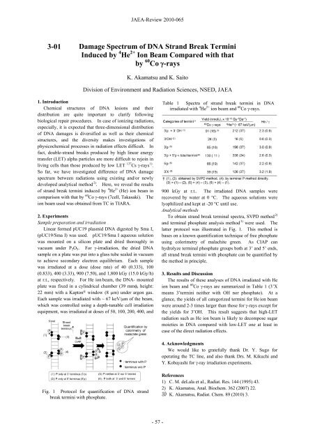
Fig. 1 Protocol for quantification of DNA strand<br />
break termini with phosphate.<br />
<strong>JAEA</strong>-<strong>Review</strong> <strong>2010</strong>-065<br />
- 57 -<br />
Table 1 Spectra of strand break termini in DNA<br />
irradiated with 4 He 2+ ion beam and 60 Co -rays.<br />
900 kGy at r.t.. The irradiated DNA samples were<br />
recovered by water at 0 °C. The aqueous solutions were<br />
lyophilized and kept at -20 °C until use.<br />
Analytical methods<br />
To obtain strand break terminal spectra, SVPD method 2)<br />
and terminal phosphate analysis method 3) were used. The<br />
latter protocol was illustrated in Fig. 1. This method is<br />
bases on a known quantification technique of free phosphate<br />
using colorimetry of malachite green. As CIAP can<br />
hydrolyze terminal phosphate groups both at 3’ and 5’ ends,<br />
all strand break termini with phosphate can be quantified by<br />
the method in principle.<br />
3. Results and Discussion<br />
The results of these analyses of DNA irradiated with He<br />
ion beam and 60 Co -rays are summarized in Table 1 (3’X<br />
means 3’termini neither with OH nor phosphate). At a<br />
glance, the yields of all categorized termini for He ion beam<br />
were around 2-3 times larger than those for -rays except for<br />
the yields for 3’OH. This result suggests that high-LET<br />
radiation such as He ion beam is likely to decompose sugar<br />
moieties in DNA compared with low-LET one at least in<br />
case of the direct radiation effects.<br />
4. Acknowledgments<br />
We would like to gratefully thank Dr. Y. Sugo for<br />
operating the TC line, and also thank Drs. M. Kikuchi and<br />
Y. Kobayashi for -ray irradiation experiments.<br />
References<br />
1) C. M. deLala et al., Radiat. Res. 144 (1995) 43.<br />
2) K. Akamatsu, Anal. Biochem. 362 (2007) 22.<br />
3) K. Akamatsu, Radiat. Chem. 89 (<strong>2010</strong>) 3.


