

JAEA-Review-2010-065.pdf:15.99MB - 日本原子力研究開発機構
JAEA-Review-2010-065.pdf:15.99MB - 日本原子力研究開発機構
JAEA-Review-2010-065.pdf:15.99MB - 日本原子力研究開発機構

Create successful ePaper yourself
Turn your PDF publications into a flip-book with our unique Google optimized e-Paper software.
3-36<br />
Difference in Bystander Lethal Effect in Human Tumor<br />
Cell Lines Depending on p53-gene Status Induced by<br />
Carbon-ion Microbeams<br />
M. Suzuki a) , T. Funayama b) , Y. Yokota b) , Y. Mutou b) , C. Tsuruoka a) ,<br />
Y. Furusawa a) and Y. Kobayashi b)<br />
a) Research Center for Charged Particle Therapy, NIRS,<br />
b) Radiation-Applied Biology Division, QuBS, <strong>JAEA</strong><br />
Since 1994, a Phase I/II clinical study and cancer<br />
radiotherapy have been begun using carbon-ion beams<br />
generated with the Heavy Ion Medical Accelerator in Chiba<br />
(HIMAC) at National Institute of Radiological Sciences 1) .<br />
In the field of fundamental biological studies for high-LET<br />
radiations, there are many reports regarding bystander<br />
cellular effects after exposure to alpha particles derived from<br />
238<br />
Pu or He-ion microbeams. However, only limited sets<br />
of studies have examined bystander effects after exposure to<br />
different ion species heavier than helium. We have been<br />
studying bystander lethal and mutagenic effects in normal<br />
human fibroblasts irradiated with carbon-ion microbeams<br />
using the 256(16 × 16)-cross-stripe irradiation method past 3<br />
years. This year we examined the bystander lethal effect<br />
using 1 normal human cell and 3 different human tumor cell<br />
lines derived from different origins and considered the<br />
relationship between bystander effect and p53-gene status.<br />
Two different astrocytoma cell lines with wild- and<br />
mutated-type p53 gene distributed by Institute for<br />
Fermentation in Japan, amelanotic melanoma with<br />
mutated-type p53 gene distributed by Health Science<br />
Research Resources Bank in Japan and normal human skin<br />
fibroblasts with wild-type p53 gene obtained from RIKEN<br />
BioResource Center in Japan were used in this study.<br />
Carbon-ion microbeams ( 12 C 5+ , 220 MeV) were generated<br />
with the HZ1 port of TIARA. Approximately 8 × 10⁵<br />
exponentially growing the 4 different human cell types were<br />
inoculated into each of microbeam dish, which was made of<br />
acrylic resin ring with 36 mm diameter and attached<br />
7.5 µm-thick polyimide film on the bottom of the ring,<br />
2 days before the microbeam irradiation. In order to block<br />
up cell-cell communication, half of the sample dishes were<br />
treated with a specific inhibitor of gap-junction mediated<br />
cell-cell communication (40 µM of gamma-isomer of<br />
hexachloro- cyclohexane) one day before the irradiation.<br />
Irradiation was carried out using the 256(16 × 16)-crossstripe<br />
irradiation method described in the previous report 2) .<br />
The linear energy transfer (LET) of carbon-ion microbeams<br />
was estimated to be 103 keV/µm at the sample position.<br />
Microbeams of 20 µm in diameter were irradiated in each<br />
point with 8 delivered ions. Cell-killing effect was<br />
detected using a colony formation assay as a reproductive<br />
cell death.<br />
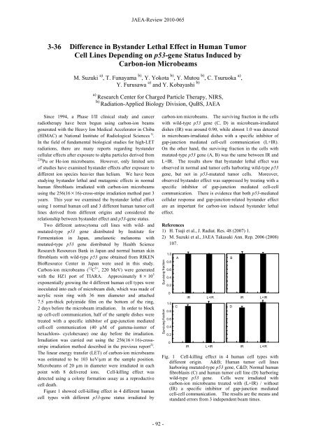
Figure 1 showed cell-killing effect in 4 different human<br />
cell types with different p53-gene status irradiated by<br />
<strong>JAEA</strong>-<strong>Review</strong> <strong>2010</strong>-065<br />
carbon-ion microbeams. The surviving fraction in the cells<br />
with wild-type p53 gene (C, D) in microbeam-irradiated<br />
dishes (IR) was around 0.90, while almost 1.0 was detected<br />
in microbeam-irradiated dishes with a specific inhibitor of<br />
gap-junction mediated cell-cell communication (L+IR).<br />
On the other hand, the surviving fraction in the cells with<br />
mutated-type p53 gene (A, B) was the same between IR and<br />
L+IR. The results show that bystander lethal effect was<br />
observed in normal and tumor cells harboring wild-type p53<br />
gene, but not in p53-mutated tumor cells. Moreover,<br />
observed bystander effect was suppressed by treating with a<br />
specific inhibitor of gap-junction mediated cell-cell<br />
communication. There is evidence that both p53-mediated<br />
cellular response and gap-junction-related bystander effect<br />
are an important for carbon-ion induced bystander lethal<br />
effect.<br />
References<br />
1) H. Tsuji et al., J. Radiat. Res. 48 (2007) 1.<br />
2) M. Suzuki et al., <strong>JAEA</strong> Takasaki Ann. Rep. 2006 (2008)<br />
107.<br />
Surviving fraction<br />
Surviving fraction<br />
- 92 -<br />
1.0<br />
0.8<br />
0.6<br />
0.4<br />
0.2<br />
0<br />
1.0<br />
0.8<br />
0.6<br />
0.4<br />
0.2<br />
0<br />
A<br />
C<br />
IR L+IR<br />
IR L+IR<br />
B<br />
D<br />
IR L+IR<br />
IR L+IR<br />
Fig. 1 Cell-killing effect in 4 human cell types with<br />
different origin. A&B; Human tumor cell lines<br />
harboring mutated-type p53 gene, C&D; Normal human<br />
fibroblasts (C) and human tumor cell line (D) harboring<br />
wild-type p53 gene. Cells were irradiated with<br />
carbon-ion microbeams treated with (L+IR) / without<br />
(IR) a specific inhibitor of gap-junction mediated<br />
cell-cell communication. The results are the means and<br />
standard errors from 3 independent beam times.


