Linear Nevus Sebaceous Syndrome
Linear Nevus Sebaceous Syndrome
Linear Nevus Sebaceous Syndrome

You also want an ePaper? Increase the reach of your titles
YUMPU automatically turns print PDFs into web optimized ePapers that Google loves.
J Med Sci 2006;26(2):077-082<br />
http://jms.ndmctsgh.edu.tw/2602077.pdf<br />
Copyright © 2006 JMS<br />
Received: March 21, 2005; Revised: September 8, 2005;<br />
Accepted: September 15, 2005.<br />
* Corresponding author: Chuen-Ming Lee, Department of<br />
Pediatrics, Tri-Service General Hospital, 325, Cheng-Gong<br />
Road Section 2, Taipei 114, Taiwan, Republic of China. Tel:<br />
+886-2-87927025; Fax: +886-2-87927293; E-mail: lcmpin<br />
@yahoo.com.tw<br />
<strong>Linear</strong> <strong>Nevus</strong> <strong>Sebaceous</strong> <strong>Syndrome</strong><br />
Cheng-Chun Chen 1 , Shao-Hung Lien 2 , Mu-Ling Hsu 2,3 , Chiung-Hsi Tien 2 , Chun-Jung Chen 2 ,<br />
Pei-Yuan Chang 1 , and Chuen-Ming Lee 2*<br />
Department of Pediatrics, 1 Armed Forces Sung Shan Hospital, Taipei,<br />
2 Tri-Service General Hospital, National Defense Medical Center, Taipei,<br />
3 Armed Forces Hualien Hospital, Hualien,<br />
Taiwan, Republic of China<br />
Cheng-Chun Chen, et al.<br />
<strong>Linear</strong> nevus sebaceous syndrome is a rare, congenital neurocutaneous syndrome comprising nevus sebaceous, seizures and<br />
mental retardation. Multiple organ system anomalies can occur. We report a male neonate with a linear-shaped nevus<br />
sebaceous over the face and scalp, whose prenatal brain sonogram revealed dilatation of the left lateral ventricle and a cysticlike<br />
lesion over the posterior cranial fossa. Postnatal magnetic resonance imaging of the brain showed hemimegalencephaly<br />
ipsilateral to the nevus sebaceous. Despite no seizures occurring during hospitalization, the diagnosis was established<br />
according to typical cutaneous lesions and brain images.<br />
Key words: colpocephaly, hemimegalencephaly, linear nevus sebaceous syndrome, neurocutaneous syndrome<br />
INTRODUCTION<br />
<strong>Linear</strong> nevus sebaceous syndrome is a rare neurocutaneous<br />
disease characterized by a midline facial skin lesion<br />
(linear nevus sebaceous), seizures and mental retardation 1-3 .<br />
Here, we report on a male infant with linear nevus sebaceous<br />
syndrome, whose prenatal brain sonogram was<br />
abnormal. Confirmation of this diagnosis was made by the<br />
presence of typical cutaneous lesions, seizures, and brain<br />
images after birth.<br />
CASE REPORT<br />
A full-term, male neonate was born to a 38-year-old<br />
mother (gravida 4, para 3, one prior abortion) at 40 weeks<br />
and 4 days of gestation. Amniocentesis had been performed<br />
at 11 weeks and 2 days of gestation. The resulting<br />
chromosome profile revealed a normal male karyotype<br />
without other chromosomal abnormalities. However, mega<br />
cisterna magnum and a cystic-like lesion were noted on a<br />
prenatal brain sonogram at 28 weeks of gestation (Figs. 1A<br />
and B). Vertex vaginal delivery was uncomplicated and<br />
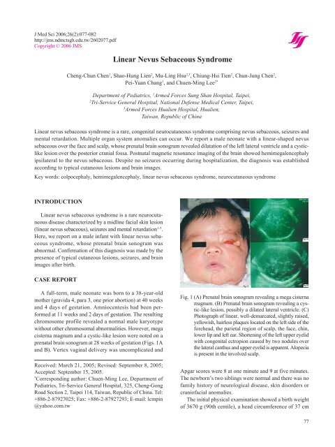
Fig. 1 (A) Prenatal brain sonogram revealing a mega cisterna<br />
magnum. (B) Prenatal brain sonogram revealing a cystic-like<br />
lesion, possibly a dilated lateral ventricle. (C)<br />
Photograph of linear, well-demarcated, slightly raised,<br />
yellowish, hairless plaques located on the left side of the<br />
forehead, the parietal region of scalp, the face, chin,<br />
lower lip and left ear. Shortening of the left upper eyelid<br />
with congenital ectropion caused by two nodules over<br />
the lateral canthus and upper eyelid is apparent. Alopecia<br />
is present in the involved scalp.<br />
Apgar scores were 8 at one minute and 9 at five minutes.<br />
The newborn’s two siblings were normal and there was no<br />
family history of neurological disease, skin disorders or<br />
craniofacial anomalies.<br />
The initial physical examination showed a birth weight<br />
of 3670 g (90th centile), a head circumference of 37 cm<br />
77
<strong>Linear</strong> nevus sebaceous syndrome<br />
(above 90th centile) and body length of 53 cm (above 90th<br />
centile). A cutaneous abnormality of the face consisted of<br />
linear, well-demarcated, raised, yellowish and hairless<br />
plaques located on the left side of the forehead, the parietal<br />
region of the scalp, the face, chin, lower lip, and left ear.<br />
Alopecia was present in the region of the scalp involved in<br />
the cutaneous abnormality. A similar, less well-formed<br />
lesion was present over the midline of the neck and the<br />
anterior chest wall (Fig. 1C). There were no other cutaneous<br />
manifestations, such as cafe-au-lait spots or hypo- or<br />
hyperpigmentation lesions. Ocular examination showed<br />
left side microphthalmia, shortage of the left upper eyelid<br />
with congenital ectropion caused by two nodules over the<br />
lateral canthus and upper eyelid, and extraocular movement<br />
restriction in the left eye. Otherwise, no limbal mass<br />
or colobomas were apparent over the bilateral eyes. The<br />
activity and muscle power of the limbs seemed normal<br />
without obvious signs of hemiparesis.<br />
A brain sonogram revealed a mega cisterna magnum<br />
and ventriculomegaly that included the third and left<br />
ventricles. Skull films showed a hypoplastic change of the<br />
greater wing of the sphenoid bone. Echocardiography<br />
revealed patent foramen ovale, closing ductal arteriosus,<br />
and mild tricuspid valve regurgitation. An abdominal<br />
sonogram showed a grade I hydronephrosis over the left<br />
kidney. An otoacoustic emissions test revealed a hearing<br />
impairment on the left side.<br />
Magnetic resonance imaging (MRI) of the brain showed<br />
asymmetrical overgrowth of the left cerebral hemisphere,<br />
with precedence of the occipital lobe pushing the midline<br />
toward the right side. These observations were compatible<br />
with hemimegalencephaly, increased gray matter in the<br />
left parahippocampus (heterotopia), and a mega cisterna<br />
magnum. Asymmetric arrangement of the facial bones and<br />
a downward orientation of the left orbital bone with a<br />
relatively small orbital cavity were also evident (Fig. 2).<br />
During hospitalization of the patient, no seizures occurred<br />
and no medication was prescribed. A chromosome<br />
study of peripheral blood showed a normal male karyotype.<br />
After being evaluated by a pediatric neurologist,<br />
ophthalmologist, dermatologist, otologist and plastic<br />
surgeon, the patient was discharged at 11 days of age. Four<br />
days after discharge (16 days old), he experienced his first<br />
seizure at home. Subsequently, numerous seizures occurred<br />
and infantile spasm was diagnosed by a pediatric<br />
neurologist. Because anomalies of other organ systems,<br />
particularly the eye, ear and bone, may also be involved, a<br />
multidisciplinary approach at a medical center was recommended<br />
to his parents.<br />
78<br />
Fig. 2 (A) T2-weighted magnetic resonance (MR) study with<br />
axial plane showing asymmetrical outgrowth of the left<br />
cerebral hemisphere with precedence of the occipital<br />
lobe pushing the midline towards the right; this is consistent<br />
with hemimegalencephaly. Additionally, underdevelopment<br />
of the left operculum was noted. (B) Increased<br />
gray matter thickness in the left parahippocampus<br />
(heterotopia). (C) T1-weighted MR study with sagittal<br />
plane showing a regional patulous cerebrospinal fluidfilled<br />
space in the posterior cranial fossa, presumably the<br />
mega cisterna magnum. (D) T1-weighted MR study with<br />
the coronal plane showing an asymmetric facial bone and<br />
a downward orientation of the left orbital bone with a<br />
relative small orbital cavity.<br />
DISCUSSION<br />
Although Jadassohn knew the characteristic cutaneous<br />
manifestation of linear nevus sebaceous syndrome in 1895,<br />
Robinson introduced the term “sebaceous nevus of Jadassohn”<br />
in 1932 4 . The syndrome was first described by<br />
Schimmelpenning in 1957 5 , and Feuerstein and Mims in<br />
1962 6 . Numerous subsequent reports have confirmed that<br />
linear nevus sebaceous syndrome is frequently associated<br />
with ocular, hearing, cardiovascular, skeletal, hepatic, and<br />
urinary tract anomalies 7 . <strong>Linear</strong> nevus sebaceous syndrome<br />
is the name most often used for this disorder, although<br />
similar associations of clinical findings have been described<br />
under the names of neuro-ocular-cutaneous<br />
syndrome, Feuerstein-Mims syndrome, Schimmelpenning-<br />
Feuerstein-Mims syndrome, Jadassohn syndrome, Solomon’s<br />
epidermal nevus syndrome, and organoid nevus phakomatosis<br />
syndrome.<br />
The etiology of linear nevus sebaceous syndrome is still
unknown. The syndrome occurs sporadically and no genetic<br />
or chromosomal abnormalities have been found.<br />
Neither sex predilection nor familial occurrence or external<br />
factors, such as drug ingestion, viral infection or<br />
radiation during pregnancy, have been linked to this<br />
syndrome 2 . However, mosaic dominant lethal gene mutation<br />
is a possible explanation 8,9 .<br />
<strong>Nevus</strong> sebaceous is estimated to occur in 0.3% of newborns<br />
and is usually present at birth or in early childhood 7 .<br />
It is the hallmark of this syndrome, and is characterized by<br />
large, often linear, sharply demarcated and slightly raised<br />
plaques, that present a waxy or pebbly surface on the skin<br />
of the head, face and neck. The plaques may extend to the<br />
trunk and extremities following the line of Blaschko, with<br />
either a yellow to tan velvety appearance or a darker<br />
verrucous look. The life history and histopathological<br />
findings of sebaceous nevus are age-dependent 7 . During<br />
infancy and in early childhood, sebaceous glands within<br />
the nevus are underdeveloped or abortive and no hair<br />
follicles are present. At puberty, the sebaceous glands<br />
within the nevus are characterized by papillomatous epidermal<br />
hyperplasia and a massive mature apocrine gland<br />
deep in the dermis. After puberty, benign and malignant<br />
tumors may develop within the nevus. These include basal<br />
cell carcinoma (15%-20%, most common), syringocystadenoma<br />
papilliferum, syringoma, nodular hidradenoma,<br />
sebaceous thelioma, keratoacanthoma, and squamous cell<br />
carcinoma. Therefore, prophylactic excision of nevus sebaceous<br />
at or before the onset of puberty, or close monitoring,<br />
is recommended 7,10 . Our patient had nevus sebaceous with<br />
a linear configuration that involved the left side of the face,<br />
the scalp, ocular region and anterior chest wall, which are<br />
the most common sites of skin lesions in linear nevus<br />
sebaceous syndrome. Because of his young age, sebaceous<br />
glands within the nevus were undeveloped and no hair<br />
follicles were present.<br />
Associated neurological abnormalities, including seizures<br />
and mental retardation, are common features of<br />
linear nevus sebaceous syndrome. Seizures occur in 44%-<br />
90% of patients during their first year of life, with mental<br />
retardation or developmental delay occurring in 40%-80%<br />
of patients 1 . The head may be enlarged and other associated<br />
neurological manifestations include progressive<br />
hemiparesis, quadriparesis, hypotonia, reflex abnormalities,<br />
gait disorders, diencephalic syndrome, microcephaly and<br />
macrocephaly 10,11 . A major finding with MRI is hemimegalencephaly<br />
(or unilateral megalencephaly), which is<br />
characterized by congenital overgrowth of one cerebral<br />
hemisphere ipsilateral to the sebaceous nevus with an<br />
increased volume of white matter and dilatation of the<br />
Cheng-Chun Chen, et al.<br />
lateral ventricle on the affected side. The affected brain has<br />
essentially no function and is frequently clinically associated<br />
with hemiparesis, early-onset seizures, mental retardation,<br />
hemimacrocephaly and severe encephalopathy 7,12 . Partial<br />
or complete hemispherectomy has been recommended in<br />
children with intractable seizures 1 . The consistent association<br />
of hemimegalencephaly with the sebaceous form of<br />
epidermal nevus provides evidence that linear nevus sebaceous<br />
syndrome is a distinct epidermal nevus syndrome.<br />
Thus, hemimegalencephaly is a sign in neurological imaging<br />
that may obviate the need for skin biopsy in patients<br />
with epidermal nevi 7 . Heterotopic gray matter is likely to<br />
be the source of a patient’s seizures, which may be the<br />
ultimate cause of death 13 . Other common neurological<br />
imaging findings including focal pachygyria, cortical<br />
atrophy, hydrocephalus, porencephaly and cerebral neoplasm<br />
have been reported 2,14,15 . In our patient, there were no<br />
seizures during the hospitalization period; however, he<br />
experienced numerous seizures after discharge. His intelligence<br />
could not be evaluated because of his young age.<br />
However, hemimegalencephaly, colpocephaly 16 , regional<br />
increased gray matter thickness, and heterotopia in the left<br />
parahippocampus, were evident upon MRI. According to<br />
the characteristic skin lesions, seizures, and these neurological<br />
findings, the diagnosis of linear nevus sebaceous<br />
syndrome was made.<br />
Ocular abnormalities are another clinical manifestation<br />
that occur in about half of patients with linear nevus<br />
sebaceous syndrome 3,10 . Indeed, it has been suggested that<br />
the syndrome could be considered an oculo-neuro-cutaneous<br />
syndrome, and ocular abnormalities should be included<br />
in the diagnostic triad 17 . The most common ocular<br />
lesions are choristoma of conjunctiva, coloboma of the globe<br />
or lid, retinal abnormalities such as optic nerve hypoplasia,<br />
pseudopapilledema, microphthalmia or macrophthalmia,<br />
corneal opacities and exotropia/esotropia 7,10 . Mechanisms<br />
of visual loss include visual inattention due to intractable<br />
seizures, subcortical visual loss secondary to unilateral<br />
hypoplasia of an optic radiation associated with hemimegalencephaly,<br />
dysplasia of the optic disc and peripapillary<br />
sclera, and visual occlusion from a corneal opacification<br />
adjacent to a limbal dermoid 7 . In our patient, left side<br />
microphthalmia and hemimegalencephaly were present.<br />
Accordingly, the prognosis of visual function was poor<br />
despite surgical treatment being conducted twice to rectify<br />
his left eyeball.<br />
Other anomalies have also been documented in linear<br />
nevus sebaceous syndrome 10,18 . In the cardiac system,<br />
these include ventricular septal defects, coarctation of the<br />
aorta, patent ductus arteriosus, hypoplastic left heart, su-<br />
79
<strong>Linear</strong> nevus sebaceous syndrome<br />
praventricular tachycardia, and pulmonary stenosis.<br />
Anomalies in the urogenital system include renal cystic<br />
dysplasia, double collecting system, cryptorchidism, inguinal<br />
hernia, hypospadias, micropenis, testicular and<br />
paratesticular tumors. Skeletal involvement has been<br />
documented 19 , including skull, facial or orbital bone<br />
deformities, limb deformities, scoliosis, kyphosis, and<br />
hypophosphatemic vitamin D-resistant rickets 20 . Furthermore,<br />
an inner ear malformation with impairment of hearing<br />
function has been described in a patient with linear<br />
nevus sebaceous syndrome. In our patient, facial or orbital<br />
bone deformities were found and otoacoustic emission<br />
screening showed an abnormal finding over the left ear.<br />
Further audiological examination of the patient was suggested<br />
to his family by an attending otologist.<br />
Additionally, developmental delay was observed in our<br />
patient and mental retardation was highly suspected, despite<br />
a measure of intelligence not being obtained because<br />
of his young age. Ataxic gait manifested when he was<br />
walking and dysarthria was present in his speech, which<br />
might be due to the involvement of the cerebellum.<br />
In a review of the literature, we found a report of a single<br />
case of linear nevus sebaceous syndrome displaying an<br />
abnormal intrauterine sonography at 18 weeks of gestation,<br />
in which the major finding was a right side intracranial<br />
cyst 1 . In another case, prenatal detection of echogenic softtissue<br />
structures external to the cranium and face was<br />
reported, along with the detection of hydrocephalus, intrathoracic<br />
mass and ascites by sonogram 21 . Nowaczyk et<br />
al. demonstrated antenatal progression of intracranial abnormalities<br />
in encephalocraniocutaneous lipomatosis by<br />
prenatal sonogram, and emphasized the difficulty with<br />
antenatal diagnosis of hamartoneoplastic conditions based<br />
on sonographic evaluation 22 . These authors considered<br />
that other modalities, such as MRI or three-dimensional<br />
sonogram, could be useful for prenatal diagnosis. Our<br />
patient received a prenatal sonogram at 28 weeks of<br />
gestation. The diagnosis was not established until birth,<br />
although several abnormal findings had been found<br />
prenatally.<br />
REFERENCES<br />
1. Herman TE, Siegel MJ. Hemimegalencephaly and<br />
linear nevus sebaceous syndrome. J Perinatol 2001;21:<br />
336-338.<br />
2. Alfonso I, Howard C, Lopez PF, Palomino JA, Gonzalez<br />
CE. <strong>Linear</strong> nevus sebaceous syndrome. A review. J<br />
Clin Neuro-ophthalmol 1987;7:170-177.<br />
3. Roth AM, Keltner JL. <strong>Linear</strong> nevus sebaceous<br />
80<br />
syndrome. J Clin Neuro- ophthalmol 1993;13:44-49.<br />
4. Robinson SS. Naevus sebaceus (Jadassohn): report of<br />
four cases. Arch Dermat Syph 1932;26:663-670.<br />
5. Schimmelpenning GW. Klinischer beitrag zur<br />
symptomatologie der phakomatosen. Fortschr<br />
..<br />
Rontgenstr 1957;87:716-720.<br />
6. Feuerstein RC, Mims LC. <strong>Linear</strong> nevus sebaceus with<br />
convulsions and mental retardation. Am J Dis Child<br />
1962;104:675-679.<br />
7. Brodsky MC, Kincannon JM, Nelson-Adesokan P,<br />
Brown HH. Oculocerebral dysgenesis in the linear<br />
nevus sebaceous syndrome. Ophthalmology 1997;104:<br />
497-503.<br />
8. Happle R. Lethal genes surviving by mosaicism: a<br />
possible explanation for sporadic birth defects involving<br />
the skin. J Am Acad Dermatol 1987;16:899-906.<br />
..<br />
9. Schworm HD, Jedele KB, Holinski E, Hortnagel K,<br />
Rudolph G, Boergen KP, Kampik A, Meitinger T.<br />
Discordant monozygotic twins with the<br />
Schimmelpenning-Feuerstein-Mims syndrome. Clin<br />
Genet 1996;50:393-397.<br />
10. Duncan JL, Golabi M, Fredrick DR, Hoyt CS, Hwang<br />
DG, Kramer SG, Howes EL Jr., Cunningham ET Jr.<br />
Complex limbal choristomas in linear nevus sebaceous<br />
syndrome. Ophthalmology 1998;105:1459-1465.<br />
11. Sarwar M, Schafer ME. Brain malformations in linear<br />
nevus sebaceous syndrome: an MR study. J Com<br />
Assist Tomogr 1998;2:338-340.<br />
12. Levy-Reis I, Casasanto DJ, Gonzalez JB, Alsop DC,<br />
Glosser G, Maldjan JA, French J, Detre JA. Cortical<br />
reorganization in linear nevus sebaceous syndrome: a<br />
multimodality neuroimaging study. J Neuroimaging<br />
2000;10:225-228.<br />
13. Hager BC, Dyme IZ, Guertin SR, Tyler RJ, Tryciecky<br />
EW, Fratkin JD. <strong>Linear</strong> nevus sebaceous syndrome:<br />
megalencephaly and heterotopic gray matter. Pediatr<br />
Neurol 1991;7:45-49.<br />
14. Leonidas JC, Wolpert SM, Feingold M, McCauley<br />
RG.K. Radiographic features of the linear nevus sebaceous<br />
syndrome Am J Roentgenol 1979;132:277-279.<br />
15. Chalhub EG, Volpe JJ, Gado MH. <strong>Linear</strong> nevus sebaceous<br />
syndrome associated with porencephaly and<br />
nonfunctioning major cerebral venous sinuses. Neurology<br />
1975;25:857-860.<br />
16. Herskowitz J, Rosman P, Wheeler CB. Colpocephaly:<br />
clinical, radiologic, and pathogenetic aspects. Neurology<br />
1985;35:1594-1598.<br />
17. Lambert HM, Sipperley JO, Shore JW, Dieckert JP,<br />
Evans R, Lowd DK. <strong>Linear</strong> nevus sebaceous syndrome.<br />
Ophthalmology 1987;94:278-282.
18. Yu KC, Lalwani AK. Inner ear malformations and<br />
hearing loss in linear nevus sebaceous syndrome. Int J<br />
Pediatr Otorhinolaryngol 2000;56:211-216.<br />
19. Muhle C, Brinkmann G, Muhle K, Heller M. Skeletal<br />
involvement and follow-up in linear nevus sebaceous<br />
syndrome. Eur Radiol 1988;8:606-608.<br />
20. Oranje AP, Przyrembel H, Meradji M, Loonen MCB,<br />
Klerk BC. Solomon's epidermal nevus syndrome (type:<br />
linear nevus sebaceus) and hypophosphatemic vitamin<br />
D-resistant rickets. Arch Dermatol 1994;130:1167-<br />
1171.<br />
Cheng-Chun Chen, et al.<br />
21. Sweeney WJ, Kuller JA, Chescheir NC, Veness-<br />
Meehan K. Prenatal ultrasound findings of linear nevus<br />
sebaceous and its association with cystic<br />
adenomatoid malformation of the lung. Obstet Gynecol<br />
1994;83:860-862.<br />
22. Nowaczyk MJ, Mernagh JR, Bourgeois JM, Thompson<br />
PJ, Jurriaans E. Antenatal and postnatal findings in<br />
encephalocraniocutaneous lipomatosis. Am J Med<br />
Genet 2000;91:261-266.<br />
81
<strong>Linear</strong> nevus sebaceous syndrome<br />
82