Alagille syndrome: family studies
Alagille syndrome: family studies
Alagille syndrome: family studies

You also want an ePaper? Increase the reach of your titles
YUMPU automatically turns print PDFs into web optimized ePapers that Google loves.
264 24Med Genet 1995;32:264-268<br />
<strong>Alagille</strong> <strong>syndrome</strong>: <strong>family</strong> <strong>studies</strong><br />
The Hospital for Sick<br />
Children, Great<br />
Ormond Street,<br />
London WClN 3JH,<br />
UK<br />
F V Elmslie<br />
H Gardiner*<br />
C Hall<br />
R M Winter<br />
Moorfields Eye<br />
Hospital, City Road,<br />
London EClV 2PD,<br />
UK<br />
A J Vivian<br />
Department of Child<br />
Health, Variety Club<br />
Children's Hospital,<br />
King's College<br />
Hospital,<br />
Denmark Hill,<br />
London SE5 8RX, UK<br />
A P Mowat<br />
* Present address:<br />
The Glenfield Hospital,<br />
Groby Road,<br />
Leicester<br />
LE3 9QP, UK.<br />
Correspondence to: Dr<br />
Elmslie, Department of<br />
Paediatrics, UCL Medical<br />
School, The Rayne Institute,<br />
5 University Street, London<br />
WC1E 6JJ, UK.<br />
Received 12 September<br />
1994<br />
Revised version accepted for<br />
publication 9 December<br />
1994<br />
Downloaded from<br />
jmg.bmj.com on July 13, 2013 - Published by group.bmj.com<br />
F V Elmslie, A J Vivian, H Gardiner, C Hall, A P Mowat, R M Winter<br />
Abstract<br />
<strong>Alagille</strong> <strong>syndrome</strong> (AGS) is one of the<br />
major forms of chronic liver disease in<br />
childhood with severe morbidity and a<br />
mortality of 10 to 20%. It is characterised<br />
by cholestasis of variable severity with<br />
paucity of interlobular bile ducts and anomalies<br />
ofthe cardiovascular system, skeleton,<br />
eyes, and face. Previous <strong>studies</strong><br />
suggest a wide variation in the expression<br />
of the disease and a high incidence of new<br />
mutations. To determine more accurately<br />
the rate of new mutations and to develop<br />
criteria for detecting the disorder in parents<br />
we systematically investigated parents<br />
in 14 families with an affected child.<br />
Clinical examination was supplemented<br />
by liver function tests, echocardiography,<br />
radiographic examination ofthe spine and<br />
forearm, ophthalmological assessment,<br />
and chromosome analysis. Six parents had<br />
typical anomalies in two or more systems<br />
pointing to the presence of autosomal<br />
dominant inheritance. Systematic screening<br />
of parents for the features defined in<br />
this study should improve the accuracy of<br />
genetic counselling.<br />
( Med Genet 1995;32:264-268)<br />
<strong>Alagille</strong> <strong>syndrome</strong> is a common cause of<br />
cholestatic liver disease in childhood, with an<br />
estimated incidence of 1 in 70 000 live births.'<br />
The association between intrahepatic cholestasis,<br />
a characteristic face, and a cardiac<br />
murmur was described in 1969 by <strong>Alagille</strong> et<br />
al2 as a new and distinct form of cholestasis<br />
in infancy. Further reports by Watson and<br />
Miller' and <strong>Alagille</strong> et al' provided more<br />
evidence for a new <strong>syndrome</strong>. Liver disease<br />
occurs in association with paucity of interlobular<br />
bile ducts (intrahepatic biliary hypoplasia),<br />
detectable on liver biopsy. It is<br />
accompanied by cardiovascular abnormalities,<br />
in particular peripheral pulmonary stenosis,<br />
skeletal anomalies, and ophthalmological defects.<br />
The characteristic skeletal abnormality<br />
is butterfly vertebrae caused by a persistent<br />
sagittal cleft through the vertebral body; these<br />
may fuse with time and therefore may not<br />
be present in older patients with AGS. Other<br />
skeletal abnormalities such as shortening of<br />
the bones of the forearm and hands and<br />
narrowed interpedicular distance of the vertebrae<br />
may be present.56 The eye abnormality<br />
usually seen is posterior embryotoxon, an<br />
abnormal prominence of Schwalbe's line.7<br />
Posterior embryotoxon is known to occur in<br />
8 to 15% of the normal population.8<br />
In a review of 80 cases in 1987, <strong>Alagille</strong><br />
et al9 suggested that there were five cardinal<br />
features of the <strong>syndrome</strong>: paucity of intrahepatic<br />
bile ducts, cardiovascular abnormalities,<br />
vertebral arch defects, posterior<br />
embryotoxon, and a characteristic face. In the<br />
same year, Mueller'" suggested that the diagnosis<br />
could be made in the presence of any three<br />
of six features: intrahepatic biliary hypoplasia,<br />
peripheral pulmonary stenosis, posterior embryotoxon,<br />
butterfly vertebrae, a characteristic<br />
face, and a first degree relative with AGS. In<br />
1986, Byrne et all' found a deletion of the<br />
short arm of chromosome 20 in a baby with<br />
intrauterine growth retardation, jejunal stenosis<br />
and dysmorphic facial features associated with<br />
peripheral pulmonary stenosis, vertebral abnormalities,<br />
and cholestasis secondary to<br />
paucity of interlobular bile ducts. Reviewing<br />
previous reports of monosomy 20p they found<br />
that all had some features of <strong>Alagille</strong> <strong>syndrome</strong>.<br />
A further 13 cases of deletions of 20p associated<br />
with AGS have been described'2 including one<br />
case in which the deletion had been transmitted<br />
from an affected mother to her daughter. This<br />
points to the existence of a locus or loci on<br />
chromosome 20p that are responsible for producing<br />
AGS.<br />
AGS is now established as being inherited<br />
in an autosomal dominant fashion,"-'6 but with<br />
extreme variability of expression and a high rate<br />
of new mutation. Several families, including<br />
Watson and Miller's original families,3 have<br />
been described in which <strong>Alagille</strong> <strong>syndrome</strong> has<br />
been transmitted from one generation to the<br />
next with variation in the phenotype. In the<br />
cases so far published, a mild phenotype in the<br />
parent has led to a more severe phenotype in<br />
the offspring, leading to the suggestion that<br />
anticipation may occur in this disorder. In addition,<br />
Shulman et all" suggested that inheritance<br />
from the mother resulted in a more<br />
severe phenotype leading this author to suggest<br />
similarities with the inheritance of myotonic<br />
dystrophy. However, there is little information<br />
to date about the proportion of affected children<br />
who inherit the disorder from a parent.<br />
Nor is there published information on the systematic<br />
evaluation of parents with regard to<br />
the abnormalities present in AGS. Knowledge<br />
of the minimal expression of the disease would<br />
enable more accurate counselling ofthe families<br />
of children with AGS.<br />
The aim of the study, therefore, was to<br />
develop criteria to aid diagnosis in an affected<br />
parent. In addition we wished to determine<br />
in what proportion of affected children there<br />
was evidence of autosomal dominant inheritance<br />
of the disease and whether maternal<br />
or paternal transmission of the disease had<br />
an effect on the severity of the phenotype of<br />
the offspring.
<strong>Alagille</strong> <strong>syndrome</strong>: <strong>family</strong> <strong>studies</strong><br />
Downloaded from<br />
jmg.bmj.com on July 13, 2013 - Published by group.bmj.com<br />
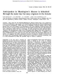
O! Liver disease<br />
O~Cardiovascular abnormality<br />
FL Posterior embryotoxon<br />
El Skeletal malformation<br />
0 Unaffected<br />
El 12 3<br />
Family 1<br />
MI : 1111:3 3 11:4 13 1:5 M<br />
Family 6<br />
1:1 1:2 I:3 I4<br />
111:1 111:2 111:3 H1:4 1111:5 1HA:<br />
Family 7<br />
:Li 1:2 1:3 1;4<br />
ILl 11:211:3 11:4 11:5 11:6 l:7 11S 11:9<br />
M1:I 111:2 M:3 111:IS<br />
Family 8<br />
M1 I:2 J 1:4<br />
MlI DL2 M3<br />
Family 12<br />
E I 1;2 1:3 1:4<br />
HA: f:2 xJ:3 tJ:4 11.5 11:6 11:7<br />
M:1111:2 111:3 1U:4 :5<br />
Family 14<br />
:1 3 1:4<br />
ILIII 211.3 11:6<br />
Figure 1 Pedigrees offamilies in which there was evidence of autosomal dominant inheritance.<br />
Patients and methods<br />
The patients studied were ascertained through<br />
the Children's Liver Unit of King's College<br />
Hospital and The Hospital for Sick Children,<br />
Great Ormond Street, London. Index cases<br />
were included in the study if they had a definite<br />
diagnosis of <strong>Alagille</strong> <strong>syndrome</strong> based on the<br />
criteria of Mueller.'0 All 14 index cases, 24<br />
parents, and four sibs were examined at The<br />
Hospital for Sick Children. In four families<br />
only one parent was examined, the remainder<br />
being unavailable for study. All index cases and<br />
their parents had the following investigations<br />
performed: liver function tests (LFTs) (serum<br />
bilirubin, alkaline phosphatase, glutamyl transpeptidase,<br />
aspartate transaminase), serum<br />
cholesterol, blood chromosomes, radiographs<br />
of the spine, forearm, and hand, echocardiography,<br />
anterior and posterior segment<br />
ophthalmic examination, and ocular electrophysiology.<br />
The bones of the hand and interpedicular<br />
distances of the spine were<br />
measured. The results were analysed using an<br />
265<br />
unpaired t test to search for minor differences<br />
between the two parent groups, affected and<br />
unaffected.<br />
Results<br />
We found definite evidence for autosomal dominant<br />
inheritance of the <strong>syndrome</strong> in six of<br />
the 14 families studied. The pedigrees of the<br />
families displaying dominant inheritance are<br />
shown in fig 1. Subject II-7 in <strong>family</strong> 7 had an<br />
interesting <strong>family</strong> history. Two sisters died in<br />
infancy and another in childhood following<br />
repeated hospital admissiors. All were known<br />
to have had heart murmurs but no other details<br />
were available. It is possible that they were also<br />
affected.<br />
In three cases the father was the affected<br />
parent and in three the mother. In only one<br />
case had the affected parent previously suspected<br />
that he was affected. Only three sibs<br />
(<strong>family</strong> 1 III-3, <strong>family</strong> 7 III-5, <strong>family</strong> 12 III-4)<br />
were examined and none of them underwent
266<br />
Downloaded from<br />
jmg.bmj.com on July 13, 2013 - Published by group.bmj.com<br />
blood tests or radiography. However, on clinical<br />
examination, there was no evidence of the disease<br />
in any of the three.<br />
In four of these six families there was a<br />
history of miscarriage, all occurring at over<br />
10 weeks' gestation. Two mothers had two<br />
miscarriages, and a further two had one each,<br />
out of a total of 22 pregnancies. In the nonfamilial<br />
group of eight families there were two<br />
miscarriages out of a total of 22 pregnancies.<br />
Although there superficially appears to be an<br />
excess of miscarriages in the familial group,<br />
this did not reach statistical significance using<br />
x2 with Fisher's exact test.<br />
The clinical findings and results of investigations<br />
in the affected parents are presented<br />
in table 1. All affected parents had<br />
posterior embryotoxon and at least one other<br />
major syndromic feature. Five had abnormalities<br />
of the spine and eye. In three,<br />
midline notches on the vertebral end plates<br />
were present representing fused butterfly vertebrae.<br />
Four also had a short ulna. Two had<br />
anomalous optic discs and a pigmentary retinopathy.<br />
Electrophysiology of the eye was normal<br />
in all cases, including the parent with<br />
pigmentary retinopathy. Three had pulmonary<br />
murmurs on clinical examination but in the<br />
two who underwent echocardiography no abnormality<br />
was detected. In only two was there<br />
any abnormality of the liver function tests, in<br />
one a mildly raised bilirubin, and in the other<br />
a mild rise of alkaline phosphatase. The mother<br />
in <strong>family</strong> 14 and the father in <strong>family</strong> 6 had<br />
a history of jaundice in infancy which was<br />
unexplained and recovered spontaneously. In<br />
all parents blood chromosomes were normal.<br />
Using an unpaired t test, no significant difference<br />
was found between affected and unaffected<br />
parents in any of the following<br />
parameters: lengths of the bones in the hand,<br />
lumbar interpedicular distance, aspartate transaminase,<br />
albumin, bilirubin, and triglycerides.<br />
However, the alkaline phosphatase levels were<br />
significantly higher in affected parents with a<br />
p value of
<strong>Alagille</strong> <strong>syndrome</strong>: <strong>family</strong> <strong>studies</strong><br />
Downloaded from<br />
jmg.bmj.com on July 13, 2013 - Published by group.bmj.com<br />
those carrying the gene express it. Complete<br />
penetrance implies that all who have the gene<br />
express it, variable penetrance or incomplete<br />
penetrance implies that there are some people<br />
who carry the gene but do not express it. The<br />
only possible historical evidence for incomplete<br />
penetrance in <strong>Alagille</strong> <strong>syndrome</strong> is the <strong>family</strong><br />
described by Mueller et al'3 in which two apparently<br />
normal parents had two affected children.<br />
In no published case has the disease been<br />
seen to skip a generation. We did not examine<br />
any of the grandparents but there was no evidence<br />
from the <strong>family</strong> histories of the unaffected<br />
parents of non-penetrance of the gene. It is<br />
possible that the expression of the disease was<br />
very mild in the <strong>family</strong> of Mueller et all3 and<br />
went undetected, but an alternative explanation<br />
would be that of gonadal mosaicism. One<br />
mechanism known for non-penetrance is imprinting.<br />
Reviewing published <strong>family</strong> data for<br />
evidence of imprinting we found no evidence<br />
for its presence in AGS. In our group three<br />
children inherited <strong>Alagille</strong> <strong>syndrome</strong> from their<br />
fathers and three from their mothers; there was<br />
no difference in phenotype according to the<br />
parent of the origin. Our findings do not support<br />
imprinting as an important mechanism in<br />
Figure 2 (Above) Proband and affected father from <strong>family</strong> 7. (Below) Proband from<br />
<strong>family</strong> 8 with affected father and unaffected mother.<br />
267<br />
AGS. Only one sib in our group appeared to<br />
be affected, out of eight, which is at odds with<br />
autosomal dominant inheritance with complete<br />
penetrance. However only three sibs were examined,<br />
and none underwent investigation. It<br />
is therefore possible that some of them may<br />
have been mildly affected and went undetected.<br />
It has been postulated that AGS may represent<br />
a contiguous gene <strong>syndrome</strong>.'8 None of<br />
the probands had dysmorphic features additional<br />
to those seen in AGS. Three of the 14<br />
probands had significant learning difficulties or<br />
motor developmental delay. Of these, one had<br />
ataxic cerebral palsy in addition to AGS attributed<br />
to hypoxic-ischaemic encephalopathy,<br />
and another had severe liver disease requiring<br />
transplantation at the age of 3 years. Posttransplantation<br />
he began to make rapid developmental<br />
progress. We found no firm evidence<br />
to suggest that AGS is a contiguous gene<br />
<strong>syndrome</strong>.<br />
In addition, we found no evidence for the<br />
existence of anticipation in this disorder. In<br />
three families the affected child appeared to<br />
have similar disease severity to the parents. If<br />
subjects II-3, II-4, and II-5 in <strong>family</strong> 7 were<br />
affected, their early deaths imply that their<br />
disease was more severe than that of both II-7<br />
and III- 1. It is more likely that those that are<br />
mildly affected survive and reproduce and those<br />
that have more severe disease die or are unable<br />
to reproduce, resulting in the superficial appearance<br />
of anticipation. In addition, there was<br />
no evidence for a "maternal factor" resulting<br />
in increased severity of disease when the disease<br />
was transmitted from an affected mother to her<br />
offspring.<br />
There is increasing evidence that AGS is not<br />
as benign as was originally thought, and there<br />
are reports of long term complications (notably,<br />
hepatocellular carcinoma and late onset liver<br />
failure) occurring in AGS.'9 It is not known<br />
whether those parents retrospectively ascertained<br />
run a risk of developing the complications<br />
associated with the <strong>syndrome</strong>,<br />
although there is no documented case of these<br />
complications occurring in a patient with no<br />
clinical or biochemical evidence of liver disease.<br />
Until more is understood about the long term<br />
natural history of this disease it will be difficult<br />
to be reassuring when counselling these families.<br />
In order to provide accurate genetic counselling<br />
to the families of children with <strong>Alagille</strong><br />
<strong>syndrome</strong> it is important to be able to distinguish<br />
between those that represent new<br />
mutations and those that have inherited AGS<br />
from an affected parent. The extreme variability<br />
of expression of the <strong>syndrome</strong> has made this a<br />
difficult task. Based on this small study a set<br />
of major and minor criteria was developed for<br />
aiding diagnosis in a <strong>family</strong> which presents with<br />
an affected child. Major criteria are established<br />
for the diagnosis of children with <strong>Alagille</strong> <strong>syndrome</strong>,910<br />
but similar criteria do not apply to<br />
adults with the disorder. The major and minor<br />
criteria are shown in table 2. All parents of<br />
children with AGS will fulfil at least one of the<br />
major criteria, that of having a first degree<br />
relative with <strong>Alagille</strong> <strong>syndrome</strong>. In addition, all
268<br />
Downloaded from<br />
jmg.bmj.com on July 13, 2013 - Published by group.bmj.com<br />
Table 2 Major and minor criteria for use in diagnosis of parents<br />
Major criteria Minor criteria<br />
History of prolonged jaundice in infancy Alkaline phosphatase > 103 U/i<br />
requiring investigation Short ulna<br />
Pulmonary murmur Pigmentary retinopathy<br />
Posterior embryotoxon Anomalous optic discs<br />
Vertebral end plate notches<br />
First degree relative with <strong>Alagille</strong><br />
<strong>syndrome</strong> (omit if proband)<br />
Table 3 Number of major and minor criteria present in<br />
affected parents<br />
Parent Major Minor<br />
1 3 1<br />
6 4 3<br />
7 3 1<br />
8 3 0<br />
12 4 3<br />
14 3 0<br />
our affected parents had at least two major<br />
criteria, and in those that had been completely<br />
investigated, between one and three minor criteria<br />
(table 3). In our group of six affected<br />
adults, this represented the minimal expression<br />
of the <strong>syndrome</strong>.<br />
The variability of expression can make it<br />
difficult to be categorical about whether a parent<br />
is affected or not. It is clear from our<br />
evaluation of 14 families that a detailed history,<br />
examination, and investigation are required to<br />
distinguish the affected group from the unaffected<br />
group, and the development of major<br />
and minor criteria for diagnosis may help in<br />
the future evaluation of families.<br />
We wish to thank all the families who participated in the study<br />
for their patience and enthusiasm, Dr Alastair Baker for his<br />
help in <strong>family</strong> ascertainment, Ms Vanda Gooch for performing<br />
echocardiography, and Dr Guan Lim for his help and encouragement.<br />
Elmslie, Vivian, Gardiner, Hall, Mowat, Winter<br />
1 Danks DM, Campbell PE, Jack I, Rogers J, Smith AL.<br />
Studies of the aetiology of neonatal hepatitis and biliary<br />
atresia. Arch Dis Child 1977;52:360-7.<br />
2 <strong>Alagille</strong> D, Habib EC, Thomassin N. L'atresie des voies<br />
biliaries intrahepatiques avec voies extrahepatiques permeable<br />
chez l'enfant. Paris: Editions Medicales Flammarion, 1969:<br />
301-18.<br />
3 Watson GH, Miller V. Arteriohepatic dysplasia: familial<br />
pulmonary arterial stenosis with neonatal liver disease.<br />
Arch Dis Child 1973;48:459.<br />
4 <strong>Alagille</strong> D, Odievre M, Gautier M, Dommergues P. Hepatic<br />
ductular hypoplasia associated with characteristic facies,<br />
vertebral malformations, retarded physical, mental and<br />
sexual development, and cardiac murmur. J Pediatr 1975;<br />
86:63.<br />
5 Singcharoen T, Partridge J, Jeans WD, Baddeley H. Arteriohepatic<br />
dysplasia. Br _J Radiol 1986;59:509-1 1.<br />
6 Rosenfield NS, Kelly MJ, Jensen PS, Cotlier E, Rosenfield<br />
AT, Riely CA. Arteriohepatic dysplasia: radiologic features<br />
of a new <strong>syndrome</strong>. A3rR 1980;135:1217-23.<br />
7 Riely CA, Cotlier E, Jensen P, Klatskin G. Arteriohepatic<br />
dysplasia: a benign <strong>syndrome</strong> of intrahepatic cholestasis<br />
with multiple organ involvement. Ann Intern Med 1979;<br />
91:520-7.<br />
8 Waring GO, Rodrigues MM, Laibson PR. Anterior chamber<br />
cleavage <strong>syndrome</strong>. A stepladder classification. Surv Ophthalmol<br />
1975;20:3-37.<br />
9 <strong>Alagille</strong> D, Estrada A, Hadchouel M, Gautier M, Odievre<br />
M, Dommergues JP. Syndromic paucity of interlobular<br />
bile ducts (<strong>Alagille</strong> <strong>syndrome</strong> or arteriohepatic dysplasia).<br />
Review of 80 cases. J Pediatr 1987;110: 195-9.<br />
10 Mueller RF. The <strong>Alagille</strong> <strong>syndrome</strong> (arteriohepatic dysplasia).<br />
_J Med Genet 1987;24:621-6.<br />
11 Byrne JLB, Harrod MLE, Friedman JM, Howard-Peebles<br />
PN. Del (20p) with manifestations of arteriohepatic dysplasia.<br />
Am J. Med Genet 1986;24:673-8.<br />
12 Anad F, Burn J, Matthews D, et al. <strong>Alagille</strong> <strong>syndrome</strong> and<br />
deletion of 20p.J Med Genet 1990;27:729-37.<br />
13 Mueller RF, Pagon RA, Pepin MG, et al. Arteriohepatic<br />
dysplasia: phenotypic features and <strong>family</strong> <strong>studies</strong>. Clin<br />
Genet 1984;25:323-31.<br />
14 Greenwood RD, Rosenthal A, Crocker AC, Nadas AS.<br />
Syndrome of intrahepatic biliary dysgenesis and cardiovascular<br />
malformations. Pediatrics 1976;58:243-7.<br />
15 Henriksen NT, Langmark F, Sorland SJ, Fausa 0, Landaas<br />
S, Aagenaes 0. Hereditary cholestasis combined with<br />
peripheral pulmonary stenosis and other anomalies. Acta<br />
Paediatr Scand 1977;66:7-15.<br />
16 Dhorne-Pollet S, Deleuze JF, Hadchouel M, Bonaiti-Pellie<br />
C. Segregation analysis of <strong>Alagille</strong> <strong>syndrome</strong>. J Med Genet<br />
1994;31:453-7.<br />
17 Shulman SA, Hyams JS, Gunta R, Greenstein RM, Cassidy<br />
SB. Arteriohepatic dysplasia (<strong>Alagille</strong> <strong>syndrome</strong>): extreme<br />
variability among <strong>family</strong> members. AmJMed Genet 1984;<br />
19:325-32.<br />
18 Schnittger S, Hofer C, Heidemann P, Beernann F,<br />
Hansmann I. Molecular and cytogenetic analysis of an<br />
interstitial 20p deletion associated with syndromic intrahepatic<br />
ductular hypoplasia (<strong>Alagille</strong> <strong>syndrome</strong>). Hum<br />
Genet 1989;83:239-44.<br />
19 Schwarzenberg SJ, Grothe RM, Sharp HL, Snover DC,<br />
Freese D. Long-term complications of arteriohepatic dysplasia.<br />
Am _t Med 1992;93:171-6.
References<br />
Email alerting<br />
service<br />
Notes<br />
<strong>Alagille</strong> <strong>syndrome</strong>: <strong>family</strong> <strong>studies</strong>.<br />
F V Elmslie, A J Vivian, H Gardiner, et al.<br />
J Med Genet 1995 32: 264-268<br />
doi: 10.1136/jmg.32.4.264<br />
Updated information and services can be found at:<br />
http://jmg.bmj.com/content/32/4/264<br />
These include:<br />
Article cited in:<br />
http://jmg.bmj.com/content/32/4/264#related-urls<br />
To request permissions go to:<br />
http://group.bmj.com/group/rights-licensing/permissions<br />
To order reprints go to:<br />
http://journals.bmj.com/cgi/reprintform<br />
To subscribe to BMJ go to:<br />
http://group.bmj.com/subscribe/<br />
Downloaded from<br />
jmg.bmj.com on July 13, 2013 - Published by group.bmj.com<br />
Receive free email alerts when new articles cite this article. Sign up<br />
in the box at the top right corner of the online article.