

Prions: Protein Aggregation and Infectious Diseases - Physiological ...
Prions: Protein Aggregation and Infectious Diseases - Physiological ...
Prions: Protein Aggregation and Infectious Diseases - Physiological ...

Create successful ePaper yourself
Turn your PDF publications into a flip-book with our unique Google optimized e-Paper software.
1116 ADRIANO AGUZZI AND ANNA MARIA CALELLA<br />
prion mechanism of disease transmission might be operating<br />
in other human diseases, some of which are highly<br />
prevalent.<br />
II. FUNCTION OF THE CELLULAR PRION<br />
PROTEIN<br />
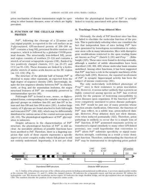
PrP C , following the cleavage of a 22-amino acid<br />
(aa) signal peptide, is exported to the cell surface as an<br />
N-glycosylated, GPI-anchored protein of 208–209 aa.<br />
PrP C contains a long NH 2-proximal flexible r<strong>and</strong>om-coil<br />
sequence, which is followed by a globular COOH-proximal<br />
domain. The structurally less-defined NH 2 proximal<br />
region consists of residues 23–124 <strong>and</strong> contains a<br />
stretch of several octapeptide repeats (OR), flanked by<br />
two positively charged clusters, CC1 (aa 23–27) <strong>and</strong><br />
CC2 (aa 95–110). These domains are linked by a hydrophobic<br />
stretch of amino acids known as the HC region<br />
(aa 111–134) (Fig. 5).<br />
The structure of the globular half of human PrP C is<br />
identical to many other mammals, as expected from the<br />
high degree of sequence identity (309). Interestingly, despite<br />
the low sequence identity between PrP C in chicken,<br />
turtle, or frog, <strong>and</strong> the mammalian isoforms, the major<br />
structural features of PrP C are remarkably preserved in<br />
nonmammalian species (88).<br />
Full-length PrP C is found in non-, mono-, or diglycosylated<br />
forms, corresponding to the variable occupancy of<br />
glycosyl groups on residues Asn-181 <strong>and</strong> Asn-197 in human<br />
<strong>and</strong> Asn-180 <strong>and</strong> Asn-196 in mice (201). A rather large<br />
variety of N-glycans are found attached to both full-length<br />
<strong>and</strong> truncated PrP C (380, 428), which may be differentially<br />
distributed in various areas of the central nervous system<br />
(49, 133). The physiological significance of PrP C glycosylation<br />
is unknown.<br />
Despite advances in the characterization of PrP C<br />
structure, the physiological function of PrP C is still unclear.<br />
An unrealistic plethora of possible functions have<br />
been ascribed to PrP. Therefore, there is a lingering suspicion<br />
that each of these reports represents a specific<br />
aspect of a more complex reality whose full picture is still<br />
not entirely understood. Importantly, it is not yet clear<br />
whether the physiological function of PrP C is actually<br />
linked to toxicity associated with prion diseases.<br />
A. Teachings From Prnp-Ablated Mice<br />
Obviously, the study of PrP knockout mice has thus<br />
far failed to elucidate the molecular function of the protein.<br />
This is particularly sobering when one considers the<br />
fact that independent lines of mice lacking PrP C have<br />
been generated by homologous recombination in embryonic<br />
stem cells in many laboratories. Mice with disruptive<br />
gene modifications restricted to the open reading frame<br />
are known as Prnp o/o (Zurich I) (85) <strong>and</strong> Prnp / (Edinburgh)<br />
(325). These mice were found to develop normally,<br />
although a number of subtle abnormalities have been<br />
described (120, 368, 498) whose molecular basis remains<br />
undefined. Among other functions, it has been suggested<br />
that PrP C may have a role in the synaptic machinery of the<br />
olfactory bulb (293). However, the reported involvement<br />
of PrP C in synaptic hippocampal activity has been the<br />
subject of intense controversy (301).<br />
The only molecularly well-defined phenotype of<br />
Prnp o/o mice is their resistance to prion inoculation<br />
(84). However, it seems rather unlikely that a protein as<br />
highly conserved among species as PrP C has evolved<br />
purely for the purpose of bestowing susceptibility to<br />
prion diseases upon organisms. If the function of PrP C<br />
were completely unrelated to prion disease pathogenesis,<br />
PrP C would be just one of many proteins whose<br />
function awaits clarification. Otherwise, the function of<br />
PrP C may reflect, in a subtle way, prion-induced damage.<br />
However, Prnp ablation does not elicit disease,<br />
even when induced postnatally (322). Therefore, prion<br />
pathology is unlikely to occur due to a simple loss of<br />
PrP C function. If PrP C possessed enzymatic activity or<br />
transduced a signal (similarly to many other GPI-linked<br />
proteins), one could hypothesize that conversion to<br />
PrP Sc alters PrP C substrate specificity or signal transduction<br />
strength, conferring as a result a toxic dominant<br />
function. Therefore, underst<strong>and</strong>ing the function of<br />
PrP C may be instrumental to deciphering prion pathol-<br />
FIG. 5. Outline of the primary structure of the cellular prion protein including posttranslational modifications. A secretory signal peptide resides<br />
at the extreme NH 2 terminus. CC 1 <strong>and</strong> CC 2 define the charged clusters. OR indicates the octapeptide repeat, <strong>and</strong> four are present. HC defines the<br />
hydrophobic core. MA denotes the membrane anchor region. S-S indicates the single disulfide bridge, <strong>and</strong> the glycosylation sites are designated as<br />
CHO. The numbers describe the position of the respective amino acids.<br />
Physiol Rev VOL 89 OCTOBER 2009 www.prv.org


