Interview with Pr Mihai Jianu p14 : Nepal Orthopedic Hospital
Interview with Pr Mihai Jianu p14 : Nepal Orthopedic Hospital
Interview with Pr Mihai Jianu p14 : Nepal Orthopedic Hospital

You also want an ePaper? Increase the reach of your titles
YUMPU automatically turns print PDFs into web optimized ePapers that Google loves.
www.argos-europe.com<br />
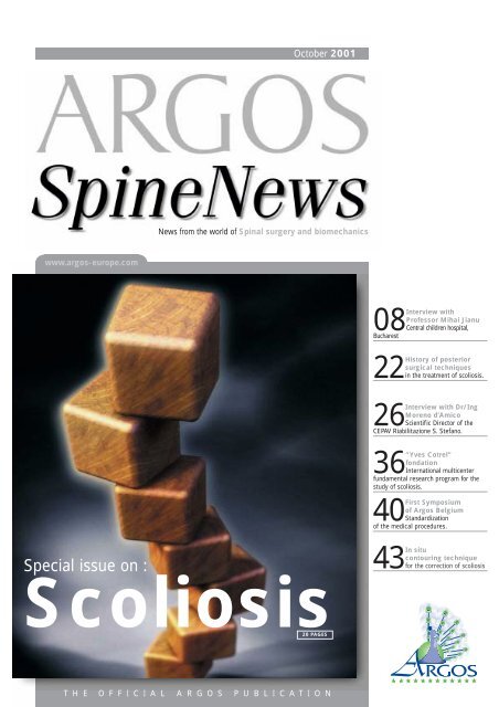
October 2001<br />
News from the world of Spinal surgery and biomechanics<br />
Special issue on :<br />
Scoliosis<br />
20 PAGES<br />
T H E O F F I C I A L A R G O S P U B L I C A T I O N<br />
<strong>with</strong><br />
<strong>Pr</strong>ofessor <strong>Mihai</strong> <strong>Jianu</strong><br />
08<strong>Interview</strong><br />
Central children hospital,<br />
Bucharest<br />
of posterior<br />
surgical techniques<br />
22History<br />
in the treatment of scoliosis.<br />
<strong>with</strong> Dr/Ing<br />
Moreno d’Amico<br />
26<strong>Interview</strong><br />
Scientific Director of the<br />
CEPAV Riabilitazione S. Stefano.<br />
Cotrel”<br />
fondation<br />
36“Yves<br />
International multicenter<br />
fundamental research program for the<br />
study of scoliosis.<br />
Symposium<br />
of Argos Belgium<br />
40First<br />
Standardization<br />
of the medical procedures.<br />
43In situ<br />
contouring technique<br />
for the correction of scoliosis
Summary<br />
Communication<br />
<strong>Interview</strong> <strong>with</strong> <strong>Pr</strong> <strong>Mihai</strong> <strong>Jianu</strong> 8<br />
<strong>Nepal</strong> <strong>Orthopedic</strong> <strong>Hospital</strong>: An opportunity to bring<br />
your support 14<br />
In the dictionary, COMPREHENSION comes before<br />
SURGERY 17<br />
First ARGOS Meeting in Belgium 40<br />
Web review 46<br />
Agenda 50<br />
Evaluation<br />
<strong>Interview</strong> <strong>with</strong> Dr. Eng. Moreno D’Amico<br />
Yves Cotrel Foundation for the research<br />
26<br />
in spinal pathologies 36<br />
Training<br />
History of Posterior Surgical Techniques<br />
in the Treatment of Scoliosis - R. Dumas, J. P. Steib 22<br />
In Situ Contouring Technique for the Correction<br />
of Scoliosis - J. P. Steib 43<br />
October 2001<br />
News from the world of Spinal surgery and Biomechanics<br />
T4<br />
L2<br />
T10<br />
T4<br />
L2<br />
T10
EDITORIAL HEADQUARTERS<br />
First surgeons, then tools…<br />
64, rue Tiquetonne 75002 Paris France<br />
Phone : +33.(0)1 42 33 06 88<br />
Fax : +33.(0)1 42 33 06 62<br />
EDITORIAL STAFF<br />
Editorial Director :<br />
Alexandre Templier, PhD<br />
Editor in chief :<br />
Anca Mitulescu, MSc<br />
<strong>Pr</strong>oduction/Art Director :<br />
Karim Boukarabila<br />
Editorial advisory board :<br />
The ARGOS committees<br />
Writer/Translators<br />
Patrick Bertranou, MD<br />
Blake W. Rodgers, MD<br />
Scientific & Technical Advisor :<br />
Alon Wolf, MSc<br />
Associate Editors :<br />
Raphaël Dumas, MSc<br />
Emeric Gallard, MSc<br />
Laurent Nodé-Langlois, MSc<br />
Virginie Lafage, MSc<br />
ARGOS COMMITTEES<br />
Communication Committee :<br />
Patrick Bertranou, MD<br />
Philippe Bedat, MD<br />
Henri Costa, MD<br />
Pierre Kehr, MD<br />
Charles Marc Laager, MD<br />
Pierre Soete, MD<br />
Training Committee :<br />
Jean-Paul Steib, MD<br />
Jean-Paul Forthomme, MD<br />
Franck Gosset, MD<br />
François Lavaste, PhD<br />
Richard Terracher, MD<br />
Jean-Marc Vital, MD<br />
Evaluation Committee :<br />
Wafa Skalli, PhD<br />
Jacques Deguise, PhD<br />
Michel Dutoit<br />
Ilhem Cherrak, PhD<br />
Alain Graftiaux<br />
Christian Mazel, MD<br />
Tony Martin, MD<br />
Henri Judet, MD<br />
ARGOS SPINE NEWS is published two<br />
times a year by Surgiview SAS. <strong>Pr</strong>inted by<br />
Imprim2000, Paris, France. It is sent for<br />
free to physicians, surgeons, researchers<br />
and industrial companies on an international<br />
scale. Single copy price is 7 Euros.<br />
Copyright 2001 by Surgiview, All rights<br />
reserved. Reproduction in any forms is forbidden<br />
<strong>with</strong>out express permission of<br />
copyright owner.<br />
Advertising sales, please contact :<br />
Alexandre Templier<br />
a.templier@argos-europe.com<br />
Anca Mitulescu<br />
anca@argos-europe.com<br />
Fax : +33 (0)1 42 33 06 62<br />
Editorial<br />
Alexandre TEMPLIER<br />
ARGOS General Manager<br />
Editorial Director<br />
( )<br />
Dear Members and readers,<br />
The last issue of Argos Spine News was dedicated to new<br />
technologies in Spinal Surgery. We have seen that most of these<br />
technologies were designed to improve the accuracy of the surgical<br />
act itself. Indeed, up to now poor technological efforts have been<br />
dedicated to help the surgeon in optimizing his clinical<br />
efficiency.Although technological aspects of surgery may be<br />
spectacular compared to clinical considerations, we have decided to<br />
focus our articles on specific clinical topics.<br />
Scoliosis is one of the most famous issue among spinal disorders,<br />
because of the complexity of its ethiology. For many years, huge<br />
research efforts have been being focused on a better comprehension<br />
of this pathology, <strong>with</strong> major surgical and clinical breakthrough.<br />
Today, scoliosis is widely recognized as a 3D deformation, thus<br />
requiring 3D exploration, analysis, and treatment. However, 3D<br />
imaging modalities remain costly and invasive, which makes 3D<br />
analysis and comprehension almost impossible in a clinical daily<br />
practice.Surgical techniques for scoliosis corrections are<br />
progressively moving to a personalized 3D correction approach, but<br />
pre-op planification and post-op evaluation systems are still missing<br />
in the daily clinical environment.<br />
We wanted to dedicate this issue of Argos Spine News to this exciting<br />
subject that is Scoliosis, which remains one of the biggest clinical,<br />
surgical and technological challenge in Spinal Surgery. Of course, it<br />
will not be possible to cover this huge subject in only one issue. We<br />
will definitely dedicate further numbers on this topic if you wish so.<br />
So please, fell free to send your feed back, remarks, and suggestions<br />
about YOUR journal at contact@argos-europe.com .<br />
We look forward to reading from you soon,<br />
Warmest regards.<br />
Christian MAZEL<br />
ARGOS <strong>Pr</strong>esident<br />
October 2001 - N° 4 ARGOS SpineNews 7
communication<br />
<strong>Interview</strong> <strong>with</strong> <strong>Pr</strong> <strong>Mihai</strong> JIANU<br />
<strong>Interview</strong> <strong>with</strong> <strong>Pr</strong>. <strong>Mihai</strong> <strong>Jianu</strong><br />
“Gr Alexandrescu” Central Children <strong>Hospital</strong>, Bucharest<br />
The “ Grigore Alexandrescu “<br />
Emergency <strong>Hospital</strong> for Children<br />
is the oldest hospital for children<br />
in Romania, founded in 1886.<br />
Today, this hospital is a public<br />
one, part of a range of six<br />
emergency hospitals in Bucharest,<br />
taking care of more than 1200000<br />
children from Bucharest and the<br />
metropolitan area. The hospital<br />
has 454 beds and five<br />
clinical departments: Pediatrics,<br />
Pediatric Surgery, <strong>Orthopedic</strong>s<br />
and Trauma Surgery, Burns and<br />
Plastic Surgery,<br />
Otorhinolaryngology.<br />
The Emergency <strong>Hospital</strong> is a<br />
clinical hospital of the University<br />
of Medicine and Farmacy of<br />
Bucharest.<br />
The Pediatric <strong>Orthopedic</strong>s<br />
department is equipped of 64<br />
beds, three surgery rooms, a<br />
postoperative care unit, a bone<br />
bank. Eight surgeons, three<br />
anesthesiologists and five<br />
residents are working in this<br />
department. Every year, more<br />
than 1000 medical students<br />
attend lectures and do clinical<br />
practice there.<br />
The diseases treated in this<br />
department are: bone trauma,<br />
congenital dislocation of the hip<br />
and other limb abnormalities,<br />
scoliosis and other spinal<br />
deformities, malignant and<br />
benign bone tumors, infections of<br />
bone and joints, limb length<br />
discrepancy, cerebral palsy and<br />
muscular dystrophy. This is the<br />
only orthopedics department in<br />
the country where the use of the<br />
botulinum toxin in the treatment<br />
of cerebral palsy is authorized.<br />
8 ARGOS SpineNews N° 4- October 2001<br />
ASN : <strong>Pr</strong> <strong>Jianu</strong>, could you<br />
introduce yourself to our readers<br />
and explain to us what made you<br />
choose spine surgery as your<br />
specialty in a country where this<br />
field has been quite neglected for<br />
a long time ?<br />
After having graduated the<br />
Medicine School in Bucharest,<br />
I became a specialist surgeon<br />
in orthopedics and trauma at<br />
the biggest hospital in<br />
Bucharest - the Central<br />
Emergency Hopital “G.<br />
Alexandrescu”. Later I<br />
became a professor at the<br />
Medicine School of Bucharest.<br />
I am now the Head of the<br />
Pediatric <strong>Orthopedic</strong>s and<br />
Trauma Department of this<br />
School.<br />
As for the second part of your question,<br />
I would say that it was especially<br />
because this discipline was highly<br />
neglected at a national scale that I<br />
oriented my activity towards spine<br />
surgery. When I started practicing, the<br />
spinal pathologies were hardly<br />
considered in clinical centers while the<br />
number of cases was dangerously<br />
increasing. So I considered that this<br />
discipline needed more attention,<br />
despite all the material difficulties we<br />
encountered to put it into practice.<br />
ASN :You prepared your PhD<br />
thesis focusing on a quite<br />
particular topic - the set up of a<br />
bone bank - so you are the<br />
creator of the first bone bank in<br />
Romania. Why having chosen<br />
this topic and which is the role of<br />
the bone bank ?<br />
Indeed, I concentrated my research<br />
activities on the bone bank set up<br />
because it is an essential tool for any<br />
orthopedics and trauma service, so my<br />
clinical activity guided my research<br />
choices. The importance of bone grafts<br />
is now well recognized, for the surgical<br />
treatment of malign tumors,<br />
pseudarthroses, dystrophies etc, in cases<br />
where the auto graft could present risks<br />
for the patient. In order to conduct my<br />
research I worked a lot <strong>with</strong> the<br />
“Pasteur” National Institute for<br />
Bioproducts and Vaccines, as I had to<br />
make a lot of experiments on animals to<br />
validate my findings. The main obstacle<br />
in my research work was the legislation<br />
void concerning this particular field of<br />
research. The corresponding laws<br />
concerning these topics have been voted<br />
only six years ago so you can imagine the<br />
difficulties researchers in this field had<br />
to overcome in the past.<br />
ASN : Could you name some of<br />
those people who influenced<br />
your professional carrier choice ?<br />
I had the chance to prepare my<br />
medicine studies <strong>with</strong> very good<br />
professors and I think that all of them<br />
played an important role in my present<br />
activity, but what really attracted me to<br />
orthopedics was on one hand its link to<br />
classical physics, which is a very<br />
practical aspect, and on the other hand<br />
its “artistic” and “feeling based” aspect,<br />
which is quite intuitive.<br />
And, most of all, what fascinated me in<br />
surgery was the relationship between<br />
surgeon and patient. We should not<br />
neglect the fact that when we operate a<br />
patient we assume a huge responsibility
towards him for the rest of his life. This<br />
is the main factor that turns surgery into<br />
a continuous challenge.<br />
ASN : At present, what are your<br />
main research activities ?<br />
I am working on a research program<br />
concerning the precocious tracking of<br />
spinal deformities in children and<br />
adolescents. My purpose is to set the<br />
basis of a national structure which<br />
should deal <strong>with</strong> this problem in order<br />
to reduce the number of cases <strong>with</strong><br />
scoliosis up to 70-80 degrees of Cobb<br />
angle at the first consultation, which is<br />
still very frequent in Romania.<br />
You are, as far as we know, the<br />
only orthopedic surgeon in<br />
Romania treating scoliosis by<br />
surgical correction. Could you<br />
explain the reasons of this lack<br />
in the orthopedic surgery field ?<br />
Is it due to a lack of training of<br />
specialists or mostly to a lack of<br />
funds ?<br />
It is true that in pediatric spinal surgery,<br />
there is no other orthopedic surgeon in<br />
Romania practicing the corrective<br />
surgery of scoliosis. In adult surgery,<br />
there are few surgeons dealing <strong>with</strong> the<br />
correction of the spine, but they use<br />
mainly the Harrington technique, which<br />
is far from being the best available<br />
technique, or even just bone<br />
arthrodesis. So we can talk about a lack<br />
of training for surgeons <strong>with</strong> regard to<br />
modern surgical correction techniques.<br />
Rehabilitation service at the "Gr ALEXANDRESCU"<br />
Central <strong>Hospital</strong><br />
It is obvious that the lack of funds<br />
dedicated to the health system in<br />
general also played a major role in this<br />
particular field. Besides high prices of<br />
metallic implants and the auxiliary tools<br />
we also have to deal <strong>with</strong> an unfavorable<br />
national context regarding the number<br />
of students following a training for this<br />
specialization. At present, in Romania,<br />
there are only 10 medical doctors per<br />
year that can be accepted for this<br />
training.<br />
ASN : In your opinion, what<br />
should be done in order to<br />
overcome this penury in spinal<br />
surgery in general and in<br />
scoliosis treatment in particular ?<br />
First of all, it seems to me that it is<br />
compulsory that the government deals<br />
<strong>with</strong> the general problem of the health<br />
funds. Starting from this point, many<br />
other possibilities to rehabilitate this<br />
field can be taken into account - training<br />
of the surgeons, acquisition of<br />
specialized material, setting up of a<br />
screening/tracking program and so on.<br />
ASN : We would like you to talk<br />
to us about your experience in<br />
the surgical treatment of<br />
scoliosis by the in situ<br />
contouring technique <strong>with</strong> the<br />
SCS instrumentation: which have<br />
been the most important steps in<br />
your training on this technique<br />
and which was the role played by<br />
the well known surgeons you<br />
have worked <strong>with</strong> ?<br />
I started my activity in the<br />
surgical correction of<br />
scoliosis in the 80s using<br />
different techniques -<br />
Harrington osteosynthesis,<br />
bone arthrodeses <strong>with</strong><br />
fibula, rib and iliac grafts.<br />
Later I started collecting<br />
information on posterior<br />
osteosynthesis of the spine<br />
<strong>with</strong> the CD and SCS<br />
instrumentations. Then I<br />
had the occasion to assist<br />
surgical corrections of the<br />
communication<br />
<strong>Interview</strong> <strong>with</strong> <strong>Pr</strong> <strong>Mihai</strong> JIANU<br />
<strong>Pr</strong> <strong>Mihai</strong> <strong>Jianu</strong>’s<br />
short background<br />
<strong>Pr</strong>ofessor of Pediatric Orthopaedics,<br />
“Carol Davilla” University of Medecine<br />
and Pharmacy, Bucharest. Head of the<br />
Department of Pediatric Orthopaedics<br />
and Trauma Surgery, “Grigore<br />
Alexandrescu” Central Children <strong>Hospital</strong>,<br />
Bucharest; Doctor of Medecine,<br />
doctorate thesis: “The creation and using<br />
of Bone Bank in Pediatric Orthopaedics”;<br />
Member of the International Society of<br />
Orthopaedics and Trauma Surgery<br />
(SOROT); Member of New York<br />
Academy of Science; Evaluation expert of<br />
“PHARE” and “SOCRATES” for the<br />
Ministry of Education.<br />
<strong>Pr</strong>ofessor <strong>Jianu</strong> has a remarcable<br />
experience in spinal surgery, but also in<br />
deformities, trauma and bone tumour<br />
surgical treatment.<br />
Pediatric <strong>Orthopedic</strong> and Trauma<br />
Surgery Department<br />
Head of the Department:<br />
<strong>Mihai</strong> JIANU, MD, PhD<br />
Phone +40 93 361 320<br />
mjianu@yahoo.com<br />
Surgeons :<br />
Vasile BRAHA, MD, PhD<br />
Marius CONSTANTINESCU, MD<br />
Catalin DUMITRESCU, MD<br />
Stefan OZSVATH, MD<br />
Pompiliu PAROTA, MD<br />
Albert STANCIU, MD<br />
Alexandru ULICI, MD<br />
Anesthesiologists:<br />
Anca SLAVILA, MD<br />
Manuela STOICESCU, MD<br />
Doina VESELU, MD<br />
Residents :<br />
Tudor NEDELCU, MD<br />
Alexandru THIERY, MD<br />
Spinal Surgery<br />
In 2000, 67 scoliosis have been operated<br />
by the spinal surgery team.<br />
Contact :<br />
Phone/ Fax : +40-1-3127938<br />
+40-1-6504194<br />
Web : www.sccfc-ga.ro<br />
October 2001 - N° 4 ARGOS SpineNews 9
communication<br />
<strong>Interview</strong> <strong>with</strong> <strong>Pr</strong> <strong>Mihai</strong> JIANU<br />
spine at the Neker <strong>Hospital</strong> for Children<br />
in Paris where I worked <strong>with</strong> <strong>Pr</strong><br />
Pouliquen, <strong>Pr</strong> Glorion and <strong>Pr</strong> Padovani.<br />
In Mai 1999, <strong>Pr</strong> Tamas Illes, from<br />
Hungary, came to Romania and<br />
performed in collaboration <strong>with</strong> our<br />
team the first osteosynthesis <strong>with</strong> the<br />
SCS instrumentation. Since then, we<br />
have already treated more than 100<br />
scoliosis cases <strong>with</strong> this technique. The<br />
whole medical staff performing this kind<br />
of surgical correction have been trained<br />
and specialized in France, during one<br />
year, at the Neker, St Vincent de Paul<br />
and Garches <strong>Hospital</strong>s in Paris as well as<br />
at the CHRU of Strasbourg.<br />
ASN : The global cost of the<br />
surgical correction of scoliosis<br />
<strong>with</strong> metallic implants is non<br />
neglectable in the specific<br />
context of a health system that<br />
can hardly afford supporting even<br />
basic health care. How come that<br />
you succeeded in treating more<br />
than 100 patients <strong>with</strong> scoliosis<br />
in less than two years ?<br />
The answer to this question could<br />
surprise you, as it seems even to myself<br />
kind of a fairy tale. In 1996, while I was<br />
at the Neker hospital in Paris, I had the<br />
chance to meet M François de Combret,<br />
the <strong>Pr</strong>esident of the SERA Foundation<br />
(Solidarité Enfants Roumains<br />
Abandonnés). We discussed about the<br />
The bone bank<br />
10 ARGOS SpineNews N° 4- October 2001<br />
dramatic situation orthopedic surgeons<br />
were facing in Romania and he proposed<br />
to us to partially support the material we<br />
needed for the scoliosis surgery. So we<br />
concluded on a sponsorship contract<br />
which is still conducted at present. We<br />
owe to this happy event the treatment of<br />
most of our patients as well as the<br />
equipment of an operating room at the<br />
Central <strong>Hospital</strong> for Children, in<br />
Bucharest. This operating room is<br />
exclusively dedicated to spinal surgery<br />
and it is now equipped of a cell-saver, an<br />
evoked potentials device and all other<br />
necessary devices. The funds provided<br />
by the SERA Foundation also allowed us<br />
to modernize in its totality the pediatric<br />
orthopedics service of this hospital.<br />
ASN : Could you give us some<br />
more details about the SERA<br />
Foundation ?<br />
The Foundation started its activity in<br />
1991 and is based in Paris, Rue de la<br />
Baume. In Bucharest, the subsidiary of<br />
the SERA Foundation is located rue<br />
Jules Michelet. The purpose of this<br />
Foundation is to provide <strong>with</strong> care in<br />
various ways the institutionalized<br />
children in Romania. They contact our<br />
service quite frequently for cases of<br />
congenital malformations and orthopedic<br />
pathologies. We already operated more<br />
than 200 children in this context.<br />
ASN : You act as an evaluation<br />
expert for the PHARE and<br />
SOCRATES programs for the<br />
National Education Ministry. Do<br />
you think that the opening up of<br />
Romania towards Europe will<br />
soon facilitate the carrying out of<br />
common research projects<br />
related to scoliosis and/or to<br />
other spinal pathologies ?<br />
Without any doubt these scientific<br />
cooperations will develop as soon as we<br />
will be able to set up well defined<br />
structures in Romania, in charge of the<br />
screening, tracking, follow-up and, why<br />
not, of the scientific research on spinal<br />
pathologies. Of couse, one of the major<br />
factors in this problem is the creation of<br />
a nucleus of specialized researchers,<br />
biomedical and medical staff able to set<br />
up, develop and promote this kind of<br />
activity in the particular field of the<br />
spinal deformities.<br />
ASN : Which aspect of spinal<br />
deformities seems, in your<br />
opinion, to be the most “urgent”<br />
to explore in terms of a research<br />
program in Romania ?<br />
Several aspects should be considered<br />
practically simultaneously - the etiology,<br />
the prognosis and evolution factors,<br />
diagnosis and surgical planning modern<br />
tools, new technologies and their impact<br />
in this field and, of course, the three<br />
dimensional aspect of spinal<br />
deformities, which has been too long<br />
neglected even though it is one of those<br />
factors best defining all spinal<br />
deformities. I personally think that<br />
completely understanding the three<br />
dimensional geometrical and also<br />
mechanical aspect of the spinal<br />
deformities could provide us <strong>with</strong> a lot<br />
of answers regarding an efficient<br />
therapy.<br />
ASN : If tomorrow you were<br />
asked to set up a research<br />
project on the spinal deformities,<br />
which would be your priority in<br />
this moment ?
In the present context of our country, it<br />
is compulsory to monitor all cases of<br />
scoliosis by geographical region and<br />
department. This requires a complete<br />
screening in schools and the exact<br />
diagnosis including etiology, localization<br />
of deformities, therapy and follow up.<br />
And further more, as I already<br />
mentioned, we need a group of<br />
specialists able to deal <strong>with</strong> all these<br />
aspects in a most efficient way.<br />
ASN : Would you imagine this<br />
group including orthopedic<br />
surgeons and also engineers or<br />
do you prefer a separation of<br />
these categories ?<br />
In my opinion, a team of orthopedic<br />
surgeons, engineers and researchers is<br />
extremely necessary for this kind of<br />
work, as their skills are really<br />
complementary. Nowadays it seems<br />
impossible to imagine a separation<br />
between these categories, as each aspect<br />
should be quantified <strong>with</strong> a high<br />
precision and accuracy, following a well<br />
defined protocol. This is the kind of<br />
requirements that can only be fulfilled<br />
by a mixed team composed both of<br />
biomedical specialists and medical staff.<br />
ASN : As we mentioned<br />
engineers, which is in your<br />
opinion their role in this field and<br />
how would a cohabitation <strong>with</strong><br />
them work in Romania ? Does it<br />
seem possible to you in this<br />
moment ?<br />
The collaboration between medical staff<br />
and engineers could be possible in<br />
Romania, because, in the particular field<br />
of orthopedic surgery, we need the help<br />
of engineers in several topics -<br />
development of accurate analyze<br />
devices and software, different diagnosis<br />
and follow up tools, surgical planning<br />
help and so on. I cited here just some of<br />
the urgent needs we address to<br />
engineers, but the list could be much<br />
longer. The contribution of engineers in<br />
this field is essential due to their<br />
scientific and mathematical approach,<br />
which could only bring accuracy in our<br />
somehow intuitive approach.<br />
ASN : Have you ever thought of<br />
the set up of a national center<br />
dedicated to the training of<br />
orthopedic surgeons on the<br />
treatment of pathologies like<br />
scoliosis and other spinal<br />
deformities ?<br />
This would be an excellent initiative as<br />
such a center would join all orthopedic<br />
surgeons wanting to specialize in the<br />
treatment of spinal pathologies. The<br />
technical staff should in this case bring<br />
together not only the orthopedic<br />
surgeons, neurosurgeons and medical<br />
staff in general, but also biomedical<br />
engineers and specialists in<br />
kinesytherapy and rehabilitation.<br />
The training should be in a first time<br />
mostly theoretical and then followed by<br />
practical sessions <strong>with</strong> participation of<br />
the fellows to surgeries.<br />
ASN : You became a member of<br />
the ARGOS Association some<br />
time ago. How did you get in<br />
contact <strong>with</strong> this association and<br />
what made you become a<br />
communication<br />
What Biomechanics is ?<br />
<strong>Pr</strong> JIANU and his assistant preparing the SCS instrumentation before surgery<br />
member ?<br />
I “met” the ARGOS Association on the<br />
Internet and I could make myself an<br />
idea about their activities. Then I<br />
participated to the ARGOS Symposiums<br />
in Paris which allowed me not only to<br />
better understand the main topics this<br />
association is dealing <strong>with</strong> but also to<br />
meet some other members of this<br />
association. The communications I<br />
attended at these symposiums seemed<br />
to me particularly impressive by their<br />
scientific rigueur and results of<br />
presented studies. I have also noticed<br />
that the background of ARGOS<br />
members in not necessarily medical,<br />
several members being scientific<br />
researchers in fields like biomechanics<br />
or medical imaging which is probably<br />
one of the strong points of this<br />
association.<br />
ASN : ARGOS is a relatively<br />
young association which<br />
promotes communication,<br />
training and evaluation in the<br />
field of spinal orthopedic surgery.<br />
What do you expect from the<br />
activity of this association ?<br />
My experience <strong>with</strong> the ARGOS<br />
October 2001 - N° 4 ARGOS SpineNews 11
communication<br />
<strong>Interview</strong> <strong>with</strong> <strong>Pr</strong> <strong>Mihai</strong> JIANU<br />
association is quite short, I became a<br />
member only two years ago, but this<br />
time was long enough to appreciate the<br />
efforts your association is making in<br />
order to facilitate the communication<br />
between orthopedic surgeons and<br />
biomedical related specialists in order to<br />
promote the orthopedic research.<br />
Please allow me to suggest that work<br />
shops be added to these extremely<br />
interesting symposiums or events<br />
organized by geographical region in<br />
order to create a practical link between<br />
surgeons performing spinal<br />
osteosynthesis, which could only be<br />
favorable to a continuous exchange and<br />
improvement of the surgical practice.<br />
ASN : Finally, which are your<br />
short term main projects -<br />
research projects or/and<br />
professional objectives ?<br />
As I mentioned before, at present I am<br />
working on the set up of a structure on a<br />
national scale allowing all specialists to<br />
monitor the whole pathology. I am also<br />
trying to create regional centers of<br />
diagnosis, surgical treatment and follow<br />
up of scoliosis. As for the orthopedic<br />
department of our hospital, we are<br />
working a lot on several accreditation<br />
programs. As an example, a result of this<br />
work is the very recent accreditation of<br />
our department for the use of botulinum<br />
toxin in the treatment of motor infirmity<br />
of cerebral origin. This accreditation<br />
given by the Health Ministry is unique<br />
in our country.<br />
ASN : And long term projects ?<br />
On a long term basis, I would say that<br />
my main objective is to be able to train<br />
as much young surgeons as possible on<br />
the spinal deformities surgical<br />
correction, and I will concentrate my<br />
efforts on recruiting mostly pediatric<br />
orthopedic surgeons as pediatric<br />
orthopedics have been recently<br />
separated from pediatric surgery in<br />
general. ■<br />
<strong>Interview</strong> by Anca Mitulescu<br />
12 ARGOS SpineNews N° 4- October 2001<br />
Argos News<br />
ARGOS Discussion Forum<br />
The ARGOS Association is pleased to announce to all its members that a<br />
discussion forum is now available online on the ARGOS Web site (www.argoseurope.com).<br />
The access to the discussion forum is private. The ARGOS<br />
Member card gives you the possibility to access the forum by using the personal<br />
member code on your card. This forum was created in order to establish a quick<br />
and easy communication between all ARGOS members. They can discuss any<br />
topic related to their daily clinical activity and even ask for advice to experts in<br />
the approach of difficult or atypical cases. For those who would also like to<br />
submit a complete medical file to one of the ARGOS experts or to all ARGOS<br />
members, we invite you to address your file to Anca MITULESCU<br />
(anca@argos-europe.com) who is in charge of contacting the experts, collecting<br />
their advice and sending it to you as well as transferring your file on the ARGOS<br />
forum so that all ARGOS members could give their advice or take part in the<br />
discussion if interested. ARGOS is not responsible of the content of these files,<br />
neither of the answers from experts and/or from ARGOS members participating<br />
to the discussion.<br />
ARGOS Thesis Award<br />
Rewarding the best thesis<br />
in spinal surgery or biomechanics<br />
The Argos Association is launching the “Argos 2002 Thesis Award”. This<br />
award is open to all persons registered for the “6th International Argos<br />
Symposium”. It will grant a prize of an amount of 1000 euros to the best thesis<br />
in spinal surgery or biomechanics of the spine. Your thesis must not have been<br />
completed prior to November 1st 1998 and must reach the Argos secretariat,<br />
in duplicate, before November 31st 2001. An analysis of the prize winning<br />
thesis will be published in the European Journal of Orthopaedic Surgery and<br />
Traumatology (EJOST).<br />
Updated addresses<br />
For a more efficient communication between the ARGOS board and the<br />
ARGOS members, please keep us informed of any change of address, telephone<br />
number and e-mail address if you wish to be kept well informed<br />
about our activities and continue to receive the ARGOS Spine News journal.<br />
This information should be sent to the ARGOS Secretary Office: Marjorie<br />
Salé, ARGOS Secretary : marjorie@argos-europe.com<br />
Phone: +33 3 21 21 59 64 - Fax: +33 3 21 21 59 70<br />
Please feel free to contact us whenever you have an interesting topic you would<br />
like us to write about in our journal.<br />
Alexandre Templier Anca Mitulescu<br />
Editorial Director Editor in Chief<br />
a.templier@argos-europe.com anca@argos-europe.com
Scientific Committee :<br />
C. Argenson<br />
EV. Buchholz<br />
B. Jeanneret<br />
P. Kehr<br />
Ch. Mazel<br />
Organization Committee :<br />
P. Antonietti - <strong>Pr</strong>esident<br />
P. Bancel<br />
B. Edouard<br />
R. Terracher<br />
Circle 6 on Reader Service Card<br />
Invited guest :<br />
June 13-14, 2002<br />
P. Anderson, MD<br />
<strong>Pr</strong>esident of CSRS, US<br />
S. Boriani, MD<br />
Orthopaedic surgeon<br />
B. Gayet, MD<br />
Digestive surgeon<br />
B. George, MD<br />
Neuro surgeon<br />
A. Grosse, MD<br />
Orthopaedic surgeon<br />
Hôtel Meridien-Montparnasse<br />
Paris FRANCE<br />
<strong>Pr</strong>esident and Host<br />
Christian Mazel, MD<br />
New address<br />
Organizing secretary : COLLOQUIUM<br />
12, rue de la Croix-Faubin<br />
F 75557 Paris Cedex 11, FRANCE<br />
Phone +33 (0) 1 44 64 15 15<br />
Fax +33 (0) 144 64 15 16<br />
eMail : z.fowdar@colloquium.fr<br />
D. Grunenwald, MD<br />
Thoracic surgeon<br />
Y. Harms, MD<br />
Orthopaedic surgeon<br />
A. Levine, MD<br />
Orth. and Oncologist surgeon<br />
P. Rabischong, MD<br />
Anatomist - Orthopaedic surgeon<br />
R. Robert, MD<br />
Anatomist - Neuro surgeon<br />
P. Thoreux, MD<br />
Orthopaedic surgeon<br />
Dear friends and dear Colleagues<br />
BEING THE PRESIDENT of the<br />
Cervical Spine Research Society -<br />
European Section is a great honour.<br />
Having to organise the next meeting of our<br />
Society in Paris is a real challenge. Many<br />
aspects anyhow make me think we should<br />
all be able to enjoy our common passion<br />
for spine.<br />
The meeting place on the left bank of the<br />
river Seine is an enjoyable position near to<br />
many fascinating places. The Meridien<br />
Montparnasse Hotel is probably one of<br />
the best modern hotels in our city.<br />
The subject of the meeting “multidisciplinary<br />
approaches to the cervical spine“<br />
should help us to understand new<br />
concepts and different approaches to it.<br />
The possibility to share the experience of<br />
an oesophagus surgeon as well as a<br />
thoracic one is a great opportunity.<br />
Famous anatomists, neuro surgeons as well<br />
as spine orthopaedic surgeons will share<br />
<strong>with</strong> us their specific knowledge<br />
and capabilities to this challenging<br />
area of spine.<br />
Different workshops sponsored<br />
by the industry will give to the<br />
participants the possibility to<br />
hands on different innovating<br />
implants and techniques.<br />
Free communications and poster presentation<br />
will be strongly supported during all<br />
the sessions to give us the possibility of<br />
increased knowledge and experience. The<br />
Gala diner should enable us to discover<br />
specific aspects of the Parisian way of life.<br />
I am looking forward to see you in June<br />
2002 in Paris and hope you will enjoy your<br />
stay <strong>with</strong> us.<br />
Christian MAZEL, MD
communication<br />
<strong>Nepal</strong> <strong>Orthopedic</strong> <strong>Hospital</strong><br />
The Kingdom of <strong>Nepal</strong> is a<br />
country hidden among the<br />
highest peaks of Himalayas,<br />
<strong>with</strong> no opening to the sea,<br />
situated between Tibet (China)<br />
and India.<br />
Variety is the main feature of<br />
this country and of the Tibetan<br />
people, in the topography and<br />
climate, passing from a<br />
tropical one in the south to a<br />
polar one on the Himalayas<br />
peaks, through a continental<br />
one in the Katmandu valley.<br />
In spite of its agriculture and<br />
natural resources, <strong>Nepal</strong> is still<br />
a very pour country: 22 % of<br />
new born babies die during<br />
their first weeks of life, 3.5 %<br />
of children die between the age<br />
of 1 to 5. Moreover, the hunger<br />
and malnutrition are still part<br />
of the rough reality in <strong>Nepal</strong>.<br />
The life hope is of only 58<br />
years and 10 % of the<br />
population suffer from<br />
different handicaps. 23 % of<br />
them suffer from physical<br />
handicaps.<br />
In order to pursue the<br />
development program, the<br />
<strong>Nepal</strong> government is still<br />
highly dependent on the<br />
international help.<br />
14 ARGOS SpineNews N° 4- October 2001<br />
<strong>Nepal</strong> <strong>Orthopedic</strong><br />
An opportunity to<br />
The <strong>Nepal</strong> <strong>Orthopedic</strong> <strong>Hospital</strong><br />
(NOH) was built in 1995 <strong>with</strong> funds<br />
obtained from the Rotary USA district<br />
7090 (225000 US$) and from the Rotary<br />
Club in Patan-Katmandu (60000 US$).<br />
The equipment of the hospital was<br />
possible due to the Rotary Foundation<br />
sponsorship (12800 US$) and to some<br />
private donations. The number of<br />
hospital beds is of only 20 in this<br />
moment.<br />
The Administrative Board is composed<br />
by <strong>Nepal</strong> Disabled Association<br />
members, Rotary Clubs members,<br />
<strong>Nepal</strong> government and private donators.<br />
Doctor ANIL SHRESTHA is the<br />
medical director of the hospital.<br />
The hospital started its activity in<br />
August 1998 (first consultations) and in<br />
October 1998 the medical staff<br />
TIBET - General Features :<br />
performed the first surgery. Since then,<br />
the activity doubled during the first year<br />
and is continuously increasing.<br />
The NOH is provided <strong>with</strong> a prosthesis<br />
manufacture department, a<br />
physiotherapy service and a social<br />
service. The NOH organizes visits and<br />
on field medical care in several villages<br />
where there are no medical care<br />
structures. The hospital is open to<br />
everybody and medical care is provided<br />
for free to poor population.<br />
In this context, Dr Pierre SOETE, one<br />
of the founders of the <strong>Orthopedic</strong>s and<br />
Trauma Department of the St Joseph<br />
Clinic in Mons, Belgium, and<br />
practitioner at the St Luc, University<br />
Clinic, Wavre, Belgium highly<br />
experimented in orthopedic, trauma and<br />
prosthetic surgery of the knee, the hip,<br />
Demography: 22 million inhabitants<br />
Alphabetization: 38%<br />
Politics: Democracy<br />
Economy: Agriculture (99% of the population live in the<br />
countryside)<br />
Tourism<br />
Health: 1 physician for 15000 inhabitants<br />
1 orthopedic surgeon for 600000 inhabitants<br />
4768 hospital beds
<strong>Hospital</strong> :<br />
bring your support<br />
the spine and the shoulder decided to<br />
bring his personal and professional<br />
support to the development of the <strong>Nepal</strong><br />
<strong>Orthopedic</strong> <strong>Hospital</strong> <strong>Pr</strong>oject, by joining<br />
the surgical team of this hospital, under<br />
the direction of Dr Anil SHRESTHA, in<br />
September 2000.<br />
His 25 year experience in the orthopedic<br />
and trauma surgery allows him not only<br />
to help <strong>Nepal</strong> surgeons acquire and<br />
master modern surgical techniques but<br />
also to develop new ones, adapted to the<br />
specific needs of the <strong>Nepal</strong> population.<br />
Indeed, <strong>Nepal</strong> medical staff face a lot of<br />
difficulties when trying to fulfill or<br />
complete their training abroad because<br />
of low financial means. Besides, the<br />
orthopedic and trauma surgery is a very<br />
young discipline in <strong>Nepal</strong> and therefore<br />
the lack of specialists in this field is quite<br />
important.<br />
The huge motivation of the orthopedic<br />
surgeons working in this hospital to<br />
reach a complete medical and financial<br />
autonomy encouraged Dr SOETE to<br />
double his efforts and energy in order to<br />
fulfill his humanitarian mission in this<br />
country. In complement of his medical<br />
activity in this hospital, he proceeded to<br />
a complete analysis of all factors that<br />
could increase the financial autonomy of<br />
this institution.<br />
He established a report on the financial<br />
situation of the hospital and also made<br />
some suggestions that could insure the<br />
autonomy if put into practice.<br />
He started searching for medical<br />
equipment and could find almost all<br />
essential devices for the orthopedic and<br />
trauma surgery that he acquired from<br />
Belgium hospitals. All devices have<br />
already been verified from a technical<br />
point of view and delivered to the <strong>Nepal</strong><br />
<strong>Orthopedic</strong> <strong>Hospital</strong>.<br />
An important factor that would ensure<br />
the financial autonomy of this hospital is<br />
the increase of the number of beds from<br />
20 to 50 beds. Only 30 more hospital<br />
beds could help the hospital become an<br />
independent institution <strong>with</strong> regard to<br />
international aid, which is not always<br />
predictable and therefore might affect<br />
the activity of the hospital staff. This<br />
increase of the number of beds would<br />
also be followed by an increase of the<br />
number of medical staff, from 33 to 59<br />
members.<br />
This might seem very easy to put into<br />
practice when living in a developed<br />
society, but it is a very hard nut to crack<br />
for <strong>Nepal</strong> medical and administrative<br />
staff.<br />
Therefore, ARGOS association invites,<br />
on behalf of Dr SOETE, all our partners<br />
as well as all those who read our journal<br />
to take the opportunity to support this<br />
project by any possible means<br />
(information, assistance, consultancy,<br />
material support, sponsorship of one or<br />
more hospital beds).<br />
EUROSURGICAL, one of our<br />
industrial partners has already<br />
established an agreement <strong>with</strong> the<br />
<strong>Nepal</strong> <strong>Orthopedic</strong> <strong>Hospital</strong> for the<br />
complete sponsorship of 1 hospital bed<br />
(250 € per month).<br />
Let this initiative be followed by several<br />
communication<br />
<strong>Nepal</strong> <strong>Orthopedic</strong> <strong>Hospital</strong><br />
similar actions from all those who can<br />
help the <strong>Nepal</strong> <strong>Orthopedic</strong> <strong>Hospital</strong><br />
become a modern and fully autonomous<br />
medical care center. ■<br />
Anca Mitulescu<br />
All sponsoring<br />
should be<br />
addressed to :<br />
Fondation Roger RIOU Asbl<br />
Mimosastraat 24<br />
B - 2600 Berchem<br />
BELGIUM<br />
or transfered on<br />
the account<br />
hereafter:<br />
Account N°: 220-0786941-16<br />
With the mention<br />
“<strong>Nepal</strong> <strong>Orthopedic</strong> <strong>Hospital</strong> -<br />
Doctor SOETE”<br />
October 2001 - N° 4 ARGOS SpineNews 15
In the dictionary,<br />
Comprehension<br />
comes before Surgery<br />
Reading this issue of our<br />
journal, entirely dedicated to<br />
scoliosis, one would probably<br />
be deceived not to find a word<br />
on the brilliant ideas of great<br />
people like Jean Dubousset,<br />
Yves Cotrel, Ginette Duval<br />
Beaupère or René Perdriolle,<br />
great surgeons but also<br />
researchers in the wide field of<br />
scoliosis study. We would<br />
therefore like to bring here a<br />
humble reminding of the ideas<br />
of these people who dedicated<br />
their lives to the complete<br />
study of scoliosis, under its<br />
complex aspects. They all<br />
began their carrier as<br />
clinicians of the spine, but soon<br />
after their way of thinking<br />
started to revolution the<br />
research in the field of spinal<br />
pathologies, especially<br />
scoliosis. They were among<br />
those who looked more<br />
towards the comprehension of<br />
the causes of the idiopathic<br />
scoliosis before making a<br />
choice <strong>with</strong> regard to the<br />
treatment, either conservative<br />
or corrective, of this pathology.<br />
“HOW can I explain to a patient<br />
suffering from scoliosis, having a double<br />
major curve that he is going to be<br />
operated while a similar patient, <strong>with</strong><br />
exactly the same curve, <strong>with</strong> an<br />
identical follow-up and conservative<br />
treatment, will probably never need to<br />
get into the operation room ?” says Dr<br />
Dubousset.<br />
The answer to this dilemma stands in<br />
the evolution factors of the idiopathic<br />
scoliosis, which still remain a hard nut to<br />
crack. Indeed, for two patients,<br />
suffering from similar idiopathic<br />
scoliosis, having practically the same<br />
Cobb angles at the beginning of the<br />
follow-up, one will develop a quick<br />
evolution of the curves while the other<br />
will keep perfectly stable.<br />
So, WHY such a paradox and HOW can<br />
it be explained ? We are not here to<br />
answer to this question, but to give to<br />
our readers a quick look to the path<br />
these researchers followed in order to<br />
first of all bring up this question and<br />
furthermore approach all possible<br />
answers.<br />
One of the major features of scoliosis is<br />
its three dimensional aspect. It might<br />
seem obvious today, but one should not<br />
neglect that it used to be considered like<br />
a “crazy” idea just some thirty years ago<br />
when Dr. Perdriolle, <strong>Pr</strong>. Dubousset, and<br />
Dr. Duval Beaupère started to claim it<br />
and to try to find reasonable solutions to<br />
what could have been called an ill posed<br />
problem until that time. One should not<br />
forget that the Harrington surgical<br />
technique is still applied to scoliosis in<br />
several countries all over the world in<br />
spite of the fact that this technique, as<br />
revolutionary as it was at its time,<br />
completely neglects the 3D aspect of<br />
the deformity.<br />
In 1979, Dr. Perdriolle dedicated a<br />
whole book to the three dimensional<br />
aspect of scoliosis, but at that time he<br />
did not have the modern technologies<br />
communication<br />
Comprehension comes before Surgery<br />
that would have probably helped him<br />
prove his theory.<br />
Some years later, <strong>Pr</strong>. Dubousset and Dr.<br />
Duval Beaupère, helped by an engineer<br />
- Jérôme Hecquet - put into practice a<br />
computer based analysis tool allowing<br />
them to evaluate 3D features of the<br />
scoliotic curvature. This tool provided<br />
the surgeon <strong>with</strong> only a qualitative<br />
evaluation, but it opened the way to<br />
long discussions on the topic. The fruit<br />
of these analyses was the development<br />
of new research projects focused on the<br />
3D aspect and treatment of scoliosis.<br />
Almost three decades of research<br />
resulted in revolutionary quantitative<br />
analysis tools, such as<br />
stereoradiographic 3D reconstruction<br />
and modeling of the healthy and<br />
pathological spine and pelvis, optoelectronic<br />
and X ray based tools for the<br />
analysis of the balance of scoliotic<br />
patients and so on.<br />
All these tools were not meant to allow a<br />
direct diagnosis or treatment, although<br />
very useful in the quantitative analysis<br />
for the choice of the corrective actions,<br />
but mostly to better understand the<br />
phenomena by standardized exams.<br />
Another major idea that these<br />
researchers tried to teach to the new<br />
generation of surgeons is that the most<br />
important factor in their work will<br />
always be the PATIENT. Each patient is<br />
unique and one should never<br />
experience a new treatment strategy or<br />
surgical technique directly on patients.<br />
Therefore, engineering came to join the<br />
medical research as a new discipline -<br />
Bio Engineering and/or Biomedical<br />
October 2001 - N° 4 ARGOS SpineNews 17
communication<br />
Comprehension comes before Surgery<br />
Engineering. Its role was to suggest,<br />
develop and validate new tools that<br />
could allow the surgeon to have a<br />
personalized quantitative and<br />
qualitative approach for each patient<br />
and also to allow a decrease of the<br />
surgery risks by pre-operative<br />
numerical simulations of corrective<br />
actions.<br />
New technologies can be applied<br />
nowadays to scoliosis due to efforts of<br />
people like Yves Cotrel, Jean<br />
Dubousset, Ginette Duval Beaupère,<br />
René Perdriolle who, by their rich<br />
experience, put the basis of many<br />
research studies in this field.<br />
Engineers and researchers like François<br />
Lavaste, Wafa Skalli, Guy Viart, Jerôme<br />
Hécquet and many others, joined their<br />
know-how to putting into practice and<br />
evaluating all these analysis tools, spinal<br />
implants and modern technologies in<br />
order to help these surgeons in their<br />
daily work.<br />
«Learn from any atypical case» - would<br />
probably be<br />
18 ARGOS SpineNews N° 4- October 2001<br />
another advice to the young generation<br />
of orthopedic surgeons, because each<br />
atypical case would strengthen and<br />
enlarge the notion of normality and<br />
abnormality and therefore help the<br />
surgeon decide whether an atypical<br />
behavior is or is not linked to a<br />
pathology and find the appropriate<br />
solution for EACH patient.<br />
On this topic, Dr Duval Beaupère<br />
dedicated her entire professional life to<br />
the study of spinal and pelvic<br />
parameters of healthy and pathological<br />
cases. Her work is a treasure left in<br />
heritage to the generations to come in<br />
order to open their eyes on the notion of<br />
normal and abnormal balance.<br />
By their joined work, <strong>Pr</strong> Dubousset and<br />
Dr Duval Beaupère proved that the<br />
spine cannot be dissociated from the<br />
pelvis, its very basis, in terms of balance.<br />
Their work yielded a new definition of<br />
the pelvis as The Pelvic Vertebra. The<br />
advances in the study of the evolution of<br />
species supported their theory by<br />
showing that one of the<br />
major factors that determined the<br />
bipedal posture of humanoids was the<br />
evolution of the pelvis, which ensured a<br />
stable balance of the trunk in the<br />
passage from the quadripedal stance to<br />
the bipedal one.<br />
Another aspect that is nowadays one of<br />
the major concerns of all orthopedic<br />
surgeons confronted to the idiopathic<br />
scoliosis treatment is the protection of<br />
the patient, usually very young, <strong>with</strong><br />
regard to the risks of the clinical<br />
examination. X-ray based exams are<br />
essential for the scoliotic patient followup<br />
but they are not completely safe<br />
when repeated at short intervals.<br />
Fundamental physics research on<br />
ionizing particles detection offers now<br />
new perspectives to the approach of this<br />
problem. Indeed, new alternatives to<br />
the conventional X ray and CT scan are<br />
now available by the low dose digital X<br />
ray devices. But a particularly<br />
innovative one is the so-called Charpak<br />
device, a new low dose digital X ray<br />
device recently developed<br />
by the BIOSPACE<br />
company based on an<br />
original idea of <strong>Pr</strong> G<br />
Charpak multiwire<br />
detectors chamber<br />
(Physics Nobel <strong>Pr</strong>ize<br />
in 1992). This new<br />
technology is quite<br />
revolutionary as it<br />
allows a higher<br />
detection of<br />
particles and<br />
therefore a better<br />
quality of the X ray<br />
image <strong>with</strong> a much<br />
lower X ray dose (10<br />
to 30 times lower<br />
<strong>with</strong> regard to<br />
conventional X rays).<br />
This device was<br />
developed in<br />
parallel to the<br />
development of<br />
the clinical
protocols of 3D personalized<br />
reconstruction and geometrical<br />
modeling by stereoradiography and will<br />
soon allow precise 3D analysis and<br />
surgical planning for scoliosis<br />
correction.<br />
One could ask why we talk about this<br />
project here. Just because this project<br />
would have probably never got out from<br />
the research lab towards the clinics if it<br />
had not had the total support and<br />
encouragement of <strong>Pr</strong>. Dubousset and <strong>Pr</strong><br />
Kalifa (St Vincent <strong>Hospital</strong> in Paris) who<br />
initiated and continuously followed its<br />
development during the last ten years.<br />
All the topics we mentioned here might<br />
seem obvious and well accepted<br />
nowadays but they seemed only utopias<br />
Bibliography<br />
1 Cotrel Y, Morel G, Rey JC:<br />
Idiopathic scoliosis. Acta Orthop<br />
Belg 1965 Sep-Oct;31(5):795-810<br />
2 Cotrel Y: Correction-fusion<br />
techniques in the treatment of<br />
idiopathic scoliosis. Acta Orthop<br />
Belg 1967 Jul-Oct;33(4):640-8<br />
3 Cotrel Y, Dubousset J : A new<br />
technique for segmental<br />
spinal osteosynthesis using the<br />
posterior approach. Rev Chir<br />
Orthop Reparatrice Appar Mot<br />
1984;70(6):489-94 Cotrel Y:<br />
<strong>Pr</strong>oceedings: Traction in the<br />
treatment of vertebral deformity.<br />
J Bone Joint Surg Br 1975<br />
May;57(2):260<br />
4 Cotrel Y: A new technique of<br />
correction and fusion. Isr J Med<br />
Sci 1973 Jun;9(6):759-66<br />
some thirty years ago.<br />
The most extraordinary achievement of<br />
people like <strong>Pr</strong>. Dubousset, Dr. Duval<br />
Beaupère, Dr. Perdriolle and others like<br />
them was to open the path towards a<br />
new way of thinking about scoliosis,<br />
towards the understanding of<br />
phenomena before choosing a<br />
treatment.<br />
At the time when brilliant mechanical<br />
engineers like François Lavaste were<br />
shun away because they dared say that<br />
engineering could and should help<br />
clinics, <strong>Pr</strong>. Dubousset, Dr. Duval<br />
Beaupère, Dr. Perdriolle listened to<br />
their “utopist” ideas and oriented their<br />
efforts towards strong co-operations<br />
5 Dubousset J, Queneau P,<br />
Lacheretz M : Diagnostic<br />
problems poses by stiff and<br />
painful scoliosis in children.Rev<br />
Chir Orthop Reparatrice Appar<br />
Mot 1971 Apr-May;57(3):215-26<br />
6 Dubousset J: <strong>Orthopedic</strong><br />
disorders of the spine. Soins Chir<br />
1983 Oct;(32):10-5<br />
7 Dubousset J: Editorial<br />
scoliosis and its pathophysiology:<br />
do we understand it? Spine 2001<br />
May 1;26(9):1001<br />
8 Dubousset J: Idiopathic scoliosis.<br />
Definition—pathology—<br />
classification—etiology. Bull<br />
Acad Natl Med 1999;183(4):699-<br />
704<br />
9 Duval-Beaupere G,<br />
Grossiord A: Contribution of<br />
poliomyelitic scoliosis to the<br />
study of idiopathic scoliosis. Acta<br />
Orthop Belg 1967<br />
Jul-Oct;33(4):575-86<br />
communication<br />
Comprehension comes before Surgery<br />
<strong>with</strong> engineering researchers.<br />
They not only listened to their ideas, but<br />
also believed in, at a time when<br />
calculating the slightest parameter by<br />
means of a computer used to take days.<br />
But their long vision and their wish to<br />
overcome the impossible (or the notyet-possible)<br />
strengthened their<br />
patience and resulted in what we call<br />
today “modern technologies” applied to<br />
clinical analysis and surgery.<br />
This is the precious heritage these<br />
people are leaving to us: a new way of<br />
thinking, a new way of looking towards<br />
the future through comprehension and<br />
thorough study of the unknown. ■<br />
Anca Mitulescu<br />
10 Duval-Beaupere G,<br />
Dubousset J, Queneau P,<br />
Grossiord A : A unique theory on<br />
the course of scoliosis. <strong>Pr</strong>esse<br />
Med 1970 May 23;78(25):1141-6<br />
passim<br />
11 Duval-Beaupere G:<br />
Maturation indices in the<br />
surveillance of scoliosis. Rev Chir<br />
Orthop Reparatrice Appar Mot<br />
1970 Jan-Feb;56(1):59-76<br />
12 Duval-Beaupere G: The<br />
growth of scoliotics. Hypothesis<br />
and preliminary study. Acta<br />
Orthop Belg 1972 Aug;38(4):365-<br />
76<br />
13 Duval-Beaupere G: Growth<br />
of the trunk and lower limbs after<br />
the menarche. Rev Chir Orthop<br />
Reparatrice Appar Mot 1976<br />
Jul-Aug;62(5):501-9<br />
14 Duval-Beaupere G:<br />
Surveillance, course, and<br />
prognosis of scoliosis. Rev <strong>Pr</strong>at<br />
1982 Nov 11;32(51-52):3261-4,<br />
3269-71<br />
October 2001 - N° 4 ARGOS SpineNews 19
communication<br />
Comprehension comes before Surgery<br />
15 Duval-Beaupere G, Barthel<br />
F: Growth in scoliotics. Rev Chir<br />
Orthop Reparatrice Appar Mot<br />
1983;69(3):201-6<br />
16 Duval-Beaupere G,<br />
Lespargot A, Grossiord A:<br />
Scoliosis and trunk muscles. J<br />
Pediatr Orthop 1984<br />
Mar;4(2):195-200<br />
17 Duval-Beaupere G,<br />
Lespargot A, Grossiord A:<br />
Flexibility of scoliosis. What does<br />
it mean? Is this terminology<br />
appropriate? Spine 1985<br />
Jun;10(5):428-32<br />
18 Duval-Beaupere G,<br />
Lamireau T : Scoliosis at less than<br />
30 degrees. <strong>Pr</strong>operties of the<br />
evolutivity (risk of progression).<br />
Spine 1985 Jun;10(5):421-4<br />
19 Duval-Beaupere G, Robain<br />
G: Visualization on full spine<br />
radiographs of the anatomical<br />
connections of the centres of the<br />
segmental body mass supported<br />
by each vertebra and measured in<br />
vivo. Int Orthop 1987;11(3):261-9<br />
20 Duval-Beaupere G: Rib<br />
hump and supine angle as<br />
prognostic factors for mild<br />
scoliosis. Spine 1992<br />
Jan;17(1):103-7<br />
21 Duval-Beaupere G, Schmidt<br />
C, Cosson P: A Barycentremetric<br />
study of the sagittal shape of<br />
spine and pelvis: the conditions<br />
required for an economic<br />
standing position. Ann Biomed<br />
Eng 1992;20(4):451-62<br />
22 Duval-Beaupere G:<br />
Measurement of gibbosity and<br />
recumbent angle as a prognostic<br />
factor in juvenile scoliosis. Acta<br />
Orthop Belg 1992;58 Suppl 1:26-<br />
20 ARGOS SpineNews N° 4- October 2001<br />
22 Duval-Beaupere G:<br />
Threshold values for supine and<br />
standing Cobb angles and rib<br />
hump measurements: prognostic<br />
factors for scoliosis. Eur Spine J<br />
1996;5(2):79-84<br />
23 Graf H, Hecquet J,<br />
Dubousset J: 3-dimensional<br />
approach to spinal deformities.<br />
Application to the study of the<br />
prognosis of pediatric scoliosis.<br />
Rev Chir Orthop Reparatrice<br />
Appar Mot 1983;69(5):407-16<br />
24 Hecquet J, Legaye J, Duval-<br />
Beaupere G: Access to a<br />
three-dimensional measure of<br />
vertebral axial rotation. Eur<br />
Spine J 1998;7(3):206-11<br />
25 Kalifa G, Charpak Y, Maccia<br />
C, Fery-Lemonnier E, Bloch J,<br />
Boussard JM, Attal M, Dubousset<br />
J, Adamsbaum C: Evaluation of a<br />
new low-dose digital x-ray<br />
device: first dosimetric and<br />
clinical results in children.<br />
Pediatr Radiol 1998 Jul;28(7):557-<br />
61<br />
26 Legaye J, Duval-Beaupere G,<br />
Hecquet J, Marty C: Pelvic<br />
incidence: a fundamental pelvic<br />
parameter for three-dimensional<br />
regulation of spinal sagittal<br />
curves. Eur Spine J 1998;7(2):99-<br />
103<br />
27 Lavaste F, Skalli W, Robin S,<br />
Roy-Camille R, Mazel C:<br />
Three-dimensional geometrical<br />
and mechanical modelling of the<br />
lumbar spine. J Biomech 1992<br />
Oct;25(10):1153-64<br />
28 Perdriolle R : La scoliose :<br />
son aspect tridimensionnel; Ed.<br />
Maloine; 1979, Paris<br />
29 Perdriolle R, Becchetti S,<br />
Vidal J, Lopez P Mechanical<br />
process and growth cartilages.<br />
Essential factors in the<br />
progression of scoliosis. Spine<br />
1993 Mar 1;18(3):343-9<br />
30 Perdriolle R, Vidal J:<br />
Morphology of scoliosis:<br />
three-dimensional evolution.<br />
<strong>Orthopedic</strong>s 1987 Jun;10(6):909-<br />
15<br />
31 Perdriolle R, Vidal J:<br />
Thoracic idiopathic scoliosis<br />
curve evolution and prognosis.<br />
Spine 1985 Nov;10(9):785-91<br />
32 Perdriolle R, Vidal J: A study<br />
of scoliotic curve. The importance<br />
of extension and vertebral. Rev<br />
Chir Orthop Reparatrice Appar<br />
Mot 1981;67(1):25-34<br />
33 Ployon A, Lavaste F, Maurel<br />
N, Skalli W, Dubousset J, Zeller<br />
R, Rolland Gosselin A: A protocol<br />
of in vivo 3D experimental evaluation<br />
of global posture and<br />
motion of the spine. Rev Chir<br />
Orthop Reparatrice Appar Mot<br />
1997;83(8):719-29<br />
34 Salanova C, Dubousset J,<br />
Moreno P, Boulot J : Segmental<br />
instrumentation in idiopathic scoliosis.<br />
Role of the upright frontal<br />
plate for determination of the<br />
area of fusion. Rev Chir Orthop<br />
Reparatrice Appar Mot 2000<br />
Sep;86(5):441-51<br />
35 Skalli W, Robin S, Lavaste<br />
F,Dubousset J: A biomechanical<br />
analysis of short segment spinal<br />
fixation using a threedimensional<br />
geometric and<br />
mechanical model. Spine 1993<br />
Apr;18(5):536-45<br />
36 Skalli W, Lavaste F,<br />
Descrimes JL : Quantification of<br />
three-dimensional vertebral<br />
rotations in scoliosis: what are the<br />
true values? Spine 1995 Mar<br />
1;20(5):546-53
training<br />
Posterior Surgical Techniques<br />
History of Posterior Surg<br />
in the Treatment of Scoli<br />
THE CONCEPT of posterior spinal<br />
fusion as a stabilisation technique<br />
for the deformed spine appears in the<br />
early 1900’s. The procedures of auto<br />
bone graft placed <strong>with</strong>in the split<br />
spinous processes were reported by<br />
Albee(1) in 1911 <strong>with</strong> bone graft from<br />
tibiae and by DeQuervain and<br />
Hoessly(9) in 1917 <strong>with</strong> bone graft from<br />
scapula.<br />
The application of spinal fusion to<br />
scoliosis was performed in 1914 by<br />
Hibbs(15). He used head-pelvic traction<br />
to obtain correction and 6 to 12 month<br />
bed rest, wearing a post operative cast.<br />
Figure 1 : Mechanical principles of Gruca’s<br />
technique<br />
22 ARGOS SpineNews N° 4- October 2001<br />
Since the 1930’s, cast correction<br />
associated to spinal fusion was widely<br />
used and improvements in fusion and<br />
casting appeared in the 1950’s.<br />
In 1953, Risser well known for his iliac<br />
apophysis sign(26) (vertebral growth),<br />
developed the localizer cast(25) for<br />
postoperative ambulation. The same<br />
mechanical principles were applied in<br />
the Milwaukee brace. This brace was<br />
initially designed in 1958 by Blount and<br />
Schmidt(3) for post operative<br />
immobilization but is generally used for<br />
non operative correction. In 1958,<br />
Moe(21) described his facet fusion<br />
technique to reduce the pseudoarthrosis<br />
rate and advised to use side bending<br />
radiographs to measure the flexibility of<br />
the lumbar curve. This completed the<br />
Cobb’s measuring of the curve<br />
magnitude(5) in frontal X ray films<br />
described in 1948.<br />
As early as 1910, Lange(17) used steel<br />
rods attached to the spinous processes<br />
by wires in the treatment of tuberculosis<br />
kyphosis and spondylolytic spine. This<br />
procedure was spread by Wilson using<br />
double plates(6) on both sides of spinal<br />
processes. But the first generation of<br />
internal fixation capable to give a<br />
correction was born in 1962 <strong>with</strong><br />
Harrington’s system(13).<br />
He was influenced by Allan’s work(2),<br />
reporting in 1955, an expanding device<br />
placed between the transverse<br />
processes. The same principles were<br />
combined <strong>with</strong> springs fastened to the<br />
Figure 2 : Mechanical principles of<br />
Harrington’s technique<br />
convex side to transverse processes or<br />
ribs, as reported by Gruca(12) in 1958<br />
(figure1).<br />
The Harrington’s vision was a “dynamic<br />
correction system”, imagined at the<br />
beginning <strong>with</strong>out additional spinal<br />
fusion. The fixation provided a good<br />
Figure 3: Harrington’s technique modified by<br />
Armstrong
ical Techniques<br />
osis<br />
stability, but rapidly the necessity of<br />
fusion and plaster immobilization<br />
became essential. The quite good frontal<br />
correction given by this technique was<br />
afterwards confronted <strong>with</strong> the bad<br />
sagittal plane effect well known today as<br />
“flatback syndrome“(18) (figure2).<br />
During the 1970’s and beginning of the<br />
1980’s, this concave distraction rod<br />
technique was widely used and<br />
modified in order to improve the<br />
stability and the corrective effect,<br />
including a convex compression rod<br />
described by Harrington(14) in 1973, a<br />
transverse traction device(31)<br />
investigated by Cotrel, in 1973, and<br />
square-end rod and hooks modified by<br />
Moe(22), in 1980. The last Harrington<br />
modified construct was imagined by<br />
Armstrong, his double rod system(27)<br />
<strong>with</strong> concave and convex distraction<br />
(figure 3).<br />
Figure 4: Mechanical principles of Lucque’s and Resina’s technique<br />
training<br />
Posterior Surgical Techniques<br />
Raphaël Dumas 1 , Jean-Paul Steib 2<br />
1 Laboratoire de Biomécanique, ENSAM, Paris, France<br />
2 Hôpitaux Universitaires de Strasbourg, France<br />
Figure 5: Mechanical principles of Cotrel’s and Dubousset’s technique<br />
In the same period, Moe also developed<br />
halo-femoral traction for correction of<br />
severe curves. Vauzelle and<br />
Stagnara(30) investigated, in 1973, the<br />
intraoperative wake-up test to monitor<br />
the spinal cord function and<br />
somatosensory evoked potentials(23)<br />
were further reported in late 1970’s.<br />
The intraoperative blood recovery was<br />
also introduced in spinal surgery(10) at<br />
the end of the 1970’s (ancestor of the<br />
actual cellsaver).<br />
The first multi-segmental and bilateral<br />
spinal fixation appeared in April 25th,<br />
1963 <strong>with</strong> the Roy-Camille’s plate<br />
technique(28), first used in a case of<br />
October 2001 - N° 4 ARGOS SpineNews 23
training<br />
Posterior Surgical Techniques<br />
spinal fracture previously operated by<br />
laminectomy. Laminae missing, he<br />
proposed the first pedicular screwing.<br />
The second generation of<br />
multisegmental instrumentation, was<br />
pioneered in scoliosis by Resina(24) and<br />
promoted, in the mid 1970’s, by<br />
Luque(20). This translation technique<br />
using sublaminar wires and precontoured<br />
rods was strong enough to<br />
avoid post operative immobilization.<br />
These two multisegmental techniques<br />
introduced the principle of two-plane<br />
correction while maintaining or<br />
improving of the sagittal plane(4)<br />
curvatures (figure 4).<br />
January, 21st 1983, Cotrel and<br />
Dubousset(7)(8) put their revolutionary<br />
instrumentation <strong>with</strong> open hooks. They<br />
rotated for the first time the concave rod<br />
to medialize the apex and to restore a<br />
physiological profile. The original idea<br />
24 ARGOS SpineNews N° 4- October 2001<br />
T4<br />
L2<br />
T10<br />
T4<br />
L2<br />
T10<br />
Figur 6: Mechanical principles of the in situ contouring technique<br />
was to design a new instrumentation<br />
<strong>with</strong> two rods and many hooks, strong<br />
enough to avoid any postoperative<br />
brace. In this third generation of<br />
instrumentation, they included the<br />
device for transverse traction (DTT) and<br />
introduced open implants.<br />
The rod derotation technique<br />
corresponds to the first attempt to<br />
three-dimensional correction (figure 5).<br />
With follow-up and experience of many<br />
cases operated around the world, most<br />
of the investigators reported in the<br />
literature(11)(19) limited correction in<br />
the axial plane.<br />
Since the 1990’s, the fourth generation<br />
of segmental instrumentation has been<br />
developed. These systems consist of<br />
multi hooks/screws and solid rod and<br />
deal <strong>with</strong> different surgical techniques:<br />
compression-distraction, translation,<br />
rod rotation and in-situ contouring.<br />
T4<br />
T10<br />
L2<br />
The in-situ contouring technique was<br />
developed by Jackson(16) in late 1980’s<br />
to correct the loss of lordosis in lumbar<br />
fractures. The same principles were<br />
adapted to scoliosis(29) and the<br />
correction is obtained directly on the<br />
malleable rod by bending maneuvers in<br />
the different planes (figure 6)<br />
Nowadays, the concepts of Risser Sign,<br />
Cobb angle and lateral bending are<br />
regularly applied in scoliosis diagnosis,<br />
to decide whether bracing treatment or<br />
surgical correction should be adopted.<br />
The intraoperative wake-up test, the<br />
cellsaver and the evoked potentials are<br />
regularly used in operation room<br />
Modern instrumentation includes open<br />
hooks and screws, bilateral rods and<br />
devices for transverse links. They are<br />
associated to spinal fusion and different<br />
surgical techniques for the reduction:<br />
compression-distraction, translation,<br />
rod rotation and in situ contouring . ■<br />
T4<br />
L2<br />
T10
Bibliography<br />
1 Albee F. Transplantation of a<br />
portion of the tibia into the spine<br />
for Pottis disease: a preliminary<br />
report. JAMA, 1911; 57:885-886<br />
2 Allan FG. Scoliosis operative<br />
correction of fixed curves. J Bone<br />
Joint Surg [Br], 1955; 37B: 92-96<br />
3 Blount WP., Schmidt AC.,<br />
Keever ED. and Leonard ET. The<br />
Milwaukee brace in the operative<br />
treatment of scoliosis.<br />
J Bone Joint Surg [Am], 1958; 40:<br />
511-525<br />
4 Bradford DS, Blatt JM, Rasp<br />
FL. Surgical management of<br />
severe thoracic lordosis. A new<br />
technique to restore normal<br />
kyphosis. Spine, 1983; 8(4):420-<br />
428<br />
5 Cobb JR. Technique for<br />
study of scoliosis. American<br />
Academy of <strong>Orthopedic</strong> Surgeons<br />
Instructional Course Lecture,<br />
1948; 5: 261-275<br />
6 Cobey MC. The value of<br />
Wilson plate in spinal fusion. Clin<br />
Orthop, 1971; 76: 138-140<br />
7 Cotrel Y. and Dubousset J.<br />
Nouvelle technique d’ostheosynthese<br />
rachidienne segmentaire<br />
par voie posterieure. Revue de<br />
Chirurgie Orthopédique,<br />
1984;70: 489-494<br />
8 Cotrel Y., Dubousset J. and<br />
Guillaumat M. A new universal<br />
instrumentation in spinal surgery.<br />
Clin Orthop, 1988; 227: 10-23<br />
9 DeQuervain F. and Hoessly<br />
H. Operative immobilization of<br />
the spine. Surg Gync Obs, 1917;<br />
24: 428-436<br />
10 Csencsitz TA. and Flynn JC.<br />
Intraoperative blood salvage in<br />
spinal deformity surgery in<br />
children. J Fla Med Assoc, 1979;<br />
66: 39-41<br />
11 Gray JM., Smith B., Ashley<br />
RK., LaGrone MO. And Mal J.<br />
Derotational analysis of Cotrel-<br />
Dubousset instrumentation in<br />
idiopathic scoliosis. Spine; 16<br />
(8S): 391-393<br />
12 Gruca A. The pathogenesis<br />
and treatment of idiopathic<br />
scoliosis. J Bone Joint Surg,1958;<br />
40A(3):570-584<br />
13 Harrington PR. Treatment<br />
of scoliosis. Correction and<br />
internal fixation by spinal<br />
instrumentation. J Bone Joint<br />
Surg [Am], 1962; 44: 591-610<br />
14 Harrington PR. and Dickson<br />
SH. An eleven year clinical<br />
investigation of Harrington<br />
instrumentation. A preliminary<br />
report of 578 cases. Clin Orthop,<br />
1973; 93:113-130<br />
15 Hibbs RA. A report of<br />
fifty-nine cases of scoliosis treated<br />
by fusion operation. J Bone Joint<br />
Surg,1924; 6(1):3-19<br />
16 Jackson R. P., Jackson sacral<br />
fixation and contouring spinal<br />
correction techniques. In<br />
“Lombosacral and spinopelvic<br />
fixation” Lippincott-Raven<br />
Publishers, Philadelphia, 1996,<br />
357-379<br />
17 Lange F. Support for the<br />
spondylolytic spine by means of<br />
buried steel bars attached to the<br />
vertebrae. Am J Orth Surg, 1910;<br />
8: 344-351<br />
18 Lagrone MO, Bradford DS,<br />
Moe JH, Lonstein JE, Winter RB,<br />
Ogilvie JW. Treatment of<br />
symptomatic flatback after spinal<br />
fusion. J Bone Joint Surg [Am],<br />
1988; 70(4):569-580.<br />
19 Lenke LG, Bridwell KH,<br />
Baldus C, Blanke K. Analysis of<br />
pulmonary function and axis<br />
rotation in adolescent and young<br />
adult idiopathic scoliosis patients<br />
treated <strong>with</strong> Cotrel-Dubousset<br />
instrumentation. J Spinal Disord,<br />
1992; 5(1): 16-25.<br />
20 Luque ER. Segmental spinal<br />
instrumentation for correction<br />
scoliosis. Clin Orthop, 1982;<br />
163:192-198<br />
21 Moe JH. A critical analysis of<br />
method of fusion for scoliosis: an<br />
evaluation of two hundred and<br />
sixty six patients. J Bone Joint<br />
Surg, 1958; 40: 529-554<br />
training<br />
Posterior Surgical Techniques<br />
22 Moe JH. Modern concepts of<br />
treatment of spinal deformities in<br />
children and adults. Clin Orthop,<br />
1980; 150:137-153<br />
23 Nash CL Jr., Loring RA.,<br />
Schatzinger LA. and brown RH.<br />
Spinal cord monitoring during<br />
operative treatment of the spine.<br />
Clin Orthop, 1977; 126: 100-105<br />
24 Resina J. and Alves AF. A<br />
technique of correction and<br />
internal fixation for scoliosis. J<br />
Bone Joint Surg [Br], 1977; 59(2):<br />
159-165<br />
25 Risser JC., Lauder CH.,<br />
Norquist DM. and Cruis WA.<br />
Three types of body casts.<br />
American Academy of<br />
<strong>Orthopedic</strong> Surgeons<br />
Instructional Course Lecture,<br />
1953; 10: 131-142<br />
26 Risser JC. The iliac<br />
apophysis: an invaluable sign in<br />
the management of scoliosis. Clin<br />
Orthop, 1958; 11:111-119<br />
27 Romana C. Michel CR.<br />
Dimnet J. Berard J. and Caton J.<br />
Le montage Armstrong dans la<br />
chirurgie de la scoliose. Etude<br />
clinique, biomécanique et<br />
tridimensionnelle. Rev Chir<br />
Orthop, 1985 ; 71 :111-118<br />
28 Roy-Camille R. and Petchot<br />
P. Le traitement des traumatismes<br />
du rachis dorsal et lombaire par<br />
plaques posterieures vissées dans<br />
les pédicules. Acta Orth<br />
Belg,1991; 57 (sup I): 184-190<br />
29 Steib JP., Averous C. and<br />
Lang G. Traitement chirurgicaux<br />
des scolioses par deux techniques<br />
de correction différentes.<br />
<strong>Pr</strong>esented at the Groupe d’Etude<br />
de la Scoliose 26th Annual<br />
Meeting Dijon France, March<br />
1995<br />
30 Vauzelle C., Stagnara P. and<br />
Jouvinroux P. Functional<br />
monitoring of spinal cord activity<br />
during spinal surgery. J Bone<br />
Joint Surg, 1973; 55: 441<br />
31 Wolf AW., Brown JC.,<br />
Bonnett CA., Nordwall A. and<br />
Sanderson R. Transverse traction<br />
in the treatment of scoliosis. A<br />
preliminary report. Spine<br />
1981;6(2) : 134-138<br />
October 2001 - N° 4 ARGOS SpineNews 25
As every year, the 6th ARGOS Meeting will<br />
take place at the “Salons des Arts & Métiers”<br />
in Paris. This beautiful private parisian mansion<br />
seems to adapt perfectly to the meeting spirit.<br />
Indeed, it favours conviviality, exchange of<br />
views and dialogue. The symposium will last<br />
from Thursday afternoon to Friday Morning.<br />
Friday afternoon will be devoted to free communications.<br />
To give the good indication of a lumbar or<br />
lumbo-sacral fusion is the guarantee of a<br />
good functionnal and clinical result. This is the<br />
key point of the treatment. Several schools,<br />
even philosophies are opposite in this field.<br />
However, it is certainly possible to access to a<br />
rather common attitude. That’s why we wish<br />
to have the opinion of few of you to help us to<br />
define easily what can be considered as the<br />
“good indication”.<br />
The second point that highly contributs to the<br />
quality of the result is the surgery’s strategy.<br />
Which levels are to be fixed? What kind of<br />
fusion to do? These are important factors,<br />
which will influence the results. The topic is<br />
wide, source of many questions and examinations.<br />
This year, we only wish to discuss the<br />
indications and the surgery’s strategy in the<br />
treatment of lumbar stenosis associated to an<br />
adult scoliosis or/and or to an arthritic spondylolisthesis,<br />
as well as in isthmic lysis and grade<br />
one spondylolisthesis. The years 2003 and 2004<br />
will allow us to study the other indications of<br />
fusion as regards of the common or post-discectomy<br />
low back pain, the disc herniation the<br />
multi operated spine and high grade spondylolisthesis.<br />
We hope to have the pleasure to meet<br />
you during these next meetings. ■<br />
Argos’ symposium<br />
> January 31st, 2002<br />
13h20 to 17h30<br />
and February 1st, 2002<br />
08h00 to 12h00<br />
Free communications<br />
> February 1st, 2002<br />
14h00 to 16h00<br />
The lumbar and lumbo-sacral<br />
degenerative spine<br />
Scientific<br />
committee :<br />
Dr Philippe BEDAT<br />
Dr Jean-Paul FORTHOMME<br />
Dr Frank GOSSET<br />
Dr Alain GRAFTIAUX<br />
<strong>Pr</strong> Pierre KEHR<br />
Dr Christian MAZEL<br />
<strong>Pr</strong> Jean-Paul STEIB<br />
Dr/Ing Alexandre TEMPLIER<br />
Dr Richard TERRACHER<br />
6 TH<br />
Argos<br />
Januar<br />
Maison des A<br />
Lumbar a<br />
which ind<br />
In lumbar<br />
in isthmic
International<br />
symposium<br />
y 31 and February 1st 2002<br />
rts et Métiers - 9bis avenue d’Iéna PARIS XVI FRANCE<br />
nd lumbo-sacral fusions :<br />
ications, which strategies ?<br />
stenosis associated to spinal deformities,<br />
lysis and grade one spondylolisthesis.<br />
www.argos-europe.com
evaluation<br />
<strong>Interview</strong> With M. D’AMICO<br />
ASN : Mr D’Amico, you recently<br />
applied for becoming an ARGOS<br />
Member. Which was your<br />
motivation in this choice ?<br />
Well first of all let me thank you and all<br />
the Editorial Staff of the ARGOS Spine<br />
News for giving me this opportunity to<br />
talk to the ARGOS scientific community<br />
about the 3D measurement optoelectronic<br />
approach for evaluation of<br />
Spine deformities and Posture.<br />
The last ARGOS Forum and Meeting of<br />
January in Paris was my first contact<br />
<strong>with</strong> this very stimulating and warm<br />
community. I had the opportunity to<br />
follow very high level presentations and<br />
debates and at the same time I felt<br />
myself as if I were among old friends<br />
<strong>with</strong> which to discuss and to exchange<br />
experiences and doubts. This is why I<br />
28 ARGOS SpineNews N° 4- October 2001<br />
immediately asked to have the<br />
possibility to become a member of the<br />
ARGOS association. So I feel myself<br />
very honoured to receive this interview.<br />
In 1994, I got the opportunity to<br />
organize and chair the 2nd International<br />
Symposium on 3D Scoliotic Deformities<br />
combined <strong>with</strong> the 8th International<br />
Symposium on Surface Topography and<br />
Spinal Deformity in Pescara. I’m very<br />
proud to remind that during this<br />
International Symposium the<br />
International Research Society of Spinal<br />
Deformities (IRSSD) was founded. This<br />
society merges Surgeons, Engineers<br />
and Scientists of different fields the<br />
main interest of whom is to study Spine<br />
and the related pathologies. There are a<br />
lot of common points between ARGOS<br />
and IRSSD and I think a contact<br />
between the two scientific communities<br />
will for sure bring new stimulating<br />
General protocol for the analysis of the posture by means of opto-electronic devices<br />
<strong>Interview</strong> <strong>with</strong> Dr.<br />
Scientific Director of the research and diagnostic center Centro<br />
General Manager of Bioengineering & Biomedicine Company (P<br />
opportunities. I intend to work on this<br />
topic to investigate any possible<br />
communication and/or common actions.<br />
ASN : From your work experience<br />
it arises you have been and are<br />
involved in a quite spread activity<br />
approaching several different<br />
fields and research topics, how<br />
do you manage it?<br />
Yes it is true! I approached many<br />
different topics and looking back, I<br />
consider myself very lucky. In fact at the<br />
beginning there had been two<br />
favourable occurrences that helped me a<br />
lot and pushed me to increase my own<br />
experience in Bioengineering field very<br />
quickly. The first was to have some<br />
experience in signal processing and data<br />
analysis, the second to study and to<br />
work at Centro di Bioingegneria<br />
Fondazione <strong>Pr</strong>o-Juventute Don<br />
Gnocchi - Politecnico di Milano CBI . In<br />
fact when I started to work on the<br />
LAMBDA algorithm, that I published<br />
more than 10 years ago, I had to study a<br />
lot in order to improve my learning and<br />
to reach some specialisation and<br />
experience in the basic discipline of<br />
“signal processing”. The signal<br />
processing techniques are very general<br />
and they are applicable in all the fields<br />
where the analysis of some 1D , 2D, 3D<br />
etc. signal is necessary. The LAMBDA<br />
algorithm itself is a very basic and<br />
general one. It can be used in different<br />
fields both to estimate velocities and<br />
accelerations from space trajectories<br />
and to obtain the 3D spine shape from a<br />
number of measured positions or even
Eng. Moreno D’Amico<br />
Valutazione Patologie Vertebrali (CEPAV)- Riabilitazione S.Stefano<br />
escara)<br />
in image processing for X-Ray analysis,<br />
so I was asked to use it in multiple<br />
situations and this just put me in a real<br />
favourable situation to “grow up”. In<br />
fact working at the CBI where a lot of<br />
projects were continuously developed I<br />
was asked to participate to many<br />
national, European and international<br />
projects. Consequently I had also the<br />
possibility to approach various issues in<br />
several different fields - rehabilitation<br />
topics concerning movement and<br />
posture analysis both in neuromotor<br />
control and orthopaedics, as well as in<br />
the study of the optimisation of sport<br />
performances or even in sport shoes<br />
design. Of course my direct<br />
involvement in so many and various and<br />
complex topics was possible only<br />
because being a signal processing<br />
specialist I had the role to take care<br />
about the signal and data treatment. It<br />
has been really a very hard and<br />
challenging period, during which I got a<br />
lot of troubles and I was somewhat<br />
drowned up by knowledge demand. But<br />
at the same time I was obliged and<br />
stimulated to learn a lot and to open my<br />
mind and in particular I learned the way<br />
to co-operate and to work in a group or<br />
even in different groups.<br />
ASN : In this context, why the<br />
spine and particularly scoliosis ?<br />
At the Don Gnocchi-<strong>Pr</strong>o Juventute<br />
Rehabilitation Foundation (one of the<br />
owner and promoter of CBI) there is a<br />
very big Department devoted to<br />
Scoliosis, Back Pain and spine related<br />
pathologies. So given the fact we were<br />
developing, at CBI, a new optoelectronic<br />
system to measure 3D spatial<br />
positions and movements of the body, it<br />
was quite natural to receive from our<br />
clinical colleagues various different<br />
solicitations to approach also the<br />
problem of spine shape measurement.<br />
We were asked to study the possibility<br />
to apply this non ionising method to<br />
spine analysis in order to have the<br />
likelihood to measure subject<br />
characteristics as many times as<br />
necessary especially in evolving<br />
deformities and also to study the<br />
relationship between spine morphology<br />
and subject’s global posture. But the<br />
most demanding questions were to find<br />
a method to measure spine shape in its<br />
3D characteristic and possibly both in<br />
static and in dynamic condition (to take<br />
into account spine functionality<br />
measurements). This has been the<br />
evaluation<br />
<strong>Interview</strong> With M. D’AMICO<br />
3D reconstruction of the skeleton for static analysis<br />
(ASAP <strong>Pr</strong>ocedure by M. D’Amico)<br />
starting point of my work of the last 10<br />
years.<br />
I think it is useful to briefly remind how<br />
an opto-electronic measurement device<br />
works. From an intuitive point of view it<br />
is quite simple, in fact the basic<br />
developing approach has been to try to<br />
reproduce the same stereoscopic<br />
viewing process present in most animals<br />
and in humans. We, as humans, are in<br />
fact capable to view the environment<br />
and to perceive its threedimensional<br />
aspect because we are provided <strong>with</strong><br />
two eyes. It is not so difficult for us to<br />
establish the position of an apple on a<br />
table and to go to catch it. This is<br />
because our brain receives an image of<br />
the environment from one eye and the<br />
same scene from the other eye but <strong>with</strong><br />
a slight shift given the distance between<br />
the two eyes. Our brain processes this<br />
information and it makes us able to<br />
April 2001 - N° 3 ARGOS SpineNews 29
evaluation<br />
<strong>Interview</strong> With M. D’AMICO<br />
perceive the height, the width but also<br />
the distance of the position of the apple<br />
<strong>with</strong> respect to that of our hand (our<br />
brain develops such ability <strong>with</strong> the<br />
experience during the first childhood<br />
producing what in maths is called<br />
triangulation). So the basic functioning<br />
of a 3D opto-electronic measurement<br />
device is to use at least two TV cameras<br />
(the eyes of the system) to view the<br />
desired environment and then to use<br />
this stereoscopic information to obtain<br />
the assessment of the 3D position in<br />
space of an object. In order to simplify<br />
and to speed up the needed<br />
mathematical process a family of optoelectronic<br />
device uses particular objects<br />
(in general little hemispheres coated by<br />
retro-reflective paper called markers) to<br />
mark up some body landmarks of<br />
interest and measures their spatial<br />
position both in static and in dynamic<br />
conditions. In this way it is possible to<br />
identify for instance the spine by<br />
putting an appropriate number of<br />
markers on palpable spinous processes<br />
and measuring their 3D positions in<br />
space. The general accuracy of the<br />
measuring process depends on the<br />
system used and on the TV Cameras<br />
30 ARGOS SpineNews N° 4- October 2001<br />
Global offset - middle point between heels (ASAP <strong>Pr</strong>ocedure by M. D’Amico)<br />
field of view. The measurement error, to<br />
measure the human posture and spine<br />
shape, can vary from fraction of<br />
millimetre to few millimetres.<br />
Not <strong>with</strong>standing this high accuracy in<br />
the measurements it has been necessary<br />
to employ almost five years in order to<br />
move from approximate spine<br />
identification to reach a reliable<br />
approach and a measurement protocol<br />
able to produce repeatable results and<br />
clinical parameter extraction<br />
comparable to X-Ray analysis. In fact<br />
accuracy was only a necessary condition<br />
in order to proceed to a correct<br />
measurement but it was not sufficient to<br />
produce the sought results. We<br />
discovered that this topic is a very high<br />
demanding signal processing task<br />
especially if, given practical clinical<br />
reasons, the number of body landmarks<br />
has to be maintained as low as possible.<br />
We were successful in limiting the<br />
necessary number to 11 spine markers<br />
starting from the position of C7 spinous<br />
process down to S3 every second<br />
vertebra.<br />
Other following five years have been<br />
necessary to develop a complete 3D<br />
self-fitting biomechanical model of the<br />
human skeleton. During this long<br />
period the parallel important task has<br />
been to study and set up a balanced<br />
clinical protocol. The challenge was to<br />
couple the necessity to obtain rigorous<br />
measurements to develop good<br />
mathematical-biomechanical model<br />
maintaining at the same time the<br />
compatibility <strong>with</strong> the general clinical<br />
necessities of time limits and<br />
manageable practical complexity of the<br />
evaluation process.<br />
Finally the advantages obtained <strong>with</strong><br />
this approach are really a lot because it<br />
is possible now to obtain different<br />
measurements both in static and in<br />
dynamic conditions and to ask the<br />
patient to perform many different<br />
postural tasks obtaining very accurate<br />
and useful measurements <strong>with</strong>out any<br />
danger. Postural and functional<br />
behaviour as well as pathology and or<br />
therapy effects can be quantitatively and<br />
continuously monitored.<br />
But of course the major limits of this<br />
approach is related to the impossibility<br />
to describe parameters like the<br />
vertebrae shape, their axial rotation the<br />
bone maturity degree and all the<br />
“inside” information.<br />
ASN : You are an engineer but<br />
you work a lot in a clinical<br />
environment. How does the<br />
cohabitation between engineer<br />
and medical doctor goes and<br />
which is the role of the engineer<br />
in this environment ?<br />
Oh really not so bad at all from an<br />
engineer point of view! I hope it is the<br />
same for my medical colleagues! Yes, in<br />
general, to work in a group formed by<br />
different competencies might generate<br />
some tension inside the group. This is<br />
particularly true when besides the<br />
competence arises also the matter of<br />
responsibility. In clinical environment it<br />
is clear for everybody that only Medical<br />
Staff has the right and also the<br />
responsibility to take decisions about<br />
clinical solutions for patients. But
3D reconstruction of the skeleton for dynamic analysis, i.e. lateral bending (ASAP <strong>Pr</strong>ocedure by<br />
M. D’Amico)<br />
Dr Ing. Moreno d’Amico’s<br />
short background<br />
Moreno D’Amico was born in Pescara-Italy in 1961. He<br />
received his doctoral degree in Electronic Engineering<br />
and a specialisation in Bioengineering from the<br />
Polytechnic University of Milan. In 1986 he became a<br />
research fellow at the Centro di Bioingegneria (CBI)<br />
Fondazione <strong>Pr</strong>o-Juventute Don Gnocchi - Politecnico<br />
di Milano where he contributed to the development of<br />
the well known and world spread opto-electronic 3D<br />
measurement device named “ELITE system”. He developed<br />
at that period an automatic filtering and derivative<br />
assessment algorithm named LAMBDA (it was the<br />
topic of his Doctoral Thesis). At CBI he started to<br />
approach Basic and Applied Biomechanics concerning<br />
Rehabilitation and Sport fields. Given the large activity<br />
of this research centre, he was involved in many different<br />
projects both at a national and an international<br />
level. In the early 90s he was involved in Biomechanics<br />
of Sport and was elected in the ISBS (International<br />
Society of Biomechanics of Sport ) board of directors<br />
for the period 1990-1992. In 1992 he contributed to the<br />
organisation of the 10th ISBS International Symposium<br />
in Milan.<br />
At the same period in 1990 he was asked to found the<br />
Bioengineering and Biomedical Technologies Research<br />
Center at Papa Paolo VI Foundation in Pescara, his<br />
home town, where he was the Scientific Director. In<br />
evaluation<br />
<strong>Interview</strong> With M. D’AMICO<br />
sometimes, in particular fields as for<br />
instance that of Biomechanics, these<br />
decisions have to be taken by medical<br />
doctors by taking into account also some<br />
measurements processing and analysis<br />
given by Engineers and this could be<br />
really critical. There are tons of books<br />
about what is defined as the “Group<br />
dynamic” and for sure they are not<br />
related only to medical doctors and<br />
Engineers interactions.<br />
The experience taught me that whatever<br />
is the group and/or the competencies<br />
expressed if the common task is<br />
approached only from a theoretical<br />
point of view, discussing about who<br />
could be charged to take decision and<br />
who is responsible for the decision to be<br />
taken, each component will try to<br />
1994, he organized and chaired the 2nd International<br />
Symposium on 3D Scoliotic Deformities combined <strong>with</strong><br />
the 8th International Symposium on Surface<br />
Topography and Spinal Deformity in Pescara. During<br />
this International Symposium the International<br />
Research Society of Spinal Deformities (IRSSD) was<br />
founded. In 1994 he became the Scientific Director of<br />
the research and diagnostic center Centro Valutazione<br />
Patologie Vertebrali (CEPAV)- Riabilitazione S.Stefano.<br />
The Riabilitazione S. Stefano is a very big private rehabilitation<br />
organization including three hospitals for<br />
high level rehabilitation and 15 ambulatory activity<br />
centers spread in various cities of the Regione Marche-<br />
Italy. The CEPAV is devoted to the evaluation of posture,<br />
spinal deformities and movement analysis by<br />
means of advanced technological opto-electronic instrumentation.<br />
The relevant part of Research activity is<br />
focused on Biomechanics both on clinical and therapheutical<br />
fields.<br />
Recently, he founded a new Company (Bioengineering<br />
& Biomedicine Company Ltd.) for the production of<br />
new HW & SW systems for biomechanical analysis<br />
specially focused on spine. And finally this year he started<br />
to give courses on Biomechanics and technological<br />
approach to clinical analysis at the Faculty of Medicine<br />
of the University G. D’Annuzio of Chieti. Moreno<br />
D’Amico is the author and the co-author of more than<br />
60 papers published in international journals and international<br />
congresses proceedings on Biomechanics,<br />
spine analysis and signal processing.<br />
April 2001 - N° 3 ARGOS SpineNews 31
evaluation<br />
<strong>Interview</strong> With M. D’AMICO<br />
defend its own role, competencies and<br />
importance, building up very strong<br />
boundaries that could destroy the group<br />
sense. On the contrary when real<br />
problems have to be solved and in the<br />
group there is the real will to solve<br />
them, all the tensions immediately<br />
disappear or even do not arise at all. At<br />
the basis there must be this “Group<br />
Will”. In this way all the members will<br />
be perfectly conscious about the<br />
importance and the necessity of their<br />
own contribution even when there<br />
could be a “little jump” over the<br />
theoretical boundaries of competence.<br />
My work <strong>with</strong> my medical colleagues<br />
has been really good and stimulating. Of<br />
course there have been a lot of quite<br />
“hot” discussions but always devoted to<br />
the search of understanding.<br />
Generally speaking the role of an<br />
engineer in medical field is well<br />
represented from a Lord Kelvin<br />
sentence: “When you can measure what<br />
you are speaking about and express it in<br />
numbers, you can say you know<br />
something about it”. In this sense the<br />
engineer’s role is to help to render as<br />
objective as possible any analysis using<br />
and developing the most accurate<br />
hardware and mathematical-statistical<br />
tools to this goal. Of course in this case<br />
the engineer has to be a Bio-engineer,<br />
i.e. to have the necessary basic<br />
knowledge on “biological systems” to<br />
correctly approach a clinical<br />
environment.<br />
I feel very honoured to have heard from<br />
my Medical Doctor colleagues that the<br />
“Engineer approach” helped them to<br />
better focus the “Evidence Based<br />
Medicine”, while on my Engineer side<br />
I got a lot of improvement in opening<br />
my view on the complexity of the<br />
“Human Body System” and on the<br />
approach of “Fuzzy Logic” when the<br />
possibility to strictly measure is limited.<br />
ASN : Let us speak a little about<br />
the posture analysis techniques.<br />
You have quite an experience in<br />
this field. Do you think that this<br />
kind of analysis is always reliable<br />
and is it compulsory ?<br />
Spinal offset (S3) and Cobb angles: the personalised model obtained by the opto-electronic<br />
procedure is superimposed on the X rays of the patient (ASAP <strong>Pr</strong>ocedure by M. D’Amico)<br />
Yes it is true, there are a lot of different<br />
techniques to analyse posture, but I<br />
should say there is also a lot of confusion<br />
about. This is due to different<br />
approaches and aims of the postural<br />
analysis performed and also due to<br />
different medicine branches involved<br />
in. Moreover sometimes there is the<br />
tendency to forget the technological<br />
limits of a given approach and to use it<br />
for not suitable tasks. Devices<br />
developed expressly for neurological<br />
analysis could be not appropriate for<br />
orthopaedic one and vice versa.<br />
Conversely it is not so infrequent to see<br />
the use of such devices in a field not<br />
completely appropriate both for clinical<br />
and research aims. Sometimes<br />
confusion starts <strong>with</strong> the definition of<br />
Posture itself. In this way I shall refer to<br />
Posture as a “continuously evolving<br />
dynamic event” that can be expressed as<br />
a position of the body maintained in<br />
space for some time under the<br />
continuous control of the Central<br />
Nervous System (CNS). This position<br />
could be characterized by an<br />
“equilibrium status” and its intrinsic<br />
variability in term of oscillations around<br />
this status. The intrinsic variability is<br />
strictly connected to a given<br />
physiological status (normal, voluntarily<br />
maintained, fatigued, under physical<br />
and/or psychological stress etc.). So<br />
changes in postural attitude and their<br />
variability are very important<br />
parameters in order to quantify the<br />
“good” functional action of body system<br />
as well as of CNS. So when analysing<br />
posture some shape measurement is of<br />
interest and there should be considered<br />
also the possible intrinsic variability<br />
connected to a particular postural status<br />
determining if the device used is able to<br />
correctly detect the whole phenomenon<br />
under study and if not to consider which<br />
could be the error level to be admitted<br />
for a given clinical evaluation. Thus, for<br />
instance, a rastereographic device is<br />
appropriate to measure back surface and<br />
rib hump but could not be able to<br />
determine sagittal spine shape changes<br />
for “continuous” postural analysis (short
term oscillations) given its low<br />
acquisition rate.<br />
ASN : How important is the work<br />
you provide for the orthopaedic<br />
surgeons in general ?<br />
As I explained before <strong>with</strong> optoelectronic<br />
approach there are many<br />
evaluation opportunities to be<br />
advantageously used. As I underlined it<br />
is possible to obtain both static and<br />
dynamic measurements conditions and<br />
to ask the patient to perform many<br />
different postural tasks obtaining very<br />
accurate and useful-meaningful<br />
measurements <strong>with</strong>out any danger.<br />
Postural and functional behaviour as<br />
well as pathology and or therapy effects<br />
can be quantitatively and continuously<br />
monitored. The orthopaedic surgeon<br />
can have an assessment of spinal<br />
stiffness at various levels focusing on<br />
hyper and hypo-mobility of different<br />
spinal segments before planning a<br />
surgical intervention for scoliosis<br />
reduction. Moreover postural and<br />
functional spine status in back-pain is<br />
really of great interest. In fact very<br />
similar lesions can be associated <strong>with</strong><br />
completely different spine shape,<br />
posture, and behaviour as for the cases I<br />
presented in a past study, where<br />
patients had similar herniated disc<br />
located at the same level and <strong>with</strong><br />
similar anatomical pattern of the hernia<br />
as measured by CAT and or MRI but<br />
presented very different spine<br />
morphology and posture <strong>with</strong><br />
hyperlordosis or flat lordosis. This could<br />
really influence both conservative and<br />
surgical treatments.<br />
ASN : We hear a lot about the<br />
scoliosis treatment, either<br />
orthopaedic or surgical<br />
treatment, but we know very few<br />
about the rehabilitation process<br />
related to this pathology. Could<br />
you explain to us what is the<br />
rehabilitation of scoliotic patients<br />
about and when can we say<br />
that the rehabilitation of a<br />
scoliotic patient is completed ?<br />
Well a very challenging question! All<br />
of us would like to have the correct<br />
and definitive answer to this question.<br />
In general it is a common opinion that<br />
when idiopathic scoliosis has to be<br />
treated by surgery some failure should<br />
have occurred in the previous process:<br />
late investigation, underestimation of<br />
pathology progression and/or not<br />
sufficiently aggressive approach etc.,<br />
but there are cases in which this<br />
unfortunately occurs even if all the<br />
precautions have been taken into<br />
account but spine attitude still remains<br />
unstable.<br />
What we analyse and concentrate on is<br />
posture re-balancing. We use optoelectronic<br />
approach to measure<br />
different “static” postural tasks as<br />
indifferent orthostasis, self-corrected<br />
orthostatic posture, under-foot wedge<br />
rebalancing when leg length inequality<br />
is detected as in our experience happens<br />
in the majority of the cases (more than<br />
95% of the analysed scoliotic population<br />
in our centre - some 800 patients).<br />
In few words, the analysis we perform in<br />
the rehabilitation centre is based on an<br />
approach focused on conservative<br />
treatment and aims to at least maintain<br />
the spine in a stable condition (i.e. stop<br />
the evolution of the deformity), even in<br />
cases where the surgery will be<br />
necessary for further correction. We try<br />
to reach this goal by “re-balancing” the<br />
subject’s posture (i.e. by inducing a<br />
reduction of postural global and spinal<br />
offsets as measured opto-electronically),<br />
so reducing asymmetric loads along the<br />
spine and the body and by “stabilising”<br />
this new posture by strengthening the<br />
posture muscles by appropriate<br />
individualised training. Sometimes we<br />
experienced clinical cases for which,<br />
when the pathology has been detected<br />
very precociously, it has been even<br />
possible to correct it simply by the use<br />
of ordinary foot wedges. We are deeply<br />
and intensively working on this topic in<br />
evaluation<br />
<strong>Interview</strong> With M. D’AMICO<br />
Movement analysis<br />
(ASAP <strong>Pr</strong>ocedure by M. D’Amico)<br />
order to study and to get some insight<br />
on the basic pathomechanism that could<br />
generate scoliosis. We hope to find some<br />
answer about.<br />
ASN : Thank you, Mr D’Amico for<br />
this quick but large overview you<br />
offered to our readers on a not<br />
so usual approach of spine<br />
study.<br />
<strong>Interview</strong> by Anca Mitulescu ■<br />
CONTACT<br />
Dr Ing Moreno D'Amico<br />
Bioengineering & Biomedicine<br />
Company<br />
Via Salara 7<br />
66020 St Giovanni Teatino (CH)<br />
Italy<br />
Ph:+39 085 4463940<br />
+39 085 4408450<br />
Email:morenodamico@tin.it<br />
October 2001 - N° 4 ARGOS SpineNews 33
evaluation<br />
Yves Cotrel Foundation<br />
“ I dedicate this Foundation to<br />
all the patients I tried to treat<br />
in the past. May it serve the<br />
new patients for a better future.<br />
May it bring a real support<br />
to my young colleagues,<br />
orthopedic surgeons from<br />
all over the world<br />
36 ARGOS SpineNews N° 4- October 2001<br />
”<br />
Yves Cotrel<br />
UNDER the aegis of the Institut de<br />
France, which continuously encourages<br />
the scientific research and creative<br />
initiatives, a new private foundation<br />
was created in January 1999. The<br />
Foundation aims to coordinate, sustain<br />
Yves Cotrel foun<br />
for the research<br />
and promote - at both national and international<br />
levels - health research, more<br />
particularly in spinal diseases. The president<br />
of the administrative Board of the<br />
Foundation is Mr Pierre Messmer, former<br />
<strong>Pr</strong>ime Minister and Chancellor of<br />
the Institut de France.<br />
Dr Yves Cotrel, the founder of this<br />
Foundation, decided to put his knowhow<br />
and his long experience at the<br />
service of the new generation of<br />
researchers in order to help them bring<br />
to success their own initiatives in the<br />
particular research field of spinal<br />
pathologies.<br />
His professional carrier was full of<br />
unexpected experiences - while he was<br />
preparing himself to become an<br />
obstetrician, he found himself detached<br />
to Berck, in the northern part of France,<br />
as an orthopedic surgeon. He spent<br />
there thirty years among young patients<br />
suffering from spinal deformities very<br />
difficult to correct at that time. Their<br />
suffering, their fear and hope made him<br />
orient all his efforts towards the<br />
understanding of phenomena related to<br />
spinal deformities and to their possible<br />
treatment as well.<br />
Up to 1975, he continuously developed<br />
orthopedic and surgical techniques,<br />
<strong>Pr</strong>. François GROS, Mr Pierre MESSMER, Dr. Yves COTREL & <strong>Pr</strong>. Yves LAPORTE at the official presentation of the Yves Cotrel Foundation<br />
Photo Brigitte Eymann - © Institut de France
dation<br />
in spinal pathologies<br />
which were very quickly evaluated as<br />
they were implemented on a great<br />
number of patients. With these<br />
techniques, national and international<br />
recognition were acquired.<br />
In 1975, he was the Harrington Guest<br />
Lecturer to the Scoliosis Research<br />
Society (S.R.S.) congress in Ottawa,<br />
where he introduced his “New<br />
correction and fusion techniques for<br />
idiopathic scoliosis”.<br />
His enthusiasm was somehow<br />
challenged at the age of 50, when severe<br />
health problems obliged him to stop any<br />
professional activity.<br />
But for this passionate and visionary<br />
man, this was only the beginning of<br />
another adventure. He launched himself<br />
in thorough researches of an optimal<br />
spinal instrumentation for scoliosis<br />
correction. Together <strong>with</strong> Dr Jean<br />
Dubousset he developed the first spinal<br />
implant that could allow a three<br />
dimensional correction of spinal<br />
deformities, while enabling an early<br />
post-surgical standing position for the<br />
patient, <strong>with</strong> no external retention <strong>with</strong><br />
cast or brace.<br />
In 1985, for the second time, he was the<br />
Harrington Guest Lecturer of the S.R.S<br />
congress, in Amsterdam, where he<br />
presented “40 years of treatment of<br />
idiopathic scoliosis“.<br />
He was the co-founder, head of the<br />
Clinical research department (1969-<br />
1977) and, since 1989, <strong>Pr</strong>esident of the<br />
Board of the “Institut de Recherche sur<br />
les Maladies du Squelette” (Skeleton<br />
Disease Research Institute), founder of<br />
the “Chaire Universitaire Marie-Lou et<br />
Yves Cotrel de Recherche en Chirurgie<br />
Orthopédique de l’Université” in<br />
Montreal, Canada (1995), author of<br />
several scientific books and<br />
publications, conferences and<br />
presentations all over the world,<br />
honorary member of several orthopedic<br />
and traumatology foreign Societies.<br />
In 1989, together <strong>with</strong> <strong>Pr</strong> Jean<br />
Dubousset, Yves Cotrel received the<br />
World Award of orthopedic Surgery<br />
from Geneva University, and in 1999,<br />
the Gold Medal of the Institut de France<br />
for his contribution to medical research.<br />
Apply for a grant :<br />
During the last 15 years, his researches<br />
and his new concepts on scoliosis<br />
correction opened the way to a rapid<br />
technological development that resulted<br />
in modern spinal instrumentations, well<br />
adapted to the three dimensional aspect<br />
of the deformed spine.<br />
“But at the beginning of the third<br />
Millennium, there is still a lot of work to<br />
be done on this topic “ says Dr Cotrel.<br />
The research programs which will be supported by the Foundation for<br />
2001-2004 will be announced in October 2001.<br />
The next deadline for receiving research programs (2002-2005) will be<br />
March 31st, 2002. Please make sure to indicate the following on your<br />
submission document :<br />
1 Name of the team leader<br />
2 Subject and description of the project<br />
3 Duration of the study<br />
4 Planned budget<br />
All files must be sent to the Yves Cotrel Foundation Secretary Office.<br />
Detailed forms will be sent to the candidates whose projects will have<br />
been pre-selected by the Scientific Board of the Foundation.<br />
For further information :<br />
Please visit : http://www.fondationcotrel.org<br />
evaluation<br />
Yves Cotrel Foundation<br />
Or contact the Yves Cotrel Foundation Secretary Office:<br />
Ms Nora Muller-Conte<br />
Institut de France - Secrétariat de la Fondation Cotrel<br />
23, Quai de Conti - 75006 Paris FRANCE<br />
Phone: +33 (0) 1 44 41 44 47 - Fax : +33 (0) 3 44 21 27 96<br />
nmc@fondationcotrel.org<br />
October 2001 - N° 4 ARGOS SpineNews 37
evaluation<br />
Yves Cotrel Foundation<br />
Therefore he decided that he will now<br />
bring all his support - scientific and<br />
material, through this new Foundation,<br />
to fundamental and applied research in<br />
spinal pathologies.<br />
Due to a lack of reliable prognosis<br />
criteria, the scoliosis follow-up and<br />
correction is still based on empiric<br />
observation and on symptomatic<br />
treatment. This empirical method of<br />
choice of a therapeutic strategy might<br />
bring prejudice to the final result, by<br />
longer or even abusive treatment in<br />
certain cases.<br />
The orthopedic treatment of scoliosis<br />
applied to progressive cases consists in<br />
corrective braces prescribed to young<br />
patients during the growing period.<br />
As for the surgical correction of<br />
scoliosis, usually needed in case of<br />
severe deformation of the spine, it<br />
consists in generating a fusion of<br />
deviated vertebrae in order to insure<br />
their stabilization, which yields a loss of<br />
mobility, one of the main functions of<br />
the spine. In other words, the surgeon<br />
38 ARGOS SpineNews N° 4- October 2001<br />
has to choose “the best compromise”.<br />
“We cannot be satisfied <strong>with</strong> this<br />
situation” says Dr Cotrel. So, rather than<br />
distribute financial support in many<br />
directions, it seems more efficient to<br />
choose a specific theme to follow over 3<br />
years periods and also bring together<br />
the most promising research programs<br />
following the same theme but coming<br />
from different disciplines, in order to<br />
create a multidisciplinary synergy on<br />
this topic.<br />
Following the advice of its Scientific<br />
Committee, the Yves Cotrel Foundation<br />
decided to support a global research<br />
program for the period 2000 - 2001<br />
focusing on the ethiopathology of<br />
idiopathic scoliosis in order to enable a<br />
better understanding of the origin and<br />
evolution mechanisms of this pathology.<br />
During the three first years of the third<br />
millennium, the Yves Cotrel Foundation<br />
has decided to select and support the<br />
most promising international research<br />
programs in Biomechanics, Genetics,<br />
Endocrinology, Neurology and<br />
Metabolism, researching the origins of<br />
idiopathic scoliosis in order to have<br />
early detection, and, furthermore, to<br />
prevent its development.<br />
* An Amercian project: Genetic linkage<br />
analysis and fine mapping of familial<br />
idiopathic scoliosis, proposed by Dr<br />
Nancy Miller, M.D., Assistant <strong>Pr</strong>ofessor<br />
- John Hopkins <strong>Hospital</strong>, School of<br />
Medicine, Baltimore, Maryland, USA.<br />
* A French-Canadian project:<br />
Biomecanical factors in idiopathic<br />
scoliosis, proposed by <strong>Pr</strong> François<br />
Lavaste and Wafa Skalli - Laboratoire de<br />
Biomécanique - Ecole Nationale des<br />
Arts et Métiers, Paris, Toulouse,<br />
FRANCE, Montréal ,CANADA.<br />
* A Japanese project : Role of<br />
melatonine in idiopathic scoliosis,<br />
proposed by <strong>Pr</strong> Masafumi Machida,<br />
Nihon University School of Medicine,<br />
Tokyo, Japan.<br />
* An English project: Changes in the<br />
endplate region in idiopathic scoliosis,<br />
proposed by Dr Jeremy Fairbanks, M.D.<br />
and Jill Urban, M.D., Nuffield<br />
Orthopaedic Centre, Oxford - United<br />
Kingdom.<br />
The work of these four teams during the<br />
three years of the research program will<br />
be presented twice a year to the<br />
scientific committee of the foundation.<br />
On behalf of the Yves Cotrel<br />
Foundation, we would like to invite our<br />
readers to submit their proposals for<br />
spinal pathology related research<br />
projects as well as to bring their<br />
contribution to the development of the<br />
research in this field by any suitable<br />
means. ■<br />
Anca Mitulescu<br />
How can you<br />
support the<br />
foundation?<br />
* By making sure that people<br />
know of its existence.<br />
* By making a donation<br />
Wherever the donations to the<br />
Foundation come from, there<br />
is only one policy : the funds<br />
are dedicated to research programs<br />
only. There is absolutely<br />
NO administrative fee <strong>with</strong>held<br />
from them.
communication<br />
ARGOS Meeting in Belgium<br />
First ARGOS Meeting<br />
in Belgium<br />
Mons, Belgium - April 21 st , 2001<br />
Dr Henri COSTA, <strong>Pr</strong>esident of<br />
ARGOS Belgium, started the<br />
First ARGOS Meeting by<br />
introducing the purpose of this<br />
meeting - the standardization<br />
of the medical procedures,<br />
essential in the present<br />
international context. He<br />
reminded that the ARGOS<br />
main purpose is to create<br />
synergies between all spine<br />
specialists, such as orthopedic<br />
surgeons, biomedical engineers<br />
and researchers, spine<br />
professionals from the<br />
industrial field.<br />
NOWADAYS, it becomes more and<br />
more important that different<br />
centers and groups could share<br />
information which directly requires<br />
comparable data. Standardization is<br />
thus a priority in the medical field and<br />
especially in orthopedic surgery where<br />
a huge amount of data is manipulated<br />
every day. The multicentric studies,<br />
Dr PALAU and Dr LEGAYE<br />
40 ARGOS SpineNews N° 3- April 2001<br />
more and more frequent, require<br />
identical protocols and procedures.<br />
In this global context, the first step<br />
towards standardization goes<br />
thorough a consensus on the<br />
acquisition of data procedures. As the<br />
medical imaging is the main tool of<br />
analysis and diagnosis in orthopedic<br />
surgery, this meeting was entirely<br />
dedicated to radiography, which is still<br />
the main imaging technique in<br />
orthopedics, in spite of the<br />
evolvement of modern techniques<br />
such as CT scan and MRI. The<br />
speakers chose the lumbar instability<br />
as an example of pathology where the<br />
X rays are the most reliable medical<br />
imaging technique.<br />
Dr Robert PALAU, from the Institut<br />
Mutualiste Montsouris (IMM) of Paris,<br />
France, presented the main advantages<br />
of the conventional X ray films in the<br />
diagnosis of spinal pathologies and<br />
especially in the lumbar instability.<br />
Ordinary static frontal and lateral incidences<br />
are preferred for most spinal X<br />
rays while dynamic lateral X rays prove<br />
to be extremely useful in lumbar<br />
instability analysis. As for spinal<br />
deformities, dynamic frontal X rays are<br />
preferred in order to detect the local<br />
stiffness of spinal segments. He also<br />
presented some X ray protocols<br />
particularly adapted to the dynamic<br />
analysis of the lumbar instability,<br />
emphasizing the importance of a good<br />
reproducibility of this protocol for the<br />
pre- and post-operative follow-up.<br />
An ordinary Swedish chair can easily be<br />
employed in order to fix the sacrum of<br />
the patient in a stable position between<br />
the two X rays. The Swedish chair had<br />
already been used in a research study<br />
Dr A. TEMPLIER<br />
protocol by A. Templier, PhD, and it is<br />
now used on a routine basis in <strong>Pr</strong> Palau<br />
service for the follow-up of lumbar<br />
instability and proved its usefulness.<br />
Mr Alexandre TEMPLIER, Managing<br />
Director of Surgiview SAS, presented<br />
SpineView, a software dedicated to the<br />
diagnosis of lumbar pathologies, based<br />
on dynamic X rays (flexion/extension)<br />
analysis. SpineView provides the user<br />
<strong>with</strong> a large range of clinical parameters<br />
automatically calculated in real time<br />
after a first step of anatomical<br />
landmarks identification, previously<br />
performed by the operator. This software<br />
is the result of a collaboration study<br />
performed at the Biomechanics<br />
Laboratory of ENSAM, Paris<br />
(Laboratoire de Biomécanique de<br />
l’ENSAM, Paris; see ARGOS Spine<br />
News n° 2 and 3) <strong>with</strong> the ETS / LIO,<br />
Montréal (Ecole de Technologie de<br />
Montréal / Laboratoire de Recherche<br />
en Imagerie et Ortopédie du CHUM,<br />
Montréal; see ARGOS Spine News<br />
n° 1). An evaluation study has already
shown that SpineView not only reduces<br />
the data processing time from 1.5 hours<br />
(for the manual procedure) to 10<br />
minutes (for the automatic procedure),<br />
but it also provides the user <strong>with</strong> very<br />
accurate data and insures a good<br />
reproducibility of the complete<br />
protocol.<br />
Moreover, the Surgiview team is now<br />
developing a clinical database which<br />
will allow the user to automatically<br />
“call” the SpineView software data as<br />
well as all other data coming from<br />
different analysis tools. The evaluation<br />
of this clinical database is still in<br />
progress, but its usefulness has already<br />
been proven by several clinical testers.<br />
The role of this database is to allow<br />
surgeons to store all data concerning<br />
their patients but also to compare their<br />
results to those obtained in other<br />
clinical centers thanks to a central<br />
server on the internet. Thus,<br />
proceeding to multicentric studies<br />
could become very easy as data will be<br />
comparable.<br />
<strong>Pr</strong> Jean LEGAYE, from the<br />
Department of orthopedic Surgery of<br />
the Univerity <strong>Hospital</strong> of Gent,<br />
Belgium, presented a study on a large<br />
set of parameters calculated from X rays<br />
for the assessment of the spine<br />
instability. Multiple correlations among<br />
several clinical parameters and indexes<br />
related to lumbar spine were found.<br />
This correlations will facilitate the<br />
analysis of the lumbar instability and<br />
especially the prognosis, diagnosis and<br />
follow-up of this pathology.<br />
This study is the result of years of<br />
research and most of the parameters<br />
calculated are inspired by <strong>Pr</strong> Duval-<br />
Beaupere and her team’s work.<br />
Dr Bart POFFYN, from the<br />
Department of orthopedic Surgery of<br />
the University <strong>Hospital</strong> of Gent, then<br />
presented several clinical cases of<br />
instability. He stressed the importance<br />
of the choice of imaging techniques for<br />
each case in order to better apprehend<br />
the pathology and to better prepare the<br />
treatment strategy. While the<br />
acquisition protocol, the data storage<br />
and processing should be standardized,<br />
the choice of a therapy strategy will<br />
always be patient dependant and<br />
therefore cannot be standardized.<br />
In conclusion, conventional<br />
radiography still remains a very useful<br />
imaging technique for certain<br />
orthopedic pathologies. As the speakers<br />
showed, for some pathologies, it might<br />
be the only reliable one, providing the<br />
surgeon <strong>with</strong> capital information. But<br />
one should not forget that X rays may be<br />
risky for the patient <strong>with</strong> regard to X ray<br />
Dr POFFYN and Dr VINCENT<br />
communication<br />
ARGOS Meeting in Belgium<br />
doses if repeated to short intervals.<br />
Another important conclusion of this<br />
meeting is that every patient is unique<br />
and the treatment cannot be<br />
generalized. Therefore the storage of<br />
the whole follow-up data is usually time<br />
consuming. The importance of new<br />
acquisition, analysis and storage tools<br />
for these data is obvious. Database and<br />
specialized software for the analysis,<br />
prognosis, diagnosis and strategy<br />
planning will soon become essential in<br />
any orthopedics department in order to<br />
better approach the standardization of<br />
these data.<br />
Once again, this ARGOS meeting<br />
showed the importance of a real and<br />
permanent synergy between orthopedic<br />
surgeons, biomedical researchers and<br />
spine professionals from various<br />
industrial fields in a global approach<br />
and apprehension of different topics<br />
related to spinal pathologies. ■<br />
Anca Mitulescu<br />
March 2001 - N° 3 ARGOS SpineNews 41
training<br />
In Situ Contouring Technique<br />
In Situ Contouring Technique<br />
for the Correction of Scoliosis<br />
Jean-Paul Steib<br />
Hôpitaux Universitaires de Strasbourg, France<br />
As we already showed in our article on the history of the posterior surgical techniques applied to<br />
scoliosis , the concept of osteosynthesis as stabilization of the deformed spine has been adopted<br />
long ago, at the beginning of last century and further developments of this concept led to the<br />
design and manufacturing of sophisticated spinal instrumentations, each one adapted to a specific<br />
surgical technique. We would like to present here a less known technique, one of the latest surgical<br />
techniques fully adapted to the correction of scoliosis - the In Situ Contouring Technique, adapted<br />
and developed by <strong>Pr</strong>ofessor J. P. Steib.<br />
THE concept of in situ contouring<br />
technique was introduced by<br />
Jackson who used to perform it for<br />
lumbar fractures treatment and is now<br />
used for several spine pathologies.<br />
The most important feature of this<br />
technique is that it combines several<br />
mechanical principles during the<br />
correction maneuvers that fully<br />
correspond to physiological normal<br />
combined movements of vertebral<br />
segments.<br />
Besides, one major advantage of this<br />
technique is the possibility to perform<br />
corrective actions step-by-step, levelby-level<br />
and plane-by-plane. Another<br />
T4<br />
L2<br />
T10<br />
essential specificity of the in situ<br />
contouring technique is the separation<br />
of the surgery in two main steps:<br />
insertions of the rod and correction by<br />
iterative maneuvers.<br />
This technique has been performed for<br />
the first time in 1993 and has been<br />
widely used ever since not only for<br />
scoliosis but also for spinal fractures,<br />
tumors, malunion <strong>with</strong> hyperkyphosis,<br />
spondylolysthesis and other spinal<br />
pathologies.<br />
Since scoliosis is an “extreme” deformity<br />
of the spine and therefore its surgical<br />
correction is a long and fastidious<br />
procedure, we will present here the<br />
T4<br />
L2<br />
T10<br />
Figure 1 : Modeling of the rod and insertion into the implants<br />
application of this surgical technique to<br />
scoliosis treatment.<br />
The first main step of the in situ<br />
contouring technique is the insertion of<br />
the rod and implants before correction.<br />
The instrumentation (insertion of rod<br />
and implants) is performed <strong>with</strong>out<br />
traction of the patient and it has no<br />
corrective purpose. The insertion of the<br />
rod is realized implant by implant in a<br />
two steps operation from one end of the<br />
construct to the other end (figure 1).<br />
The rod is modeled before being<br />
inserted into the implants. Once<br />
inserted, the closing of the screws<br />
and/or hooks can be achieved easily.<br />
T4<br />
L2<br />
T10<br />
October 2001 - N° 4 ARGOS SpineNews 43<br />
T4<br />
L2<br />
T10
training<br />
In Situ Contouring Technique<br />
The second step of the surgery consists<br />
in corrective maneuvers performed<br />
plane by plane and segment by segment<br />
by contouring the rod in two planes<br />
(sagittal and frontal), at different levels<br />
(figure 2).<br />
In the specific case of the Spinal Clip<br />
System (SCS), the mechanical features<br />
of the rod allow the transformation from<br />
the plastic domain into the elastic one,<br />
in order to insure an optimal behavior<br />
during the main phases of the surgical<br />
procedure: pre-constraining of the rod,<br />
correction by successive contouring,<br />
stability of the final correction. This last<br />
requisite is insured by the second rod<br />
44 ARGOS SpineNews N° 4-October 2001<br />
insertion, in the case of scoliosis. In the<br />
specific cases of fractures, tumors and<br />
degenerative pathologies treatment, the<br />
Course of the thoracic vertebra allowed<br />
by the progressive reduction plane by<br />
plane and segment by segment along the<br />
curve of the scoliotic deformity. The<br />
vertebra is pulled back and medially<br />
Figure 2: In situ contouring maneuvers of correction<br />
contouring is performed simultaneously<br />
on both rods, mostly in the sagittal<br />
plane.<br />
As for the vertebral movement (figure<br />
3), the in situ contouring technique<br />
presents a major particularity <strong>with</strong><br />
regard to other techniques: it is a<br />
composed movement, which does not<br />
Figure 3: Movement of vertebrae during the<br />
contouring maneuvers
correspond to any pure movement<br />
(translation, rotation, distraction,<br />
compression). The vertebra is mobile in<br />
space and the corrective action follows a<br />
path that allows the surgeon to bring it<br />
in a physiological location by copying<br />
the path of the deformation.<br />
With regard to other surgical techniques<br />
of correction of scoliosis, the number of<br />
implants used for the in situ contouring<br />
is higher than in other techniques in<br />
order to better share the loads along the<br />
instrumentation to avoid local excessive<br />
stress. An important consequence of this<br />
implant distribution along the spine is<br />
the possibility to instrument the apical<br />
zone, in the case of scoliosis, and<br />
therefore to better handle the local<br />
correction of this segment.<br />
The successive manoeuvres allow an<br />
optimal distribution of corrective forces<br />
on the whole length of the rod, in the<br />
frontal plane as well as in the sagittal<br />
plane. Furthermore, the experience<br />
proved that the segmental step-by-step<br />
correction is more adapted and easier to<br />
put into practice than other techniques.<br />
The three-dimensional correction, the<br />
correction in the apical zone, the<br />
possibility of obtaining derotation<br />
<strong>with</strong>out distraction, the separation of<br />
the surgery into two main steps -<br />
instrumentation and correction are the<br />
main strong points of this technique.<br />
Evaluation studies in progress show that<br />
the in situ technique is a continuously<br />
developing technique, safe and easy to<br />
put into practice for well-trained<br />
surgeons. Moreover, it seems perfectly<br />
adaptable to several pathologies,<br />
allowing both two-dimensional and<br />
three-dimensional mastering of the<br />
correction during the surgery. ■<br />
We will present, in one of our<br />
next issues, a global<br />
feedback, including quantified<br />
results of statistical analysis,<br />
obtained from the evaluation<br />
studies of the In situ<br />
Contouring technique applied<br />
to scoliosis as well as<br />
to several other spinal<br />
pathologies.<br />
Bibliography<br />
training<br />
In Situ Contouring Technique<br />
1 Steib JP : Une autre façon de réduire les déformations<br />
rachidiennes : le cintrage in situ. EUROPEAN JOURNAL OF<br />
ORTHOPAEDIC SURGERY & TRAUMATOLOGY, 1994, 4 , n° 2,<br />
70-72.<br />
2 Steib JP : Y a-t-il une alternative à la rotation de la tige dans le<br />
traitement chirurgical des scolioses ? 25 ème RECUEIL DU<br />
GROUPE D’ETUDE DE LA SCOLIOSE, 1994, 193-198.<br />
3 Steib JP, Ducrocq X., Averous Ch., Bogorin I : Correction<br />
chirurgicale des scolioses lombaires : comparaison de différentes<br />
techniques : résultats. 27 ème RECUEIL DU GROUPE D’ETUDE<br />
DE LA SCOLIOSE, 1996, 51-55.<br />
4 Steib JP., Ducrocq X., Avérous Ch., Bogorin J: Surgical correction<br />
of lumbar scoliosis : a comparison of different techniques results. Eur.<br />
J. Orthop. Surg. Traumatol, 1999, 9, 3 : 151-156.<br />
5 Steib JP : Traitement chirurgical du spondylolisthésis. In :<br />
DIETMANN JL, DOSCH JC, STEIB JP, IMAGERIE DU<br />
SPONDYLOLISTHESIS, COLLECTION D’IMAGERIE<br />
RADIOLOGIQUE. MASSON ed, Paris/Milan/Barcelone, 1995, 115-<br />
126.<br />
6 Steib JP : Les “nouveaux” systèmes d’instrumentation<br />
rachidienne postérieure : l’instrumentation “SCS”. CAHIERS<br />
D’ENSEIGNEMENT DE LA SOCIETE FRANCAISE<br />
D’ORTHOPEDIE ET TRAUMATOLOGIE : INSTRUMENTA-<br />
TION RACHIDIENNE, 1995, 53, 121-124.<br />
7 Steib JP : Moyens de fixation sacrée et lombo-sacrée dans la<br />
chirurgie du rachis dégénératif. CAHIERS D’ENSEIGNEMENT<br />
DE LA SOCIETE FRANCAISE D’ORTHOPEDIE ET<br />
TRAUMATOLOGIE : INSTRUMENTATION RACHIDIENNE,<br />
1995, 53, 229-235.<br />
8 Steib JP: Spine-Contouring System in lumbosacral arthrodesis.<br />
In : MARGULES JY et al, LUMBOSACRAL AND SPINOPELVIC<br />
FIXATION. LIPPINCOTT-RAVEN PUBLISHERS ed, Philadelphia,<br />
1996, 421-430.<br />
9 Steib JP: Le cintrage in situ dans le traitement des scolioses. Un<br />
autre chemin pour la réduction chirurgicale des scolioses. In : DE<br />
SEZE S, DEBEYRE J, HELD JP, REVEL M, REEDUCATION.<br />
Expansion Scientifique ed, Paris 1997, 158-161.<br />
October 2001 - N° 4 ARGOS SpineNews 45
www.asb-biomech.org<br />
The American Society of Biomechanics<br />
(ASB) was founded in October 1977. The<br />
purpose of the Society is to provide a<br />
forum for the exchange of information<br />
and ideas among researchers in<br />
biomechanics. The term biomechanics<br />
is used here to mean the study of the<br />
structure and function of biological<br />
systems using the methods of<br />
mechanics. There are several regional<br />
and national associations - the American<br />
College of Sports Medicine, the<br />
American Society of Mechanical<br />
Engineers, and the Orthopaedic<br />
Research Society, for example, which<br />
regularly or occasionally assign portions<br />
46 ARGOS SpineNews N° 4- Octoberl 2001<br />
internet<br />
Web review<br />
Web review<br />
<strong>Orthopedic</strong> surgery appears on the internet in a variety of contexts ranging from academic institutional<br />
websites and websites for commercial ventures to personal webpages for individual surgeons. Educational<br />
material and product information is now avalaible around the clock.<br />
of their meetings to biomechanics.<br />
These however, are generally attended<br />
by those people associated <strong>with</strong> the<br />
parent field. As a result, there is little<br />
opportunity for the kind of exchange<br />
between people working in different<br />
fields of application that would clearly<br />
benefit all concerned. The American<br />
Society of Biomechanics was formed to<br />
meet this need. The Society<br />
is affiliated <strong>with</strong> the Journal of<br />
Biomechanics.<br />
www.coa-aco.org/<br />
“Achieve excellence in orthopaedic care<br />
for Canadians”<br />
The COA, in partnership <strong>with</strong> the<br />
Arthritis Society, launches a crosscountry<br />
public awareness initiative<br />
called “Canada In Motion” <strong>with</strong> the<br />
hopes to raise general awareness of<br />
orthopaedic and musculoskeletal issues<br />
in Canada. Dr. Rorabeck, in one of his<br />
final addresses as <strong>Pr</strong>esident of the<br />
Association, provides an overview of this<br />
important initiative and how it could<br />
benefit orthopaedic surgeons and<br />
patients nationwide.<br />
www.sicot.org<br />
The International Society of<br />
Orthopaedic Surgery and Traumatology<br />
SICOT is the world orthopaedic<br />
organisation. It is a truly global<br />
association of specialised surgeons and<br />
researchers devoted to the advancement<br />
of the orthopaedics and traumatology on<br />
an international level. SICOT aims to<br />
improve the quality of care world-wide<br />
and thus improve the quality and<br />
functional capacity of people’s lives<br />
www.orthogate.org/<br />
Orthogate.com - Home<br />
The concept of the gateway is that from
here in no more than three clicks you<br />
should be able to reach any of the<br />
resources on the Orthopaedic Internet<br />
which you would like. While we are still<br />
a long way from that ideal and need all<br />
the help you can give collecting<br />
information, this site is committed to the<br />
task.<br />
Orthogate supports the OMNI Advisory<br />
Group for Evaluation Criteria. These<br />
can be used as a guide to assessment of<br />
high quality trauma and orthopaedic<br />
websites.<br />
www.mrcas.ri.cmu.edu/<br />
The Center for Medical Robotics and<br />
Computer Assisted Surgery (MRCAS -<br />
pronounced “Mister Kass”) was<br />
organized in 1993 as a joint venture<br />
between Shadyside <strong>Hospital</strong> and<br />
Carnegie Mellon Univerity.<br />
The Center for MRCAS was formed to<br />
foster the application of robotic<br />
technologies <strong>with</strong>in medicine. MRCAS<br />
has three primary goals:<br />
Raise the awareness and support for<br />
robotics and computer assisted<br />
techniques <strong>with</strong>in medicine.<br />
<strong>Pr</strong>omote collaboration between<br />
physicians and researchers <strong>with</strong>in the<br />
Robotics Institute.<br />
Perform application-oriented research<br />
aimed at current clinical needs <strong>with</strong>in<br />
the medical system.<br />
www.cor.ssh.edu/<br />
The Center for Orthopaedic Research at<br />
UPMC Shadyside <strong>Hospital</strong> is helping to<br />
provide better treatment to orthopaedic<br />
patients though applied technology. In<br />
cooperation <strong>with</strong> the Carnegie Mellon<br />
University, COR investigates the use of<br />
computers and robotics to improve<br />
surgical technique and patient<br />
outcomes. Research and development<br />
efforts at COR are carried out internally<br />
in one of three different groups or at our<br />
sister lab at Carnegie Mellon University.<br />
The current “hot project” at COR is the<br />
HipNav system, however a number of<br />
projects of impact are in the works at<br />
any given time.<br />
www.efort.org<br />
European Federation Of National<br />
Associations of Orthopaedics and<br />
Traumatology<br />
This non commercial Homepage<br />
provides information as statutes, aims<br />
and history of EFORT. Additionally it<br />
contains actual adresses of national<br />
associations and will list activities such<br />
as congresses, meetings and<br />
internet<br />
Web review<br />
instructional courses etc. In addition we<br />
offer a discussion platform for<br />
communication.<br />
The Homepage will be updated every<br />
first Friday of each month.<br />
www.icoe.aoassn.org<br />
The International Center for<br />
Orthopaedic Education is a free<br />
program that promotes exchange in<br />
postgraduate orthopaedic education.<br />
The ICOE’s mission is to provide better<br />
patient care by coordinating<br />
educational, teaching and service<br />
experiences between countries around<br />
the world.<br />
If you would like to find an opportunity,<br />
simply apply for opportunities <strong>with</strong> the<br />
ICOE’s online database. The database<br />
lists clinical, research, service, teaching<br />
and observership opportunities around<br />
the world. Experiences can be shortterm<br />
or long-term, and can take place at<br />
hospitals, clinics or universities.<br />
To locate high-quality applicants, list<br />
your opportunity <strong>with</strong> the ICOE.<br />
Applicants hail from all over the globe<br />
and specialize in areas such as<br />
arthroscopy, hip reconstruction,<br />
pediatric orthopaedics, sports medicine<br />
and trauma, to name just a few.<br />
To remain a free program, the<br />
International Center for Orthopaedic<br />
Education depends on the generosity of<br />
its supporters. You can support ICOE<br />
through financial contributions as well<br />
as helping to publicize the program. ■<br />
October 2001 - N° 4 ARGOS SpineNews 47
Wafa<br />
Skalli, PhD<br />
<strong>Pr</strong>esident of the<br />
committee<br />
Christian<br />
Mazel, MD<br />
<strong>Pr</strong>esident<br />
48 ARGOS SpineNews N° 4- October 2001<br />
Organization chart<br />
Jean-Paul<br />
Steib, MD<br />
<strong>Pr</strong>esident of the<br />
committee<br />
Jacques<br />
De Guise, PhD<br />
Philippe<br />
Bedat, MD<br />
Jean-Paul<br />
Forthomme, MD<br />
Jean-Paul<br />
Forthomme, MD<br />
Vice <strong>Pr</strong>esident<br />
Michel<br />
Dutoit, MD<br />
Alexandre<br />
Templier, Msc, PhD<br />
General manager<br />
Pierre<br />
Kehr, MD<br />
Executive secretary<br />
Communication committee<br />
Pierre<br />
Kehr, MD<br />
Franck<br />
Gosset, MD<br />
Training committee<br />
Evaluation committee<br />
Alain<br />
Graftiaux, MD<br />
Charles-Marc<br />
Laager, PhD<br />
François<br />
Lavaste, PhD<br />
Christian<br />
Mazel, MD<br />
Jean-Paul<br />
Forthomme, MD<br />
Richard<br />
Terracher, MD<br />
Alain<br />
Graftiaux, MD<br />
Treasurer<br />
Juan Antonio<br />
Martin, MD<br />
Henri<br />
Costa, MD<br />
Jean-Marc<br />
Vital, MD<br />
Henry<br />
Judet, MD
ARGENTINA<br />
Dr Ivan R. AYERZA<br />
Dr Juan Pablo BERNASCONI<br />
Dr Pedro Ariel COLL<br />
Dr Alysudro Cesar D'INNOCENZO<br />
Dr Osvaldo FERNANDEZ BOAN<br />
Dr Frederic J. GELOSI<br />
Dr Felipe LANARI ZUBIAUR<br />
Dr Aroldo Carlos LEGARRETA<br />
Dr Jose Luis MONAYER<br />
Dr Luis A. PATALANO<br />
Dr Pablo PLATER<br />
Dr Victor G. RAMANZIN<br />
Dr Gustavo RAMIREZ<br />
Dr Roberto Carlos RODRIGUEZ<br />
Dr Victor ROSITTO<br />
Dr Gabriel ROSITTO<br />
Dr Tomas RÜDT<br />
Dr Jorge Guillermo SELSER<br />
Dr Eduardo SEMBER<br />
Dr Pablo Mario SIRNA<br />
Dr Carlos A. SOLA<br />
Dr Eduardo Angel SOSA<br />
Dr Roberto Gustavo ZISUELA<br />
BELGIUM<br />
Dr Henri COSTA<br />
Dr Guido DELEFORTRIE<br />
Dr Damien DESMETTE<br />
Dr Sabri EL BANNA*<br />
Dr Jean-Paul FORTHOMME*<br />
Dr Jean LEGAYE<br />
Dr Frédéric MATHEI<br />
Dr Yves RYSSELINCK<br />
BRAZIL<br />
Dr André Rafaël HÜBNER<br />
CANADA<br />
<strong>Pr</strong> Jacques DE GUISE*<br />
CHINA<br />
<strong>Pr</strong> John LEONG<br />
EGYPT<br />
Dr Talaat EL-HADIDI<br />
Dr Hossam SALAH<br />
Argos’ members list<br />
FRANCE<br />
Dr Joseph ABIKHALIL<br />
Dr Pierre ANTONIETTI<br />
<strong>Pr</strong> Claude ARGENSON<br />
Dr Xavier ARTIERES<br />
Dr Mohamed Kamel BENCHENOUF<br />
<strong>Pr</strong> Robert BOUVET<br />
Dr Emmanuel BRAUN<br />
Dr Ihem CHERRAK<br />
<strong>Pr</strong> Denis CORDONNIER*<br />
<strong>Pr</strong> Alain DEBURGE<br />
Dr Jean-François DESROUSSEAUX*<br />
<strong>Pr</strong> Jean-Claude DOSCH<br />
Dr Ing Raphäl DUPUIS<br />
Dr Brice EDOUARD<br />
Dr Gilles GAGNA<br />
Dr Franck GOSSET*<br />
Dr Alain GRAFTIAUX*<br />
Dr Pierre GUIGUI<br />
Dr Michel GUILLAUMAT<br />
Dr Pierre HEISSLER<br />
Dr Etienne HOVERKA<br />
Dr Yves JABY<br />
Dr Henri JUDET*<br />
<strong>Pr</strong> Pierre KEHR*<br />
<strong>Pr</strong> François LAVASTE*<br />
Dr Jean-Philippe LEMAIRE<br />
Dr LEONARD<br />
<strong>Pr</strong> René LOUIS<br />
Dr Jean-Luc MARMORAT<br />
Dr Christian MAZEL*<br />
Dr Anca MITULESCU<br />
Dr Serge NAZARIAN<br />
<strong>Pr</strong> Michel ONIMUS<br />
Dr François PODDEVIN<br />
Dr Stéphane RAMARE<br />
Dr Olivier RICART<br />
<strong>Pr</strong> Gérard SAILLANT<br />
Dr Jacques SENEGAS<br />
<strong>Pr</strong> Wafa SKALLI*<br />
Dr Joël SORBIER*<br />
<strong>Pr</strong> Jean-Paul STEIB*<br />
Dr David Hamid TALEGHANI<br />
Dr Ing Alexandre TEMPLIER*<br />
Dr Richard TERRACHER*<br />
<strong>Pr</strong> Jean-Marc VITAL*<br />
GERMANY<br />
Dr Jens DANNENBERG<br />
Dr Ferdinand KRAPPEL<br />
<strong>Pr</strong> Andreas WEIDNER<br />
GRECE<br />
Dr Panagiotis KOROVESSIS<br />
Dr Demetre KORRES*<br />
<strong>Pr</strong> George SAPKAS<br />
HUNGARY<br />
Dr Zoltan CSERNATONY<br />
Dr Tamas ILLES*<br />
ISRAEL<br />
Dr Alexander BRUSKIN<br />
Dr CASPI<br />
Dr Moshe LEVINKOPF<br />
Dr Jacob NERUBAY<br />
Dr Moshe SHOHAM<br />
Dr Alon WOLF<br />
ITALY<br />
Dr Flavio BADO<br />
Dr Paolo BONACINA<br />
Dr Luigi CATANI<br />
Dr Ing Moreno D'AMICO<br />
<strong>Pr</strong> Vincenzo DENARO<br />
Dr Charles-Marc LAAGER*<br />
Dr Tonino MASCITTI<br />
<strong>Pr</strong> Giovanni PERETTI*<br />
Dr Carlo PIERGENTILI<br />
Dr Dario RODIO<br />
Dr Michele Attilio ROSA<br />
JAPAN<br />
Dr Akira DEZAWA<br />
Dr Kiyoshi KUMANO<br />
Dr Jun-Ichi KUNOGI<br />
Dr Nobuaki TSUNODA<br />
LUXEMBOURG<br />
Dr Adrien WIJNE*<br />
POLAND<br />
Dr Daniel ZARZYCKI<br />
PORTUGAL<br />
Dr Joao CANNAS<br />
Dr Luis DE ALMEIDA<br />
ROMANIA<br />
<strong>Pr</strong> <strong>Mihai</strong> JIANU<br />
SENEGAL<br />
Dr Seydina Issa Laye SEYE<br />
SLOVENIA<br />
Dr Samo K. FOKTER<br />
SOUTH AFRICA<br />
Dr Johan WASSERMAN<br />
SPAIN<br />
Dr Fernando ALVAREZ RUIZ<br />
Dr Diego BRAGADO NAVARRO<br />
Dr Sergio CABRERA MEDINA<br />
Dr Alfonso CAMPUZANO<br />
Dr J.M. CASAMITJANA FERRANDIZ<br />
Dr J. Ignacio CIMARA DIAZ<br />
Dr Jose Maria CORBACHO GIRONES<br />
Dr Alvaro DE BLAS ORLANDO<br />
Dr Jose Antonio DE MIGUEL VIELBA<br />
Dr Angel Jorge ECHEVERRI BARREIRO*<br />
Dr Manuel FERNANDEZ GONZALEZ<br />
Dr Fernando FERNANDEZ MANCILLA<br />
Dr Frederic FONT VILA<br />
Dr Luis Antonio GARCIA<br />
Dr Antonio GIMENEZ<br />
Dr Francisco GONZALEZ<br />
Dr Ernesto GONZALEZ RODRIGUEZ<br />
Dr Angel GONZALEZ SAMANIEGO<br />
Dr Cesar HERNANDEZ GARCIA<br />
Dr Carlos HERNANDO ARRIBAS<br />
Dr Eduardo HEVIA<br />
Dr Juan HUERTA<br />
Dr Alberto ISLA GUERRERO<br />
Dr Manuel LAGUIA<br />
Dr Rafael LLOMBART AIS<br />
Dr Juan Antonio LOZANO-REQUENA<br />
Dr Carlos LUNA<br />
Dr Antonio MARTIN BENLLOCH*<br />
Dr Jose Ignacio MARUENDA<br />
Dr Cesar PEREZ JIMENEZ<br />
Dr Enrique RODA FRADE<br />
Dr Manuel SANCHEZ VERA<br />
Dr Miguel SANFELIU GINER<br />
Dr Hugo SANTOS BENITEZ<br />
Dr Jose Luis SOPESEN MARIN<br />
Dr Agustin VELLOSO LANUZA<br />
Dr Javier VICENTE THOMAS<br />
Dr Julio Alfonso VILAR PEREZ<br />
SWITZERLAND<br />
Dr Philippe BEDAT*<br />
<strong>Pr</strong> Michel DUTOIT*<br />
Dr Bernhard JEANNERET<br />
Dr Denis KAECH<br />
Dr Thomas Walter LUTZ<br />
Dr Thierry SELZ<br />
SYRIA<br />
Dr Taha ALOMAR<br />
THE NETHERLANDS<br />
Dr Willem F. LUITJES<br />
TUNISIA<br />
Dr Mohamed Habib KAMOUN<br />
Dr Fathi KHOUADJA<br />
Dr M. Fethi LADEB<br />
Dr Mondher M'BAREK<br />
Dr Mongi MILADI*<br />
UK<br />
Dr John P. O'BRIEN<br />
Dr Constantin SCHIZAS<br />
USA<br />
Dr Michael ALBERT<br />
Dr Fabien BITAN*<br />
Dr Jean-Pierre FARCY*<br />
<strong>Pr</strong> Kamal IBRAHIM<br />
Dr George JALLO<br />
Dr Eric JONES<br />
Dr David LANGE<br />
Dr Vincent J. LEONE<br />
Dr Robert W. LOWE<br />
<strong>Pr</strong> Joseph MARGULIES<br />
Dr Kee PARK<br />
<strong>Pr</strong> S.M. REZAIAN<br />
Dr William RODGERS<br />
Dr Michael SCHNEIER<br />
Dr Frank SCHWAB<br />
Dr Michael L. SWANK<br />
Dr Kostas P. VELIS<br />
* Full members being entitled to sponsor<br />
October 2001 - N° 4 ARGOS SpineNews 49
communication Agenda<br />
Agenda<br />
5th International Symposium on Computer<br />
Methods in Biomechanics and Biomedical<br />
Engineering<br />
Oct 31 - Nov 3, 2001<br />
Rome, Italy<br />
Ph/Fax: +44(0)292068 2161<br />
middletonJ2@Cardiff.ac.uk<br />
www.uwcm.ac.uk/biorome<br />
North American Spine Society’s 16th Annual<br />
Meeting<br />
October 31 - November 3, 2001<br />
Seattle, IL, USA<br />
Ph: (877)Spine-Dr<br />
info@spine.org<br />
www.spine.org/<br />
10th Multidisciplinary<br />
Conference: Osteoporosis<br />
November 01-04, 2001<br />
Sulphur Springs, WV, USA<br />
Ph: 336-716-2011<br />
4th Interdisciplinary World Congress on<br />
Low Back and Pelvic Pain: Moving from<br />
structure to function<br />
Nov. 8-10 nov. 2001<br />
Montreal, QC, Canada<br />
Ph: 1-858-534-3940<br />
Fax: 1-858-534-7672<br />
ocme@ucsd.edu<br />
cme.ucsd.edu<br />
2nd Annual Fall Meeting of the Southern<br />
Orthopaedic Association<br />
November 08- 10, 2001<br />
Nashville, TN, USA<br />
Ph: 1-800-423-4992<br />
Fax: 205-945-1548<br />
pwilliams@sma.org<br />
2001 Annual Meeting of the Academic<br />
<strong>Orthopedic</strong> Society<br />
November 9 & 10, 2001<br />
Washington, DC, USA<br />
Ph: 847-698-1694<br />
Fax: 847-823-0536<br />
www.a-o-s.org/<br />
50 ARGOS SpineNews N° 4- October 2001<br />
3rd CAESARIUM Computer<br />
Aided Medicine<br />
November 12 - 13, 2001 - Bonn, Germany<br />
www.caesar.de<br />
MEDICA 2001<br />
21-24 nov 2001<br />
Dusseldorf, Germany<br />
Ph: +49(0)711/7 65 14 54<br />
Fax: +49(0)711/76 69 92<br />
medica@medicacongress.de<br />
www.medica.de<br />
29th Anual Meeting of the Cervical Spine<br />
Research Society<br />
Nov 29 - Dec 1st, 2001<br />
Monterey, CA, USA<br />
www.csrs.org<br />
14th Annual Disorders of the Spine Course<br />
(USF College of Medicine)<br />
Jan 19-25 jan. 2002<br />
Whistler, BC, Canada<br />
Ph: (813) 974-1414<br />
Fax: (813) 974-3217<br />
ckenyon@hsc.usf.edu<br />
6th International ARGOS Symposium,<br />
January 31st - February 1st, 2002<br />
Paris, France<br />
Ph: +33 (0)3 21 21 59 64<br />
Fax: +33 (0)3 21 21 56 70<br />
marjorie@argos-europe.com<br />
www.argos-europe.com<br />
The Orthopaedic Research Society 48th<br />
Annual Meeting<br />
February 10-13, 2002<br />
Dallas, TX, USA<br />
www.ors.org<br />
69th Annual Meeting of the American<br />
Academy of Orthopaedic Surgeons (AAOS)<br />
February 13-17, 2002<br />
Dallas, TX , USA<br />
Ph: (847) 823-7186<br />
Fax (847) 823-8031<br />
niesen@aaos.org<br />
5th Annual <strong>Pr</strong>actical Spine and Image<br />
Guided Interventional Imaging Symposium<br />
February 26- March 01 , 2002<br />
Nassau, Bahamas<br />
Ph: 770-641-9773<br />
Fax: 770-552-9859<br />
info@ryalsmeet.com<br />
<strong>Orthopedic</strong>s 2002<br />
March 28-29, 2002<br />
Mashantucket, CT, USA<br />
Ph: 508-856-3041<br />
continuing.education@umassmed.edu<br />
2002 American Association of Neurological<br />
Surgeons Annual Meeting<br />
April 6-11, 2002<br />
Chicago, IL, USA<br />
www.neurosurgery.org<br />
La 8eme Èdition du Congres de l’A.O.L.F<br />
May 14-17, 2002<br />
Bucharest, Romania<br />
Ph : 40-12-113-060<br />
Fax : 40-12-113-060<br />
office@aolf2002.ro<br />
The International Society For The Study Of<br />
The Lumbar Spine<br />
May 14-18, 2002<br />
Cleveland, OH, USA<br />
www.issls.org<br />
International Research Society of Spinal<br />
Deformities Meeting<br />
May 24 - 27, 2002<br />
Athens, Greece<br />
Ph : 30-175-622-567<br />
Fax: 30-17-562-258<br />
citycongress@yahoo.com