1. Inflammatory cell infiltration
1. Inflammatory cell infiltration
1. Inflammatory cell infiltration

Create successful ePaper yourself
Turn your PDF publications into a flip-book with our unique Google optimized e-Paper software.
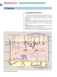
c. Dermis<br />
<strong>1.</strong> <strong>Inflammatory</strong> <strong>cell</strong> <strong>infiltration</strong><br />
<strong>Inflammatory</strong> <strong>cell</strong> <strong>infiltration</strong> occurs when inflammatory <strong>cell</strong>s<br />
such as neutrophils, eosinophils, lymphocytes, plasmacytes,<br />
macrophages and mast <strong>cell</strong>s infiltrate around the blood vessels<br />
(perivascular <strong>infiltration</strong>). There are several <strong>infiltration</strong> patterns,<br />
such as lichenoid <strong>infiltration</strong> (the <strong>cell</strong>s infiltrate in a band resembling<br />
that in lichen planus), vasculitis (the <strong>cell</strong>s cause fibrinoid<br />
degeneration, blood clots, or bleeding in the blood vessels), and<br />
nodular <strong>infiltration</strong>.<br />
The principal infiltrating <strong>cell</strong>s and the causative diseases are<br />
shown in Table. 2.2.<br />
2. Granuloma<br />
A granuloma is a thick aggregation of histiocytes (mostly<br />
macrophages) that form focal chronic <strong>infiltration</strong>. The<br />
macrophages in granulomas are called epithelioid <strong>cell</strong>s. Besides<br />
macrophages, in granulomas one can observe lymphocytes,<br />
fibroblasts, degenerated connective tissue, and blood vessels.<br />
Granulomas are classified according to the distribution patterns<br />
and subtypes of inflammatory <strong>cell</strong>s, as below.<br />
Sarcoidal granuloma: The main components are epitheliod <strong>cell</strong>s<br />
Table 2.2 Diseases with inflammatory <strong>infiltration</strong> into the skin.<br />
Infiltrated<br />
<strong>cell</strong>s<br />
Neutrophils<br />
Eosinophils<br />
Lymphocytes<br />
Plasma <strong>cell</strong>s<br />
Histiocytes<br />
Mast <strong>cell</strong>s<br />
Go Back to the Top To Order, Visit the Purchasing Page for Details<br />
Disorders<br />
early-stage inflammation; irritant contact dermatitis,<br />
erythema nodosum, etc.<br />
infections; impetigo, candidiasis, etc.<br />
disorders associated with reactions of immunocomplex and<br />
complements; cutaneous small-vessel vasculitis, Sweet’s<br />
disease, Behçet’s disease<br />
early inflammation; incontinentia pigmenti<br />
autoimmune diseases; pemphigus, bullous pemphigoid,<br />
etc.<br />
type I allergy<br />
malignant diseases; mycosis fungoides, Langerhans <strong>cell</strong><br />
histiocytosis<br />
inflammations; allergic diseases, etc.<br />
infections; syphilis, lymphogranuloma venereum deep<br />
fungal infection<br />
actinic keratosis, syringocystadenoma papilliferum, etc.<br />
granulomatous diseases; sarcoidosis, granuloma annulare,<br />
etc.<br />
inflammations; atopic dermatitis, chronic eczema, lichen<br />
planus, etc.<br />
other; wounds (especially during healing), neurofibroma, etc.<br />
Fig. 2.18 Vacuolar degeneration.<br />
Graft-versus-host disease. Dyskeratosis is also<br />
seen, from the apotosis of the epidermal keratinocytes.<br />
Fig. 2.19 Sarcoidal granuloma.<br />
Cutaneous sarcoidosis. In sarcoidosis, epithelioid<br />
<strong>cell</strong> granuloma is accompanied by few inflammatory<br />
<strong>cell</strong> <strong>infiltration</strong>, which is also called “naked<br />
granuloma.”<br />
2
2<br />
34 2 Histopathology of the Skin<br />
Fig. 2.20 Foreign-body granuloma. Cholesterin<br />
deposition (arrows).<br />
foreign body giant <strong>cell</strong> Langhans giant <strong>cell</strong><br />
Touton giant <strong>cell</strong><br />
Fig. 2.21 Giant <strong>cell</strong>s originating from histiocytes.<br />
and giant <strong>cell</strong>s. It contains a few necrotic foci and slight lymphocytic<br />
<strong>infiltration</strong>. This is the typical epithelioid <strong>cell</strong> granuloma<br />
observed in sarcoidosis (Fig. 2.19).<br />
Tuberculoid granuloma: Epithelioid <strong>cell</strong> granuloma with<br />
caseous necrosis in the center and abundant lymphocytic <strong>infiltration</strong><br />
at the periphery is observed.<br />
Palisading granuloma: The granuloma contains degenerated<br />
collagen fibers and mucin deposition in the center, with peripheral<br />
macrophages in a palisade or circular pattern. It is found in<br />
granuloma annulare and rheumatoid nodules.<br />
Suppurative granuloma: An abscess (neutrophilic <strong>infiltration</strong>)<br />
surrounded by macrophages and lymphocytes, it is found in deep<br />
mycoses.<br />
Foreign-body granuloma: Macrophages, neutrophils and lymphocytes<br />
accumulate around an extrinsic body (e.g., glass, suture<br />
thread, animal hair, plant fiber) or an intrinsic body (e.g., elastic<br />
fiber, calcium deposits, cholesterin crystal). It is a normal reaction<br />
to foreign bodies (Fig. 2.20). Giant <strong>cell</strong>s that have phagocytosed<br />
a foreign body are often observed; however, the foreign<br />
substance becomes buried in fibrous tissues over time.<br />
3. Giant <strong>cell</strong><br />
Giant <strong>cell</strong> is the general term for <strong>cell</strong>s that contain a characteristically<br />
large nucleus. Most giant <strong>cell</strong>s derive from macrophages<br />
and are multinuclear from the fusion of macrophages or repeated<br />
nuclear division (Fig. 2.21). Ballooning observed in viral diseases<br />
and Reed-Sternberg <strong>cell</strong>s found in Hodgkin’s disease are<br />
types of giant <strong>cell</strong>s.<br />
These are other types:<br />
Foreign-body giant <strong>cell</strong>: Macrophages grow large by phagocytosing<br />
foreign substances The nuclei are irregularly arranged<br />
(Fig. 2.20).<br />
Langhans giant <strong>cell</strong>: Syncytial macrophages with regularly<br />
arranged nuclei in a circular or horseshoe-shaped arrangement.<br />
These often found in tuberculosis, sarcoidosis, and lichen nitidus.<br />
Touton giant <strong>cell</strong>: These macrophages phagocytose fat tissue.<br />
The eosinophilic cytoplasm at the center of the <strong>cell</strong> is surrounded<br />
by a nucleus that is further surrounded by foamy light cytoplasm.<br />
Touton giant <strong>cell</strong>s are found in juvenile xanthogranuloma and<br />
xanthoma.<br />
4. Changes in connective tissue<br />
Fibrosis (irregular proliferation of fibroblasts and collagen<br />
fibers such as in scarring and dermatofibroma) and sclerosis<br />
(decrease of fibroblasts, swelling or homogenization of collagen<br />
fibers, radiation dermatitis, scleredema) are observed in changes<br />
of collagen fibers. Elastic fibers decrease in size and number,<br />
fracture, and degenerate in senile skin and pseudoxanthoma elasticum.<br />
Edema with detachment of connective tissue and accumu-
lation of serous fluid (scleredema) and dermis elevation caused<br />
by projected dermal papillae (papillomatosis) are also changes of<br />
connective tissues.<br />
5. Deposition of foreign substances<br />
Substances that deposit in the dermis include amyloids (e.g., in<br />
macular amyloidosis, lichen amyloydosis), mucins (e.g., myxedema,<br />
lupus erythematosus), calcium (e.g., in carcinosis cutis, pseudoxanthoma<br />
elasticum, CREST syndrome), hemosiderins (e.g., in<br />
bruising, angiitis, hemochromatosis), uric acid, porphyrin and<br />
hyaline.<br />
d. Subcutaneous fat tissue<br />
<strong>1.</strong> Panniculitis<br />
Panniculitis is an inflammation of the subcutaneous fat tissue<br />
(Figs. 2.22 and 2.23). It is categorized by the site of inflammation.<br />
In septal panniculitis, inflammation occurs mostly in the<br />
septa of the subcutaneous fat tissue, such as seen in erythema<br />
nodosum. In lobular panniculitis, inflammation occurs in the lobules<br />
of the fat tissue, such as seen in erythema induratum. Panniculits<br />
can also occur in acute pancreatitis from the fat necrosis<br />
that occurs as a complication.<br />
2. Other changes in fat tissue<br />
Lipogranuloma, lipoatrophy, liponecrosis, lipolysis, lipoma<br />
and liposarcoma are other changes of fat tissue.<br />
epidermis dermis subcutaneous tissue epidermis dermis subcutaneous tissue<br />
B. Dermatopathology 35<br />
septal panniculitis<br />
lobular panniculitis<br />
Fig. 2.22 Differences between septal panniculitis<br />
and lobular panniculitis.<br />
Black dots are the infiltrated inflammatory <strong>cell</strong>s.<br />
Fig. 2.23 Septal panniculitis.<br />
Erythema nodosum.<br />
Go Back to the Top To Order, Visit the Purchasing Page for Details<br />
2