Corel Ventura - ELANSARY.CHP - SIBioC
Corel Ventura - ELANSARY.CHP - SIBioC
Corel Ventura - ELANSARY.CHP - SIBioC

You also want an ePaper? Increase the reach of your titles
YUMPU automatically turns print PDFs into web optimized ePapers that Google loves.
CONTRIBUTI SCIENTIFICI SCIENTIFIC PAPERS<br />
sponse plays an important role in the pathogenesis of<br />
clinical manifestations and control of tuberculosis. Tuberculous<br />
pleural effusions have an increased percentage<br />
and an increased absolute number of T-lymphocytes compared<br />
with peripheral blood. Other types of effusions also<br />
have increased percentages of T-lymphocytes but the<br />
absolute number of lymphocytes is not elevated (35).<br />
Pleural infections by Mycobacterium tuberculosis are<br />
accompanied by a lymphocytic infiltrate and formation of<br />
an exudates rich in T-lymphocytes, predominantly T4<br />
lymphocytes. In vitro stimulation with purified protein derivative<br />
(PPD) leads to a proliferative response (36). Shiratsuchi<br />
and Tsuyuguchi (37) proved that PPD-induced<br />
proliferating lymphocytes mainly belonged to the T4 subset.<br />
The stimulation of T-lymphocytes also is accompanied<br />
by the production of IFN- γ (38). Some authors proved that<br />
different T-cell subsets could produce IFN- γ, with results<br />
depending on the technique, stimulus and lymphocytes<br />
used (39). Shimokata et al (40) showed that T4<br />
lymphocytes are responsible for in vitro production of IFN-γ<br />
when the tuberculous pleural lymphocytes are stimulated<br />
by PPD.<br />
The elevated IFN-γ levels found in tuberculous pleural<br />
fluids might be the equivalent in vivo of the production<br />
observed in vitro after PPD stimulation . IFN-γ detected in<br />
pleural fluid may be the result of the in situ stimulation of<br />
T4 lymphocytes by tuberculous antigens. IFN-γ is known<br />
to activate macrophages, increasing their bactericidal capacity<br />
against Mycobacterium tuberculosis (41), and when<br />
they are treated with CD4 monoclonal antibodies and<br />
complement, IFN-γ levels decreases (34). Thus the reason<br />
for the increase is considered to be production by CD4+<br />
lymphocytes reacting against tubercle bacilli . In fact , the<br />
concentration of tubercle bacilli in pleural liquid correlates<br />
with the amount of IFN-γ (34). A number of reports have<br />
demonstrated that IFN-γ levels in patients with tuberculous<br />
pleurisy are high, with sensitivities and specificities ran-<br />
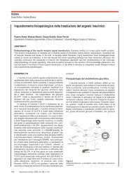
Figure 6<br />
Correlation between ADA and IFN-gamma levels in tuberculous pleural effusion<br />
18 biochimica clinica, 2005, vol. 29, n. 1<br />
ging from 90% to 100% (34,42:47). Valdes et al. (11)<br />
conducted research on pleural fluid samples obtained from<br />
145 patients and reported two with small volumes out of<br />
35 tuberculous cases to be false-negative, while nine out<br />
of 110 non-tuberculous cases were false-positives ( one ;<br />
parapneumonic pleural effusion, three; pulmonary embolism,<br />
three; lymphoma, one; lymphocytic leukemia, one;<br />
neuroblastoma) (28). In our study there were no false-positives<br />
with the use of IFN- γ, and only one case with a<br />
small volume of pleural fluid presented as a false-negative.<br />
These findings provide strong support for the conclusion<br />
that IFN-γ is a reliable marker of tuberculous pleurisy.<br />
Our finding of high levels of these cytokines in tuberculous<br />
pleural effusion together with the finding of very low<br />
levels in the serum , may support the suggestion that these<br />
cytokines are produced locally by inflammatory<br />
cells(12,28,41) .<br />
ROC curves can profile sensitivity and specificity of<br />
markers, and are regarded as useful for analyzing and/or<br />
comparing diagnostic accuracy (48) . While ADA was here<br />
found to have good values for both parameters , IFN-γ was<br />
superior as a marker for tuberculous pleurisy. All of the<br />
three cases with false -positive ADA findings were cases<br />
with pyothorax, and their IFN-γ levels were all lower than<br />
the cut-off value. There were two cases showing false-negatives<br />
for ADA , but they had high IFN-γ levels. In these<br />
two cases , pleural effusion was aspirated after one month<br />
of onset. ADA levels in tuberculous pleurisy may decrease<br />
long after onset, but, even if this is also the case for<br />
IFN-γ, elevation above the cut-off value seems to be<br />
maintained.<br />
Although the rises in ADA and IFN-γ levels in tuberculous<br />
pleural effusion have different origins (infected macrophages<br />
in the case of ADA and sensitized CD4+ cells<br />
in that of IFN-γ), ’’in this study and another study (49)<br />
although not in certain others (50)’’ ADA and IFN-γ were<br />
correlated.<br />
As we have found gamma<br />
interferon to be a highly<br />
specific finding in tuberculous<br />
effusions, an assay for<br />
IFN-γ may prove to be useful<br />
as a screening test for tuberculous<br />
pleurisy. Nevertheless,<br />
the measurement of<br />
IFN-γ is an expensive technique<br />
when compared with<br />
adenosine deaminase determinations<br />
, an other excellent<br />
method for rapid screening<br />
of tuberculous effusions.<br />
The cost of a test becomes<br />
an important consideration<br />
when one is dealing<br />
with a disease with a higher<br />
incidence in less developed<br />
countries.<br />
In conclusion , Levels of<br />
the pleural fluid cytokines