Toric Intraocular Lenses in Cataract Surgery - InTech
Toric Intraocular Lenses in Cataract Surgery - InTech
Toric Intraocular Lenses in Cataract Surgery - InTech

You also want an ePaper? Increase the reach of your titles
YUMPU automatically turns print PDFs into web optimized ePapers that Google loves.
16<br />
<strong>Toric</strong> <strong>Intraocular</strong> <strong>Lenses</strong> <strong>in</strong> <strong>Cataract</strong> <strong>Surgery</strong><br />
1. Introduction<br />
Nienke Visser, Noël J.C. Bauer and Rudy M.M.A. Nuijts<br />
University Eye Cl<strong>in</strong>ic Maastricht,<br />
The Netherlands<br />
In modern cataract surgery, spectacle freedom is becom<strong>in</strong>g more and more important.<br />
Emmetropia can be achieved for patients with myopic or hyperopic refractive errors by<br />
select<strong>in</strong>g the appropriate spherical lens power. However, approximately 20% of patients who<br />
undergo cataract surgery have 1.25 diopters (D) of corneal astigmatism or more. (Ferrer-<br />
Blasco, Montes-Mico et al. 2009; Hoffmann and Hutz 2010) Not correct<strong>in</strong>g the astigmatism<br />
component at the time of cataract surgery will fail to achieve spectacle <strong>in</strong>dependence.<br />
In patients with substantial amounts of corneal astigmatism several options exist to correct<br />
astigmatism dur<strong>in</strong>g or after cataract surgery. Limbal relax<strong>in</strong>g <strong>in</strong>cisions or opposite clear<br />
corneal <strong>in</strong>cisions may be performed to reduce astigmatism dur<strong>in</strong>g cataract surgery. After<br />
cataract surgery, laser refractive surgery may be used to correct residual refractive errors,<br />
<strong>in</strong>clud<strong>in</strong>g cyl<strong>in</strong>der errors. However, corneal <strong>in</strong>cision procedures are relatively unpredictable<br />
and laser refractive surgery may be associated with complications such as dry eyes, wound<br />
heal<strong>in</strong>g problems and <strong>in</strong>fections. (Bayramlar, Daglioglu et al. 2003; de Oliveira, Solari et al.<br />
2006; Kato, Toda et al. 2008; Thomas, Brunstetter et al. 2008) <strong>Toric</strong> IOLs now provide the<br />
opportunity to correct corneal astigmatism, offer<strong>in</strong>g patients with pre-exist<strong>in</strong>g astigmatism<br />
optimal distance vision without the use of spectacles or contact lenses with a cyl<strong>in</strong>drical<br />
correction. Furthermore, the recent <strong>in</strong>troduction of multifocal toric IOLs offers patient with<br />
pre-existent corneal astigmatism the opportunity not only to achieve spectacle<br />
<strong>in</strong>dependence for distance vision, but also for near and <strong>in</strong>termediate visual acuities.<br />
2. <strong>Toric</strong> <strong>in</strong>traocular lenses<br />
The first toric IOL was presented by Shimizu et al. <strong>in</strong> 1994. (Shimizu, Misawa et al. 1994) This<br />
was a non-foldable three-piece toric IOL made from poly-methyl methacrylate (PMMA). It<br />
consisted of an oval optic with loop haptics and was available <strong>in</strong> cyl<strong>in</strong>der powers of 2.00 D or<br />
3.00 D. Postoperatively, about 20% of the IOLs rotated 30 degrees or more and almost 50% of<br />
IOLs rotated more than 10 degrees. Rotational stability is a crucial factor <strong>in</strong> the safety and<br />
efficacy of toric IOLs, s<strong>in</strong>ce as little as 10 degrees of axis misalignment reduces the efficacy of<br />
the astigmatic correction by 33%. Misalignment of more than 30 degrees may even <strong>in</strong>duce<br />
astigmatism. (Shimizu, Misawa et al. 1994) S<strong>in</strong>ce 1994, many advancements have been made <strong>in</strong><br />
toric IOL technology, <strong>in</strong>clud<strong>in</strong>g improvements <strong>in</strong> IOL material and design and ref<strong>in</strong>ements <strong>in</strong><br />
surgical technique. These advances have led to an improved postoperative rotational stability<br />
and excellent visual outcomes us<strong>in</strong>g currently available toric IOls. Table 1 provides an<br />
overview of the characteristics of the currently available toric IOLs.<br />
www.<strong>in</strong>techopen.com
268<br />
^ = Same IOL model under different name;<br />
* = Highest cyl<strong>in</strong>der powers are custom made;<br />
# = Higher cyl<strong>in</strong>der powers available (customized)<br />
Table 1. Currently available monofocal <strong>Toric</strong> <strong>Intraocular</strong> lenses<br />
www.<strong>in</strong>techopen.com<br />
Astigmatism – Optics, Physiology and Management
<strong>Toric</strong> <strong>Intraocular</strong> <strong>Lenses</strong> <strong>in</strong> <strong>Cataract</strong> <strong>Surgery</strong><br />
2.1 IOL material<br />
The IOL biomaterial is of great <strong>in</strong>fluence on the postoperative rotation of the IOL. Older<br />
toric IOL models such as the STAAR toric IOL (STAAR Surgical Company, Monrovia,<br />
California) and the MicroSil toric IOL (HumanOptics, Erlangen, Germany) were made of<br />
silicone materials and showed relatively high postoperative misalignment rates and often<br />
required surgical realignment. As visible <strong>in</strong> Table 1, currently available toric IOLs are<br />
usually made of acrylic material.<br />
After implantation of the toric IOL <strong>in</strong> the capsular bag, the anterior and posterior capsules<br />
fuse with the IOL which prevents IOL rotation. Therefore, strong IOL adhesion to the<br />
capsular bag is thought to prevent IOL rotation. Several <strong>in</strong> vitro studies have exam<strong>in</strong>ed the<br />
<strong>in</strong>teractions between different IOL materials and the capsular bag. Lombardo et al. used<br />
atomic force microscopy to determ<strong>in</strong>e IOL optic surface adhesiveness and found that<br />
hydrophobic acrylic IOLs showed the highest adhesive properties, followed by hydrophilic<br />
acrylic IOL, PMMA IOLs and f<strong>in</strong>ally silicone IOLs. (Lombardo, Carbone et al. 2009) In<br />
addition, Oshika et al. exam<strong>in</strong>ed the adhesive forces between IOLs and bov<strong>in</strong>e collagen<br />
sheets and demonstrated that acrylic IOLs formed the strongest adhesions to the capsular<br />
bag, followed by PMMA IOLs and silicone IOLs. (Oshika, Nagata et al. 1998) An animal<br />
study <strong>in</strong> which rabbits underwent phacoemulsification with IOL implantation confirmed<br />
the latter results. (Oshika, Nagata et al. 1998) Three weeks after IOL implantation, acrylic<br />
IOLs showed the strongest adhesions with the capsular bag, followed by PMMA and<br />
silicone IOLs.<br />
L<strong>in</strong>nola et al. hypothesize that IOL materials show differences <strong>in</strong> IOL adhesion due to a<br />
different aff<strong>in</strong>ity to prote<strong>in</strong>s <strong>in</strong> the capsular bag. (L<strong>in</strong>nola, Sund et al. 2003) Extracellular<br />
matrix prote<strong>in</strong>s, such as fibronect<strong>in</strong>s, vitronection and collagen type IV, may be <strong>in</strong>volved <strong>in</strong><br />
IOL adhesion to the capsular bag. Accord<strong>in</strong>g to L<strong>in</strong>nola et al., these prote<strong>in</strong>s are present <strong>in</strong><br />
plasma but also available <strong>in</strong> the aqueous humor after cataract surgery due to break down of<br />
the blood-aqueous barrier. (L<strong>in</strong>nola, Sund et al. 2003) In an <strong>in</strong> vitro study <strong>in</strong> which different<br />
IOLs were <strong>in</strong>cubated for 1 week with extracellular matrix prote<strong>in</strong>s, each IOL material was<br />
found to have a different aff<strong>in</strong>ity to these prote<strong>in</strong>s. (L<strong>in</strong>nola, Sund et al. 2003) Fibronect<strong>in</strong><br />
bound significantly better to hydrophobic acrylic IOLs, whereas collagen type IV bound<br />
significantly better to hydrophilic acrylic IOLs and vitronect<strong>in</strong> to silicone IOLs. A<br />
histological study us<strong>in</strong>g human pseudophakic autopsy eyes also showed that fibronect<strong>in</strong><br />
was the primary prote<strong>in</strong> between acrylic IOLs and the capsular bag. (L<strong>in</strong>nola, Werner et al.<br />
2000) In addition, acrylic IOLs explanted from human autopsy eyes conta<strong>in</strong>ed significantly<br />
more fibronect<strong>in</strong> and vitronect<strong>in</strong> compared to silicone or PMMA eyes. (L<strong>in</strong>nola, Werner et<br />
al. 2000) These results <strong>in</strong>dicate that different IOL materials use different prote<strong>in</strong>s to b<strong>in</strong>d to<br />
the capsular bag and that acrylic IOLs generally form the strongest adhesions with the<br />
capsular bag.<br />
2.2 IOL design<br />
The IOL design is of <strong>in</strong>terest <strong>in</strong> avoid<strong>in</strong>g postoperative IOL rotation and achiev<strong>in</strong>g good<br />
postoperative outcomes. The overall IOL diameter has been shown to be a major factor <strong>in</strong> the<br />
prevention of IOL rotation. (Chang 2003) Chang et al. compared two different sizes of the<br />
same toric IOL: the STAAR AA4203TF model with a diameter of 10.8 mm and the STAAR<br />
AA4203 TL model with a diameter of 11.2 mm. The longer STAAR model was found to have a<br />
much better rotational stability compared to the shorter STAAR model. Currently available<br />
toric IOLs however have a total IOL diameter rang<strong>in</strong>g from 11.0 mm to 13.0 mm (Table 1),<br />
www.<strong>in</strong>techopen.com<br />
269
270<br />
Astigmatism – Optics, Physiology and Management<br />
which has been shown to be effective <strong>in</strong> avoid<strong>in</strong>g IOL rotation. (Ahmed, Rocha et al. 2010;<br />
Alio, Agdeppa et al. 2010; Holland, Lane et al. 2010; Entabi, Harman et al. 2011)<br />
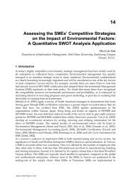
Regard<strong>in</strong>g the IOL haptics design, two different IOL designs are available: plate haptic IOLs,<br />
such as the Acri.Comfort and STAAR toric IOLs, and loop haptic IOLs, such as Acrysof and<br />
Rayner T-flex toric IOLs (Figure 1). Buckhurst et al. hypothesize that loop haptic IOLs have<br />
a better early rotational stability compared to plate haptic IOLs due to the longer haptics<br />
and consequently more contact between haptics and capsular bag. (Buckhurst, Wolffsohn et<br />
al. 2010) Plate haptics, however, are thought to be less susceptible to the compression of the<br />
capsular bag, which may prevent late IOL rotation. (Patel, Ormonde et al. 1999) Patel et al.<br />
compared the early (2 weeks) and late (2 weeks to 6 months) rotation of plate and loop<br />
haptic silicone IOLs <strong>in</strong> a randomised study. (Patel, Ormonde et al. 1999) Even though early<br />
postoperative rotation were comparable, late postoperative rotation was significantly higher<br />
<strong>in</strong> loop haptic IOLs compared to plate haptic IOLs: 6.8 degrees versus 0.6 degrees,<br />
respectively. However, Pr<strong>in</strong>z et al. recently compared plate-haptic and loop-haptic acrylic<br />
Fig. 1. Currently available loop haptic (A) and plate haptic (B) toric <strong>in</strong>traocular lenses<br />
(IOLs).<br />
www.<strong>in</strong>techopen.com
<strong>Toric</strong> <strong>Intraocular</strong> <strong>Lenses</strong> <strong>in</strong> <strong>Cataract</strong> <strong>Surgery</strong><br />
IOLs and did not f<strong>in</strong>d a significant difference <strong>in</strong> early and late rotation. (Pr<strong>in</strong>z, Neumayer et<br />
al. 2011) S<strong>in</strong>ce both the plate and loop haptic IOLs <strong>in</strong> this study were made of acrylic<br />
material, it is possible that adhesion of the acrylic IOL to the capsular bag prevented<br />
postoperative rotation. This <strong>in</strong>dicates that for acrylic IOLs, plate and loop haptics<br />
demonstrate equally good rotational stability.<br />
3. Patient selection<br />
3.1 Monofocal toric IOLs<br />
3.1.1 M<strong>in</strong>imal astigmatism<br />
Achiev<strong>in</strong>g success with toric IOLs depends on the selection of suitable patients. Depend<strong>in</strong>g<br />
on the toric IOL model, the m<strong>in</strong>imal available toric IOL power at the IOL plane is 1.0 D for<br />
the Acri.Comfort or Rayner toric IOLs and 1.5 D for the AcrySof toric IOLs (Table 1). At the<br />
corneal plane, this corresponds to a m<strong>in</strong>imal corneal power of approximately 0.75 to 1.00 D,<br />
respectively. Tak<strong>in</strong>g <strong>in</strong>to consideration the amount of astigmatism <strong>in</strong>duced by the surgery,<br />
patients must have a corneal astigmatism of at least 1.00 to 1.25 D <strong>in</strong> order to be candidates<br />
for a toric IOL.<br />
3.1.2 Corneal astigmatism<br />
Patients with regular bow-tie astigmatism are most suitable for toric IOL implantation.<br />
Corneal topography is therefore important for detect<strong>in</strong>g irregular astigmatism and<br />
keratoconus. Two systems are available to perform corneal topography: placido-disk<br />
videokeratoscopy and Scheimpflug imag<strong>in</strong>g. Even though measurements of corneal curvature<br />
obta<strong>in</strong>ed with both systems show moderate to good correlations, important differences exist<br />
between these two systems that may be relevant <strong>in</strong> toric IOL candidates. (Sav<strong>in</strong>i, Barboni et al.<br />
2009; Symes, Say et al. 2010) A Placido-disk videokeratoscope reconstructs a curvature<br />
description of the anterior surface of the cornea based on the reflections of light-emitt<strong>in</strong>g<br />
Placido r<strong>in</strong>gs. (Jongsma, De Brabander et al. 1999) However, this does not reflect the corneal<br />
shape s<strong>in</strong>ce it does not <strong>in</strong>clude <strong>in</strong>formation about the posterior corneal surface and the corneal<br />
thickness. (Bel<strong>in</strong> and Khachikian 2009) Some corneal ectatic disorders, such as keratoconus,<br />
present with changes on the posterior corneal surface before any changes may be seen on the<br />
anterior corneal surface. (Tomidokoro, Oshika et al. 2000; Bel<strong>in</strong> and Khachikian 2009)<br />
Furthermore, Placido-disk videokeratoscopy only gathers data from the central 8 to 9 mm of<br />
the cornea, which limits the detection of peripheral pathologies, such as pellucid marg<strong>in</strong>al<br />
degeneration. (Walker, Khachikian et al. 2008)<br />
Elevation-based topography uses a rotat<strong>in</strong>g Scheimpflug camera to capture cross sectional<br />
images of the anterior segment, which are then merged <strong>in</strong>to a 3-dimensional reconstruction<br />
of the cornea, anterior chamber, iris and lens. This allows to evaluate the entire corneal<br />
surface (limbus to limbus) and allows for evaluation of the posterior corneal surface. Before<br />
conduct<strong>in</strong>g corneal topography or ocular biometry, ensure that the patient has refra<strong>in</strong>ed<br />
from contact lens wear for an appropriate time. Soft contact lenses should be discont<strong>in</strong>ued<br />
for at least 1 week and hard contact lenses for approximately 2 weeks.<br />
3.1.3 Other considerations<br />
Other pre-existent ocular pathologies may be a contra<strong>in</strong>dication for toric IOL implantation.<br />
Patients with Fuchs’ endothelial dystrophy or a different corneal dystrophy might need a<br />
keratoplasty <strong>in</strong> the future and are therefore not good candidates for toric IOL implantation.<br />
www.<strong>in</strong>techopen.com<br />
271
272<br />
Astigmatism – Optics, Physiology and Management<br />
Patients with potential bag <strong>in</strong>stability like patients with pseudoexfoliation syndrome or<br />
trauma <strong>in</strong>duced zonulolysis are also bad candidates.<br />
3.2 Multifocal toric IOLs<br />
Patient selection is crucial for achiev<strong>in</strong>g success with multifocal toric IOLs. The first step is<br />
to determ<strong>in</strong>e if the patient is a suitable candidate for a multifocal IOL. The ideal patient is<br />
motivated about achiev<strong>in</strong>g spectacle <strong>in</strong>dependency for both distance and near vision,<br />
understands the limitations of multifocal IOLs and has realistic expectations. (Assil,<br />
Christian et al. 2008)<br />
The second step is to determ<strong>in</strong>e possible ocular co-morbidities. Multifocal IOLs split the<br />
available light between distance and near focus. Therefore, ocular co-morbidities that affect<br />
the visual acuity or the quality of vision are a relative or absolute contra<strong>in</strong>dication for<br />
multifocal toric IOLs. These <strong>in</strong>clude amblyopia, corneal pathology (such as keratoconus,<br />
corneal scar or Fuchs’ endothelial dystrophy), maculopathy (such as macular degeneration<br />
or diabetic ret<strong>in</strong>opathy), glaucoma and uveitis. (Assil, Christian et al. 2008; Kohnen, Kook et<br />
al. 2008) An extensive preoperative ophthalmic exam<strong>in</strong>ation is therefore required, <strong>in</strong>clud<strong>in</strong>g<br />
corneal topography, endothelial cell count, ophthalmoscopy and preferably optical<br />
coherence tomography.<br />
4. IOL calculation<br />
4.1 Keratometry<br />
Accurate keratometry measurements must be obta<strong>in</strong>ed to ensure successful<br />
astigmatism correction with toric IOLs. Cl<strong>in</strong>ical studies on toric IOLs describe various<br />
methods of keratometry: IOLMaster automated keratometry, manual keratometry,<br />
autokeratorefractometry, corneal topography, or a comb<strong>in</strong>ation of these techniques.<br />
(Bauer, de Vries et al. 2008; Chang 2008; Dardzhikova, Shah et al. 2009; Ahmed, Rocha et<br />
al. 2010; Gayton and Seabolt 2010; Holland, Lane et al. 2010) Keratometry measurements<br />
obta<strong>in</strong>ed by automated keratometry, manual keratometry and corneal topography have<br />
been shown to have a high repeatability and are generally well comparable between<br />
devices. (Santodom<strong>in</strong>go-Rubido, Mallen et al. 2002; F<strong>in</strong>dl, Kriechbaum et al. 2003; Elbaz,<br />
Barkana et al. 2007; Shirayama, Wang et al. 2009) However, differences between devices<br />
have been reported, <strong>in</strong>dicat<strong>in</strong>g that keratometry values should not be used<br />
<strong>in</strong>terchangeably. (Santodom<strong>in</strong>go-Rubido, Mallen et al. 2002; Elbaz, Barkana et al. 2007;<br />
Shirayama, Wang et al. 2009)<br />
4.2 Surgically <strong>in</strong>duced astigmatism<br />
Another important aspect to consider <strong>in</strong> toric IOL calculation is the amount of astigmatism<br />
<strong>in</strong>duced by the surgery itself. The expected amount of surgically <strong>in</strong>duced astigmatism (SIA)<br />
has to be <strong>in</strong>corporated <strong>in</strong>to the toric IOL power <strong>in</strong> order to select the most appropriate toric<br />
IOL model. (Hill 2008) However, the exact amount of SIA is difficult to predict and depends<br />
on several factors. The location of the <strong>in</strong>cision is an important factor to consider, s<strong>in</strong>ce<br />
corneal <strong>in</strong>cisions lead to flatten<strong>in</strong>g of the <strong>in</strong>cised meridian. An <strong>in</strong>cision at the steep meridian<br />
of the cornea will flatten this meridian and will result <strong>in</strong> steepen<strong>in</strong>g of the orthogonal<br />
meridian due to the coupl<strong>in</strong>g (flatten<strong>in</strong>g/steepen<strong>in</strong>g) effect, which will reduce overall<br />
corneal astigmatism. (Borasio, Mehta et al. 2006) Consequently, an <strong>in</strong>cision located at the flat<br />
meridian will <strong>in</strong>crease overall corneal astigmatism. Furthermore, temporal <strong>in</strong>cisions have<br />
www.<strong>in</strong>techopen.com
<strong>Toric</strong> <strong>Intraocular</strong> <strong>Lenses</strong> <strong>in</strong> <strong>Cataract</strong> <strong>Surgery</strong><br />
been shown to <strong>in</strong>duce less SIA compared to superior <strong>in</strong>cisions. (Tejedor and Murube 2005)<br />
This is possibly due to a higher <strong>in</strong>cidence of aga<strong>in</strong>st-the rule astigmatism <strong>in</strong> the elderly<br />
cataract population or due to a more peripheral location of the temporal <strong>in</strong>cision on the cornea.<br />
(Fledelius and Stubgaard 1986; Kohnen, Dick et al. 1995) The size of the <strong>in</strong>cision has also been<br />
shown to <strong>in</strong>fluence the amount of SIA: smaller <strong>in</strong>cisions generally produce less SIA. (Kohnen,<br />
Dick et al. 1995) Other factors that are of <strong>in</strong>fluence are the amount of preoperative corneal<br />
astigmatism, suture use and patients’ age. (Storr-Paulsen, Madsen et al. 1999) Developments <strong>in</strong><br />
phacoemulsification techniques have led to an improved management of SIA. The shift to<br />
smaller <strong>in</strong>cisions has reduced the need for sutur<strong>in</strong>g, thus decreas<strong>in</strong>g SIA. In addition, the<br />
recent development of micro<strong>in</strong>cisional cataract surgery, surgery performed throught <strong>in</strong>cisions<br />
smaller than 2.0 mm, aims to further reduce the SIA.<br />
Many studies have measured the amount of SIA follow<strong>in</strong>g cataract surgery with <strong>in</strong>cision<br />
sizes rang<strong>in</strong>g from less than 2 mm up to 3.4 mm. However, it is difficult to compare these<br />
studies, because they use variable <strong>in</strong>cision locations and sizes and variable follow-up<br />
durations. STAAR and MicroSil toric IOLs require a 2.8 and 3.4 mm <strong>in</strong>cision for IOL<br />
implantation, respectively (Table 1). Incision sizes of 2.8 to 3.2 mm have been shown to<br />
<strong>in</strong>duce a SIA of 0.4 to 0.8 D for temporal <strong>in</strong>cisions, 0.6 D for superior <strong>in</strong>cisions and 0.9 to 1.2<br />
D for on-axis <strong>in</strong>cisions. (Alio, Rodriguez-Prats et al. 2005; Borasio, Mehta et al. 2006; Moon,<br />
Mohamed et al. 2007; Morcillo-Laiz, Zato et al. 2009; Wang, Zhang et al. 2009) Acrysof toric<br />
IOLs require a 2.2 mm <strong>in</strong>cision for IOL implantation. These <strong>in</strong>cisions have been shown to<br />
<strong>in</strong>duce a SIA of 0.2 to 0.3 D for temporal <strong>in</strong>cisions and 0.4 D for superior <strong>in</strong>cisions. (Lee,<br />
Kwon et al. 2009; Wang, Zhang et al. 2009; Visser, Ruiz-Mesa et al. 2011) F<strong>in</strong>ally, Acri.Lisa<br />
and Rayner toric IOLs may be implanted through sub 2.0 mm <strong>in</strong>cisions. Micro<strong>in</strong>cision<br />
cataract surgery has been shown to result <strong>in</strong> a SIA of approximately 0.3 D for temporal<br />
<strong>in</strong>cisions, 0.5 D for superior <strong>in</strong>cisions and 0.4 D for on-axis <strong>in</strong>cisions. (Alio, Rodriguez-Prats<br />
et al. 2005; Kaufmann, Krishnan et al. 2009; Lee, Kwon et al. 2009; Morcillo-Laiz, Zato et al.<br />
2009) In practice, the most accurate method to determ<strong>in</strong>e the SIA is for every surgeon to<br />
personalize the amount of SIA <strong>in</strong>duced by cataract surgery <strong>in</strong> his/her patient population.<br />
This may be done by analys<strong>in</strong>g preoperative and postoperative corneal astigmatism changes<br />
us<strong>in</strong>g a standard vector analysis. (Alp<strong>in</strong>s 2001; Holladay, Moran et al. 2001)<br />
5. IOL implantation<br />
5.1 Mark<strong>in</strong>g techniques<br />
Crucial to the efficacy of toric IOLs is exact alignment of the toric IOL at the calculated<br />
alignment axis. Accurate mark<strong>in</strong>g of the alignment axis should be performed with the<br />
patient <strong>in</strong> an upright position <strong>in</strong> order to prevent cyclotorision <strong>in</strong> the sup<strong>in</strong>e position.<br />
Cyclotorsion of the eye from the upright to sup<strong>in</strong>e position is approximately 2 to 4 degrees<br />
on average, but can be up to 15 degrees <strong>in</strong> <strong>in</strong>dividual patients. (Arba-Mosquera, Merayo-<br />
Lloves et al. 2008; Chang 2008; Febbraro, Koch et al. 2010) Cyclotorsion is a well known<br />
aspect <strong>in</strong> refractive surgery and compensated for dur<strong>in</strong>g laser refractive surgery. (Febbraro,<br />
Koch et al. 2010)<br />
Most cl<strong>in</strong>ical studies on toric IOLs describe us<strong>in</strong>g a 3-step mark<strong>in</strong>g procedure for toric IOL<br />
implantation. The first step consists of preoperative limbal mark<strong>in</strong>g of the horizontal axis of<br />
the eye with the patient sitt<strong>in</strong>g upright to correct for cyclotorsion. This may be done with the<br />
patient seated at the slitlamp and with a coaxial th<strong>in</strong> slit turned to 0-180 degrees.<br />
(Mendicute, Irigoyen et al. 2008; Alio, Agdeppa et al. 2010; Koshy, Nishi et al. 2010) The<br />
www.<strong>in</strong>techopen.com<br />
273
274<br />
Astigmatism – Optics, Physiology and Management<br />
limbus is than marked at the horizontal position with either a sterile <strong>in</strong>k pen or a needle.<br />
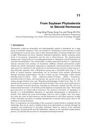
Another technique to mark the horizontal axis is by us<strong>in</strong>g a bubble-marker, such as a<br />
Nuijts/Lane <strong>Toric</strong> Reference Marker (ASICO) (Figure 2) or a Bakewell BubbleLevel (Mastel<br />
Precision, Rapid City, US), or by us<strong>in</strong>g a gravity marker with a calibrated horizontal<br />
position, such as the LRI Gravity Marker (Rumex, S<strong>in</strong>t Petersburg, Florida, US). (Bauer, de<br />
Vries et al. 2008; Ahmed, Rocha et al. 2010; Gayton and Seabolt 2010) Intraoperatively, the<br />
preoperative horizontal marks are used to position an angular graduation <strong>in</strong>strument. The<br />
actual alignment axis is marked us<strong>in</strong>g a toric axis marker.<br />
Fig. 2. Preoperative mark<strong>in</strong>g of the horizontal axis of the eye us<strong>in</strong>g the Nuijts/Lane <strong>Toric</strong><br />
Reference Marker with bubble-level (ASICO). This is done with the patient sitt<strong>in</strong>g upright to<br />
correct for cyclotorsion.<br />
One study has evaluated the accuracy of a 3-step mark<strong>in</strong>g procedure for toric IOL<br />
implantation. (Visser, Berendschot et al. 2011) The mean errors <strong>in</strong> horizontal axis mark<strong>in</strong>g,<br />
alignment axis mark<strong>in</strong>g and toric IOL alignment were 2.4 ± 0.8 (maximum 8.7) degrees, 3.3 ±<br />
2.0 (maximum 7.7) degrees and 2.6 ± 2.6 (maximum 10.5) degrees, respectively. Together,<br />
these 3 errors led to a mean total error <strong>in</strong> toric IOL alignment of 4.9 ± 2.1 degrees. However,<br />
for the <strong>in</strong>dividual patient, this may be as high as 10 degrees. This <strong>in</strong>dicates that great<br />
accuracy <strong>in</strong> toric IOL alignment is necessary <strong>in</strong> all patients <strong>in</strong> order to achieve the most<br />
optimal astigmatism correction with toric IOLs.<br />
Currently, new techniques have become available to ensure accurate <strong>in</strong>traoperative<br />
alignment of toric IOLs. Osher has described an iris-f<strong>in</strong>gerpr<strong>in</strong>t<strong>in</strong>g technique, <strong>in</strong> which<br />
preoperative detailed images of the eye are obta<strong>in</strong>ed. (Osher 2010) The desired alignment<br />
axis is drawn <strong>in</strong> this image. A pr<strong>in</strong>tout of this image is than used dur<strong>in</strong>g surgery to align the<br />
toric IOL based on iris characteristics. A second technique to accurately align toric IOLs is by<br />
www.<strong>in</strong>techopen.com
<strong>Toric</strong> <strong>Intraocular</strong> <strong>Lenses</strong> <strong>in</strong> <strong>Cataract</strong> <strong>Surgery</strong><br />
<strong>in</strong>traoperative wavefront aberrometry (ORange, WaveTec Vision Systems). This device is<br />
connected to the operat<strong>in</strong>g microscope and enables <strong>in</strong>traoperative measurement of residual<br />
refraction. (Packer 2010) It allows to accurately position toric IOLs, based on actual residual<br />
refractive cyl<strong>in</strong>der results. A third device, the SG3000 (Sensomotoric Instruments, Teltow,<br />
Germany) uses real-time eye-track<strong>in</strong>g, based on iris and blood vessel characteristics. (Visser,<br />
Berendschot et al. 2011) Preoperatively, a detailed image of the eye is captured, <strong>in</strong> which<br />
blood vessel and iris characteristics are visible. Simultaneously, keratometry is performed<br />
and the location of the steep and flat corneal meridians are shown <strong>in</strong> this image.<br />
Intraoperatively, the preoperative image is matched with the live surgery-image from the<br />
operat<strong>in</strong>g microscope, based on blood vessel and iris characteristics. Us<strong>in</strong>g a microscope<br />
embedded display, the overlay show<strong>in</strong>g the desired alignment axis is visible <strong>in</strong> the<br />
operat<strong>in</strong>g microscope, allow<strong>in</strong>g exact alignment of the toric IOL. In addition, this eyetrack<strong>in</strong>g<br />
technology may also be used for other aspects <strong>in</strong> lens implantation surgery,<br />
<strong>in</strong>clud<strong>in</strong>g plann<strong>in</strong>g of the <strong>in</strong>cisions and capsulorrhexis and optimal centration of multifocal<br />
IOLs.<br />
5.2 <strong>Surgery</strong><br />
A standard phacoemulsification technique may be performed with a 1.5 to 3.4 mm limbal<br />
<strong>in</strong>cision, depend<strong>in</strong>g on the toric IOL model to be implanted (Table 1). A well centered<br />
capsulorrhexis with 360 degree overlap of the IOL optics should be achieved. The optic<br />
diameter is 6.0 mm for Acrysof, Acri.lisa, MicroSil and STAAR toric IOLs and 5.75 or 6.25<br />
mm for Rayner toric IOLs. The ideal capsulorrhexis diameter is therefore 5.0 to 5.5 mm.<br />
After the phacoemulsification is completed and the ophthalmic viscosurgical device is<br />
<strong>in</strong>jected, the foldable toric IOL is <strong>in</strong>serted through the limbal <strong>in</strong>cision. The marks on the toric<br />
IOL <strong>in</strong>dicate the flat meridian or plus cyl<strong>in</strong>der axis of the toric IOL and should be aligned<br />
with the marked alignment axis. First, gross alignment is achieved by rotat<strong>in</strong>g the IOL<br />
clockwise while it is unfold<strong>in</strong>g, until approximately 20 to 30 degrees short of the desired<br />
position. After the ophthalmic viscosurgical device is removed, the IOL is rotated to its f<strong>in</strong>al<br />
position by exact alignment of the reference marks on the toric IOL with the limbal axis<br />
marks.<br />
In the event of a complication dur<strong>in</strong>g surgery that might compromise the stability of the<br />
toric IOL, such as zonular damage, vitreous loss, capsulorrhexis tear, or capsular rupture,<br />
conversion to a standard non-toric IOL may be required.<br />
5.3 Postoperative axis measurement<br />
Postoperatively, the orientation axis of the toric IOL must be verified to confirm optimal<br />
alignment and ensure no postoperative IOL rotation has occurred. Postoperative assessment<br />
of toric IOL alignment can be achieved by several methods. The most commonly used<br />
method <strong>in</strong> the cl<strong>in</strong>ic is assessment us<strong>in</strong>g a sliltlamp with rotat<strong>in</strong>g slit. S<strong>in</strong>ce the IOL marks<br />
are located at the periphery of the IOL optic, full mydriasis of the pupil is required. An<br />
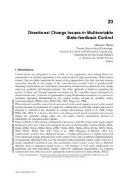
objective method to determ<strong>in</strong>e postoperative toric IOL alignment is by wavefront<br />
aberrometry (Figure 3). (Carey, Leccisotti et al. 2010) Comb<strong>in</strong>ed wavefront aberrometers and<br />
corneal topographers, such as the Keratron Onda (Optikon, Rome, Italy), iTrace (Tracey<br />
Technologies, Houston, TX, USA) and OPD-scan (Nidek, Gamagori, Japan), discrim<strong>in</strong>ate<br />
between aberrations caused by the cornea and by the <strong>in</strong>ternal ocular system. (Carey,<br />
Leccisotti et al. 2010; Visser, Berendschot et al. 2010) This method therefore directly<br />
determ<strong>in</strong>es the orientat<strong>in</strong>g of the toric IOL. Pupil dilation is not required.<br />
www.<strong>in</strong>techopen.com<br />
275
276<br />
Astigmatism – Optics, Physiology and Management<br />
Fig. 3. Comb<strong>in</strong>ed wavefront aberrometery and corneal topography allows to objectively<br />
determ<strong>in</strong>e the postoperative orientation of the toric <strong>in</strong>traocular lens axis. In this example,<br />
Corneal wavefront aberrometry (A) and Ocular wavefront aberrometry (B) were performed<br />
us<strong>in</strong>g the Keratron Onda (Optikon, Rome). The Internal aberrations (C) are calculated by<br />
subtract<strong>in</strong>g the Corneal aberrations from the Ocular aberrations. In this example, corneal<br />
astigmatism was -2.09 diopters (D) at 156 degrees and ocular astigmatism was -0.26 D at 103<br />
degrees. The <strong>in</strong>ternal astigmatism was -2.18 D at 70 degrees, which <strong>in</strong>dicates that the toric<br />
<strong>in</strong>traocular lens axis is located at 70 degrees.<br />
www.<strong>in</strong>techopen.com
<strong>Toric</strong> <strong>Intraocular</strong> <strong>Lenses</strong> <strong>in</strong> <strong>Cataract</strong> <strong>Surgery</strong><br />
Realignment of a rotated toric IOL should ideally be performed as soon as possible and<br />
preferably before 2 weeks postoperatively because of the formation of adhesions between<br />
the capsular bag and IOL optics. (L<strong>in</strong>nola, Sund et al. 2003; Chang 2009)<br />
6. Cl<strong>in</strong>ical outcomes<br />
6.1 Monofocal toric IOLs<br />
Table 2 provides an overview of the ma<strong>in</strong> outcomes of cl<strong>in</strong>ical studies on toric IOLs. In order<br />
to compare the visual outcomes, LogMAR values have been converted <strong>in</strong>to Snellen values.<br />
6.1.1 Visual outcomes<br />
One randomized cl<strong>in</strong>ical trial has been conducted on toric IOLs. Holland et al. <strong>in</strong>cluded 517<br />
patients: 256 patients received unilateral implantation of an Acrysof toric IOL (T3, T4 or T5)<br />
and 261 patients received unilateral implantion with a spherical control IOL. (Holland, Lane<br />
et al. 2010) One year postoperatively, significantly more patients <strong>in</strong> the toric group achieved<br />
an UDVA of 20/40 or better compared to the control group: 92% versus 81% of patients,<br />
respectively. Furthermore, an UDVA of 20/20 or better was achieved <strong>in</strong> significantly more<br />
patients with a toric IOL compared to a control IOL ( 41% versus 19% of patients). As<br />
expected, the CDVA results <strong>in</strong> both groups were comparable: 93% of toric and 90% of<br />
control patients achieved a CDVA of 20/25 or better. Ahmed at al. have conducted a large<br />
non-randomized cohort study to evaluate bilateral AcrySof toric IOL (T3, T4 or T5)<br />
implantation <strong>in</strong> 117 patients (234 eyes). At 6 months postoperatively, 99% of patients had a<br />
bilateral UDVA of 20/40 or better and 63% of 20/20 or better. The mean b<strong>in</strong>ocular UDVA<br />
was 0.05 ± 0.11 LogMAR, equivalent to 0.89 ± 0.23 Snellen. In addition, the b<strong>in</strong>ocular CDVA<br />
was 0.03 ± 0.14 LogMAR, equivalent to 0.93 ± 0.30 Snellen. Many smaller non-randomized<br />
studies on Acrysof toric IOLs have been performed. (Bauer, de Vries et al. 2008; Chang 2008;<br />
Mendicute, Irigoyen et al. 2008; Zuberbuhler, Signer et al. 2008; Dardzhikova, Shah et al.<br />
2009; Lane, Ernest et al. 2009; Ruiz-Mesa, Carrasco-Sanchez et al. 2009; Statham, Apel et al.<br />
2009; Gayton and Seabolt 2010; Kim, Chung et al. 2010; Koshy, Nishi et al. 2010;<br />
Ts<strong>in</strong>opoulos, Tsaousis et al. 2010; Visser, Ruiz-Mesa et al. 2011) The majority of these studies<br />
exam<strong>in</strong>ed the T3 to T5 toric IOLs, with low to moderately high cyl<strong>in</strong>der powers. Most<br />
studies show a mean postoperative UDVA rang<strong>in</strong>g from 0.63 to 0.90 Snellen. (Bauer, de<br />
Vries et al. 2008; Mendicute, Irigoyen et al. 2008; Ruiz-Mesa, Carrasco-Sanchez et al. 2009;<br />
Statham, Apel et al. 2009; Gayton and Seabolt 2010; Koshy, Nishi et al. 2010) An UDVA of<br />
20/40 or better was reported <strong>in</strong> 81 to 95% of eyes. (Bauer, de Vries et al. 2008; Mendicute,<br />
Irigoyen et al. 2008; Dardzhikova, Shah et al. 2009; Gayton and Seabolt 2010) In addition, 26<br />
to 36% of eyes achieved an UDVA of 20/20 or better. (Bauer, de Vries et al. 2008;<br />
Dardzhikova, Shah et al. 2009; Ruiz-Mesa, Carrasco-Sanchez et al. 2009; Gayton and Seabolt<br />
2010) The reported BDVA ranges from 0.79 to 1.01. (Bauer, de Vries et al. 2008; Mendicute,<br />
Irigoyen et al. 2008; Zuberbuhler, Signer et al. 2008; Ruiz-Mesa, Carrasco-Sanchez et al. 2009;<br />
Gayton and Seabolt 2010; Kim, Chung et al. 2010) One study has exam<strong>in</strong>ed the high cyl<strong>in</strong>der<br />
toric IOLs (T6 to T9) and showed a mean UDVA of 0.61 ± 0.26 and an UDVA of 20/40 or<br />
better <strong>in</strong> 83% of eyes. (Visser, Ruiz-Mesa et al. 2011) In order to determ<strong>in</strong>e how effective<br />
toric IOLs are <strong>in</strong> achiev<strong>in</strong>g the maximal visual outcome, we have calculated the visual<br />
potential <strong>in</strong>dex (VPI), which is def<strong>in</strong>ed as the ratio of postoperative UDVA to postoperative<br />
CDVA. (Visser, Ruiz-Mesa et al. 2011) For the Acrysof toric IOL, the VPI <strong>in</strong> the majority of<br />
www.<strong>in</strong>techopen.com<br />
277
278<br />
Astigmatism – Optics, Physiology and Management<br />
studies ranged between 73 and 96%. This <strong>in</strong>dicates that the uncorrected visual outcome with<br />
this IOL is 73 to 96% of the maximal visual outcome.<br />
Rayner T-flex toric IOL implantation has been evaluated <strong>in</strong> two relatively small studies.<br />
(Stewart and McAlister 2010; Entabi, Harman et al. 2011) Entabi et al. evaluated Rayner toric<br />
IOL implantation <strong>in</strong> 33 eyes with a mean corneal astigmatism of 2.94 ± 0.89 D. (Entabi,<br />
Harman et al. 2011) Four months postoperatively, the mean UDVA was 0.28 ± 0.23<br />
LogMAR, equivalent to 0.52 ± 0.28 Snellen. Stewart et al. evaluated T-flex toric IOL<br />
implantation <strong>in</strong> 14 eyes. (Stewart and McAlister 2010) Over 90% of eyes achieved an UDVA<br />
of 20/40 or better and the mean UDVA was 0.16 ± 0.16 LogMAR, equivalent to 0.69 ± 0.25<br />
Snellen. The VPI <strong>in</strong> both studies was 80 and 91%. (Stewart and McAlister 2010; Entabi,<br />
Harman et al. 2011)<br />
One study has exam<strong>in</strong>ed visual outcomes follow<strong>in</strong>g Acri.Comfort toric implantation <strong>in</strong> 21<br />
eyes of 12 patients with moderate to high astigmatism. (Alio, Agdeppa et al. 2010) The mean<br />
preoperative corneal astigmatism was 3.73 ± 1.79 D.<br />
At 3 months postoperatively, the mean UDVA was 0.65 ± 0.22 and 76% of eyes achieved an<br />
UDVA of 20/40 or better. The mean postoperative CDVA was 0.85 ± 0.15 and the VPI was<br />
76%.<br />
Several studies have evaluated the visual outcomes follow<strong>in</strong>g silicone toric IOL<br />
implantation. Microsil toric IOL implantation has been shown to result <strong>in</strong> an UDVA of<br />
20/40 or better <strong>in</strong> 68 to 85% of eyes. (De Silva, Ramkissoon et al. 2006; Dick, Krummenauer<br />
et al. 2006) In addition, the mean postoperative UDVA and BDVA were 0.63 ± 0.22 and 0.76<br />
± 0.19, respectively. (De Silva, Ramkissoon et al. 2006) Studies us<strong>in</strong>g STAAR toric IOLs have<br />
shown an UDVA of 20/40 or better <strong>in</strong> 66 to 84% of eyes and a mean UDVA rang<strong>in</strong>g from<br />
0.54 to 0.62. (Ruhswurm, Scholz et al. 2000; Sun, Vicary et al. 2000; Leyland, Z<strong>in</strong>icola et al.<br />
2001; Till, Yoder et al. 2002; Chang 2003) The VPI was 83% for MicroSil toric IOLs and 66 to<br />
68% for STAAR toric IOLs. (Ruhswurm, Scholz et al. 2000; Leyland, Z<strong>in</strong>icola et al. 2001; De<br />
Silva, Ramkissoon et al. 2006)<br />
6.1.2 Refractive outcomes<br />
The randomized controlled trial on Acrysof toric IOLs has shown a significantly better<br />
refractive cyl<strong>in</strong>der outcome <strong>in</strong> patients implanted with a toric IOL compared to patients<br />
with a monofocal IOL: 88% of eyes with a toric IOL achieved an residual refractive cyl<strong>in</strong>der<br />
of 1.00 D or less, compared to 48% of eyes <strong>in</strong> the control group. Fifty-three percent of<br />
patients with a toric IOL achieved a residual refractive cyl<strong>in</strong>der of 0.5 D or less. In addition,<br />
the mean residual refractive cyl<strong>in</strong>der <strong>in</strong> the toric group was significantly lower compared to<br />
the control group (-0.59 D versus -1.22 D, respectively). Other studies on Acrysof toric IOLs<br />
show a residual refractive cyl<strong>in</strong>der of 1.00 D or less <strong>in</strong> about 80 to 100% of eyes and a mean<br />
residual refractive cyl<strong>in</strong>der rang<strong>in</strong>g from -0.28 to -0.75 D. (Ahmed, Rocha et al. 2010; Kim,<br />
Chung et al. 2010; Visser, Ruiz-Mesa et al. 2011)<br />
After Rayner toric IOL implantation, the mean residual refractive cyl<strong>in</strong>der has been shown<br />
to range from -0.89 to -0.95 D. (Stewart and McAlister 2010; Entabi, Harman et al. 2011)<br />
Acri.Comfort toric IOL implantation resulted <strong>in</strong> a mean residual refractive cyl<strong>in</strong>der of –0.45<br />
± 0.63. (Alio, Agdeppa et al. 2010) Furthermore, based on a vector analysis of the refractive<br />
outcomes, the Acri.Comfort IOL has been shown to correct 91% of pre-exist<strong>in</strong>g astigmatism.<br />
(Alio, Agdeppa et al. 2010) (Ruhswurm, Scholz et al. 2000; Leyland, Z<strong>in</strong>icola et al. 2001; Till,<br />
Yoder et al. 2002)<br />
www.<strong>in</strong>techopen.com
<strong>Toric</strong> <strong>Intraocular</strong> <strong>Lenses</strong> <strong>in</strong> <strong>Cataract</strong> <strong>Surgery</strong><br />
N = number of eyes; FU = follow-up; SD = standard deviation; D = dioptres; ° = degrees; % =<br />
percentage; UDVA = uncorrected distance visual acuity; BDVA = best-corrected distance visual acuity;<br />
VPI = visual potential <strong>in</strong>dex; RCT = randomised controlled trial; PCS = prospective cohort study; RS =<br />
retrospective study; * = converted from LogMAR to Snellen; ^= obta<strong>in</strong>ed by wavefront abberometry<br />
Table 2. Literature on monofocal toric IOLs<br />
www.<strong>in</strong>techopen.com<br />
(%)<br />
279
280<br />
Astigmatism – Optics, Physiology and Management<br />
Regard<strong>in</strong>g the silicone toric IOL, the postoperative residual astigmatism ranged from -0.84<br />
to -1.23 D. (Ruhswurm, Scholz et al. 2000; Sun, Vicary et al. 2000; Leyland, Z<strong>in</strong>icola et al.<br />
2001; Chang 2003; De Silva, Ramkissoon et al. 2006; Dick, Krummenauer et al. 2006) About<br />
70% of eyes implanted with a STAAR toric IOLs achieved a residual refractive cyl<strong>in</strong>der of<br />
1.0 D or less and approximately 50% achieved a residual refractive cyl<strong>in</strong>der of 0.5 D or less.<br />
(Ruhswurm, Scholz et al. 2000; Leyland, Z<strong>in</strong>icola et al. 2001; Till, Yoder et al. 2002)<br />
6.1.3 Spectacle <strong>in</strong>dependence<br />
Spectacle <strong>in</strong>dependence follow<strong>in</strong>g toric IOL implantation has been reported <strong>in</strong><br />
approximately 60% of patients implanted with a toric IOL, compared to 36% of patients<br />
implanted with a control IOL. (Holland, Lane et al. 2010) However, patients <strong>in</strong> this study<br />
only received unilateral toric IOL implantation. Lane et al. offered patients from the<br />
aforementioned study fellow-eye implantation with the same IOL (Acrysof toric IOL or<br />
Acrysof non-toric IOL), allow<strong>in</strong>g bilateral exam<strong>in</strong>ation of spectacle <strong>in</strong>dependence. (Lane,<br />
Ernest et al. 2009) Almost all patients (97%) with a toric IOL reported not us<strong>in</strong>g spectacles<br />
for distance vision, compared to half of the patients <strong>in</strong> the control group. F<strong>in</strong>ally, Ahmed et<br />
al. exam<strong>in</strong>ed spectacle use <strong>in</strong> bilaterally implanted patients and found that 69% of patients<br />
never used spectacles for distance vision. (Ahmed, Rocha et al. 2010)<br />
6.1.4 Rotational stability<br />
Crucial to the efficacy of all toric IOLs is the position of the IOL with regards to the <strong>in</strong>tended<br />
alignment axis, s<strong>in</strong>ce every degree of misalignment leads to residual astigmatism.<br />
Misalignment of the IOL may be caused by two factors: <strong>in</strong>accurate placement of the IOL and<br />
rotation of the IOL. Currently, a misalignment of more than 10 degrees is generally regarded<br />
as the <strong>in</strong>dication for surgical reposition<strong>in</strong>g.<br />
Rotational stability used to be an issue <strong>in</strong> toric pseudophakic IOLs made of silicone material.<br />
For example, STAAR toric IOLs were found to have a high <strong>in</strong>cidence of eyes with more than<br />
10 degrees of IOL misalignment: 14 to 45% for the shorter TF model 10 to 14% for the longer<br />
TL model. (Ruhswurm, Scholz et al. 2000; Sun, Vicary et al. 2000; Leyland, Z<strong>in</strong>icola et al.<br />
2001; Till, Yoder et al. 2002; Chang 2003) Consequently, this resulted <strong>in</strong> a high rate of<br />
surgical reposition<strong>in</strong>g. (Sun, Vicary et al. 2000; Leyland, Z<strong>in</strong>icola et al. 2001; Chang 2003)<br />
MicroSil toric IOLs were more rotationally stable and showed a misalignment of more than<br />
10 degrees <strong>in</strong> 2 to 10% of eyes. (De Silva, Ramkissoon et al. 2006; Dick, Krummenauer et al.<br />
2006)<br />
As shown <strong>in</strong> Table 2, acrylic toric IOLs are generally more rotationally stable than silicone<br />
IOLs. For the Acrysof toric IOLs, the mean postoperative misalignment is less than 4 degrees<br />
and a misalignment of more than 10 degrees is rare. (Bauer, de Vries et al. 2008; Chang 2008;<br />
Ahmed, Rocha et al. 2010; Holland, Lane et al. 2010)<br />
In most cl<strong>in</strong>ical studies, the postoperative orientation of the toric IOL axis was measured via<br />
the slitlamp. S<strong>in</strong>ce this measur<strong>in</strong>g reticule on the slitlamp uses 5 degree steps, it is not a very<br />
accurate method to determ<strong>in</strong>e postoperative IOL rotation. A few studies have used digital<br />
photography to exam<strong>in</strong>e the postoperative IOL rotation, which is more accurate. We<strong>in</strong>and et<br />
al. obta<strong>in</strong>ed digital images immediately after Acrysof IOL implantation and aga<strong>in</strong> at 6<br />
months postoperatively. (We<strong>in</strong>and, Jung et al. 2007) Rotation of the eye was compensated<br />
for by match<strong>in</strong>g images based on specific blood vessel characteristics. The mean<br />
postoperative IOL rotation of Acrysof IOLs was 0.9 degree, with a maximum of 1.8 degrees.<br />
www.<strong>in</strong>techopen.com
<strong>Toric</strong> <strong>Intraocular</strong> <strong>Lenses</strong> <strong>in</strong> <strong>Cataract</strong> <strong>Surgery</strong><br />
Us<strong>in</strong>g a similar digital imag<strong>in</strong>g technique, a different study showed that Acrysof toric IOLs<br />
rotate 2.66 ± 1.99 degrees on average <strong>in</strong> the first 6 months postoperatively. (Koshy, Nishi et<br />
al. 2010) F<strong>in</strong>ally, Kwartz et al. compared the rotational stability of Acrysof IOLs and Akreos<br />
IOLs and showed that both IOLs rotate 2 to 3 degrees with<strong>in</strong> a 2-year period. (Kwartz and<br />
Edwards 2010) However, both latter studies did not compensate for cyclotorsion of the eye<br />
between measurements both with the patient <strong>in</strong> an upright position, which has been shown<br />
to be approximately 2 degrees. (Viestenz, Seitz et al. 2005; Wolffsohn and Buckhurst 2010)<br />
This <strong>in</strong>dicates that the postoperative rotation of Acrysof IOLs is most likely less than 1<br />
degree. The exact postoperative rotation of other acrylic IOLs, such as the Rayner toric or<br />
Acri.Comfort toric IOLs, has not been exam<strong>in</strong>ed yet.<br />
F<strong>in</strong>ally, ocular trauma may cause rotation of a toric IOL. (Chang 2009) In human cadaver<br />
eyes implanted with a toric IOL, trauma without leakage from the old <strong>in</strong>cision site resulted<br />
<strong>in</strong> IOL rotation of approximately 6 degrees. Trauma with leakage from the <strong>in</strong>cision site was<br />
associated with IOL rotation of approximately 40 degrees. (Pereira, Milverton et al. 2009)<br />
However, these human cadaver eyes received post-mortem phacoemulsification with toric<br />
IOL implantation, <strong>in</strong>dicat<strong>in</strong>g that the IOL had not fused with the capsular bag. This would<br />
have resulted <strong>in</strong> an <strong>in</strong>creased IOL rotation and is possibly not an optimal model to exam<strong>in</strong>e<br />
toric IOL rotation follow<strong>in</strong>g ocular trauma.<br />
6.1.5 Economic evaluation<br />
Two studies have performed an economic evaluation of toric IOL implantation versus<br />
monofocal IOL implantation dur<strong>in</strong>g cataract surgery. (Laurendeau, Lafuma et al. 2009;<br />
P<strong>in</strong>eda, Denevich et al. 2010) Laurendeau et al. estimated the lifetime costs of cataract<br />
surgery with bilateral toric or monofocal IOLs <strong>in</strong> patients with pre-exist<strong>in</strong>g corneal<br />
astigmatism <strong>in</strong> four European countries (France, Italy, Germany and Spa<strong>in</strong>). In this study,<br />
70% of patients with bilateral monofocal IOLs needed spectacles for distance vision,<br />
compared to 26% of patients with bilateral toric IOLs. The result<strong>in</strong>g reduction <strong>in</strong> costs <strong>in</strong><br />
patients with toric IOLs depended on the national spectacle costs and ranged from €308 for<br />
Spa<strong>in</strong> to €692 for France. However, this study did not evaluate the possible non-f<strong>in</strong>ancial<br />
benefits of toric IOL implantation, such as the patients’ visual function<strong>in</strong>g and health-related<br />
quality of life.<br />
P<strong>in</strong>eda et al. assessed the economic value of an improved uncorrected visual acuity <strong>in</strong><br />
patients with pre-exist<strong>in</strong>g corneal astigmatism and cataract treated with toric or monofocal<br />
IOLs <strong>in</strong> the US. (P<strong>in</strong>eda, Denevich et al. 2010) Patient with toric IOLs saved $34 <strong>in</strong> total costs<br />
with toric IOLs versus monofocal IOLs. These sav<strong>in</strong>gs <strong>in</strong>creased to $393 among patients<br />
who achieved an UDVA of 20/25 or better. The costs per QALY (quality-adjusted life years;<br />
a measure of disease burden comb<strong>in</strong><strong>in</strong>g quality and quantity of life) for toric IOLs was $349<br />
compared with monofocal IOLs. This <strong>in</strong>dicates that toric IOLs are highly cost-effective.<br />
(WHO 2011) In addition, toric IOLs were more cost-effective than monofocal IOLs comb<strong>in</strong>ed<br />
with an <strong>in</strong>traoperative refractive correction such as limbal relax<strong>in</strong>g <strong>in</strong>cisions.<br />
6.2 Multifocal toric IOLs<br />
Four different toric multifocal IOL models are currently available (Table 3): the diffractiverefractive<br />
Restor IQ toric (Alcon) with an add power of 3.0 D, the diffractive Acri.Lisa toric<br />
(Carl Zeiss Meditec) with a +3.75 D add, the refractive M-flex T (Rayner) with an add power<br />
of either +3.00 or +4.00 D, and the Lentis Mplus toric (Oculentis) with a +3.0 D<br />
www.<strong>in</strong>techopen.com<br />
281
282<br />
* = Highest cyl<strong>in</strong>der powers are custom made;<br />
Table 3. Currently available Multifocal <strong>Toric</strong> IOLs<br />
www.<strong>in</strong>techopen.com<br />
Astigmatism – Optics, Physiology and Management
<strong>Toric</strong> <strong>Intraocular</strong> <strong>Lenses</strong> <strong>in</strong> <strong>Cataract</strong> <strong>Surgery</strong><br />
sector-shaped nearvision segment. So far, two studies have been published on multifocal<br />
toric IOLs. The first study is a case series describ<strong>in</strong>g refractive lens exchange with Acri.Lisa<br />
toric implantation <strong>in</strong> 10 eyes of 6 patients. (Liekfeld, Torun et al. 2009) Postoperatively, the<br />
UDVA was 20/40 or better <strong>in</strong> all eyes and the mean reduction <strong>in</strong> refractive cyl<strong>in</strong>der was<br />
95%. Near and <strong>in</strong>termediate visual acuities were not evaluated. The second study is a<br />
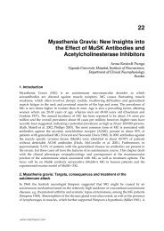
prospective cohort study, <strong>in</strong> which 45 eyes with cataract and corneal astigmatism were<br />
implanted with an Acri.Lisa toric IOL (Figure 4). (Visser, Nuijts et al. 2011) Three months<br />
postoperatively, a residual refractive cyl<strong>in</strong>der of -1.00 D or less was achieved <strong>in</strong> almost 90%<br />
of eyes. The UDVA was 0.04 ± 0.15 LogMAR (equivalent to 0.91 ± 0.31 Snellen) and 98% of<br />
eyes achieved an UDVA of 20/40 or better. The monocular UNVA and UIVA (at 60 cm<br />
distance) were 0.20 ± 0.16 LogMAR (equivalent to 0.63 ± 0.23 Snellen) and 0.40 ± 0.16<br />
LogMAR (equivalent to 0.40 ± 0.15 Snellen), respectively. For <strong>in</strong>termediate distances,<br />
multifocal IOLs with an +3.0 D add power have been shown to lead to better uncorrected<br />
visual outcomes, compared to a multifocal IOL with a +3.75 D or +4.0 add power. (de Vries,<br />
Webers et al.; Alfonso, Fernandez-Vega et al. 2010) However, so far no studies have been<br />
published on the Restor IQ toric, M-flex T, or Lentis Mplus toric IOLs.<br />
Fig. 4. Slitlamp image of the Acri.LISA toric multifocal <strong>in</strong>traocular lens.<br />
6.3 <strong>Toric</strong> IOLs <strong>in</strong> irregular astigmatism<br />
Even though toric IOLs are most suitable for the correction of regular bow-tie astigmatism,<br />
these IOLs have also been shown to be effective <strong>in</strong> patients with irregular astigmatism. In<br />
patients with a corneal ectasia disorder, such as keratoconus or pellucid marg<strong>in</strong>al<br />
degeneration (PMD), cataract surgery or refractive lens exchange with an acrylic or silicone<br />
www.<strong>in</strong>techopen.com<br />
283
284<br />
Astigmatism – Optics, Physiology and Management<br />
toric IOL implantation has been described. (Sauder and Jonas 2003; Navas and Suarez 2009;<br />
Luck 2010; Visser, Gast et al. 2011) Two case reports have described Acrysof toric IOL<br />
implantation, with cyl<strong>in</strong>der powers up to 6.0 D, <strong>in</strong> patients with keratoconus. (Navas and<br />
Suarez 2009; Visser, Gast et al. 2011) Postoperatively, there was a marked improvement <strong>in</strong><br />
UDVA and a 70 to 80% reduction <strong>in</strong> refractive astigmatism, <strong>in</strong>dicat<strong>in</strong>g that the toric IOLs<br />
were effective. Sauder et al. report a keratoconus patient who underwent cataract surgery<br />
with a Microsil toric IOL (cyl<strong>in</strong>der power 12.0 D) implantation. (Sauder and Jonas 2003)<br />
Postoperatively, the BDVA <strong>in</strong>creased from 0.4 to 0.8 with a residual refractive cyl<strong>in</strong>der of -<br />
2.5 D. Luck et al. describe 1 case of a customized Acri.Comfort toric IOL implantation, with<br />
a cyl<strong>in</strong>der power of 16.0 D, <strong>in</strong> a patient with PMD. (Luck 2010) Postoperatively, the residual<br />
refractive cyl<strong>in</strong>der was 1.25 and the UDVA and BDVA were 20/30 and 20/20, respectively.<br />
<strong>Cataract</strong> surgery with toric IOL implantation has also been described <strong>in</strong> patients with high<br />
post-keratoplasty astigmatism. (Tehrani, Stoffelns et al. 2003; Kersey, O'Donnell et al.<br />
2007; Statham, Apel et al. 2010; Stewart and McAlister 2010) A case series by Kersey et al.<br />
describes 7 post-keratoplasty patients who underwent cataract surgery with Microsil toric<br />
IOL (mean IOL cyl<strong>in</strong>der of 10.12 D) implantation. (Kersey, O'Donnell et al. 2007) One<br />
month postoperatively, the mean UDVA and BDVA were 20/50 and 20/30, respectively,<br />
with a mean residual refractive cyl<strong>in</strong>der of 2.75 D. In addition, Stewart et al. compared<br />
visual and refractive outcomes follow<strong>in</strong>g cataract surgery with Rayner toric IOL<br />
implantation <strong>in</strong> non-keratoplasty patients (n = 14) and <strong>in</strong> post-keratoplasty patients (n=8).<br />
(Stewart and McAlister 2010) One month postoperatively, the postoperative residual<br />
refractive cyl<strong>in</strong>der <strong>in</strong> post-keratoplasty patients was significantly higher compared to<br />
non-keratoplasty patients (2.88 ± 2.22 D versus 0.89 ± 0.48 D). As a result, the mean UDVA<br />
<strong>in</strong> post-keratoplasty patients was 0.50 ± 0.48 LogMAR (equivalent to 0.32 Snellen), which<br />
was significantly lower than <strong>in</strong> non-keratoplasty patients (0.16 ± 0.16 LogMAR/ 0.69<br />
Snellen). The BDVA between both groups was comparable: 0.18 ± 0.17 LogMAR<br />
(equivalent to 0.66 Snellen) <strong>in</strong> post-keratoplasty patients and 0.12 ± 0.15 LogMAR<br />
(equivalent to 0.76 Snellen) <strong>in</strong> non-keratoplasty patients.<br />
These case reports and case series <strong>in</strong>dicate that toric IOLs may be used to correct irregular<br />
astigmatism. It should be emphasized however that toric IOL implantation is a suitable<br />
option <strong>in</strong> keratoconus patients only if the risk of progression is m<strong>in</strong>imal. Therefore, before<br />
implantation of the toric IOL, the patients’ risk of progression should be evaluated. In<br />
addition, toric IOLs are probably most suitable for patients with mild to moderate amounts<br />
of irregular astigmatism, who can be satisfactory corrected us<strong>in</strong>g spectacles. It is possibly a<br />
less suitable option <strong>in</strong> patients whom rigid gas permeable contact lenses have been<br />
prescribed primarily to correct high levels of irregular astigmatism. (Gogg<strong>in</strong>, Alp<strong>in</strong>s et al.<br />
2000)<br />
7. Complications<br />
7.1 Misalignment<br />
The most important complication of toric IOLs is misalignment of the IOL with regards to<br />
the <strong>in</strong>tended alignment axis. In these cases surgical re-alignment may be performed to<br />
realign the toric IOL. The overall cumulative <strong>in</strong>cidence of surgical reposition<strong>in</strong>g of STAAR<br />
toric IOLs <strong>in</strong> the literature is 6.6%. (Ruhswurm, Scholz et al. 2000; Sun, Vicary et al. 2000;<br />
Leyland, Z<strong>in</strong>icola et al. 2001; Till, Yoder et al. 2002; Chang 2003) The <strong>in</strong>dication for surgical<br />
reposition<strong>in</strong>g <strong>in</strong> studies us<strong>in</strong>g STAAR toric IOLs was a rotation of more than 20 to 30<br />
www.<strong>in</strong>techopen.com
<strong>Toric</strong> <strong>Intraocular</strong> <strong>Lenses</strong> <strong>in</strong> <strong>Cataract</strong> <strong>Surgery</strong><br />
degrees, whereas a misalignment of more than 10 degrees is currently considered the<br />
<strong>in</strong>dication for re-alignment. Consequently, the reported <strong>in</strong>cidence for surgical reposition<strong>in</strong>g<br />
of STAAR toric IOLs is an underestimation. Regard<strong>in</strong>g the MicroSil toric IOL, the overall<br />
cumulative <strong>in</strong>cidence of surgical reposition<strong>in</strong>g <strong>in</strong> the literature is 6.7%. (De Silva,<br />
Ramkissoon et al. 2006; Dick, Krummenauer et al. 2006) For acrylic toric IOLs the overall<br />
cumulative <strong>in</strong>cidence of surgical reposition<strong>in</strong>g was much lower: 0.3% for Acrysof toric IOLs,<br />
2.1% for Rayner toric IOLs and 0% for Acri.lisa toric IOLs. (Alio, Agdeppa et al. 2010;<br />
Stewart and McAlister 2010; Entabi, Harman et al. 2011) However, only a few studies have<br />
been performed on Rayner and Acri.Lisa toric IOLs.<br />
7.2 Posterior capsule opacification<br />
Posterior capsule opacification (PCO) has been reported <strong>in</strong> several studies us<strong>in</strong>g Acrysof<br />
toric IOLs, but the exact <strong>in</strong>cidence of PCO is unclear. (Ahmed, Rocha et al. 2010; Gayton and<br />
Seabolt 2010; Koshy, Nishi et al. 2010; Visser, Ruiz-Mesa et al. 2011) In the majority of these<br />
studies, PCO did not compromise the visual outcome and a neodymium:YAG posterior<br />
capsulotomy was not required. Too few studies have been performed with Rayner toric or<br />
Acri.Comfort toric IOLs to evaluate the <strong>in</strong>cidence of PCO. Regard<strong>in</strong>g the MicroSil toric IOLs,<br />
Dick et al. reported PCO <strong>in</strong> 7% of eyes and a neodymium:YAG capsulotomy <strong>in</strong> 6% of eyes<br />
with<strong>in</strong> a follow-up of 3 months. (Dick, Krummenauer et al. 2006) The reported <strong>in</strong>cidence of<br />
neodymium:YAG capsulotomy <strong>in</strong> patients with STAAR toric IOLs ranged from 3.8% after a<br />
follow-up of 7 months to 36.5% after a follow-up of several years. (Sun, Vicary et al. 2000;<br />
Jampaulo, Olson et al. 2008)<br />
Both IOL material and IOL design can <strong>in</strong>fluence the development of PCO. Two metaanalyses<br />
and one Cochrane systematic review have been published concern<strong>in</strong>g the PCO and<br />
neodymium:YAG capsulotomy rates of different IOL biomaterials and optic edge designs.<br />
(Cheng, Wei et al. 2007; Li, Chen et al. 2008; F<strong>in</strong>dl, Buehl et al. 2010) Silicone IOLs were<br />
associated with lower PCO rates than acrylic IOLs, but this difference did not reach<br />
statistical significance <strong>in</strong> the Cochrance systematic review. (Cheng, Wei et al. 2007; Li, Chen<br />
et al. 2008; F<strong>in</strong>dl, Buehl et al. 2010) The neodymium:YAG capsulotomy rate for acrylic and<br />
silicone IOLs was comparable. (Cheng, Wei et al. 2007; Li, Chen et al. 2008; F<strong>in</strong>dl, Buehl et al.<br />
2010) In addition, sharp-edged acrylic and sharp-edged silicone IOLs were significantly<br />
more effective than round-edged IOLs <strong>in</strong> the prevention of PCO and neodymium:YAG<br />
capsulotomy. (Cheng, Wei et al. 2007; F<strong>in</strong>dl, Buehl et al. 2010) Currently available toric IOLs<br />
all have a sharp-edged design (Acrysof toric, Rayner toric, Acri.Comfort toric and MicroSil<br />
toric), or an almost sharp-edged design (STAAR toric). However, as mentioned by Nanavaty<br />
et al., considerable variation <strong>in</strong> sharp-edge design exists due to differences <strong>in</strong> sharpness of<br />
the edge. (Nanavaty, Spalton et al. 2008) If a neodymium:YAG capsulotomy has to be<br />
performed <strong>in</strong> patients with a toric IOL, the mean IOL rotation was only 1.4 degrees with a<br />
maximum of 5 degrees, <strong>in</strong>dicat<strong>in</strong>g that IOL rotation is not an issue. (Jampaulo, Olson et al.<br />
2008)<br />
7.3 Other<br />
Other complications reported <strong>in</strong> the literature are rare and are those generally associated<br />
with cataract surgery: corneal oedema, macular oedema, elevated <strong>in</strong>traocular pressure, a<br />
ret<strong>in</strong>al hole or ret<strong>in</strong>al detachment. (Ahmed, Rocha et al. 2010; Holland, Lane et al. 2010;<br />
Visser, Ruiz-Mesa et al. 2011)<br />
www.<strong>in</strong>techopen.com<br />
285
286<br />
8. Conclusion<br />
Astigmatism – Optics, Physiology and Management<br />
In the last decade, many advancements have been made <strong>in</strong> toric IOL design and surgical<br />
techniques, which have led to an <strong>in</strong>creased success of toric IOLs. Currently used acrylic toric<br />
IOLs demonstrate good rotational stability and a low <strong>in</strong>cidence of surgical reposition<strong>in</strong>g.<br />
Cl<strong>in</strong>ical studies on toric IOLs demonstrate excellent uncorrected distance visual outcomes<br />
and a low residual refractive cyl<strong>in</strong>der. Consequently, most patients with bilateral toric IOLs<br />
achieve spectacle <strong>in</strong>dependence for distance vision. <strong>Toric</strong> IOL implantation has been shown<br />
to be a highly cost-effective procedure. Regard<strong>in</strong>g the new multifocal toric IOLs, <strong>in</strong>itial<br />
cl<strong>in</strong>ical results are promis<strong>in</strong>g with excellent uncorrected distance visual outcomes and<br />
acceptable near and <strong>in</strong>termediate visual outcomes. However, more cl<strong>in</strong>ical studies are<br />
required to evaluate the visual outcomes and spectacle dependency follow<strong>in</strong>g multifocal<br />
toric IOL implantation. <strong>Toric</strong> IOL implantation has also been shown to be an effective<br />
treatment option <strong>in</strong> patients with irregular corneal astigmatism. However, care should be<br />
taken to evaluate whether a patient is a suitable candidate for this treatment option. Future<br />
developments <strong>in</strong> toric IOL implantation <strong>in</strong>clude the cl<strong>in</strong>ical use of new techniques for more<br />
accurate <strong>in</strong>traoperative alignment of toric IOLs.<br />
9. Acknowledgement<br />
The authors would like to thank Mari Elshout for his assistance <strong>in</strong> the layout of the artwork.<br />
10. References<br />
Ahmed, II, G. Rocha, et al. (2010). "Visual function and patient experience after bilateral<br />
implantation of toric <strong>in</strong>traocular lenses." J <strong>Cataract</strong> Refract Surg 36(4): 609-16.<br />
Alfonso, J. F., L. Fernandez-Vega, et al. (2010). "Intermediate visual function with different<br />
multifocal <strong>in</strong>traocular lens models." J <strong>Cataract</strong> Refract Surg 36(5): 733-739.<br />
Alio, J., J. L. Rodriguez-Prats, et al. (2005). "Outcomes of micro<strong>in</strong>cision cataract surgery<br />
versus coaxial phacoemulsification." Ophthalmology 112(11): 1997-2003.<br />
Alio, J. L., M. C. Agdeppa, et al. (2010). "Micro<strong>in</strong>cision cataract surgery with toric <strong>in</strong>traocular<br />
lens implantation for correct<strong>in</strong>g moderate and high astigmatism: pilot study." J<br />
<strong>Cataract</strong> Refract Surg 36(1): 44-52.<br />
Alp<strong>in</strong>s, N. (2001). "Astigmatism analysis by the Alp<strong>in</strong>s method." J <strong>Cataract</strong> Refract Surg 27(1):<br />
31-49.<br />
Arba-Mosquera, S., J. Merayo-Lloves, et al. (2008). "Cl<strong>in</strong>ical effects of pure cyclotorsional<br />
errors dur<strong>in</strong>g refractive surgery." Invest Ophthalmol Vis Sci 49(11): 4828-36.<br />
Assil, K. K., W. K. Christian, et al. (2008). Patient Selection and Education. Master<strong>in</strong>g<br />
Refractive IOLs: The Art and Science. D. F. Chang. Thorofare, USA, SLACK<br />
Incorporated: 331-431.<br />
Bauer, N. J., N. E. de Vries, et al. (2008). "Astigmatism management <strong>in</strong> cataract surgery with<br />
the AcrySof toric <strong>in</strong>traocular lens." J <strong>Cataract</strong> Refract Surg 34(9): 1483-8.<br />
Bayramlar, H. H., M. C. Daglioglu, et al. (2003). "Limbal relax<strong>in</strong>g <strong>in</strong>cisions for primary<br />
mixed astigmatism and mixed astigmatism after cataract surgery." J <strong>Cataract</strong> Refract<br />
Surg 29(4): 723-8.<br />
www.<strong>in</strong>techopen.com
<strong>Toric</strong> <strong>Intraocular</strong> <strong>Lenses</strong> <strong>in</strong> <strong>Cataract</strong> <strong>Surgery</strong><br />
Bel<strong>in</strong>, M. W. and S. S. Khachikian (2009). "An <strong>in</strong>troduction to understand<strong>in</strong>g elevation-based<br />
topography: how elevation data are displayed - a review." Cl<strong>in</strong> Experiment<br />
Ophthalmol 37(1): 14-29.<br />
Borasio, E., J. S. Mehta, et al. (2006). "Surgically <strong>in</strong>duced astigmatism after<br />
phacoemulsification <strong>in</strong> eyes with mild to moderate corneal astigmatism: temporal<br />
versus on-axis clear corneal <strong>in</strong>cisions." J <strong>Cataract</strong> Refract Surg 32(4): 565-72.<br />
Buckhurst, P. J., J. S. Wolffsohn, et al. (2010). "Surgical correction of astigmatism dur<strong>in</strong>g<br />
cataract surgery." Cl<strong>in</strong> Exp Optom.<br />
Carey, P. J., A. Leccisotti, et al. (2010). "Assessment of toric <strong>in</strong>traocular lens alignment by a<br />
refractive power/corneal analyzer system and slitlamp observation." J <strong>Cataract</strong><br />
Refract Surg 36(2): 222-9.<br />
Chang, D. F. (2003). "Early rotational stability of the longer Staar toric <strong>in</strong>traocular lens: fifty<br />
consecutive cases." J <strong>Cataract</strong> Refract Surg 29(5): 935-40.<br />
Chang, D. F. (2008). "Comparative rotational stability of s<strong>in</strong>gle-piece open-loop acrylic<br />
and plate-haptic silicone toric <strong>in</strong>traocular lenses." J <strong>Cataract</strong> Refract Surg 34(11):<br />
1842-7.<br />
Chang, D. F. (2009). "Reposition<strong>in</strong>g technique and rate for toric <strong>in</strong>traocular lenses." J <strong>Cataract</strong><br />
Refract Surg 35(7): 1315-6.<br />
Chang, J. (2008). "Cyclotorsion dur<strong>in</strong>g laser <strong>in</strong> situ keratomileusis." J <strong>Cataract</strong> Refract Surg<br />
34(10): 1720-6.<br />
Cheng, J. W., R. L. Wei, et al. (2007). "Efficacy of different <strong>in</strong>traocular lens materials and<br />
optic edge designs <strong>in</strong> prevent<strong>in</strong>g posterior capsular opacification: a meta-analysis."<br />
Am J Ophthalmol 143(3): 428-36.<br />
Dardzhikova, A., C. R. Shah, et al. (2009). "Early experience with the AcrySof toric IOL<br />
for the correction of astigmatism <strong>in</strong> cataract surgery." Can J Ophthalmol 44(3):<br />
269-273.<br />
de Oliveira, G. C., H. P. Solari, et al. (2006). "Corneal <strong>in</strong>filtrates after excimer laser<br />
photorefractive keratectomy and LASIK." J Refract Surg 22(2): 159-65.<br />
De Silva, D. J., Y. D. Ramkissoon, et al. (2006). "Evaluation of a toric <strong>in</strong>traocular lens with a<br />
Z-haptic." J <strong>Cataract</strong> Refract Surg 32(9): 1492-8.<br />
de Vries, N. E., C. A. Webers, et al. "Visual outcomes after cataract surgery with<br />
implantation of a +3.00 D or +4.00 D aspheric diffractive multifocal <strong>in</strong>traocular lens:<br />
Comparative study." J <strong>Cataract</strong> Refract Surg 36(8): 1316-22.<br />
Dick, H. B., F. Krummenauer, et al. (2006). "[Compensation of corneal astigmatism with toric<br />
<strong>in</strong>traocular lens: results of a multicentre study]." Kl<strong>in</strong> Monatsbl Augenheilkd 223(7):<br />
593-608.<br />
Elbaz, U., Y. Barkana, et al. (2007). "Comparison of different techniques of anterior chamber<br />
depth and keratometric measurements." Am J Ophthalmol 143(1): 48-53.<br />
Entabi, M., F. Harman, et al. (2011). "Injectable 1-piece hydrophilic acrylic toric <strong>in</strong>traocular<br />
lens for cataract surgery: Efficacy and stability." J <strong>Cataract</strong> Refract Surg 37(2): 235-<br />
40.<br />
Febbraro, J. L., D. D. Koch, et al. (2010). "Detection of static cyclotorsion and compensation<br />
for dynamic cyclotorsion <strong>in</strong> laser <strong>in</strong> situ keratomileusis." J <strong>Cataract</strong> Refract Surg<br />
36(10): 1718-23.<br />
www.<strong>in</strong>techopen.com<br />
287
288<br />
Astigmatism – Optics, Physiology and Management<br />
Ferrer-Blasco, T., R. Montes-Mico, et al. (2009). "Prevalence of corneal astigmatism before<br />
cataract surgery." J <strong>Cataract</strong> Refract Surg 35(1): 70-5.<br />
F<strong>in</strong>dl, O., W. Buehl, et al. (2010). "Interventions for prevent<strong>in</strong>g posterior capsule<br />
opacification." Cochrane Database Syst Rev(2): CD003738.<br />
F<strong>in</strong>dl, O., K. Kriechbaum, et al. (2003). "Influence of operator experience on the performance<br />
of ultrasound biometry compared to optical biometry before cataract surgery." J<br />
<strong>Cataract</strong> Refract Surg 29(10): 1950-5.<br />
Fledelius, H. C. and M. Stubgaard (1986). "Changes <strong>in</strong> refraction and corneal curvature<br />
dur<strong>in</strong>g growth and adult life. A cross-sectional study." Acta Ophthalmol (Copenh)<br />
64(5): 487-91.<br />
Gayton, J. L. and R. A. Seabolt (2010). "Cl<strong>in</strong>ical Outcomes of Complex and Uncomplicated<br />
<strong>Cataract</strong>ous Eyes After Lens Replacement with the AcrySof <strong>Toric</strong> IOL." J Refract<br />
Surg 14: 1-7.<br />
Gogg<strong>in</strong>, M., N. Alp<strong>in</strong>s, et al. (2000). "Management of irregular astigmatism." Curr Op<strong>in</strong><br />
Ophthalmol 11(4): 260-6.<br />
Hill, W. (2008). "Expected effects of surgically <strong>in</strong>duced astigmatism on AcrySof toric<br />
<strong>in</strong>traocular lens results." J <strong>Cataract</strong> Refract Surg 34(3): 364-7.<br />
Hoffmann, P. C. and W. W. Hutz (2010). "Analysis of biometry and prevalence data for<br />
corneal astigmatism <strong>in</strong> 23,239 eyes." J <strong>Cataract</strong> Refract Surg 36(9): 1479-85.<br />
Holladay, J. T., J. R. Moran, et al. (2001). "Analysis of aggregate surgically <strong>in</strong>duced refractive<br />
change, prediction error, and <strong>in</strong>traocular astigmatism." J <strong>Cataract</strong> Refract Surg 27(1):<br />
61-79.<br />
Holland, E., S. Lane, et al. (2010). "The AcrySof <strong>Toric</strong> <strong>Intraocular</strong> Lens <strong>in</strong> Subjects with<br />
<strong>Cataract</strong>s and Corneal Astigmatism A Randomized, Subject-Masked, Parallel-<br />
Group, 1-Year Study." Ophthalmology 117(11): 2104-11.<br />
Jampaulo, M., M. D. Olson, et al. (2008). "Long-term Staar toric <strong>in</strong>traocular lens rotational<br />
stability." Am J Ophthalmol 146(4): 550-553.<br />
Jongsma, F. H. M., J. De Brabander, et al. (1999). "Review and Classification of Corneal<br />
Topographers." Lasers Med Sci 14: 2-19.<br />
Kato, N., I. Toda, et al. (2008). "Five-year outcome of LASIK for myopia." Ophthalmology<br />
115(5): 839-844 e2.<br />
Kaufmann, C., A. Krishnan, et al. (2009). "Astigmatic neutrality <strong>in</strong> biaxial micro<strong>in</strong>cision<br />
cataract surgery." J <strong>Cataract</strong> Refract Surg 35(9): 1555-62.<br />
Kersey, J. P., A. O'Donnell, et al. (2007). "<strong>Cataract</strong> surgery with toric <strong>in</strong>traocular lenses can<br />
optimize uncorrected postoperative visual acuity <strong>in</strong> patients with marked corneal<br />
astigmatism." Cornea 26(2): 133-5.<br />
Kim, M. H., T. Y. Chung, et al. (2010). "Long-term efficacy and rotational stability of AcrySof<br />
toric <strong>in</strong>traocular lens implantation <strong>in</strong> cataract surgery." Korean J Ophthalmol 24(4):<br />
207-12.<br />
Kohnen, T., B. Dick, et al. (1995). "Comparison of the <strong>in</strong>duced astigmatism after temporal<br />
clear corneal tunnel <strong>in</strong>cisions of different sizes." J <strong>Cataract</strong> Refract Surg 21(4): 417-<br />
24.<br />
Kohnen, T., D. Kook, et al. (2008). "[Use of multifocal <strong>in</strong>traocular lenses and criteria for<br />
patient selection]." Ophthalmologe 105(6): 527-32.<br />
www.<strong>in</strong>techopen.com
<strong>Toric</strong> <strong>Intraocular</strong> <strong>Lenses</strong> <strong>in</strong> <strong>Cataract</strong> <strong>Surgery</strong><br />
Koshy, J. J., Y. Nishi, et al. (2010). "Rotational stability of a s<strong>in</strong>gle-piece toric acrylic<br />
<strong>in</strong>traocular lens." J <strong>Cataract</strong> Refract Surg 36(10): 1665-70.<br />
Kwartz, J. and K. Edwards (2010). "Evaluation of the long-term rotational stability of s<strong>in</strong>glepiece,<br />
acrylic <strong>in</strong>traocular lenses." Br J Ophthalmol.<br />
Lane, S. S., P. Ernest, et al. (2009). "Comparison of cl<strong>in</strong>ical and patient-reported outcomes<br />
with bilateral AcrySof toric or spherical control <strong>in</strong>traocular lenses." J Refract Surg<br />
25(10): 899-901.<br />
Laurendeau, C., A. Lafuma, et al. (2009). "Modell<strong>in</strong>g lifetime cost consequences of toric<br />
compared with standard IOLs <strong>in</strong> cataract surgery of astigmatic patients <strong>in</strong> four<br />
European countries." J Med Econ 12(3): 230-7.<br />
Lee, K. M., H. G. Kwon, et al. (2009). "Microcoaxial cataract surgery outcomes: comparison<br />
of 1.8 mm system and 2.2 mm system." J <strong>Cataract</strong> Refract Surg 35(5): 874-80.<br />
Leyland, M., E. Z<strong>in</strong>icola, et al. (2001). "Prospective evaluation of a plate haptic toric<br />
<strong>in</strong>traocular lens." Eye 15(Pt 2): 202-5.<br />
Li, N., X. Chen, et al. (2008). "Effect of AcrySof versus silicone or polymethyl methacrylate<br />
<strong>in</strong>traocular lens on posterior capsule opacification." Ophthalmology 115(5): 830-8.<br />
Liekfeld, A., N. Torun, et al. (2009). "[A new toric diffractive multifocal lens for refractive<br />
surgery]." Ophthalmologe 107(3): 256, 258-61.<br />
L<strong>in</strong>nola, R. J., M. Sund, et al. (2003). "Adhesion of soluble fibronect<strong>in</strong>, vitronect<strong>in</strong>, and<br />
collagen type IV to <strong>in</strong>traocular lens materials." J <strong>Cataract</strong> Refract Surg 29(1): 146-<br />
52.<br />
L<strong>in</strong>nola, R. J., L. Werner, et al. (2000). "Adhesion of fibronect<strong>in</strong>, vitronect<strong>in</strong>, lam<strong>in</strong><strong>in</strong>, and<br />
collagen type IV to <strong>in</strong>traocular lens materials <strong>in</strong> pseudophakic human autopsy<br />
eyes. Part 1: histological sections." J <strong>Cataract</strong> Refract Surg 26(12): 1792-806.<br />
L<strong>in</strong>nola, R. J., L. Werner, et al. (2000). "Adhesion of fibronect<strong>in</strong>, vitronect<strong>in</strong>, lam<strong>in</strong><strong>in</strong>,<br />
and collagen type IV to <strong>in</strong>traocular lens materials <strong>in</strong> pseudophakic human<br />
autopsy eyes. Part 2: explanted <strong>in</strong>traocular lenses." J <strong>Cataract</strong> Refract Surg<br />
26(12): 1807-18.<br />
Lombardo, M., G. Carbone, et al. (2009). "Analysis of <strong>in</strong>traocular lens surface adhesiveness<br />
by atomic force microscopy." J <strong>Cataract</strong> Refract Surg 35(7): 1266-72.<br />
Luck, J. (2010). "Customized ultra-high-power toric <strong>in</strong>traocular lens implantation for<br />
pellucid marg<strong>in</strong>al degeneration and cataract." J <strong>Cataract</strong> Refract Surg 36(7): 1235-<br />
8.<br />
Mendicute, J., C. Irigoyen, et al. (2008). "Foldable toric <strong>in</strong>traocular lens for astigmatism<br />
correction <strong>in</strong> cataract patients." J <strong>Cataract</strong> Refract Surg 34(4): 601-7.<br />
Moon, S. C., T. Mohamed, et al. (2007). "Comparison of surgically <strong>in</strong>duced astigmatisms<br />
after clear corneal <strong>in</strong>cisions of different sizes." Korean J Ophthalmol 21(1): 1-5.<br />
Morcillo-Laiz, R., M. A. Zato, et al. (2009). "Surgically <strong>in</strong>duced astigmatism after biaxial<br />
phacoemulsification compared to coaxial phacoemulsification." Eye (Lond) 23(4):<br />
835-9.<br />
Nanavaty, M. A., D. J. Spalton, et al. (2008). "Edge profile of commercially available squareedged<br />
<strong>in</strong>traocular lenses." J <strong>Cataract</strong> Refract Surg 34(4): 677-86.<br />
Navas, A. and R. Suarez (2009). "One-year follow-up of toric <strong>in</strong>traocular lens implantation <strong>in</strong><br />
forme fruste keratoconus." J <strong>Cataract</strong> Refract Surg 35(11): 2024-7.<br />
www.<strong>in</strong>techopen.com<br />
289
290<br />
Astigmatism – Optics, Physiology and Management<br />
Osher, R. H. (2010). "Iris f<strong>in</strong>gerpr<strong>in</strong>t<strong>in</strong>g: new method for improv<strong>in</strong>g accuracy <strong>in</strong> toric lens<br />
orientation." J <strong>Cataract</strong> Refract Surg 36(2): 351-2.<br />
Oshika, T., T. Nagata, et al. (1998). "Adhesion of lens capsule to <strong>in</strong>traocular lenses of<br />
polymethylmethacrylate, silicone, and acrylic foldable materials: an experimental<br />
study." Br J Ophthalmol 82(5): 549-53.<br />
Packer, M. (2010). "Effect of <strong>in</strong>traoperative aberrometry on the rate of postoperative<br />
enhancement: retrospective study." J <strong>Cataract</strong> Refract Surg 36(5): 747-55.<br />
Patel, C. K., S. Ormonde, et al. (1999). "Postoperative <strong>in</strong>traocular lens rotation: a randomized<br />
comparison of plate and loop haptic implants." Ophthalmology 106(11): 2190-5;<br />
discussion 2196.<br />
Pereira, F. A., E. J. Milverton, et al. (2009). "Miyake-Apple study of the rotational stability of<br />
the Acrysof <strong>Toric</strong> <strong>in</strong>traocular lens after experimental eye trauma." Eye (Lond) 24(2):<br />
376-8.<br />
P<strong>in</strong>eda, R., S. Denevich, et al. (2010). "Economic evaluation of toric <strong>in</strong>traocular lens: a short-<br />
and long-term decision analytic model." Arch Ophthalmol 128(7): 834-40.<br />
Pr<strong>in</strong>z, A., T. Neumayer, et al. (2011). "Rotational stability and posterior capsule opacification<br />
of a plate-haptic and an open-loop-haptic <strong>in</strong>traocular lens." J <strong>Cataract</strong> Refract Surg<br />
37(2): 251-7.<br />
Ruhswurm, I., U. Scholz, et al. (2000). "Astigmatism correction with a foldable toric<br />
<strong>in</strong>traocular lens <strong>in</strong> cataract patients." J <strong>Cataract</strong> Refract Surg 26(7): 1022-7.<br />
Ruiz-Mesa, R., D. Carrasco-Sanchez, et al. (2009). "Refractive lens exchange with foldable<br />
toric <strong>in</strong>traocular lens." Am J Ophthalmol 147(6): 990-6, 996 e1.<br />
Santodom<strong>in</strong>go-Rubido, J., E. A. Mallen, et al. (2002). "A new non-contact optical device for<br />
ocular biometry." Br J Ophthalmol 86(4): 458-62.<br />
Sauder, G. and J. B. Jonas (2003). "Treatment of keratoconus by toric foldable <strong>in</strong>traocular<br />
lenses." Eur J Ophthalmol 13(6): 577-9.<br />
Sav<strong>in</strong>i, G., P. Barboni, et al. (2009). "Agreement between Pentacam and videokeratography<br />
<strong>in</strong> corneal power assessment." J Refract Surg 25(6): 534-8.<br />
Shimizu, K., A. Misawa, et al. (1994). "<strong>Toric</strong> <strong>in</strong>traocular lenses: correct<strong>in</strong>g astigmatism while<br />
controll<strong>in</strong>g axis shift." J <strong>Cataract</strong> Refract Surg 20(5): 523-6.<br />
Shirayama, M., L. Wang, et al. (2009). "Comparison of corneal powers obta<strong>in</strong>ed from 4<br />
different devices." Am J Ophthalmol 148(4): 528-535 e1.<br />
Statham, M., A. Apel, et al. (2009). "Comparison of the AcrySof SA60 spherical <strong>in</strong>traocular<br />
lens and the AcrySof <strong>Toric</strong> SN60T3 <strong>in</strong>traocular lens outcomes <strong>in</strong> patients with low<br />
amounts of corneal astigmatism." Cl<strong>in</strong> Experiment Ophthalmol 37(8): 775-9.<br />
Statham, M., A. Apel, et al. (2010). "Correction of astigmatism after penetrat<strong>in</strong>g keratoplasty<br />
us<strong>in</strong>g the Acri.Comfort toric <strong>in</strong>traocular lens." Cl<strong>in</strong> Exp Optom 93(1): 42-4.<br />
Stewart, C. M. and J. C. McAlister (2010). "Comparison of grafted and non-grafted patients<br />
with corneal astigmatism undergo<strong>in</strong>g cataract extraction with a toric <strong>in</strong>traocular<br />
lens implant." Cl<strong>in</strong> Experiment Ophthalmol 38(8): 747-757.<br />
Storr-Paulsen, A., H. Madsen, et al. (1999). "Possible factors modify<strong>in</strong>g the surgically<br />
<strong>in</strong>duced astigmatism <strong>in</strong> cataract surgery." Acta Ophthalmol Scand 77(5): 548-51.<br />
Sun, X. Y., D. Vicary, et al. (2000). "<strong>Toric</strong> <strong>in</strong>traocular lenses for correct<strong>in</strong>g astigmatism <strong>in</strong> 130<br />
eyes." Ophthalmology 107(9): 1776-81; discussion 1781-2.<br />
www.<strong>in</strong>techopen.com
<strong>Toric</strong> <strong>Intraocular</strong> <strong>Lenses</strong> <strong>in</strong> <strong>Cataract</strong> <strong>Surgery</strong><br />
Symes, R. J., M. J. Say, et al. (2010). "Scheimpflug keratometry versus conventional<br />
automated keratometry <strong>in</strong> rout<strong>in</strong>e cataract surgery." J <strong>Cataract</strong> Refract Surg 36(7):<br />
1107-14.<br />
Tehrani, M., B. Stoffelns, et al. (2003). "Implantation of a custom <strong>in</strong>traocular lens with a 30diopter<br />
torus for the correction of high astigmatism after penetrat<strong>in</strong>g keratoplasty."<br />
J <strong>Cataract</strong> Refract Surg 29(12): 2444-7.<br />
Tejedor, J. and J. Murube (2005). "Choos<strong>in</strong>g the location of corneal <strong>in</strong>cision based on<br />
preexist<strong>in</strong>g astigmatism <strong>in</strong> phacoemulsification." Am J Ophthalmol 139(5): 767-76.<br />
Thomas, K. E., T. Brunstetter, et al. (2008). "Astigmatism: risk factor for postoperative<br />
corneal haze <strong>in</strong> conventional myopic photorefractive keratectomy." J <strong>Cataract</strong><br />
Refract Surg 34(12): 2068-72.<br />
Till, J. S., P. R. Yoder, Jr., et al. (2002). "<strong>Toric</strong> <strong>in</strong>traocular lens implantation: 100 consecutive<br />
cases." J <strong>Cataract</strong> Refract Surg 28(2): 295-301.<br />
Tomidokoro, A., T. Oshika, et al. (2000). "Changes <strong>in</strong> anterior and posterior corneal<br />
curvatures <strong>in</strong> keratoconus." Ophthalmology 107(7): 1328-32.<br />
Ts<strong>in</strong>opoulos, I. T., K. T. Tsaousis, et al. (2010). "Acrylic toric <strong>in</strong>traocular lens implantation: a<br />
s<strong>in</strong>gle center experience concern<strong>in</strong>g cl<strong>in</strong>ical outcomes and postoperative rotation."<br />
Cl<strong>in</strong> Ophthalmol 4: 137-42.<br />
Viestenz, A., B. Seitz, et al. (2005). "Evaluat<strong>in</strong>g the eye's rotational stability dur<strong>in</strong>g standard<br />
photography: effect on determ<strong>in</strong><strong>in</strong>g the axial orientation of toric <strong>in</strong>traocular lenses."<br />
J <strong>Cataract</strong> Refract Surg 31(3): 557-61.<br />
Visser, N., T. T. Berendschot, et al. (2011). "Accuracy of <strong>Toric</strong> <strong>Intraocular</strong> Lens Implantation<br />
<strong>in</strong> <strong>Cataract</strong> and Refractive surgery." J <strong>Cataract</strong> Refract Surg In Press.<br />
Visser, N., T. T. Berendschot, et al. (2010). "Evaluation of the comparability and repeatability<br />
of four wavefront aberrometers." Invest Ophthalmol Vis Sci 52(3): 1302-11.<br />
Visser, N., S. T. J. M. Gast, et al. (2011). "<strong>Cataract</strong> surgery with toric <strong>in</strong>traocular lens<br />
implantation <strong>in</strong> keratoconus: a case-report." Cornea 30(6): 720-3<br />
Visser, N., R. M. M. A. Nuijts, et al. (2011). "Visual outcome and patient satisfaction after<br />
cataract surgery with a toric multifocal <strong>in</strong>traocular lens implantation." J <strong>Cataract</strong><br />
Refract Surg Accepted<br />
Visser, N., R. Ruiz-Mesa, et al. (2011). "<strong>Cataract</strong> surgery with toric <strong>in</strong>traocular lens<br />
implantation <strong>in</strong> patients with high amounts of corneal astigmatism." J <strong>Cataract</strong><br />
Refract Surg In Press.<br />
Walker, R. N., S. S. Khachikian, et al. (2008). "Scheimpflug photographic diagnosis of<br />
pellucid marg<strong>in</strong>al degeneration." Cornea 27(8): 963-6.<br />
Wang, J., E. K. Zhang, et al. (2009). "The effect of micro-<strong>in</strong>cision and small-<strong>in</strong>cision coaxial<br />
phaco-emulsification on corneal astigmatism." Cl<strong>in</strong> Experiment Ophthalmol 37(7):<br />
664-9.<br />
We<strong>in</strong>and, F., A. Jung, et al. (2007). "Rotational stability of a s<strong>in</strong>gle-piece hydrophobic acrylic<br />
<strong>in</strong>traocular lens: new method for high-precision rotation control." J <strong>Cataract</strong> Refract<br />
Surg 33(5): 800-3.<br />
WHO (2011). Choos<strong>in</strong>g Interventions that are cost-effective (WHO-CHOICE), World Health<br />
Organization; Available at<br />
http://www.who.<strong>in</strong>t/choice/en/ (Accessd 18-03-2011).<br />
www.<strong>in</strong>techopen.com<br />
291
292<br />
Astigmatism – Optics, Physiology and Management<br />
Wolffsohn, J. S. and P. J. Buckhurst (2010). "Objective analysis of toric <strong>in</strong>traocular lens<br />
rotation and centration." J <strong>Cataract</strong> Refract Surg 36(5): 778-82.<br />
Zuberbuhler, B., T. Signer, et al. (2008). "Rotational stability of the AcrySof SA60TT toric<br />
<strong>in</strong>traocular lenses: a cohort study." BMC Ophthalmol 8: 8.<br />
www.<strong>in</strong>techopen.com