JCDA - Canadian Dental Association
JCDA - Canadian Dental Association
JCDA - Canadian Dental Association

Create successful ePaper yourself
Turn your PDF publications into a flip-book with our unique Google optimized e-Paper software.
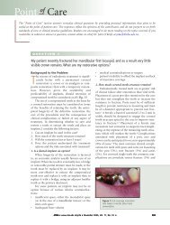
Figure 4: The access hole for the lingual<br />
screw has been sealed with red boxing wax<br />
to protect the abutment screw from the<br />
acrylic to be used.<br />
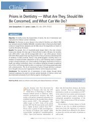
Figure 7: Acrylic monomer and polymer are<br />
applied with a brush technique.<br />
–––– Clinical Showcase ––––<br />
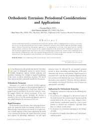
Figure 5: The lingual surface of the acrylic<br />
denture tooth is hollowed to fit around a<br />
temporary cylinder; the labial surface of the<br />
veneer remains intact.<br />
surface in reasonably good esthetic orientation<br />
(Fig. 6).<br />
5) Position the labial veneer of the shaped acrylic<br />
denture tooth around the seated temporary cylinder<br />
and then fasten it to the temporary cylinder with<br />
autopolymerizing acrylic using a brush technique<br />
(Figs. 7 and 8).<br />
6) Remove the temporary cylinder with the attached<br />
labial veneer surface from the fixture<br />
platform and immediately replace it with a conventional<br />
healing abutment (Fig. 9).<br />
7) Complete the fabrication of the desired<br />
final tooth form by adding and shaping the<br />
autopolymerizing acrylic. The acrylic provisional<br />
restoration will need to be tried in<br />
and adjusted a few times to achieve the<br />
desired shape; polishing will then be required<br />
(Fig. 10).<br />
By varying the subgingival contour of the<br />
provisional restoration, the peri-implant gingival<br />
610 <strong>JCDA</strong> • www.cda-adc.ca/jcda • September 2008, Vol. 74, No. 7 •<br />
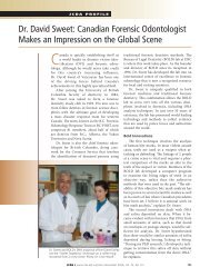
Figure 6: The labial veneer of the<br />
acrylic denture tooth is fitted around<br />
a temporary cylinder in preparation<br />
for fastening to the cylinder with<br />
autopolymerizing acrylic.<br />
Figure 8: The labial veneer of the acrylic<br />
denture tooth is attached to the temporary<br />
cylinder with autopolymerizing acrylic,<br />
which is then allowed to set.<br />
tissue may be moulded and manipulated. On the<br />
labial surface of the provisional restoration, the<br />
more convex the subgingival contour, the further<br />
the gingival tissue may be moved in the apical<br />
direction. Conversely, the less convex or flatter<br />
the subgingival surface, the more the tissue may<br />
be moved in the coronal direction. The papilla<br />
may be moved incisally by making the subgingival<br />
interproximal acrylic contour more convex, which<br />
pushes the tissue toward the proximal surface of<br />
the adjacent tooth and moves it incisally (within<br />
reason, taking biologic constraints into account).<br />
Proceed with caution, as too great a pressure will<br />
restrict the vascular supply to the papillary tissue<br />
and cause necrosis. Adjust the tooth contour and<br />
form to establish appropriate esthetics, occlusion<br />
and soft-tissue support.<br />
The soft tissue, both on the ridge and on the<br />
adjacent structures, will play a role in how much<br />
tissue moulding and manipulation are possible. 4,5