Case 5 winged scapula - Bonefix
Case 5 winged scapula - Bonefix Case 5 winged scapula - Bonefix
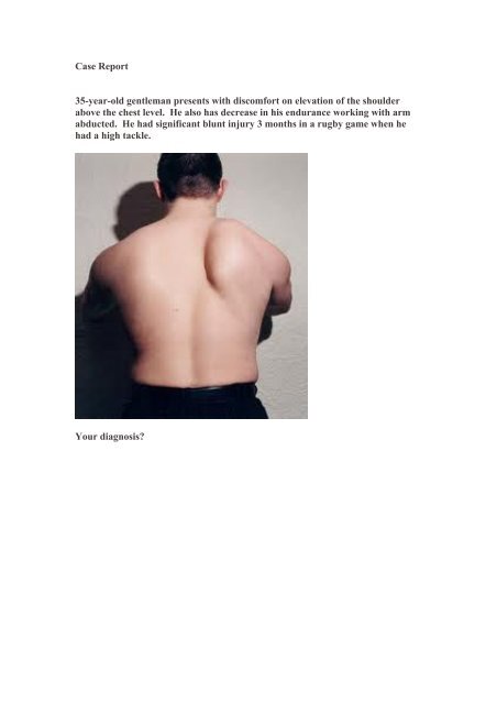
Case Report 35-year-old gentleman presents with discomfort on elevation of the shoulder above the chest level. He also has decrease in his endurance working with arm abducted. He had significant blunt injury 3 months in a rugby game when he had a high tackle. Your diagnosis?
- Page 2 and 3: Diagnosis: SCAPULAR WINGING Scapula
- Page 4 and 5: innervates the rhomboid muscles, wh
- Page 6 and 7: 6. Paralysis of the trapezius muscl
<strong>Case</strong> Report<br />
35-year-old gentleman presents with discomfort on elevation of the shoulder<br />
above the chest level. He also has decrease in his endurance working with arm<br />
abducted. He had significant blunt injury 3 months in a rugby game when he<br />
had a high tackle.<br />
Your diagnosis?
Diagnosis: SCAPULAR WINGING<br />
Scapular winging is a rare disorder often caused by neuromuscular imbalance<br />
in the scapulothoracic stabilizer muscles.<br />
Lesions of the long thoracic nerve and spinal accessory nerves are the most common<br />
cause.<br />
Patients report diffuse neck, shoulder girdle, and upper back pain, which may be<br />
debilitating, associated with abduction and overhead activities.<br />
Accurate diagnosis and detection depend on appreciation on comprehensive<br />
physical examination.<br />
Although most cases resolve nonsurgically, surgical treatment of <strong>scapula</strong>r winging<br />
has been met with success.<br />
True incidence is largely unknown because of under diagnosis.<br />
Most commonly it is categorized anatomically as medial or lateral shift of the inferior<br />
angle of the <strong>scapula</strong>.<br />
Primary winging occurs when muscular weakness disrupts the normal balance of the<br />
scapulothoracic complex.<br />
Secondary winging occurs when pathology of the shoulder joint pathology.<br />
Delay in diagnosis may lead to traction brachial plexopathy, peri<strong>scapula</strong>r muscle<br />
spasm, frozen shoulder, subacromial impingement, and thoracic outlet syndrome.<br />
Anatomy and Biomechanics<br />
Scapula is rotated 30° anterior on the chest wall; 20° forward in the sagittal plane; the<br />
inferior angle is tilted 3° upward.<br />
It serves as the attachment site for 17 muscles.<br />
The trapezius muscle accomplishes elevation of the <strong>scapula</strong> in the cranio-caudal axis
and upward rotation. The serratus anterior and pectoralis major and minor muscles<br />
produce anterior and lateral motion, described as <strong>scapula</strong>r protraction.<br />
Normal Scapulothoracic abduction:<br />
As the limb is elevated, the effect is an upward and lateral rotation of the<br />
inferior pole of <strong>scapula</strong>. Peri<strong>scapula</strong>r weakness resulting from overuse may manifest<br />
as <strong>scapula</strong>r dysfunction (ie, winging).<br />
Serratus Anterior Muscle<br />
Origin From the first 9 ribs<br />
Insert The medial border of the <strong>scapula</strong>.<br />
The upper portion originates from the first<br />
and second ribs and inserts onto the supero-<br />
medial aspect of the <strong>scapula</strong>. This portion f<br />
acilitates lateral rotation of the inferior <strong>scapula</strong>r angle.<br />
Nerve supply: Long thoracic nerve C5,6,7,<br />
Trapezius Muscle<br />
Origin The external occipital protuberance, the medial one third of the<br />
nuchal line (ie, the ligamentum nuchae), and the spines of the 7<br />
cervical and 12 thoracic vertebrae.<br />
Insertion The superior fibers insert onto the posterior clavicle.<br />
The medial fibers insert onto the medial aspect of the acromion.<br />
The inferior component: onto the spine of the <strong>scapula</strong>.<br />
Nerve supply Cranial nerve XI, the spinal accessory nerve.<br />
Rhomboid Muscles<br />
Deep to the trapezius, the rhomboid minor muscle originates from the<br />
ligamentum nuchae and the spinous processes of C7 and T1. Inferior to this<br />
muscle lies the rhomboid major, which originates from the spinous processes<br />
of T2 through T5. The rhomboid minor muscle inserts at the level of the<br />
<strong>scapula</strong>r spine, whereas the rhomboid major inserts distally on the medial<br />
<strong>scapula</strong> to the inferior angle. Both muscles serve to retract and elevate the<br />
<strong>scapula</strong> while rotating the inferior angle medially. The dorsal <strong>scapula</strong>r nerve
innervates the rhomboid muscles, which derive mainly from the C5 nerve root.<br />
Levator Scapulae<br />
The C5 nerve root via the dorsal also innervates the levator <strong>scapula</strong>e<br />
<strong>scapula</strong>r nerve. These muscles originate from the first four cervical vertebrae<br />
and insert onto the medial borders of the <strong>scapula</strong>e. The levator <strong>scapula</strong>e<br />
elevate the <strong>scapula</strong> and assist in rotating the glenoid inferiorly.<br />
Etiology of Winging<br />
Serratus Anterior Muscle Palsy<br />
The most common cause of primary <strong>scapula</strong>r winging<br />
1. Compression, traction, and laceration of the nerve<br />
2. The most common injury is neurapraxia after blunt or stretch injury:<br />
Repetitive activities with the head tilted away from the nerve and the arm overhead—<br />
as occurs in baseball pitchers, javelin throwers, and tennis servers—may place the<br />
long thoracic nerve on stretch. Similarly, upper extremity overuse in industrial<br />
laborers and homemakers may also contribute to serratus anterior muscle palsy.<br />
Atraumatic: Paralysis of the serratus anterior muscle has been associated with C7<br />
radiculopathy,. Transient brachial neuritis, Guillain-Barré syndrome,<br />
Trapezius Muscle Palsy<br />
Blunt trauma, such as assault or a direct blow from a football tackle or a lacrosse or<br />
hockey stick, may be associated with spinal accessory nerve palsy. Similarly,<br />
penetrating gunshot wounds, stabbings, and bite injuries have been implicated.<br />
The most common cause of trapezius palsy is iatrogenic injury to the spinal<br />
accessory nerve during cervical lymph node biopsy.<br />
Rhomboid Muscle Palsy<br />
Avulsion of the C5 nerve root may occur with heavy lifting and<br />
motor vehicle accidents.<br />
Fascioscapulohumeral Dystrophy<br />
Fascioscapulohumeral dystrophy (FSHD) is an autosomal dominant genetic
neuromuscular dystrophy linked to chromosome 4q35 that predominantly<br />
affects the face, shoulder girdle, and upper limb muscles. Weakness in the<br />
trapezius, levator <strong>scapula</strong>e, and rhomboids relative to preserved strength in<br />
the deltoid and rotator cuff muscles leads to severe <strong>scapula</strong>r winging.<br />
Secondary Causes<br />
Painful intra-articular conditions of the glenohumeral joint cause patients to<br />
compensate for lost motion with the scapulothoracic articulation. Scapular<br />
stabilizers, such as the serratus anterior and trapezius muscles, quickly<br />
fatigue under the increased demands, producing winging. Common causes<br />
include subacromial bursitis, adhesive capsulitis, rotator cuff tears, and<br />
shoulder instability.<br />
Patient Evaluation<br />
1. Fatigue, or muscle weakness, especially with elevation<br />
2. Difficulty with ADL<br />
3. Feel discomfort while sitting against hard surfaces<br />
Serratus Anterior Trapezius winging<br />
4. Atrophy of the peri<strong>scapula</strong>r muscles<br />
5. Paralysis of the serratus anterior muscle causes medial winging.
6. Paralysis of the trapezius muscle causes the inferior angle is rotated laterally<br />
7. A palpable clunk with abduction may be a sign of an underlying osteochondroma.<br />
8. If winging produced with forward elevation is eliminated with glenohumeral<br />
external rotation, then posterior instability is confirmed. Persistent winging with this<br />
maneuver suggests a complete serratus anterior muscle paralysis.<br />
9. MCR Trapezius muscle function: a resisted shoulder shrug.<br />
Investigations<br />
Rhomboid and levator <strong>scapula</strong>e: evaluated by having the patient place his or<br />
her hands on the hips and pushing the elbows posterior against resistance.<br />
Serratus anterior muscle can be easily appreciated with the shoulder flexed<br />
to 90° while the patient performs push-ups or push against the wall.<br />
1. CT may help to better characterize osteochondromas.<br />
2. MRI: cervical disk disease, shoulder instability, or rotator cuff tears.<br />
3. NCV and EMG are valuable adjuncts to confirm neuromuscular causes<br />
Management<br />
1. For neurapraxia, observation of 6 to 12 months is undertaken. Physio exercises.<br />
2. Acute transections are best managed with repair and nerve grafting, when<br />
necessary.<br />
3. For recalcitrant neurapraxia, surgical muscle transfer is recommended.
Patients with serratus anterior muscle palsy are treated with pectoralis major<br />
muscle transfer augmented with autogenous hamstring graft.<br />
5. In patients with recalcitrant trapezius palsy who require a muscle transfer, a<br />
modified Eden-Lange procedure is used. Originally described in 1924, the procedure<br />
involves lateralization of the rhomboid major, rhomboid minor, and levator <strong>scapula</strong>e<br />
insertions on the <strong>scapula</strong>, thereby allowing them to act synergistically for the three<br />
components of the trapezium


