

Case 7 Groin Pain in 16 Y - Bonefix
Case 7 Groin Pain in 16 Y - Bonefix
Case 7 Groin Pain in 16 Y - Bonefix

Create successful ePaper yourself
Turn your PDF publications into a flip-book with our unique Google optimized e-Paper software.
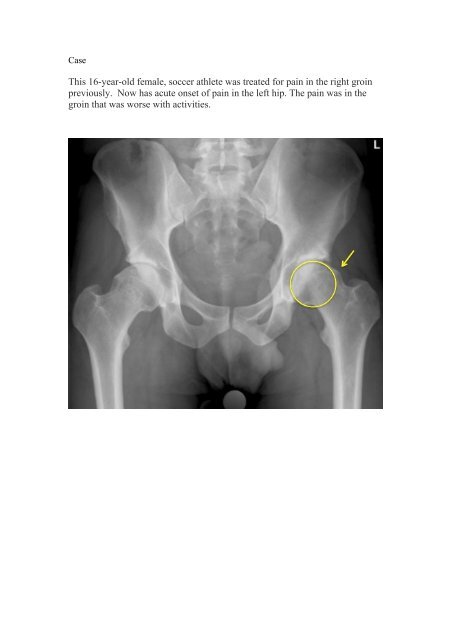
<strong>Case</strong><br />
This <strong>16</strong>-year-old female, soccer athlete was treated for pa<strong>in</strong> <strong>in</strong> the right gro<strong>in</strong><br />
previously. Now has acute onset of pa<strong>in</strong> <strong>in</strong> the left hip. The pa<strong>in</strong> was <strong>in</strong> the<br />
gro<strong>in</strong> that was worse with activities.
Diagnosis FAI syndrome with or without labral tear.<br />
Other <strong>in</strong>vestigations required<br />
1.X ray:<br />
1. Anterolateral bony prom<strong>in</strong>ence on the femoral neck (AP or lateral radiograph)<br />
2. Lateral subluxation<br />
3. CE angle [<strong>in</strong> AP
2. CT assessment for retroversion<br />
3. MRI<br />
MRI: Of the 13 labral tears found at arthroscopy, 85% were detected by conventional<br />
MRI, whereas 100% were identified via <strong>in</strong>direct MR arthrography.<br />
Direct Arthrogram<br />
A, Coronal proton density fast sp<strong>in</strong>-echo fatsuppressed<br />
unenhanced image obta<strong>in</strong>ed<br />
without exercise.
Indirect Arthrogram<br />
IV contrast-enhanced <strong>in</strong>direct MR<br />
arthrography appears to be an effective means of hip evaluation for labral tears. It<br />
does not appear to improve detection of cartilage abnormalities when compared with<br />
conventional MRI. [AJR:194, March 2010: 719]<br />
However, presently noncontrast magnetic resonance imag<strong>in</strong>g (MRI) has been the gold<br />
standard us<strong>in</strong>g 1.5-T MRI for detection of labral tear.<br />
4. Arthroscopic Classification[Wardell]<br />
Stage 0: contusion of the labrum with adjacent synovitis<br />
I: Discrete labral free marg<strong>in</strong> tear with <strong>in</strong>tact articular cartilage<br />
II: Labral tear with damage to the subjacent femoral head<br />
III: II + Acetabular cartilage damage<br />
IV: Diffuse labral and acetabular cartilage damage<br />
The patient received IV gadol<strong>in</strong>ium contrast<br />
medium (0.2 mL/kg) and was <strong>in</strong>structed to<br />
exercise for 15 m<strong>in</strong>utes.<br />
B, Coronal T1-weighted image with fat<br />
suppression after IV gadol<strong>in</strong>ium enhancement<br />
performed after exercise and delay. The<br />
acetabular labrum tear (arrow) is more clearly<br />
depicted on the contrast-enhanced portion of the<br />
SUMMARY<br />
The acetabular labrum has various functions <strong>in</strong>clud<strong>in</strong>g enhanc<strong>in</strong>g jo<strong>in</strong>t stability,<br />
shock absorption, proprioception, jo<strong>in</strong>t lubrication, pressure distribution, deepen<strong>in</strong>g of<br />
the jo<strong>in</strong>t, and creat<strong>in</strong>g a seal.<br />
Intra-articular abnormalities of the hip are be<strong>in</strong>g diagnosed with <strong>in</strong>creased frequency<br />
by diagnostic methods such as conventional MRI, conventional MR arthrography, and<br />
hip arthroscopy.<br />
However, labral tear can be seen <strong>in</strong> asymptomatic young population. In a recent<br />
study [Am J Sports Med. 2012 Jun;40(6):1337-41]. Acetabular paralabral cysts were<br />
identified <strong>in</strong> 25% with an <strong>in</strong>terobserver reliability of 90.5% and <strong>in</strong>traobserver<br />
reliability of 95%. In addition, acetabular labral tears were identified <strong>in</strong> 85%.
More recently, many of these lesions have been shown to be due to underly<strong>in</strong>g<br />
femoral acetabular imp<strong>in</strong>gement [FAI]. Recently, FAI has been diagnosed more<br />
frequently <strong>in</strong> younger active <strong>in</strong>dividuals.<br />
FAI is commonly classified as a cam-type, p<strong>in</strong>cer type, or mixed cam-p<strong>in</strong>cer.<br />
Cam-type imp<strong>in</strong>gement occurs when the femoral<br />
head is abnormally shaped and contacts a normal<br />
acetabulum. This “bump” abuts the acetabular<br />
rim creat<strong>in</strong>g shear<strong>in</strong>g and compressive forces that<br />
may result <strong>in</strong> tear<strong>in</strong>g of the acetabular labrum and<br />
chondral <strong>in</strong>jury. Males have a predilection for<br />
cam-type imp<strong>in</strong>gement.<br />
Asymptomatic male subjects us<strong>in</strong>g MRI found a<br />
cam-type deformity <strong>in</strong> every fourth male<br />
[Arthritis Care & Res. 2010;62 (9): 1319-1327.]<br />
P<strong>in</strong>cer-type imp<strong>in</strong>gement occurs when the acetabular<br />
rim is abnormally shaped, deep, or retroverted<br />
and contacts a normally shaped femoral head. This<br />
“over-coverage” often results <strong>in</strong> labral degeneration<br />
and tear<strong>in</strong>g. P<strong>in</strong>cer-type imp<strong>in</strong>gement is more<br />
common <strong>in</strong> females.<br />
In 18-19 years-old, its prevalence is high <strong>in</strong> girls.<br />
[Arthritis Care & Res. 2010;62 (9): 1319-1327.]<br />
Studies have shown that mixed imp<strong>in</strong>gement can<br />
occur <strong>in</strong> 57-85% of younger <strong>in</strong>dividuals.<br />
Damage to the acetabular labrum from FAI can affect<br />
overall hip function.<br />
The presence of FAI <strong>in</strong> younger football players may be more prevalent than previously<br />
suspected. In fact, Kapron [J Bone Jo<strong>in</strong>t Surg AM. 2011;93:e111(1-10)] looked at the<br />
radiographic prevalence of FAI <strong>in</strong> collegiate football players (age, 21 Y) and found that 95%<br />
had a least one sign of cam or p<strong>in</strong>cer imp<strong>in</strong>gement and 57% had signs of both (mixed).<br />
Although a labral tear may occur with a s<strong>in</strong>gle traumatic event, often another<br />
underly<strong>in</strong>g cause may be already present, predispos<strong>in</strong>g the <strong>in</strong>dividual to <strong>in</strong>jury [Cl<strong>in</strong><br />
Sports Med. 2011 Apr;30(2):293-315].<br />
Labral tears occur most frequently <strong>in</strong> the anterior quadrant. This is due to<br />
(1) the anterior labrum has a low vascular supply which results <strong>in</strong> poor heal<strong>in</strong>g;<br />
(2) this region is mechanically weaker than other regions;<br />
(3) the anterior labrum experience higher loads and shear forces than other regions.<br />
How Does a Tear Occur <strong>in</strong> the Hip Labrum?<br />
There are two general types of hip labral tears: degenerative tears and traumatic<br />
<strong>in</strong>juries.<br />
A degenerative tear is a chronic <strong>in</strong>jury that occurs as a result of repetitive use and<br />
activity. Degenerative labral tears can be seen <strong>in</strong> the early stages of hip arthritis.
A traumatic hip labral tear is usually an acute <strong>in</strong>jury as a result of a sports <strong>in</strong>jury,<br />
fall, or accident. Hip labral tears can be seen <strong>in</strong> association with episodes of hip<br />
dislocation or subluxation. They are commonly associated with sudden, twist<strong>in</strong>g<br />
maneuvers that cause immediate pa<strong>in</strong> <strong>in</strong> the hip. Isolated athletic <strong>in</strong>jury or repetitive<br />
traumatic activity can lead to labral tears [Cl<strong>in</strong> Sports Med. 2006 Apr; 25(2):279-92].<br />
Classification Arthroscopy. 1996 ;12(3):269-72.<br />
a. Etiology<br />
Traumatic, 18.9%<br />
Degenerative, 48.6%;<br />
Idiopathic, 27.1%;<br />
Congenital, 5.4%.<br />
b. Morphology<br />
Radial flap, 56.8%;<br />
Radial fibrillated, 21.6%;<br />
Longitud<strong>in</strong>al peripheral, <strong>16</strong>.2%;<br />
Unstable, 5.4%).<br />
What Are the Symptoms of a Hip Labral Tear?<br />
A hip labral tear can be difficult to diagnose.<br />
Many of the symptoms of a hip labral tear are similar to symptoms of a gro<strong>in</strong> stra<strong>in</strong>,<br />
snapp<strong>in</strong>g hip syndrome, sports hernia, or other athletic <strong>in</strong>juries of the hip jo<strong>in</strong>t.<br />
Furthermore, just because a tear is seen <strong>in</strong> the hip labrum on an MRI, it does not mean<br />
the tear is necessarily the cause of the pa<strong>in</strong>.<br />
Typical symptoms of a hip labral tear <strong>in</strong>clude:<br />
• <strong>Gro<strong>in</strong></strong> pa<strong>in</strong><br />
• Click<strong>in</strong>g and snapp<strong>in</strong>g sensations <strong>in</strong> the hip<br />
• Limited motion of the hip jo<strong>in</strong>t<br />
Cl<strong>in</strong>ical tests:<br />
1. Provocation imp<strong>in</strong>gement sign<br />
Flexion, adduction and <strong>in</strong>ternal<br />
rotation cause pa<strong>in</strong>.<br />
This comb<strong>in</strong>ation br<strong>in</strong>gs the<br />
proximal and anterior part of the<br />
femoral neck <strong>in</strong>to contact with<br />
the rim of the acetabulum
2. Hip Apprehension Test<br />
McCarthy [Cl<strong>in</strong> Orthop Relat Res. 2001 Dec;(393):25-37]<br />
(1) FAI excessively loads the acetabular labrum at the extremes of jo<strong>in</strong>t motion,<br />
(2) Fray<strong>in</strong>g of the articular marg<strong>in</strong> of acetabular labrum, tear<strong>in</strong>g along the articular<br />
marg<strong>in</strong> of the acetabular labrum,<br />
(3) delam<strong>in</strong>ation of the cartilage from the articular marg<strong>in</strong> adjacent to the labrum<br />
(4) more global labral and articular cartilage degeneration.<br />
Treatment<br />
Arthroscopic surgery for FAI & labral tears has become an accepted procedure for many<br />
athletes of varied ages.<br />
Hip arthroscopy: assessment<br />
Traction 1cm distraction on the fracture table<br />
Entry po<strong>in</strong>t: 1 Cm: anterior and posterior to the trochanter<br />
Labral tear: isolated tear or with cartilage<br />
Debride or reattach the labrum and cartilage<br />
Open method [ Ganz ]<br />
Capsulotomy close to the acetabulum<br />
Dislocate the hip<br />
Cam anterior: imp<strong>in</strong>ges è excise<br />
Excise any osteophytes anterior<br />
Debride cartilage<br />
This surgery meant to improve ROM<br />
• In the patient with classic dysplasia,<br />
pa<strong>in</strong> is reproduced anteriorly as the<br />
extremity is forcefully externally<br />
rotated with hip <strong>in</strong> extension and slight<br />
abduction.<br />
•<br />
• In the patient with degenerative<br />
changes of the posterior acetabulum,<br />
pa<strong>in</strong> is reproduced posteriorly.<br />
Post op:<br />
There is little evidence regard<strong>in</strong>g the rehabilitation of younger athletes who undergo<br />
arthroscopic hip surgery. This case study described a four phase rehabilitation<br />
program for a high school football player who underwent hip arthroscopy and labral<br />
repair. The patient achieved positive outcomes with a full return to athletic activity<br />
and football. [The International Journal of Sports Physical Therapy | Volume 7,
Number 2 | April 2012 | Page 173]<br />
For younger football players, future studies should focus on risk factors for the<br />
development of FAI that may be related to position, tra<strong>in</strong><strong>in</strong>g methods, and maturation.<br />
Future research should also focus on the outcomes of the four phase rehabilitation<br />
program for younger athletes <strong>in</strong> specific sports <strong>in</strong>clud<strong>in</strong>g: dance, hockey, and soccer.<br />
Recent review: The American Journal of Sports Medic<strong>in</strong>e, Vol. 38, No. 11<br />
1.Femoroacetabular imp<strong>in</strong>gement as the cause of primary idiopathic osteoarthritis <strong>in</strong><br />
young patients<br />
2.Treatment for FAI succeed <strong>in</strong> improv<strong>in</strong>g patient symptoms? Both arthroscopic and<br />
open treatment methods for FAI significantly improved mean postoperative scores.<br />
3. Avoid : Surgery <strong>in</strong> Outerbridge grade III or IV cartilage <strong>in</strong>jury<br />
3. Labral refixation superior to labral resection? The studies exam<strong>in</strong>ed varied <strong>in</strong> their<br />
treatment of labral <strong>in</strong>jury.<br />
4. Does treatment for FAI affect the progression of osteoarthritis? Because all case<br />
series were relatively recent and degenerative jo<strong>in</strong>t disease is generally a gradual<br />
process, it was difficult to elucidate the effect of surgical correction of FAI on the<br />
progression of osteoarthritis with radiographic imag<strong>in</strong>g.<br />
In addition to a lack of high level of evidence studies, there has yet to be a direct<br />
comparison between open and arthroscopic methods.<br />
Patients with advanced osteoarthritic change as shown on preoperative radiographs<br />
will likely benefit less from treatment of FAI.

![Vol [Aug 2007] 1.3 Vasu Pai Editor Orthopaedic Surgeon ... - Bonefix](https://img.yumpu.com/17158213/1/184x260/vol-aug-2007-13-vasu-pai-editor-orthopaedic-surgeon-bonefix.jpg?quality=85)


![CARPO-METACARPAL [CMC] ARTHRITIS CMC joint is a ... - Bonefix](https://img.yumpu.com/17157176/1/184x260/carpo-metacarpal-cmc-arthritis-cmc-joint-is-a-bonefix.jpg?quality=85)


