

Read more - Bonefix
Read more - Bonefix
Read more - Bonefix

You also want an ePaper? Increase the reach of your titles
YUMPU automatically turns print PDFs into web optimized ePapers that Google loves.
SCAPULAR FRACTURE Vasu Pai<br />
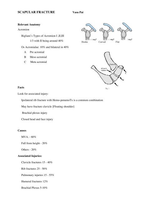
Relevant Anatomy<br />
Acromion<br />
Facts<br />
Bigliani’s Types of Acromion I ,II,III<br />
1/3 with II being around 40%<br />
Os Acromialae: 10% and bilateral in 40%<br />
A Pre acromial<br />
B Meso acromial<br />
C Meta acromial<br />
Look for associated injury:<br />
Ipsilateral rib fracture with Hemo-penumoTx is a common combination<br />
May have fracture clavicle [Floating shoulder]<br />
Brachial plexus injury<br />
Closed head and face injury<br />
Causes<br />
MVA. - 60%<br />
Fall from height - 20%<br />
Others - 20%<br />
Associated Injuries:<br />
Clavicle fractures 15 - 40%<br />
Rib fractures 25 - 50%<br />
Pulmonary injuries 15 - 55%<br />
Humeral fractures 12%<br />
Brachial Plexus 5-10%<br />
Skull fractures 25%<br />
Hooke Curved Flat
Of scapular fractures, the glenoid 40% fractures.<br />
36% of anterior rim fractures, 19% of posterior rim fractures, and<br />
45% of short oblique fractures<br />
Clinical Evaluation<br />
These are typically high energy injuries, assessment should begins with the A,B,C's.<br />
C/O shoulder pain after trauma.<br />
Shoulder often held in abduction and external rotation Evaluate for tenderness, ecchymosis, soft<br />
tissue injury. Document axillary, median, ulnar,<br />
radial nerve function and radial pulse.<br />
Xray / Diagnositc Tests<br />
AP, scapular lateral and axillary views.<br />
CT scan with 2-3 mm cuts<br />
Chest X ray to rule out associated injuries.<br />
Classification [Idelberg] of scapula<br />
Type I Glenoid rim [Ant or Post]<br />
Type II Glenoid to the lateral border<br />
Type III Superior 30-50% of the glenoid<br />
surface with the coracoid,<br />
Type IV Involve medial border of the<br />
scapula,<br />
Type V Combination<br />
Type VI Combination and severe<br />
comminuted.
Glenoid Rim<br />
I Nondisplaced / minimally displaced : immobilization in a sling for 1 week followed by<br />
progressive ROM and physical therapy.<br />
II Displaced (subluxation of humeral head<br />
>10 mm displacement,<br />
>1/4 of glenoid cavity anteriorly<br />
>1/3 of the glenoid cavity posteriorly<br />
>5mm articular step-off)<br />
Treatment<br />
Consider: Intra-articular or extra-articular<br />
Displaced or undisplaced<br />
Associated injury: Presence of floating shoulder<br />
Joint stability<br />
Age of the patient and medical comorbidities<br />
Recommended treatment<br />
Ia – Anterior rim fracture: If unstable - Ant. Henry’s approach - ORIF with a screw<br />
If comminuted - excise & tricorticate iliac graft(Eden)<br />
Ib Fix through posterior approach<br />
II – Glenoid fracture: When step is <strong>more</strong> than 5 mm and joint is incongruous<br />
Posterior approach and IFS fixation<br />
III IV, V- Post-sup approach and Lag screw<br />
VI Non-operative<br />
Repair of displaced anterior glenoid rim fractures has been advocated: to restore articular<br />
congruity, glenoid concavity, and glenohumeral stability.<br />
DePalma recommended that fractures that involve at least 25% of the glenoid area or are<br />
displaced at least 10 mm should be repaired. Itoi showed that a fracture width greater than 21% of<br />
the glenoid length created instability and that capsulolabral repair after excision of these fractures.<br />
Fracture exposure and visualization can be challenging, and subscapularis dysfunction can occur
postoperatively.<br />
Arthroscopic glenoid fracture repair offers<br />
advantages of enhanced visualization and<br />
diminished morbidity, but published studies<br />
into the arthroscopic treatment of glenoid rim<br />
fractures have been limited to a few case<br />
reports.<br />
Posterior Approach<br />
Patient on lateral position<br />
Incision along cranial to caudal along the spine of the scapula<br />
Elevated the flap: elevating deltoid subperiosteally from the scapular spine<br />
Internervous plane between the teres minor and infraspinatus<br />
[Do not go between Minor and major to avoid damage to axillary nerve<br />
Posterior capsule is incised from the humerus and elcvated superiorly exposing the glenoid.<br />
Fracture is identified, reduced and fixed using standard AO techniques.<br />
Fixation is generally provided with 3.5 mm reconstruction plates, and / or 3.5mm or 4.0mm<br />
cannulated screws.<br />
Keep in mind the majority of the scapula is paper-thin.<br />
Adequate bone stock to hold screws can be found: in the glenoid neck, coracoid process, base of<br />
the scapular spine, and lateral border of the scapular body. Irrigate<br />
Closure: stitch the aponeurotic flap with drill holes in the spine
Glenoid Fx Follow-up Care<br />
Shoulder immobilizer with gentle pendulum, elbow/wrist/hand ROM immediately. Generally<br />
Plane is between Infraspinatus and teres minor<br />
patients remain in the sling for 6 weeks. Has limited use of the extremity for 10-12 weeks and<br />
must refrain from heavy physical activity for 4-6 months. 90% G-E results<br />
Concept of floating shoulder<br />
Stabilise at one level or both levels or no levels. Not clear.<br />
Trend: fix both or Fix clavicle and check Glenoid reduction.<br />
However recent: report on 20 patients treated Non-op: 17 had excellent and 3 had good results.<br />
[Edwards. JBJS 82A: 774]
Complications<br />
Heterotopic Ossification<br />
Nerve injury: axillary N<br />
Glenohumeral arthritis<br />
Nonunion and Malunion<br />
Infection<br />
Stiffness<br />
REFERENCES<br />
1. Goss JAAOS 3:22;1995 Ada JR, CORR 1991;269:174<br />
2. Jaeger. J Shoulder Elbow Surg. 2012 Sep 27.<br />
3. Arthroscopic fixation. J Shoulder Elbow Surg (2010) 19, e16-e19<br />
4. Goss: Fractures of the Glenoid JBJS 74 A: 299<br />
5. Boyer. Instruction course lecture 52.591<br />
6. KUD 7<br />
7. Ada JR, CORR 1991;269:174<br />
8. Rockwood and Green's Fractures in Adults 6th ed, 2006<br />
9. OKU - Shoulder and Elbow 2nd ed, 2002<br />
10. ORIF (Kavanaugh BF, JBJS 1993;75A:479).

![Vol [Aug 2007] 1.3 Vasu Pai Editor Orthopaedic Surgeon ... - Bonefix](https://img.yumpu.com/17158213/1/184x260/vol-aug-2007-13-vasu-pai-editor-orthopaedic-surgeon-bonefix.jpg?quality=85)


![CARPO-METACARPAL [CMC] ARTHRITIS CMC joint is a ... - Bonefix](https://img.yumpu.com/17157176/1/184x260/carpo-metacarpal-cmc-arthritis-cmc-joint-is-a-bonefix.jpg?quality=85)


