


Create successful ePaper yourself
Turn your PDF publications into a flip-book with our unique Google optimized e-Paper software.
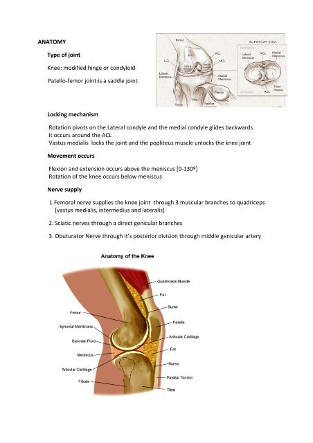
ANATOMY<br />
Type of joint<br />
Knee: modified hinge or condyloid<br />
Patello‐femor joint:is a saddle joint<br />
Locking mechanism<br />
Rotation pivots on the Lateral condyle and the medial condyle glides backwards<br />
It occurs around the ACL<br />
Vastus medialis locks the joint and the popliteus muscle unlocks the knee joint<br />
Movement occurs<br />
Flexion and extension occurs above the meniscus [0‐130º]<br />
Rotation of the knee occurs below meniscus<br />
Nerve supply<br />
1.Femoral nerve supplies the knee joint through 3 muscular branches to quadriceps<br />
[vastus medialis, intermedius and lateralis]<br />
2. Sciatic nerves through a direct genicular branches<br />
3. Obuturator Nerve through it’s posterior division through middle genicular artery
Bursae<br />
4 Anterior bursae<br />
2 Medial bursae<br />
2 Lateral bursae<br />
4 Posterior bursae<br />
Suprapatellar bursa<br />
Prepatellar bursa<br />
Superficial and deep infrapatellar bursa<br />
Between MCL and Pes anserinus<br />
Between MCL and Semimembranous and capsule<br />
Between Lateral collateral ligament and biceps tendon<br />
Between Popliteus and Capsule<br />
2 Between lateral and medial gastrocnemius and joint capsule<br />
1 Between Semimembranous and Medial Gastrocnemius<br />
1 Popliteus and back of the tibia (synovial extension)
ACL [Anterior cruciate ligament]<br />
Intra‐articular and extrasynovial ligament<br />
Length and width: 30‐40 mm x 11mm<br />
Fibers rotate by 90° between its attachments<br />
Tibial attachment is anterior and stronger than<br />
femoral.<br />
Femoral attachment is to the medial side of the<br />
lateral femoral condyle<br />
Anterior fibres: blend with Anterior horn of the<br />
lateral Meniscus<br />
2 bundles Anteromedial [tight in flexion]<br />
Posterolateral [tight in extension]<br />
When the knee is extended, the ACL is a relatively flat, ribbon‐like ligament composed of<br />
many parallel fibers.<br />
Blood Supply Middle Genicular Artery<br />
Nerve supply Golgi tendon receptors<br />
Load to failure 1700N [Normal walking 170N; Running 500N]<br />
Strain rate plays a role in the location of ligament failure<br />
High strain rate Midsubstance tears [common]<br />
Low strain rate Avulsion from tibial attachment [seen in children with tibial eminence<br />
fracture]<br />
Tension in cruciate ligament<br />
Tension is constant in all position. This is due to different fibers in the ACL experience<br />
different tension from flexion and extension and the whole ligament is not in constant<br />
state of tension.<br />
Function of ACL<br />
1. ACL Carries loads throughout flexion<br />
2. Normally it carries small load ie., 500 N [fails at 2500 N]<br />
3. Highest loading of ACL is during quadriceps powered extension moving from 40°<br />
flexion<br />
4. Tunes roll back with PCL. 4 bar linkage<br />
5. Prevents anterior translation<br />
6. Important for rotational instability
ACL VS MCL<br />
MCL ACL<br />
Macroscopic Extra‐articular;Flat;<br />
uniform<br />
Collagen Fibres are parallel<br />
Densely packed<br />
Electron Micro Mean fibril diameter is<br />
larger<br />
Elastic Modulus<br />
Tensile strength<br />
2 fold more than ACL<br />
110 MPa<br />
Healing Heals.<br />
No surgery required<br />
Posterior cruciate ligament [PCL]<br />
Intra‐articular and extrasynovial ligament<br />
Its mean length is 38 mm and mean width 13 mm<br />
2 bundles Anterolateral [larger and 85%]<br />
Posteromedial bands<br />
Intra‐articular; varied<br />
Varied and Nonparallel<br />
Less fibers<br />
Is smaller<br />
Less than MCL<br />
70 MPa (Medial bundle)<br />
Always need surgery<br />
Anterolateral band is tight in flexion and lax in<br />
extension while the posteromedial bundle is tight in extension and lax in Flexion<br />
Division of PCL increases in the Patellofemoral joint forces. This may lead to Patello‐<br />
femoral arthritis.
Medial structures of the knee<br />
3 layers: Superficial, middle and deep layers<br />
Grouped: into anterior, middle and posterio third<br />
Anterior Middle Posterior<br />
Superficial Fascia Fascia Layer of fascia<br />
Middle None Superficial MCL Posteromedial capsule<br />
Deep None Deep MCL Posteromedial capsule<br />
3 important medial ligamentous structure<br />
1. Superficial MCL<br />
Femur attachment: 1cm anterior and distal to Adductor tubercle<br />
Tibia attachment: Under Pes Anserinus [ 6 cm distal to the joint]<br />
Fibers are grouped into<br />
a. Anterior fibers Parallel fibers<br />
b. Posterior fibers Oblique<br />
Posterior oblique ligament<br />
2. Deep MCL<br />
Femur: Femur just below Superficial<br />
Tibia: close to the articular margin<br />
3. Posteromedial capsule<br />
Blending of superficial and deep MCL and Semimbranous extension<br />
Posterior oblique ligament probably not distinct and is as above<br />
Appears to dynamically stabilised by Semimembranous<br />
Lateral structures of the knee [Seebacher]<br />
I layer Biceps Femoris; Iliotibial band<br />
II layer Quadriceps retinaculum; Patellofemoral ligament<br />
III layer Superficial: Lateral collateral ligament; Fibulo‐fabellar ligament<br />
Deep: Coronary ligament, Arquate ligament, Popliteo‐fibular ligament
Anatomic variation:<br />
67% have both arcuate and Fabello‐ fibular ligament<br />
20% only Fabello‐ fibular ligament; 13% only arcuate
III. BIOMECHANICS<br />
a. Anatomic and mechanical axis<br />
Mechanical axis: Centre of the hip to the centre of the ankle<br />
Anatomic axis of femur is approx 6 degrees of valgus from<br />
mechanical axis or 9 degrees of valgus from vertical axis<br />
Anatomic axis of tibia is 3 degrees of varus from mechanical<br />
axis<br />
Lines that intersect the tibia and the femur intersect at knee 6°<br />
Posterior slope in the tibial plateau is 9 º<br />
2. The “screw home mechanism<br />
Rotation between the tibia and femur occurs<br />
automatically between full extension (0 o ) and 20 o of knee flexion.<br />
External rotation of the tibia on femur during full extension is<br />
Obligatory external rotation<br />
This occurs because of differential radii. Medial femoral larger<br />
than lateral (by 17mm). At full extension, medial tibial plateau<br />
has to cover more distance and this causes external rotation of<br />
the tibia.<br />
This screw home: very stable and both Cruciates are tight and<br />
very minimal movement occurs between tibia and femur<br />
1.During knee extension<br />
The tibia glides anteriorly on the femur.<br />
2.During the last 20 degrees of knee extension<br />
Anterior tibial glide persists on the tibia's medial condyle because<br />
its articular surface is longer in that dimension than the lateral<br />
condyle's.<br />
3.Prolonged anterior glide<br />
On the medial side produces external tibial rotation, the "screw‐home" mechanism<br />
c. Sliding and rolling<br />
With 0‐15 º of flexion: [Rolling prominent]<br />
Sliding::Rolling is 1:2<br />
With 15 º‐130 º of flexion: [Sliding prominent]<br />
Sliding::Rolling is 4:1
From kinematics studies,<br />
Flexion and extension do not occur about a fixed transverse axis of rotation but rather about<br />
a constantly changing center of rotation (polycentric rotation).<br />
Motion is therefore achieved by a complex coupled mechanism in which the femoral<br />
condyles simultaneously glide and roll back on the tibial plateaus<br />
D. 4 Bar linkage<br />
4 Bars ACL, PCL, Femur: Roof of the intercondylar notch, Tibia intercondylar<br />
eminence<br />
Relation to Femoral link<br />
Extension: ACL is parallel to the Femoral link<br />
Flexion PCL is parallel to the femoral link.<br />
Isometry Only Some fibers in ACL and PCL is isometric during any ROM.<br />
With flexion and extension<br />
None of the length of bars changes with movement but angle between ACL and<br />
PCL changes.<br />
e. Instant centre<br />
Centre is at the intersection of ACL and PCL.<br />
Therefore do not produce moment .<br />
Line from instant centre to tibifemoral contact<br />
point forms perpendicular to the tangential to<br />
the tibia.
f. Movement<br />
Normal range 0‐140º<br />
Functional range ‐3 to 120 º<br />
Walk : Heel strike 15 º flexion<br />
Sprint: Heel strike 30 º<br />
Swing 60 º<br />
Getting in and out of chair 115 º<br />
Climbing stair 90 to 100 º<br />
g. Primary joint restraints<br />
Primary Secondary<br />
Anterior ACL Deep MCL<br />
Posterior PCL Posterolateral<br />
complex<br />
Varus LCL Posterolateral<br />
complex<br />
Valgus MCL Cruciates<br />
Internal Rotation MCL ACL<br />
External Rotation LCL & posterior<br />
complex<br />
H. Posterior roll back<br />
On knee flexion, both femoral condyles<br />
roll back. This is due to four bar linkage.<br />
PCL retaining supposed favour normal roll back<br />
Roll back: increases maximum knee flexion.<br />
And it increases patello‐femoral moment arm<br />
PCL
1I. Tibio‐femoral force<br />
Moment: Fq x 2.5 = W x 7.5 ie., Fq=3 x W<br />
Sum of Forces: F q+ Fj+ Fw = 0<br />
Fq = Quadriceps force; Fj = Joint reaction force

![Vol [Aug 2007] 1.3 Vasu Pai Editor Orthopaedic Surgeon ... - Bonefix](https://img.yumpu.com/17158213/1/184x260/vol-aug-2007-13-vasu-pai-editor-orthopaedic-surgeon-bonefix.jpg?quality=85)


![CARPO-METACARPAL [CMC] ARTHRITIS CMC joint is a ... - Bonefix](https://img.yumpu.com/17157176/1/184x260/carpo-metacarpal-cmc-arthritis-cmc-joint-is-a-bonefix.jpg?quality=85)


