of fetal rat liver - Digitum
of fetal rat liver - Digitum
of fetal rat liver - Digitum

Create successful ePaper yourself
Turn your PDF publications into a flip-book with our unique Google optimized e-Paper software.
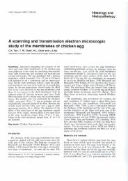
Histol Histopath (1991) 6: 217-228 Histology and<br />
Histopathology<br />
Morphology, differentiation and matrix production <strong>of</strong><br />
<strong>liver</strong> cells in organoid cultures (high density cultures)<br />
<strong>of</strong> <strong>fetal</strong> <strong>rat</strong> <strong>liver</strong>s<br />
A. Elkasabyl, D. Xul, Chr. Schroter-Kermanil and H.-J. Merker1s2<br />
'Institut fur Toxikologie und Embryonal-Pharmakologie and 'Institut fur Anatomie, Freie Universitat Berlin, Berlin, FRG<br />
Summary. Tlie aim <strong>of</strong> this study \\as to dcn~onst<strong>rat</strong>e tlie isolation ant1 tlicil- I-apicl cledifferentintion. Tlie first<br />
morpl~ology and matrix synthesis <strong>of</strong> ernl~ryonic <strong>rat</strong> <strong>liver</strong> prolJlem could 121-gely be solved by improving the<br />
cc.11\ (clay IS <strong>of</strong> gestation) in organoid cultures (high tecliniq~~e. e.g. optinii~ation <strong>of</strong> tlie perfusion technique<br />
densit!, cultures) with electron microscopic and and tlie perfusion solution as well us by application <strong>of</strong><br />
imm~~nomorphological techniclues. For this purpose tlie tlie gradient cenLrifugation method (Berry ancl Friend.<br />
cells <strong>of</strong> embryonic <strong>rat</strong> <strong>liver</strong>s were isolated enzyniaticall! IVhO: Segcln. 1973: Williams et al.. 1077: Schaefl'cr<br />
and gro~~i i11 ;in organoid culture (high clensit!, c~~lture) ancl Kcssler. IC)SO: Sirica and Pitot. I1)SO: Carlsen et al..<br />
l'or .; weeks in n Trowell systern. During the first 48 h n 108 I : Kreanier et al.. 1986). For a better ~lnderstnncling<br />
sorting-o~~t process took place. i.e. <strong>liver</strong> and blood- <strong>of</strong> tlic second point tlie term i
21 8<br />
influence or even to stimulate the differentiation<br />
behaviour <strong>of</strong> epithelia1 cells via a direct contact. via<br />
the secretion <strong>of</strong> diffusible substances or via matrix<br />
components (Fleischmajer and Billingham, 1968: Le<br />
Dourin, 1975: Rojkind et al., 1980: Kleinnian et al.,<br />
1981; Riso. 1983: Sawada et al., 19x7: Bissell anti Choun,<br />
1988). This interaction is missing in monola!.er cultures.<br />
To a certain degree it may be present in co-cultures. In<br />
organ cultures as in vivo-like situation is observed. but<br />
this cuIture technique involves other difficultics, e.g.<br />
lesions during cutting and problems in tlie supply <strong>of</strong><br />
central areas (Sipes et al.. 1987).<br />
The search for liepatocyte cultures that liave the<br />
capability <strong>of</strong> metabolization over a long enough period<br />
has therefore still a great topicality. For this purpose<br />
mainly monolayer cultures have so far bcen uscd. Organ<br />
or slice cultures have been applied to a negligible extent<br />
only (Sipcs et al.. 1987). The organoid or high density<br />
culture has so far not been used in this connection.<br />
However, in the case <strong>of</strong> other tissues the high density<br />
tcclinique has proved to be very suitable for tlie study <strong>of</strong><br />
differentiation processes, for example with hlastcmal<br />
cells (choncirogenesis), with calvarial osteoblasts<br />
(osteogenesis) and <strong>fetal</strong> lung cells (differentiation into<br />
pneumocytes type 11) (Merker et al.. 1981: Kistler et al..<br />
1985: Flint. 1986: Zimmermann, 1987; Merker. 1991).<br />
WC thcreforc studied the behaviour <strong>of</strong> hepatocytes in<br />
this cultlire system ~lsing morphological means.<br />
Several possibilities exist to study the functions <strong>of</strong><br />
hepatocytes in culture. Firstly. we liave to consider the<br />
demonst<strong>rat</strong>ion <strong>of</strong> the metabolization capability (Lake<br />
and Paine. 1983; Begue ct al.. 1984: Menncs et al.. 1988).<br />
Secondly. the production <strong>of</strong> plasma proteins can be<br />
measured (Hannah et al., 1980; Guguen-Guillouzo et<br />
al.. 1983a; Guillouzo, 1986). And finally. hepatocytes<br />
arc involved in the formation <strong>of</strong> the connective tissue<br />
matrix (Hata anci Ninomiya, 1984: Diegelmann. 1986:<br />
Xu et al.. 1989. 1991). The aim <strong>of</strong> this study is to<br />
demonst<strong>rat</strong>e thc morphology <strong>of</strong> <strong>fetal</strong> hepatocytes in high<br />
density cultures depending on time and to investigate the<br />
production <strong>of</strong> connective tissue matrix components.<br />
Materials and methods<br />
Livers from 18-day-oId <strong>rat</strong> fetuses (Wistarstrain) were<br />
used for the experiments. Thc animals were kept at a<br />
revcrscd daylnight cycle. Two female animals each, at<br />
cstrus. were matcd with one male betwccn 7 a.m. and<br />
9 a.m. Only animals showing copulatory plugs ser\.ed for<br />
our investigations. The first 24 h after mating was<br />
designated as day O <strong>of</strong> gestation. Immediately after<br />
removal the <strong>fetal</strong> <strong>liver</strong>s were mechanically minced and the<br />
pieces rinsed 3 times with Hanks solution. Suhsequently.<br />
the pieces were placed for 15 min at 4" C into a buffer<br />
solution consisting <strong>of</strong> 1 mM EDTA, I0 mM KHCO; and<br />
155 mM NH, Cl, in distilled water. pH 7.4 to remove the<br />
blood cells. his-was followed by enzymatic isolation <strong>of</strong><br />
the cells with 0.1% dispase (grade 11) in buffer solution<br />
(see above) for 30 min in a water-bath (37" C) using a mag-<br />
netic stirrer, and filt<strong>rat</strong>ion through a nylon network (mesh<br />
Fetal <strong>rat</strong> <strong>liver</strong><br />
width 20 pin). The isolated cells were ccntrif~igcd for 10<br />
inin at 700 rpm. One drop each <strong>of</strong> thc scdinicnt was<br />
dropped onto the filter paper resting 011 a KV4A steel<br />
bridge. The medium rcaclied tlie filter from beneath<br />
(Trowcll technique). The ISCOVE-Modificd-Dul1~ecc(><br />
solution with tlie addition <strong>of</strong> 10% <strong>fetal</strong> calf scruni. 17%<br />
pcnicillinlstreptoin~in and 0.1%~ ampholin B. I"%<br />
vitamin C, glutamine and NEA served as medium.<br />
Tlie culture period was 7, 11 or 2 1 days. Thc cultui-CS<br />
were fixed in a modified Karnovsky solution (3%<br />
glutaraldehyde. 3%) paraforinaldeliyde in 0.1 M<br />
phosphate buffer. pH 7.2) or in a 2%) gl~ltaraldeliyde<br />
solution with thc addition <strong>of</strong> 0.5% tannic ncicl.<br />
Postfixation in buffered 1% 050, and dch!~d<strong>rat</strong>ion in<br />
alcohol series were followed by embedding in Epon. Tlie<br />
sections were cut on Rcichcrt Ultracut microtonies and<br />
contrasted with ~~ranyl acetate and lead cit<strong>rat</strong>c. Pictures<br />
were taken and evaluated on a Siemens Elmiskop 101 or<br />
a Zciss EM 10.<br />
For the iminun<strong>of</strong>luorescence microscopic investigation<br />
the cultures on the filters were coated with Histosol.<br />
frozen in liquid nitrogen and cut in an almost transverse<br />
direction on a cryotome. Antibodies against the<br />
following antigens were used: collagen type I. 111, IV.<br />
VI. laminin. nidogen. heparan sulfatc proteoglycan ancl<br />
fibronectin. Collagen type I and type 111 wcrc obtained<br />
from <strong>fetal</strong> mouse skin (Trclstad et al.. 1970). collagen<br />
type IV from human placenta according to Sage et al.<br />
(1979). collagen type V1 was a gift from Fa. Hc!/l (Berlin.<br />
FRG). laminin. nidogen and hcparan sulfate proteoclvcan<br />
were obtained from ESH tumour according to<br />
L a<br />
Hassell et al. (1980) and Timpl et al. (1979). and fibroncctin<br />
from <strong>rat</strong> scrum (Engvall and Ruoslahti. 1977).<br />
Antisera were prepared in rabbits following standarci<br />
protocols (Kittelberger-Ewert ct al., 1988) using 0.5 mg<br />
antigen per injection. The sera were purified by crossabsorption<br />
on affinity columns consisting <strong>of</strong> each <strong>of</strong> the<br />
other antigens and additionally collagen type I and t!pe<br />
I1 and fibronectin coupled to CNBr-activated Sepharose<br />
4B (Pharmacia. Uppsala. Sweden) and proccsscd<br />
according to March ct al. (1974).<br />
The monospecificity <strong>of</strong> the antibodies was<br />
established by ELISA (Engvall and Pcrlman, 1971) on<br />
microtiter plates, coated with laminin. nidogen.<br />
fibroncctin and collagen types I, 11, 111. IV. V ancl VI.<br />
The antibodies against Iaminin. collagen type IV and<br />
type V1 reacted only with their col-respoiidiiig antigens.<br />
A cross-reaction <strong>of</strong> the anti-nidogen antibody with<br />
laminin in ELISA could be attributed to contaminations<br />
<strong>of</strong> thc laniinin prepa<strong>rat</strong>ion with nidogen by immunobIotting<br />
(Towbin et al., 1979).<br />
The unfixed cryosections (frozen in liquid nitrogen)<br />
were covered with thc antibodies and their binding was<br />
demonst<strong>rat</strong>ed with FITC-labelled IgG-antibodies<br />
(Merker et al.. 1987).<br />
Results<br />
Electron microscopy<br />
The mixture <strong>of</strong> isolated cells obtained after enz! me
Fetal <strong>rat</strong> <strong>liver</strong><br />
tre:rtment ancl filt<strong>rat</strong>ion. \vhich is tlic h;r\ic material<br />
~rsecl I'or tlie organoicl cult~rres. slio~vs all the cell tkpes<br />
str~~clu~-e <strong>of</strong> ~ L I ag21-egiltc~<br />
C ~<br />
\\.;IS not clcar.<br />
Arouncl ancl <strong>of</strong>ten <strong>rat</strong>her close to the aggregate5<br />
lli:~~ can ;rIsi) hc dcmonst~.:rted in vi\,o in thc <strong>fetal</strong> \viclc. c;rpill:~r!-likc c:~\~itiea occurrecl. The endothclium<br />
li\.cr: liepntocytes. hloocl cells ancl their prec~~rsors. hncl :I lliickncss <strong>of</strong>200-500 nm and dicl not slio\\, an!. pores<br />
enclo~heliul cells. fibroc! tcs. K~~pfl'er's cells. ITO-cells. o~.gaps. A II~Is~II lamina (BL) could not be clemonstr;rtc~l.<br />
epitlielial cell\ <strong>of</strong> hilc ducts and smooth m~lsclc cell\.<br />
klan!. <strong>of</strong> lllese cell types ob\!iousl!. die or ase not able to<br />
In the \-kinit! <strong>of</strong> the hel>atoc),te aggregates the<br />
enilotheli~~rn <strong>of</strong>ten atlaclietl to the clescrihecl clcctron-<br />
:rcllierc. Other cell types. especially Iicp:rtoc!~cs. LII-~ tlense mnlris rim. O~~tsicle these regions :I comp:rrable<br />
cIia~-:rc~eri~ed b the ~,hennmenc~n <strong>of</strong> a > structure \vns missing or collagenous fibrils \vel-c in<br />
p1-0~~44. i.e.. the cells :~pproacli one another and nc1Iic1-c direct conl:rct \\,it11 the enclotlicliun~. Outside and<br />
to form aggregates. Endothclial and I'ihrublast-like cells inside tlie \essel-like ca\,itics colonies <strong>of</strong> hloocl cells<br />
are localized Iwtween ancl arc)uncl these aggregate\. <strong>of</strong> \ar.ious matu<strong>rat</strong>ion stages <strong>of</strong> tlie red ancl the<br />
This sitnation \vns observed as earl!. as -IS h after \\.liite scries (~lct~tr~pliil. hi~~~>phil :11ic1 cosinopliil<br />
the heginning <strong>of</strong> the culture period and rcmoinecl in and ~iicg:rl\~rryc>c~~t~s ~~~CLII.SC)SS incluclcd) occur^-ed. ,A<br />
this I'orni for principally 3 \vceks (pcriocl <strong>of</strong> tlic clear-cut morphological clemarcation <strong>of</strong> these colonies<br />
\tud! ). 1-io\vever. sonic morphologicall! clemonatr:rhlc<br />
;~ltcri~tions occ~~rrecl clepcndiny on the time.<br />
\\'as rare. Tlie! were occasion;rlly su1-ro~1nde~l<br />
cntlothelium-likc c.ells.<br />
After 6 clays (Figs. 2. .3) in vitro the Iiepatoc!tes<br />
rlitl not \how an\. lesion\. Tlic nucleus \\,as l-o~~nd :rnd the<br />
.J\\o ollier cell t!,pes were recogniz:tble in tlic<br />
organoid cultures <strong>of</strong> <strong>fetal</strong> <strong>rat</strong> <strong>liver</strong>s in addition ~o<br />
k. ,r~!c~plasrn . . had a loose appearance e\-c.t'pt for sonic thcse hepattlc!-te Lrggregates. cnclotlielial cells and<br />
sm:rll pl:rclues <strong>of</strong> marginal chl-omatin ant1 1-2 large I~lood cells: mcscnchymal cells ancl liistioc!tes. Tlie<br />
~iucleoli. 7'he qtoplasm exhibitecl numesotrs proi'iles <strong>of</strong> mescncli!.mal cell were characterized by a greatl!,<br />
tlie ro~~gli ~1i~1c~pItrs1i~ic retie~~lurn. Struct~rrcx 01' smooth \,arying shape \vitli plump pl-oeesses ancl a wellcnclopl:rsmie<br />
retic~rl~ini could only rarcl!- be idcntifiecl prono~rncr~l rough endoplasmic 1-ctic~11~1ni. t11~1s<br />
\\ it11 ccrt~rinty. l'he mitochonclria cont:rincd an electron- resembling fibrohlast-like cells. These cells occurred<br />
dense matrix: swellings were not ohscr\-ccl. The otlicr sin$!, or in ;I group filling areas <strong>of</strong> varying sizc<br />
cell or~:rnelles did not s1ic)w an!. ch~rnges either. l.etwccn the licpntocyte aggregates. The histiocytcs<br />
Numerous polysomes ancl some glycogcn ~ ~ ~ I I ~ \\,ere L I ~ C S also occ~~rrecl singly or in groups. They were<br />
loc:rtcrl between tlie orgunelles. Aceorcling to this c1i:rr:rcterizccl h!, an ir~.cgular surface with numerous<br />
morl>lioIogic:rl str~~cturc the ~iescrihed cells can e~rsil! I>I'C>CC\WS i111~l riclges <strong>of</strong> varying size anrl by numerous<br />
Iw iclc~itificcl as hepatocytcs. Only some memt~r~rnc- inclu\ions. The mo~.phology <strong>of</strong> the 500 to 3000 nn-hig<br />
I~orclcred incluions <strong>of</strong> -300-600 nni in cliameter tliat mcmt>r;rnc-hot-clercd inclusions varied from cell to cell.<br />
cont~~inccl c!.toplasmic components or other pt~orl!' Tlicir content ma! he <strong>of</strong> average electron density ancl<br />
irlcntifi;rhlc m:rterial dicl not show the morpliolog!~ <strong>of</strong> mtrinl! honiogenco~~\. In other cells gr:rnul:rr or<br />
:I no~.ninl <strong>liver</strong> cell. 111 ;111;1Iogy to sinlil;rt- str~rc1~11.e~ membrane-lik \tr~rct~~re\ nredo1iiin;rtee1. Inclu\ion4<br />
in other c~~lt~rrccl cells these inclusions ma!, he consiclered with :I distinct ~>ol!.morpli colitent also occurred.<br />
as a~~topIi;rgic \~~rcuolcs (c!~tol!~sosomes) tliat cle\-clop d ~ ~ e Electron microscopic inspection revealeel matrix<br />
to an ircl;rptation to the neu 111 \ itro conditions.<br />
>II.LIC~LI~C\ bet\\een the different cell types. CTsing the<br />
PoI:rri/ation <strong>of</strong> tlie hcpatocytcs \vas prono~~~lcerl. In Karno\,sk! or tannic acid fixation techniq~re m:rinl\<br />
the puiplrery <strong>of</strong> tlic epithclial ccll aggregates the collageno~rs fil2rils were observecl in uclditio~l to the si~n<br />
licpaloc!.tes with their smooth cell membr:rncs \vel-e ~rround the licpaloc!,te aggregates (Figs. 5c. (h). They<br />
resting on an almost contin~~ons matsix rim <strong>of</strong> \raving ive1.c 20--30 nm thick. hacl 21 cross-striation allcl appeaser1<br />
tllickness (50-300 nm). After tannic acid fixation this rim singl! nncl irreg~~ln~-ly or occurred in bundle\. i.c..<br />
sl~o\\.ccl a Iiigli electson density. Higher magnification ~:rrallel to onc anothes. Moreo\.er. Uilamcnts <strong>of</strong> 12- l$ nm<br />
re\,calccl densely packccl fine-filamento~rs ant1 I'ine- in cliametcr coulcl be denionst<strong>rat</strong>ed. These filaments<br />
granular niatcri:rl in which clcnsely packeel coll;~genous occurred :rlso singl!, or in bundles ancl <strong>of</strong>tcn shu\vecl a<br />
fibrils \\ere c~ilbeclcled. C;r\:ities \Yere pcl-ceptihle in the clear-cut spatial relationship to the collagenous fibrillar<br />
ccntl-;rI areas ol'the ;rggreg:rtcs. These cavities rescnihled bundles. Fin;rll!. irreg~rl;rrl\; bordcrecl. electron-clcnse<br />
hilt capill:rries. Stump-like microvilli reached into 1~lac1~1esoi' I00 nm to 1 pm were seen. l'hey consisteel <strong>of</strong>;\<br />
them. Thc~ ndj~rccnt intcrcelluliu gap was sc:rle
Fetal <strong>rat</strong> <strong>liver</strong><br />
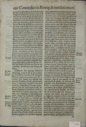
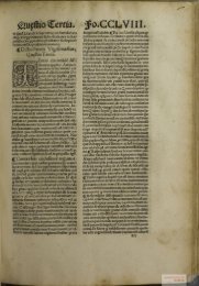
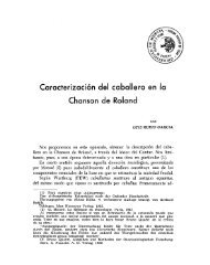
Fig. 1. Organold cultures <strong>of</strong> <strong>fetal</strong> <strong>rat</strong> <strong>liver</strong>s. oblique sections (a. b. e. fj 01 cross-sectiorls \c d. g, tij a. CLI~LII~ pe~lcc.<br />
- 3 weeks; plastic embedd~ng. 1 pm section. G~emsa sta~ning. Liver cell aggregates (.I with numerous vacuoles (s~??al<br />
arrow -= swollen mitochondria and autophag~c vacuoles), single necroses (arrow) 7 250 (b-h). Frozen sections o:<br />
2-week-old cultures. lmm~~n<strong>of</strong>luorescence mlcroscoplc demonst<strong>rat</strong>ion <strong>of</strong> connective tissue matrix components<br />
b. Collagen type Ill. c. Collagen type VI d. Collagen type IV e. Nidogeri. f. Laminin. g. Heparan sulfate proteoglycaii<br />
h. Fibronectin. x 150
Fetal <strong>rat</strong> <strong>liver</strong><br />
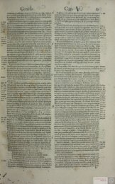
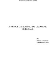
Fig. 2. Culture per~od 7 days a. Hepatocyte aggregate with tannlc ac~d-posit~ve ~natr~x coat (arrow) B~le cap~llar~es ~n the centre<br />
(small arrow), glycogen areas (') w~th ~nd~cat~ons <strong>of</strong> loosen~ng S = s~nus-l~ke cav~ty f 6,000 b. Cell contact between ~IIO hepatocytes<br />
In an aggregate Next to contact areas w~th an ~ntercellular space <strong>of</strong> 20 nm (arrow head), gap junct~on (small arrow) and tannic ac~dnegat~ve<br />
t~ght junct~on (arrow) are seen x 60,000<br />
22 1
Fetal <strong>rat</strong> <strong>liver</strong><br />
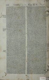
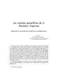
Fig. 3. Culture period = 7 days a. Hepatocyte with two nucle~ In an aggregate show~ng m~tochondr~a<br />
(m) rough ER ( ) and an<br />
autophaglc vacuole (arrow) One nucleus wlth a large nucleolus (n) In a loosened karyoplasm, cell contacts (small arrow) and a b~le<br />
capillary (X) In a nelghbourlng cell X 12,000 b. Secretion granules (arrow) ~n the aplcal reglon <strong>of</strong> 3 contacting hepatocytes (1 2 3)<br />
X 30,000 c. Rough ER <strong>of</strong> a hepatocyte from an aggregate r 60 000
Fetal <strong>rat</strong> <strong>liver</strong><br />
Fig. 4. Culture period = 2 weeks a. Centre <strong>of</strong> a hepatocyte aggregate w~th two b~le cap~llar~es (') Narrowing <strong>of</strong> the ~ntercellulal<br />
space and reduct~on ~n the tann~c ac~d sta~n~ng (arrow) ~n the v~c~n~ty <strong>of</strong> the b~le cap~llar~es A well-developed rough ER (e) and normal<br />
(1) and swollen (2) m~tochondr~a r 24,000 b. B~le cap~llar~es ~n the contact areas <strong>of</strong> four hepatocytes (1-4) In an aggregate<br />
u 35 000 c. Contact area between two hepatocytes In the vlc~n~ty <strong>of</strong> a b~le cap~llary (*) No tann~c ac~d-pos~t~ve sta~nlng (arrow =<br />
t~ght junct~on) ,: 60 000<br />
223
Fetal <strong>rat</strong> <strong>liver</strong><br />
Fig. 5a. Culture per~od 3 weeks Typical polygo,ial <strong>liver</strong> cell with well developed rough ER ( ) and mitochondria (small arroiv)<br />
In neighbouring cells nucleus (n) wlth large nucleolus and numerous swollen mitochondria (m) In the periphery closely attached<br />
bundles <strong>of</strong> collagenous fibrils (arrow) At the edge a vessel-like cav~ty (v) with endothelial lining (e) Between hepatocyte aggregates<br />
and endothel~um a fibroblast-like cell (f) x 9 000 b. Culture period - 7 days Hepatocytes in an aggregate wlth mitochondria (m)<br />
and rough ER At the periphery <strong>of</strong> the cell glycogen areas (g) The aggregate surrounded by a highly tannic acid-positive matrix rlm<br />
(arrow) I = lumen <strong>of</strong> a vessel-like cavity lined by endothel~um (e) 1 9 000 c. Culture period = 2 weeks Periphery <strong>of</strong> a hepatocyte<br />
aggregate (h) with closely attached collagenous fibrils (arrow) Fibroblast-like cell (f) border~ng<br />
on thls aggregate from the outside .( 25 000
Fetal <strong>rat</strong> <strong>liver</strong><br />
Fig. 6. a. Culture pel~od - 2 weeks Vessel-like cav~ty (') betih/een lhepatocyte aggregates (h) w~th granulal tannlc ac~d-pos~t~\ie<br />
structures (serum prote~ns?) erythrocytes (arroih/) and sect~ons <strong>of</strong> h~st~ocytes (H) Cont~nuous lhn~ng by endothel~um-l~ke cells (e)<br />
Between hepatocytes and endothellum f~broblast-l~ke cells (f) or hlstiocytes (Hi 7 6 000 b. Vessel-l~ke cav~ty (*) showing a neutroph~l<br />
granulocyte (G) and an Immature erythrocyte (er) Lln~ng by endothel~al cells (e) At the outside mesenchymal cells (m) w~th collagenous<br />
f~br~ls ~n the ~ntercellular space h = hepatocytes i 12,000<br />
225
Fetal <strong>rat</strong> <strong>liver</strong><br />
After a .i-\\cck culture pcriod (Fig. 5) tlie picture <strong>of</strong> aggregates. BL-niaterilrl can i~ntlouhtecll!, he<br />
these culti~res had principally not changed. But s\\ollcn demonstr.lrtecI imm~~nomorphol~~gic~rlly in otlicr arct~\<br />
mitochondria occurred to an increasing extent in the also. In these \\c ol>\.iou\ly see the electron-dense<br />
liepatocytes. The numher <strong>of</strong> cell necroses. c\peciall! in placlue\ crl I'ila~iientous ancl grani~lar material.<br />
the liematopoictic foci. had risen and tlie ~rmount <strong>of</strong> Tlie mat]-i~ componcnts clemonst<strong>rat</strong>ecl \\,it11 the<br />
connective tissue matrix had increased f~~rtlicr as had tlic inimunomorpllological tcchniq~~e correspond in man!<br />
number <strong>of</strong> cytol!~sosorncs in tlie liepatoc!~tes.<br />
aspect\ to the finclings obtainccl l'or intact <strong>rat</strong> li\,crs.<br />
Collagcn t!,pe I i\ nii\sing. types 111. V. V1 irncl<br />
lmmun<strong>of</strong>luorescence microscopy<br />
fihronectin arc prcscnt. The BL component\ art. also<br />
clcnlonstrahle in sufficient amounts: coll:rgcn [!,pc I\'.<br />
As earlv as after 6 davs in culture different matrix laminin. niclogen ancl lieparan sult'atc protcogl!,can.<br />
components could be demonst<strong>rat</strong>etl imm~~nomorplio- Some differences clo. liowe\~er. exist. Aclult li\,t.r<br />
logically. Their quality and clistril>~rtiv~i tlid not change contains small amounts <strong>of</strong> collagen t!pc I: laminin i\<br />
during the 3-week investigated period (Fig. 1 ). It \s,a\ missing. Tlic\c difi'crences ma!, he due to the <strong>fetal</strong> origin<br />
only the amount that increascd during tlie ~ LI~~LII-C pcriocl. <strong>of</strong> the liepatoc! tes. Our prc\lious result\ (XLI et al.. 1089.<br />
The distribution was similar for a11 antigens. The!. 1001) ha1.e slio~vn that in the ncu-\\orItl prim~rtt. Callithrix<br />
surroiuided the hepatocyte aggregate\ a\ ;I clear-cut rim, jacchus tlie cl~~alitati\~c spectrum <strong>of</strong> the ~iiatrix<br />
occasionally and less clearly defining the hacmatopoietic components <strong>of</strong> tlie <strong>liver</strong> changes tinit.-~lcl>cnclc~itl!.:<br />
foci. Smaller amounts coulcl also be demonst<strong>rat</strong>ecl withi11 before birtli collagen t!,pe 1 is nli\sing. hilt lamillin is<br />
the foci. Between the rims around tlic aggregates expressed. In tlic organoid culturc the licpatoc!tes<br />
antibody binding was seen in the form <strong>of</strong> loosel!, maintain their <strong>fetal</strong> character a\ t'ar a\ this aspect is<br />
distributed, network-like fibres or niorc lionio, ere llt.0LlS conccrnecl. Another problem i\ thc ~~llocation <strong>of</strong> the<br />
areas. Tlie following matrix antigens coulcl be<br />
dcmonstr:rted: collagens type 111, IV. VI. laniinin.<br />
nidogen. 1iep:lran sulfatc proteoglycan, fihronectin.<br />
matrix procluction to certain cdll t!,pcs. Stuclies<br />
performed during tlie last fe\\. !curs li:~\.c \lio\\,n tliat<br />
most cell t!pes are able to pracluce rnnt1.i~ (XLI et al..<br />
1089. 1001 ). Tlie 1iepntoc)~te is especially cap:rhlc <strong>of</strong> tliis<br />
Discussion<br />
function. 13~1t I'ihroI>lasts. ITO-cells ancl cnclotlieli~~l cells<br />
mu\t al\o he mentionccl. The n~u~iihcr <strong>of</strong> fihrohla\t-like<br />
Thc orgnnoid culture (high densit!, culture) <strong>of</strong> the cclls is \el-! lo\v: [TO-cells could not he clenion\trotecl.<br />
vrrriouc cnzyrnutically isolated cell t!,pes <strong>of</strong> <strong>fetal</strong> <strong>rat</strong> li\,er Sincc ncitlier tlie number nor the morpliolog! <strong>of</strong>'<br />
yielded in 100'1.; <strong>of</strong> the cases a <strong>liver</strong>-like ti\sue as earl!, as the c.ndothclial cclls suggest a pronoi~ncecl matri~<br />
after :I ?-day culture pcriod given n sufficient or optimal production. tlie cpitliclial cells in the aggregates<br />
cell densit!,. An inlportant mcclianism for tliis liistiot! pie n2~1st he ~.csponsible for tlic formation <strong>of</strong> the rn~ljor<br />
picture is a sorting-out process where tlic Iiepatoc),te\ arc part cif tlie cpitlielial cells in tlie aggregate\. Thih<br />
attached to one another to form aggregates concept is also s~~pportecl h!, tlic nccurn~~lation <strong>of</strong> m;rtri\<br />
(Zinimerman, 1987). Although tlie compo5ition <strong>of</strong> these 111;rterial ;rt tlic periphery <strong>of</strong> the aggregates.<br />
aggregates is <strong>of</strong>ten difficult to follow in tlie thin slice\. \VC<br />
are in most cases obviousl> dealing \\it11 :I one-la!ered<br />
The electron niicrosconic nicture <strong>of</strong> the c13itheli;1l<br />
I I<br />
cclls in the aggregate\ largcl! corrcsponcls to that 01'<br />
unit <strong>of</strong> polarized cells. In the peripher! the! usually nonii;~l hel3;1toc!,tcs. After 6 c1;1ys or 7 \vecks Ic\ion\ ;~rc<br />
horder on the connective tissue ancl apically the!, are not !.et c;h\er\~etl in tlie cell organelles. Inclu\ions<br />
involved in tlie composition <strong>of</strong> tlie \\all <strong>of</strong> the central<br />
cavities. Thcsc central cavitics clo not hasically cliffer<br />
(c!,tol!~sosomes. autopliagic . - \.acuolcs) lio\~c\.cr. arc not<br />
detectable in vivo. It can he :rssumed that \\c ;Ire dealing<br />
from tlie bile capillaries in vivo.<br />
with prowsses <strong>of</strong> ;rcl;rptation to the culture conclitions<br />
Tlie morphology <strong>of</strong> the border het\vcen epitlieliuni :uid to tlie non-optin1;rl conclitions in vitro. Tlie increa\e<br />
ancl connective tissue in vivo ancl in v~tro \Iio\vs cIc;rr-c~~t <strong>of</strong> inclusions after 3 \\leeks ancl the start <strong>of</strong> the form:~tion<br />
differences. In viva a rim-like clensific:rtion <strong>of</strong> tlie matrix <strong>of</strong> necroses wggest tliat the temporal limit <strong>of</strong> thi\ in vitro<br />
at the sinus pole <strong>of</strong> licputocytes is rnihsing. But this rim model is reached after 3 weeks. Until tliis stage<br />
does not represent a typical hasal lamina. A clearl!, the typical morpholog!~ <strong>of</strong> a licpntoc!'tc is largcl!<br />
defined Laniinrl rara and Lamina densa clo not cle\ elop. maintained: declifferentiatian processes art. not<br />
A striking observation is a \ar!,ing thickne\s. perccptihlc. Froni tliis point oi view the organoicl culture<br />
Collagenous fibrils radiate into this rim. Higher has an ad\,antage c>\.er tlie other culture tecliniclues <strong>of</strong><br />
magnification reveals collagen fihrils and a hasal lamina- 1ic~~;rtoc~~tcs. l'lic main aim <strong>of</strong> thc\c culti\ation<br />
like material that consists <strong>of</strong> fine fila~nents ancl granular experiments is undoubtedly to acliie\,e ancl maintain a<br />
material. We are. therefore. obviousl!. dealing with :I nictnholization in vitro for an cxtcndcd period <strong>of</strong> time.<br />
nii~ture <strong>of</strong> BL-material and the other components <strong>of</strong> the<br />
connecti\,e tissue matrix. This li!~potlicsis i\ supported<br />
Basecl on tlie electron microsco~ic nicture thi\ aim coulcl<br />
1 ,<br />
bc achie\.ed \\lit11 the describecl organoi~l culti~rc<br />
17). the oecurrcncc <strong>of</strong> the different matrix and teclinicl~~c. But one problem ni~~\t fir\[ he solvcci. It is not<br />
BL-antigens. as rcvcaled b), imn~~1nomo1~p1~~~1ogic~1l possil7le to est:rhlisli an organoicl ciilt~~re lc<br />
shown tliat they are mainly located at the border <strong>of</strong> tlie \\,it11 <strong>liver</strong> cells from <strong>fetal</strong> <strong>liver</strong>s. This rai\c\ tlic cl~~c\tion
\\ lictlic~l- tlic\c hepatoc!~tes arc. able to met:rholize. i.e..<br />
\\.l~ctlics the! ;llre:ld! lia\.c the P-450 s!stcm. According<br />
to thC hitlicrto oht;rinecl findings this system dc\.clops in<br />
sat\ sl~o~.tl!. befo~.c or al'ter birth. Howe\er, more recent<br />
fincling\ indic;rle that the cnpatlility to metaholizc can<br />
alrc;rd!. he in~l~rcccl hcforc birth (C'l.c\tcil et ;)I.. l9S2:<br />
Schulr ancl Ncullc~.t. IOSS). First res~~lts inclicate the<br />
possihilit! <strong>of</strong> an incluction ~vith barbitul-atcs. If these<br />
finding\ can hc confirmed. a stahlt. in vitro<br />
mctnlloli~aticln \!stem \L.OLIIC~ be available. Since the<br />
1icl~;rtoc~tes [xoclucc a large a~iiount <strong>of</strong> niatris material.<br />
it can be a\sumcd that :rt least this step ot'difl'crcntiation<br />
can he acliicvcd \kit11 thi.; ol-ganoicl cult~~re tcchnicl~~c.<br />
Acknowledgements. Supported by grants from the Deutsche<br />
Forschungsgemeinschaft awarded to Sfb 174.<br />
References<br />
Begue J.M.. Guguen-Guillouzcco C.. Pasdeloup N.<br />
and Guillouzco A. (1984). Prolonged maintenance <strong>of</strong><br />
act~ve cytochrome P-450 in adult <strong>rat</strong> hepatocytes<br />
cocultered with another <strong>liver</strong> cell type. Hepatology 4,<br />
839-842.<br />
Berry M.N. and Frlend D.S. (1969). High-yield prepa<strong>rat</strong>~on <strong>of</strong><br />
~solated <strong>rat</strong> llver parenchyma1 cells. A biochemical and fine<br />
structural study. J. Cell Biol. 43, 506-520.<br />
B~ssell D.M. and Guzelian P.S. (1 980). Phenotypic stab~lity <strong>of</strong> adult<br />
<strong>rat</strong> hepatocytes in primary monolayer culture. Ann. N.Y. Acad.<br />
Sci. 349. 85-98.<br />
Blssell D.M. and Choun M.O. (1988). The role <strong>of</strong> extracellular<br />
matrix in normal <strong>liver</strong>. Scand. J. Gastroenterol. 23 (Suppl.<br />
151), 1-7.<br />
Carlsen S.A.. Schmell E., Weigel P.H. and Roseman S. (1981).<br />
The effect <strong>of</strong> the method <strong>of</strong> isolation on the surface properties<br />
<strong>of</strong> Isolated <strong>rat</strong> hepatocytes. J. Biol. Chem. 256, 8058-8062.<br />
Cresteil T.. Beaune P,, Kremers P., Flino~s J.P. and Leroux J.P.<br />
(1982). Drug metabolizing enzymes in human foetal <strong>liver</strong>:<br />
part~al resolution <strong>of</strong> mult~ple cytochrome P. 450. Pediatr.<br />
Pharmacol. 2. 199-207.<br />
Diegelmann R.F. (1986). Synthesis <strong>of</strong> extracellular<br />
matrlx components by cultured hepatocytes. In: Research<br />
~n Isolated and Cultured Hepatoyctes. Gillouzo A.<br />
and Guguen-Gu~llouzo C. (eds). John Libbey Eurotext<br />
Ltd./lNSERM. pp 209-224.<br />
Engvall E. and Perlmann P. (1971). Enzyme-linked Immunosorbent<br />
assay. Quantitative assay <strong>of</strong> immunoglobulin G.<br />
lmmunochemistry 8, 871 -874.<br />
Engvall E. and Ruoslahtl E. (1977). Binding <strong>of</strong> a soluble form<br />
<strong>of</strong> f~broblast surface proteln fibronectin to collagen. Int.<br />
J. Cancer 20, 1-5.<br />
Flelschmajer R. and B~ll~ngham R. (1968). Epithelial-<br />
Mesenchymal Interactions. W~lliams and Williams. Baltimore.<br />
Flint O.P. (1986). An in vitro test for te<strong>rat</strong>ogens: Its practical<br />
application. Food. Chem. Toxicol. 24, 627-631.<br />
Guguen-Gu~llouzo C., Seignoux D., Courtois Y.. Br~ssot P,,<br />
Marceau N., Glaise D. and Guillouzo A. (1982). Amplified<br />
phenotyp~c changes in adult <strong>rat</strong> and baboon hepatocytes<br />
Fetal <strong>rat</strong> <strong>liver</strong><br />
cultured on a complex b~omatrix. B~ol. Cell 46, 11-20.<br />
Guguen-Guillouzo C., Clement B., Baffet C., Beaummont C.,<br />
Morel-Chany E.. Glaise D. and Guillouzo A. (1983a).<br />
Maintenance and reversibility <strong>of</strong> active albumin secretion by<br />
adult <strong>rat</strong> hepatocytes CO-cultured with another <strong>liver</strong> epithellal<br />
cell type. Exp. Cell Res. 143, 47-54.<br />
Guguen-Guillouzo C., Baffet G., Clement B.. Begue J.M., Gla~se<br />
D. and Gu~llouzo A. (1983b). Human adult hepatocytes:<br />
isolation and ma~ntenance at high levels <strong>of</strong> speciflc functions<br />
in a CO-cultured system. In: Isolation, characterizat~on and use<br />
<strong>of</strong> hepatocytes. Harrls R.A. and Cornell N.W. (eds). Elsevier.<br />
New York. pp 105-1 10.<br />
Guillouzo A. (1 986). Plasma prote~n production by cultured adult<br />
hepatocytes. In: Research in isolated and cultured<br />
hepatocytes. Gu~llouzo A. and Guguen-Guillouzo C. (eds).<br />
John Libbey Eurotext LTDANSERM. pp 155-170.<br />
Guzellan P.S.. Bissell D.M. and Meyer U.A. (1977). Drug<br />
metabolism in adult <strong>rat</strong> hepatocytes in primary monolayei<br />
culture. Gastroenterology 72, 1232-1 239.<br />
Hannah R., Simkins R. and Eisen H.J. (1980). Synthesis <strong>of</strong> 0-<br />
fetoproteln and album~n by <strong>fetal</strong> mouse <strong>liver</strong> cultured in<br />
chemically defined medium. Dev. Biol. 77, 244-252.<br />
Hassell ,I.R.. Gehron R.P., Barrach H.J., Wilszek J., Rennard S.J.<br />
and Martin G.R. (1 980). Isolation <strong>of</strong> heparan sulfate-contain~ng<br />
proteoglycan from basement membrane. Proc. Natl. Acad.<br />
SCI. USA 77. 4494-4498.<br />
Hata R. and N~nom~ya Y. (1984). Hepatocytes (hepatic<br />
parenchyma1 cells) produce a major part <strong>of</strong> <strong>liver</strong> collagen in<br />
vivo. Biochem. Int. 8, 181-186.<br />
Kistler A., Mislin M. and Gehring A. (1985). Chondrogenesis <strong>of</strong><br />
limb bud cells: improved culture method and the effect <strong>of</strong> the<br />
potent te<strong>rat</strong>ogen retinoic acid. Xenobiotika 15. 673-679.<br />
Kittelberger-Ewert R., Hlnz N.. Oeschner l., Schroter-Kermani C.<br />
and Barrach H.J. (1988). Production and specific~ty <strong>of</strong><br />
antibodies agalnst the central region <strong>of</strong> type II collagen.<br />
Immunol. Invest. 17, 49-61.<br />
Kle~nman H.K.. Klebe R.J. and Martin G.R. (1981). Role <strong>of</strong><br />
collagenous matrlces in the adhesion and growth <strong>of</strong> the cells.<br />
J. Cell Biol. 88, 473-485.<br />
Kreamer B.L.. Staecker J.L., Sawada N., Sattler G.L., Stephen<br />
Hsia M.T. and Pitot H.C. (1986). Use <strong>of</strong> a low-speed, [sodensity<br />
percoll centrifugation method to increase the viability<br />
<strong>of</strong> Isolated <strong>rat</strong> hepatocyte prepa<strong>rat</strong>ions. In vitro 22, 201-21 1.<br />
Lake B.G. and Palne A.J. (1 983). lnduct~on <strong>of</strong> hepatic cytochrome<br />
P-450 and drug metabolism by metyrapone ~n the <strong>rat</strong>:<br />
relevance to ~ts effects in <strong>rat</strong> <strong>liver</strong> cell culture. Xenobiotika 13.<br />
725-730.<br />
Le Dourin N.M. (1975). An experimental analysis <strong>of</strong> <strong>liver</strong><br />
development. Med. Biol. 53, 427-455.<br />
Leffert H.L. and Paul D. (1972). Studies on primary cultures <strong>of</strong><br />
differentiated <strong>fetal</strong> <strong>liver</strong> cells. J. Cell Biol. 52, 559-568.<br />
Lescoat G., Theze N., Clement B., Guillouzo A. and Guguen-<br />
Gu~llouzo C. (1985). Control <strong>of</strong> albumin and U-fetoprotein<br />
secretion by <strong>fetal</strong> and neonatal <strong>rat</strong> hepatocytes maintained In<br />
CO-culture. Cell Differ. 16, 259-269.<br />
Marceau N.. Noel M. and Deschenes J. (1982). Growth and<br />
funct~onal activ~t~es <strong>of</strong> neonatal and adult <strong>rat</strong> hepatocytes<br />
cultured on fibronectin-coated subst<strong>rat</strong>um in serum-free<br />
medlum. In Vitro 18, 1-1 1.<br />
March S.C.. Parikh J. and Cuatrecasas P. (1974). A simplif~ed
method for cyanogen bromide activation <strong>of</strong> agarose for affinity<br />
chromatography. Anal. Biochem. 60, 149-1 52.<br />
Maslansky C.J. and Williams G.M. (1982). Primary cultures and<br />
the levels <strong>of</strong> cytochrome P-450 in hepatocytes from mouse,<br />
<strong>rat</strong>, hamster and rabbit <strong>liver</strong>. In vitro 18, 683-693.<br />
Mennes W.C., Blaanboer B.J. and Van Holsteijn C.W.M. (1988).<br />
Biotransformation and cytotoxicity studies in primary<br />
hepatocyte cultures derived from different mammalian<br />
species. In: Liver Cells and Drugs. Vol. 164. Guillouzo A. and<br />
Guguen-Guillouzo. C. (eds). John Libbey Eurotext. Ltd./<br />
INSERM. pp. 351 -355.<br />
Merker H.-J. (1 986). Die Bedeutung dersog. >-Kultur fiir<br />
dieexperimentelle Embryologie. Verh. Anat. Ges. 80.137-1 49.<br />
Merker H.-J. (1991). Testung von Te<strong>rat</strong>ogenen in vitro. In:<br />
Te<strong>rat</strong>ologie, die Lehre von den Fehlbildungen. VEB Verlag fiir<br />
Medizin. Berlin. (in press).<br />
Merker H.-J., Krijger U. and Zimmermann B. (1 981). Simulation <strong>of</strong><br />
limb bud skeletogenesis in vitro. In: Culture Techniques.<br />
Neubert D. and Merker H.-J. (eds). Walter de Gryuter Verlag.<br />
Berlin. pp 11 9-133.<br />
Merker H.-J., Bremer D., Barrach H.-J. and Gossrau R. (1987).<br />
The basement membrane <strong>of</strong> the persisting maternal blood<br />
vessels in the placenta <strong>of</strong> Callithrix jacchus. Anat. Embryol.<br />
176, 87-97.<br />
Michalopoulos G. and Pitot H.C. (1975). Primary culture <strong>of</strong><br />
parenchymal cells on collagen membranes: morphological<br />
and biochemical observations. Exp. Cell Res. 94, 70-78.<br />
Michalopoulos G., Sattler C.L. and Pitot H.C. (1976). Maintenance<br />
<strong>of</strong> microsomal cytochromes b5 and P-450 in primary cultures<br />
<strong>of</strong> parenchymal <strong>liver</strong> cells on collagen membranes. Life Sci.<br />
18, 1139-1144.<br />
Michalopoulos G., Russel F. and Bile C. (1 979). Primary cultures<br />
<strong>of</strong> hepatocytes on human fibroblasts. In Vitro 15, 796-806.<br />
NakamuraT., Yoshimoto K., NakayamaY., TomitaY. and lchihara<br />
A. (1983). Reciprocal modulation <strong>of</strong> growth and differentiated<br />
functions <strong>of</strong> mature <strong>rat</strong> hepatocytes in primary culture by<br />
cell-cell contact and cell membranes. Proc. Natl. Acad. Sci.<br />
USA 80, 7229-7233.<br />
Paine A.J. and Hockin L.J. (1982). The maintenance <strong>of</strong><br />
cytochrome P-450 in <strong>liver</strong> cell culture: recent studies <strong>of</strong> P-450<br />
mediated mechanisms <strong>of</strong> toxicity. Toxicology. 25, 41 -45.<br />
Paine A.J., Villa P. and Hockin L.J. (1980). Evidence that ligand<br />
formation is a mechanism underlying the maintenance<strong>of</strong> cytochrome<br />
P-450 in <strong>rat</strong> <strong>liver</strong> cell culture. Biochem. J. 188,937-939.<br />
Ratanasavanh D., Beaune P,, Baffet G., Rissel M.. Kremers P,,<br />
Guengerich F.P. and Guillouzo A. (1986). Immunocytochemical<br />
evidence for the maintenance <strong>of</strong> cytochrome P-450<br />
isozymes, NADPH cytochrome C reductase and epoxide<br />
hyd<strong>rat</strong>ase in pure and mixed primary cultures <strong>of</strong> adult human<br />
hepatocytes. Food Chem. Toxicol. 24, 577-578.<br />
Reid L.M. and Rojkind M. (1979). New techniques for culturing<br />
differentiated cells: reconstituted basement membrane rafts.<br />
Methods Enzymol. 58, 263-303.<br />
Riso B. (1983). Morphology <strong>of</strong> the epitelio-mesenchymal<br />
Interaction during lung development <strong>of</strong> the mouse. Cell Differ<br />
13,309-31 8.<br />
Rojkind M., Gatmaitan Z., Mackensen S., Giambrone M.N.,<br />
Ponce P. and Reid L.M. (1980). Connective tissue biomatrix.<br />
Its isolation and utilization for long-term cultures <strong>of</strong> normal <strong>rat</strong><br />
hepatocytes. J. Cell B~ol. 87, 255-263.<br />
Fetal <strong>rat</strong> <strong>liver</strong><br />
Sage H., Woodbury R.G. and Bornstein P. (1979). Structural stu-<br />
dies on human type IVcollagen. J. Biol. Chem. 254,9893-9900.<br />
Sattler C.A., Michalopoulos G., Sattler C.L., and Pitot H.C. (1978).<br />
Clltrastructure <strong>of</strong> adult <strong>rat</strong> hepatocytes cultured on floating<br />
collagen membranes. Cancer Res. 38,1539-1 549.<br />
Sawada N., Tomornura A., Sattler C.A., Sattler C.L., Kleinman<br />
H.K. and Pitot H.C. (1987). Effects <strong>of</strong> extracellular matrix<br />
components on the growth and different~ation <strong>of</strong> cultured <strong>rat</strong><br />
hepatocytes. In Vitro 23, 267-273.<br />
Schaeffer W.I. and Kessler D.J. (1980). Human <strong>liver</strong> cells in<br />
culture. Methods Cell Biol. 21 B, 457-473.<br />
Schulz T. and Neubert D. (1988). Compa<strong>rat</strong>ive studies on the<br />
activity <strong>of</strong> monooxygenases during the perinatal period in<br />
the marmoset and <strong>rat</strong>. in: Non-Human Prlmates-<br />
Developmental Bioology and Toxicology. Neubert D., Merker<br />
H.-J. Hendrickx A.G. (eds). Ueberreuter Wissenschaftsverlag.<br />
Wien - Berlin. pp 353.<br />
Schwarz L.R., Gotz R., Wolff Th. and Wiebel F.J. (1979).<br />
Monooxygenase and glucuronyl transferase activities In<br />
short-term cultures <strong>of</strong> isolated <strong>rat</strong> hepatocytes. FEBS Lett. 98,<br />
203-206.<br />
Segeln P.O. (1973). Prepa<strong>rat</strong>ion <strong>of</strong> <strong>rat</strong> <strong>liver</strong> cells. Ill. Enzymatic<br />
requirementsfor tissue dispersion. Exp. Cell Res. 82,391 -398.<br />
Sipes I.G., Fisher R.L., Smith P.F., Stine E.R.. Gandolfi A.J. and<br />
Brendel K. (1987). A dynamic <strong>liver</strong> culture system: a tool for<br />
studying chemical biotransformation and toxicity. Arch.<br />
Toxicol. Suppl. 11. 20-33.<br />
Sirica E.A. and Pitot H.C. (1980). Drug metabolism and effects <strong>of</strong><br />
carcinogens in cultured hepatic cells. Pharmacol. Rev. 31.<br />
205-228.<br />
Steward A.R., Dannan G.A., Guzelian P. and Guengerich F.P.<br />
(1985). Changes in the concent<strong>rat</strong>ion <strong>of</strong> seven forms <strong>of</strong><br />
cytochrome P-450 in primary cultures <strong>of</strong> adult <strong>rat</strong><br />
hepatocytes. Mol. Pharmacol. 27, 125-132.<br />
Tlmpl R., Rohde H., Geron R.P., Rennard S.J., Foidart J.-M. and<br />
Martin G.R. (1979). Laminin- a glycoprotein from basement<br />
membranes. J. Biol. Chem. 254, 9933-9937.<br />
Towbin H., Staehelin T. and Gordon T. (1979). Electrophoret~c<br />
transfer <strong>of</strong> proteins from polyacrylamide gels to nitrocellulose<br />
sheets: procedure and some applications. Proc. Natl. Acad.<br />
Sci. USA 76,4350-4354.<br />
Trelstad R.L., Catanase V.M. and Rubin D.F. (1976). Collagen<br />
fractionation: sepa<strong>rat</strong>ion <strong>of</strong> native types I, II and Ill by<br />
differential precipitation. Anat. Biochem. 71. 114-1 18.<br />
Williams G.M., Bermudez E. and Scarmuzzino D. (1977). Rat<br />
hepatocyte primary cell cultures. Ill. Improved dissociation<br />
and attachment techniques and the enhancement <strong>of</strong> survival<br />
by culture medium. In Vitro 13, 809-81 7.<br />
Xu Daqun, Schroter-Kermani C., Hinz N. and Merker H.-J. (1989).<br />
Connective tissue <strong>of</strong> the <strong>liver</strong>s <strong>of</strong> newborn and adult<br />
marmosets (jacchus). Histol. Histopath. 4, 479-492.<br />
Xu Daqun, Schroeter-Kermani C., Hinz N. and Merker H.-J.<br />
(1991). lmmun<strong>of</strong>luorescence microscopic localization <strong>of</strong><br />
collagen type IV and VI, laminin and nidogen in the <strong>liver</strong>s <strong>of</strong><br />
Callithrix jacchus during pre- and postnatal development.<br />
Acta Anat. (in press).<br />
Zimmermann B. (1987). Lung organoid culture. Differentiation 36.<br />
86-1 09.<br />
Accepted November 25,1990