Exploring Mushroom Anatomy Lab 2009-2010
Exploring Mushroom Anatomy Lab 2009-2010
Exploring Mushroom Anatomy Lab 2009-2010

You also want an ePaper? Increase the reach of your titles
YUMPU automatically turns print PDFs into web optimized ePapers that Google loves.
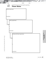
Names: ______________________________________ Date: ___________________ Period: ______ Group #: ______<br />
______________________________________<br />
______________________________________<br />
______________________________________<br />
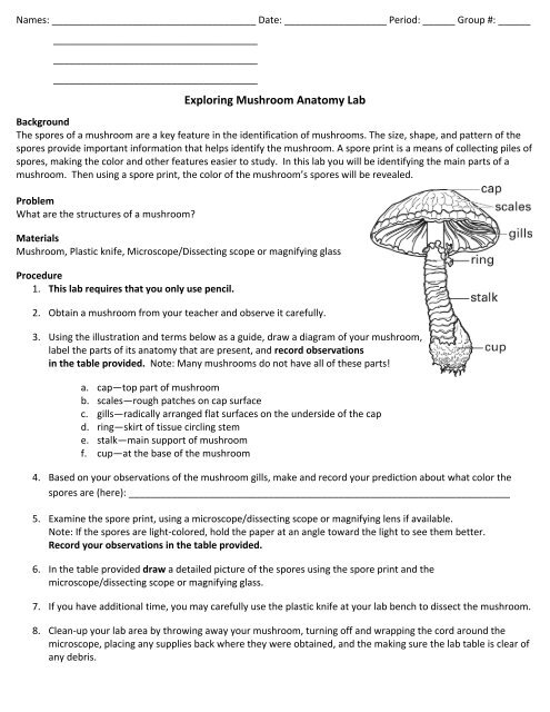
<strong>Exploring</strong> <strong>Mushroom</strong> <strong>Anatomy</strong> <strong>Lab</strong><br />
Background<br />
The spores of a mushroom are a key feature in the identification of mushrooms. The size, shape, and pattern of the<br />
spores provide important information that helps identify the mushroom. A spore print is a means of collecting piles of<br />
spores, making the color and other features easier to study. In this lab you will be identifying the main parts of a<br />
mushroom. Then using a spore print, the color of the mushroom’s spores will be revealed.<br />
Problem<br />
What are the structures of a mushroom?<br />
Materials<br />
<strong>Mushroom</strong>, Plastic knife, Microscope/Dissecting scope or magnifying glass<br />
Procedure<br />
1. This lab requires that you only use pencil.<br />
2. Obtain a mushroom from your teacher and observe it carefully.<br />
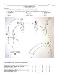
3. Using the illustration and terms below as a guide, draw a diagram of your mushroom,<br />
label the parts of its anatomy that are present, and record observations<br />
in the table provided. Note: Many mushrooms do not have all of these parts!<br />
a. cap—top part of mushroom<br />
b. scales—rough patches on cap surface<br />
c. gills—radically arranged flat surfaces on the underside of the cap<br />
d. ring—skirt of tissue circling stem<br />
e. stalk—main support of mushroom<br />
f. cup—at the base of the mushroom<br />
4. Based on your observations of the mushroom gills, make and record your prediction about what color the<br />
spores are (here): _______________________________________________________________________<br />
5. Examine the spore print, using a microscope/dissecting scope or magnifying lens if available.<br />
Note: If the spores are light‐colored, hold the paper at an angle toward the light to see them better.<br />
Record your observations in the table provided.<br />
6. In the table provided draw a detailed picture of the spores using the spore print and the<br />
microscope/dissecting scope or magnifying glass.<br />
7. If you have additional time, you may carefully use the plastic knife at your lab bench to dissect the mushroom.<br />
8. Clean‐up your lab area by throwing away your mushroom, turning off and wrapping the cord around the<br />
microscope, placing any supplies back where they were obtained, and the making sure the lab table is clear of<br />
any debris.
Analyze and Conclude<br />
Directions: As a group, answer the following questions in complete sentences unless otherwise noted.<br />
These will be given a group grade, so be sure you are a part of the collaboration.<br />
All incomplete sentences could lose up to half credit per answer.<br />
1. Why might it be difficult to predict spore color?<br />
__________________________________________________________________________________________<br />
__________________________________________________________________________________________<br />
__________________________________________________________________________________________<br />
__________________________________________________________________________________________<br />
2. Why do you think the spores are located where they are in the mushroom?<br />
__________________________________________________________________________________________<br />
__________________________________________________________________________________________<br />
__________________________________________________________________________________________<br />
__________________________________________________________________________________________<br />
3. Why was it necessary for your teacher to cover the mushroom cap when it was left out overnight?<br />
__________________________________________________________________________________________<br />
__________________________________________________________________________________________<br />
__________________________________________________________________________________________<br />
__________________________________________________________________________________________<br />
4. Under what type of environmental conditions do you think these spores would be most likely to grow in to<br />
new mycelia?<br />
__________________________________________________________________________________________<br />
__________________________________________________________________________________________<br />
__________________________________________________________________________________________<br />
__________________________________________________________________________________________<br />
5. The color of the external mushroom parts can vary within one species since they may be affected by<br />
environmental conditions. Knowing this, why do you think spores prints are a valuable technique for<br />
mushroom identification?<br />
__________________________________________________________________________________________<br />
__________________________________________________________________________________________<br />
__________________________________________________________________________________________<br />
__________________________________________________________________________________________<br />
__________________________________________________________________________________________
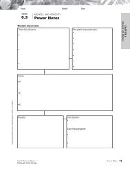
Name: _____________________________________ Date: ______________________ Period: _____ Group #: ______<br />
<strong>Mushroom</strong> <strong>Anatomy</strong> Table<br />
Directions: Fill in the table as neatly and observantly as possible. EACH group member will complete a table of their<br />
own. I know you will be working together, so your answers may be similar, but be sure to use your own original<br />
observations. Staple this sheet (along with your group members’) to the back of the lab.<br />
<strong>Mushroom</strong><br />
Parts<br />
Cap<br />
Scales<br />
Gills<br />
Ring<br />
Stalk<br />
Cup<br />
Spores<br />
Observations<br />
Diagram of <strong>Mushroom</strong><br />
Drawing of Spores<br />
Was your prediction of spore color correct? (Circle one) Yes or No