Hypoplasia-Hyperplasia
Hypoplasia-Hyperplasia
Hypoplasia-Hyperplasia

You also want an ePaper? Increase the reach of your titles
YUMPU automatically turns print PDFs into web optimized ePapers that Google loves.
ע"<br />
שת/<br />
תבט/<br />
ד"<br />
י•<br />
1•<br />
<strong>Hypoplasia</strong> / <strong>Hyperplasia</strong><br />
Syndromes<br />
Post-Graduate Course<br />
Plastic Surgery<br />
Tel Aviv University<br />
Presentation:<br />
Noam Hai – Sha’are Zedek M.C, Jerusalem<br />
Instruction:<br />
Meir Cohen – Schneider’s Children Hospital, Petah Tiqwa<br />
Alex Margulis – Hadassah Ein-Kerem M.C, Jerusalem<br />
Craniofacial Subjects<br />
Clefts<br />
Synostosis<br />
<strong>Hypoplasia</strong>/Atrophy<br />
<strong>Hyperplasia</strong><br />
Combined hyperplasia/hypoplasia<br />
Tumors<br />
Trauma
ע"<br />
שת/<br />
תבט/<br />
ד"<br />
י•<br />
2•<br />
<strong>Hypoplasia</strong>/Dysgenesis<br />
Atrophy<br />
Congenital (Primary)<br />
Craniofacial microsomia / Goldenhar syndrome<br />
Treacher Collins syndrome<br />
Nager, Miller syndromes<br />
Binder syndrome<br />
Pierre-Robin Sequence<br />
Moebius syndrome<br />
Acquired<br />
Hemifacial atrophy (Romberg’s Disease)<br />
Branchial Arches Syndromes<br />
Craniofacial microsomia / Goldenhar<br />
Treacher-Collins Syndrome<br />
Nager<br />
Miller<br />
Moebius<br />
Orofaciodigital Syndromes (I-VIII)<br />
Wildervanck
ע"<br />
שת/<br />
תבט/<br />
ד"<br />
י•<br />
3•<br />
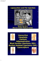
Age - 28 days
ע"<br />
שת/<br />
תבט/<br />
ד"<br />
י•<br />
4•
ע"<br />
שת/<br />
תבט/<br />
ד"<br />
י•<br />
5•<br />
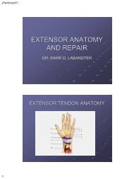
Tensor tympani<br />
Tensor veli palatini<br />
Branchial Arches Derivatives<br />
Stapedius
ע"<br />
שת/<br />
תבט/<br />
ד"<br />
י•<br />
6•<br />
•<br />
Branchial arch<br />
derivatives<br />
Visceral arch Cranial Nerve Muscles Skeletal<br />
structures<br />
I<br />
V (mandibular<br />
branch)<br />
Mastication, mylohyoid,<br />
anterior belly of<br />
digastric, tensor tympany<br />
and t. of palatine curtain<br />
II VII Muscles of facial<br />
expression, stapedius,<br />
stylohyoid, posterior<br />
belly of digastric<br />
III IX Stylopharyngeal and<br />
upper pharyngeal<br />
IV, V, VI<br />
X (superior laryngeall<br />
and recurrent<br />
laryngeal branches)<br />
Meckel’s cartilage,<br />
malleus, incus, mandible,<br />
sphenomandibular<br />
ligament<br />
Reichert’s cartilage,<br />
stapes, styloid process,<br />
lesser cornu of hyoid,<br />
upper part of hyoid bone,<br />
stylohyoid ligament<br />
Greater cornu of hyoid,<br />
lower part of body of<br />
hyoid<br />
Pharyngeal and laryngeal Thyroid, arytenoid,<br />
corniculate and<br />
cuneiform cartilages
ע"<br />
שת/<br />
תבט/<br />
ד"<br />
י•<br />
7•<br />
Congenital / Primary<br />
<strong>Hypoplasia</strong><br />
Atrophy<br />
Dysgenesis<br />
Craniofacial<br />
Microsomia
ע"<br />
שת/<br />
תבט/<br />
ד"<br />
י•<br />
8•<br />
Craniofacial Microsomia<br />
Craniofacial Microsomia<br />
Hemifacial Microsomia (HFM)<br />
Dysostosis Otomandibularis<br />
1 0 and 2 0 Branchial arch<br />
syndrome
ע"<br />
שת/<br />
תבט/<br />
ד"<br />
י•<br />
9•<br />
Introduction<br />
The most common major craniofacial<br />
malformation of hypoplasia of the<br />
skeleton and overlying soft tissue.<br />
Second most common after CLP/CP<br />
Not Genetic<br />
Incidence = 1/ 4000-5500 L.B<br />
15% are bilateral<br />
Pathogenesis<br />
Vascular theory - Stapedial artery:<br />
Temporary embryonic collateral of the hyoid artery<br />
Forms connections with the pharyngeal artery<br />
Replaced by the external carotid artery<br />
Hemorrhagic insult in the 1 0 & 2 0 branchial arches<br />
Hematoma formation<br />
Subsequent ischemia & maldevelopment.
ע"<br />
שת/<br />
תבט/<br />
ד"<br />
י•<br />
10•<br />
Mandible hypoplasia is the key to<br />
diagnosis and management<br />
Pruzansky’s Classification<br />
(Pruzansky, 1969 and Mulliken and<br />
Kaban, 1987). A: Type I: the condyle and<br />
ramus are reduced in size but the overall<br />
morphology is maintained. B: Type IIa:<br />
the ramus and condyle demonstrate<br />
abnormal morphology but the glenoid<br />
fossa has maintained a position in the<br />
temporal bone similar to that of the<br />
contralateral side. C: Type IIb: The<br />
ramus/condyle is hypoplastic and<br />
malformed and displaced outside the<br />
plane of that of the contralateral side. D:<br />
Type III: The ramus is essentially absent<br />
without any evidence of a<br />
temporomandibular joint.
ע"<br />
שת/<br />
תבט/<br />
ד"<br />
י•<br />
11•
ע"<br />
שת/<br />
תבט/<br />
ד"<br />
י•<br />
12•<br />
Munro and Lauritzen<br />
Classification (1985)<br />
The circle in Figure 1A designates the usual site of<br />
skeletal involvement. The midsagittal, midincisor,<br />
occlusal and orbital planes are designated.<br />
Type IA, The craniofacial skeleton is only mildly<br />
hypoplastic and the occlusal plane is horizontal;<br />
Type IB, the skeleton is as in IA, but the occlusal<br />
plane is canted. Type II, the condyle and part of the<br />
affected ramus are absent; Type III, in addition to<br />
the findings in Type II, the zygomatic arch and<br />
glenoid fossa are absent; Type IV, This is an<br />
uncommon type with hypoplasia of the zygoma<br />
and medial and posterior displacement of the<br />
lateral orbital wall; Type V, the most extreme type<br />
has inferior displacement of the orbit with a<br />
decrease in orbital volume.<br />
(from McCarthy, J. G., Grayson, B. H., Coccaro, P.<br />
J., Wood-Smith, D. Craniofacial microsomia. In<br />
McCarthy J. G., ed. Plastic Surgery. Philadelphia:<br />
W. B. Saunders, 1990.)<br />
Clinical Findings<br />
Jaws - The most obvious deformity is the mandible,<br />
esp. ascending ramus, can be absent or reduced in<br />
the vertical dimension.<br />
Other skeletal components - the maxilla,<br />
zygoma, temporal, orbit and cervical vertebrae may<br />
be deformed and hypoplastic.<br />
Muscles of mastication - hypoplastic<br />
Ears - hypoplasia<br />
Nervous system - hypoplasia and facial palsy<br />
Soft tissue - clefts, hypoplasia of the skin, skin tags
ע"<br />
שת/<br />
תבט/<br />
ד"<br />
י•<br />
13•<br />
Clinical Findings<br />
OMENS<br />
Orbital & Other skeletal<br />
Mandible & muscles of mastication<br />
Ear hypoplasia<br />
Nerve (Facial N. palsey)<br />
Soft tissue<br />
Clinical Findings<br />
Mandibular <strong>Hypoplasia</strong> – 89-100%<br />
Muscle hypoplasia – 85-95%<br />
Microtia – 66-99%<br />
Ear tags – 40%<br />
Macrostomia – 17-62%<br />
Facial N. palsy – 10-45%<br />
Palatal deviation - 39%<br />
Orbital discrepancy – 15%<br />
Frontal Plagiocephaly – 5%
ע"<br />
שת/<br />
תבט/<br />
ד"<br />
י•<br />
14•<br />
Clinical Findings<br />
OMENS Plus<br />
Cardiac<br />
Skeletal<br />
Pulmonary<br />
Renal<br />
Gastrointestinal<br />
Limb<br />
Goldenhar Syndrome<br />
Goldenhar
ע"<br />
שת/<br />
תבט/<br />
ד"<br />
י•<br />
15•<br />
Goldenhar Syndrome<br />
Spectrum: Hemifacial microsomia- Goldenhar<br />
Also includes -<br />
Epibulbar dermoids<br />
Lipodermoids or lipomas of the orbital region<br />
Vertebral anomalies<br />
98% - Sporadic 2% - Familial (AD)<br />
Incidence: 1:3500 – 1:25,000<br />
AKA – Oculo-Auriculo-Vertebral (OAV) Dysplasia<br />
Epibulbar Dermoid
ע"<br />
שת/<br />
תבט/<br />
ד"<br />
י•<br />
16•<br />
Facial:<br />
Goldenhar Syndrome<br />
Unilateral <strong>Hypoplasia</strong> Rt.:Lt. = 60:40 1/3 Bilat.<br />
Mandible, Zygoma, Maxilla, Temporal<br />
Muscles of mastication, Soft tissue<br />
Parotid – Agenesis / Hypoplastic / Displaced<br />
Cleft Lip/Palate – 10%<br />
Cardiac: VSD, PDA, TOF, Ao. Coarctation<br />
CNS: Retardation : 5-15% esp. Micro/Anophthalmia<br />
Renal: Ectopy, Hydronephrosis<br />
Skeletal: Club foot & other anomalies<br />
Craniofacial Microsomia<br />
Bilateral, Asymmetric
ע"<br />
שת/<br />
תבט/<br />
ד"<br />
י•<br />
17•<br />
Differential Diagnosis<br />
Treacher Collins<br />
Inheritance.<br />
Symmetry<br />
Other features: (absent in craniofacial microsomia)<br />
Absence of the medial lower eyelashes<br />
Antegonial notching of the mandible<br />
Micrognathia (developmental or posttraumatic)<br />
Underdevelopment restricted to the mandible<br />
No evidence of facial paralysis, ear anomalies, or<br />
soft tissue hypoplasia of the cheeks.
ע"<br />
שת/<br />
תבט/<br />
ד"<br />
י•<br />
18•<br />
Preoperative Assessment<br />
Complete clinical evaluation is mandatory<br />
Look for other organ systems: kidneys, heart.<br />
Photographs: frontal, lateral, oblique, submental<br />
vertex and occlusal views.<br />
Cephalograms (posteroanterior, lateral, and basilar)<br />
Panoramic (Panorex).<br />
3-D CT - Optimal.<br />
Dental models from the Orthodentist<br />
Other: Renal US, Audiometry, Temporal bone CT.<br />
Treatment<br />
ע"<br />
שת/<br />
תבט/<br />
ד"<br />
י•<br />
19•<br />
Treatment<br />
> 14 y :<br />
(1) limited autogenous bone grafting of deficient<br />
portions of the craniofacial skeleton<br />
(2) combined Le Fort I osteotomy, bilateral<br />
mandibular ramisection and genioplasty<br />
(3) bilateral mandibular advancement in patients with<br />
mild to moderate mandibular micrognathia<br />
(4) microvascular free flap to augment the soft<br />
tissues of the face on the affected side<br />
Mandibular Distraction<br />
An intraoral incision is made along<br />
the oblique line of the mandibular<br />
remnant.<br />
Sites of the pinholes and proposed osteotomy<br />
(interrupted line).
ע"<br />
שת/<br />
תבט/<br />
ד"<br />
י•<br />
20•<br />
The pins have been<br />
inserted.<br />
The osteotomy is<br />
performed.<br />
Commencement of distraction with the appliance in position. The arrows<br />
designate the movement of the mandibular segments with formation of bony<br />
regenerate in the resulting gap.
ע"<br />
שת/<br />
תבט/<br />
ד"<br />
י•<br />
21•<br />
Vertical Distraction<br />
Costochondral rib grafts, Le Fort I Osteotomy<br />
Sagittal split contralateral ramus & genioplasty<br />
A: The skeletal deformity in unilateral<br />
(left) craniofacial microsomia. Note the<br />
absent ramus with deviation of the chin to<br />
the affected side and an occlusal cant<br />
upward toward the left side. The arrows<br />
designate the proposed skeletal movements<br />
of the mandible and maxilla. The dots<br />
designate the midpoints of the upper face,<br />
midface and chin. The dotted area<br />
represents the area where bone is removed<br />
from the maxilla and the interrupted lines<br />
designate the proposed maxillary and chin<br />
osteotomy sites. (Modified from Munro, I.<br />
R., Lauritzen C. G. Classification and<br />
treatment of hemifacial microsomia. In:<br />
Caronni, E. P., (ed.). Craniofacial Surgery.<br />
Boston: Little, Brown & Co., 391–400,<br />
1985.)<br />
B: At the completion of the<br />
osteotomies with plate and screw<br />
fixation applied. Note that the dots<br />
have recreated the midsagittal<br />
plane of the craniofacial skeleton.<br />
The ramus has been reconstructed<br />
with costochondral rib grafts. The<br />
Le Fort I segment has been<br />
impacted on the right side and a<br />
bone graft has been inserted on the<br />
left side. The sagittal split<br />
osteotomy has been fixated with<br />
screws and the genioplasty has<br />
been secured with lag screws.
ע"<br />
שת/<br />
תבט/<br />
ד"<br />
י•<br />
22•<br />
C: Lateral view illustrating<br />
the reconstruction of the<br />
zygomatic arch with a rib<br />
graft, the undersurface of<br />
which serves as the<br />
neoglenoid fossa for the<br />
reconstructed ramus/condyle.<br />
D: Close-up view of the<br />
reconstruction of the zygomatic<br />
arch and the glenoid fossa. A cap<br />
of costal cartilage is sutured to the<br />
undersurface of the rib graft to<br />
serve as a glenoid fossa. A<br />
resorbable suture temporarily<br />
secures the condyle to the fossa.<br />
The Le Fort I osteotomy cannot be<br />
performed until the permanent<br />
maxillary teeth have erupted.
ע"<br />
שת/<br />
תבט/<br />
ד"<br />
י•<br />
23•<br />
The combined Le Fort I osteotomy,<br />
bilateral sagittal split of the mandible<br />
and genioplasty in a patient with rightsided<br />
hemifacial microsomia<br />
A: left, Lines of osteotomy. The<br />
ostectomy and site of vertical<br />
impaction are illustrated on the left<br />
maxilla. The solid circles designate<br />
the midpoints of the chin, maxilla,<br />
and orbital region. The arrow shows<br />
the direction of the jaw movements.<br />
B: right, Following movement of the<br />
maxillary, mandibular, and chin<br />
segments and the establishment of<br />
rigid skeletal fixation. Note the<br />
interposition bone graft in the right<br />
maxilla. The solid circles line up<br />
along the craniofacial midsagittal<br />
plane.
ע"<br />
שת/<br />
תבט/<br />
ד"<br />
י•<br />
24•<br />
14 years old male with left<br />
hemifacial microsomia.<br />
An overgrowth of a rib<br />
graft was found 4 years<br />
folowing insertion.<br />
Treacher-Collins<br />
Syndrome (TCS)
ע"<br />
שת/<br />
תבט/<br />
ד"<br />
י•<br />
25•<br />
Introduction<br />
Treacher Collins syndrome (TCS)<br />
AKA Franceschetti-Zwahlen-Klein Syndrome<br />
Mandibulofacial dysotosis<br />
Bilateral 6,7,8 clefts<br />
AD Chr #5<br />
High Penetrance ; Variable expressivity.<br />
Incidence: 1/25,000 - 1/50,000 L.B<br />
Symmetric Bilateral anomalies<br />
1 0 & 2 0 branchial arches derivatives<br />
Nonprogressive - stable deformities with growth<br />
Descriptive criteria<br />
Convex facial profile<br />
“Prominent” nasal dorsum<br />
Depressed Malar bones<br />
Antimongoloid slant<br />
Inferolateral orbital dystopia.<br />
Hypoplastic lower eyelids &<br />
Colobomata<br />
Absence of medial eyelashes<br />
Microtia 60-77%<br />
Long “tongue-shaped”<br />
sideburns
ע"<br />
שת/<br />
תבט/<br />
ד"<br />
י•<br />
26•<br />
Descriptive criteria<br />
Macrostomia (wide commissures)<br />
Parotid hypoplasia<br />
Retrusive lower jaw and chin<br />
Small nasopharynx AW problems<br />
Choanal atresia<br />
Other temporal bone anomalies <br />
Hearing loss (Deafness)<br />
Cleft palate & VPI in 33%<br />
Robin sequence<br />
Teardrop Orbits<br />
Hypoplastic Malar Bones<br />
Hypoplastic\Absent Zygomata<br />
Clefting in most severe cases<br />
Bilateral 6,7,8 clefts<br />
Flat coronoid process<br />
Malformed condyles<br />
X-Ray
ע"<br />
שת/<br />
תבט/<br />
ד"<br />
י•<br />
27•<br />
Description<br />
The most characteristic skeletal<br />
finding is hypoplasia of the<br />
malar bones, often with clefting<br />
through the zygomatic arches.<br />
Description<br />
Maxillary and mandibular bones:<br />
Hypoplastic, antigonial notching of mandibular angle<br />
Variable effects on the TMJs.<br />
Occlusion: Angle class II Ant. open-bite<br />
Steep (clockwise rotated) occlusal plane<br />
Accentuated curve of Spee.<br />
Cranium: Synostosis is not a feature of TCS<br />
But often the neurocranium has abnormal shape<br />
evident during childhood through adulthood:<br />
Decreased A-P length<br />
Diminished bitemporal width
ע"<br />
שת/<br />
תבט/<br />
ד"<br />
י•<br />
28•<br />
Treacher Collins syndrome is an AD condition with variable expressivity. This mother and daughter<br />
demonstrate the extent of variation within a family. The mother (A) was not aware that she carried the<br />
Treacher Collins syndrome gene until after the birth of her daughter (B).
ע"<br />
שת/<br />
תבט/<br />
ד"<br />
י•<br />
29•<br />
An infant with a severe form of Treacher Collins syndrome shown at 2 months of age<br />
and 2 years of age without treatment interventions. There is no progression of the<br />
deformity.<br />
C, D: Three-dimensional CT lateral views at 2 months of age (C) and at 2 years of age (D).<br />
Growth of the condyle, coronoid, and ascending ramus of the mandible is evident.
ע"<br />
שת/<br />
תבט/<br />
ד"<br />
י•<br />
30•<br />
Treatment<br />
Infancy<br />
At birth concerns center on:<br />
AW, Swallowing & feeding, Hearing, Vision,<br />
the presence of cleft palate, with or without<br />
cleft lip and any associated malformations.<br />
Begining in childhood (5-7 years)<br />
3 stage repair<br />
(1) zygomatic and orbital region<br />
(2) maxillomandibular region<br />
(3) nasal region, soft tissues, external ears<br />
auditory canal & middle ear structures.<br />
Soft tissue<br />
Eyelids - Lateral canthopexy and upper eyelid<br />
flaps, grafts (bad results)<br />
Malar hypoplasia –<br />
Pericranial, Temporoparietal flaps (hypoplastic)<br />
Bad results<br />
Auricle reconstruction
ע"<br />
שת/<br />
תבט/<br />
ד"<br />
י•<br />
31•<br />
Zygomatico-Orbital<br />
reconstruction<br />
A 7-year-old boy before and one year after zygomatico-orbital reconstruction.
ע"<br />
שת/<br />
תבט/<br />
ד"<br />
י•<br />
32•<br />
E, F: Contoured full-thickness autogenous cranial bone grafts before inset. Three-dimensional<br />
craniofacial CT scan reviews before and one year after surgery. E: Oblique views. F: Frontal views.<br />
Despite well-intended surgical attempts to<br />
improve the soft tissue deficiencies of the<br />
eyelid-adnexal regions in the patient with<br />
Treacher Collins syndrome, few aesthetically<br />
pleasing soft tissue eyelid reconstructions<br />
have resulted. A: Frontal view of teenager<br />
who underwent transposition of pedicled<br />
upper eyelid skin/muscle flaps to the lower<br />
eyelid deficient regions.<br />
Soft Tissue Deficiency
ע"<br />
שת/<br />
תבט/<br />
ד"<br />
י•<br />
33•<br />
Despite well-intended surgical attempts to improve<br />
the soft tissue deficiencies of the eyelid-adnexal<br />
regions in the patient with Treacher Collins<br />
syndrome, few aesthetically pleasing soft tissue<br />
eyelid reconstructions have resulted. B: A 4-yearold<br />
child with Treacher Collins syndrome after<br />
placement of full-thickness skin grafts to the lower<br />
eyelids. The grafts leave a “patchy,” “operated-on”<br />
look that generally detracts from the result.<br />
Pedicled Osteopericranial<br />
Calvarial flap
ע"<br />
שת/<br />
תבט/<br />
ד"<br />
י•<br />
34•<br />
Distraction Osteogenesis
ע"<br />
שת/<br />
תבט/<br />
ד"<br />
י•<br />
35•<br />
Nager Syndrome<br />
Acrofacial dysostosis AD<br />
Face:<br />
Downward slant of palpebrae<br />
Absent lower eyelashes<br />
Scalp hair on cheeks<br />
Absent/Hypoplastic Mandible<br />
Microtia/Anotia<br />
Middle ear anomalies<br />
Cleft palate<br />
Limb:<br />
Absent Radial Limb<br />
Missing Thumbs<br />
Elbow Ankylosis
ע"<br />
שת/<br />
תבט/<br />
ד"<br />
י•<br />
36•
ע"<br />
שת/<br />
תבט/<br />
ד"<br />
י•<br />
37•<br />
Miller Syndrome<br />
Postaxial acrofacial dysostosis AR<br />
Face:<br />
Downward slant of palpebrae<br />
Retrognathia<br />
Small cup ears<br />
Broad nasal bridge<br />
Cleft palate<br />
Limb:<br />
Short Bowed forearm<br />
Preaxial (Ulnar) hypoplasia<br />
Webbed/Missing fingers<br />
Also fibula/tibia hypoplasia
ע"<br />
שת/<br />
תבט/<br />
ד"<br />
י•<br />
38•<br />
Miller Syndrome<br />
Postaxial acrofacial dysostosis AR<br />
Occasionally:<br />
Cardiac anomalies<br />
Pulmonary anomalies<br />
GE Reflux<br />
Vesicourethral (V-U) Reflux<br />
Undescended testes<br />
DDH<br />
Extra Nipples
ע"<br />
שת/<br />
תבט/<br />
ד"<br />
י•<br />
39•<br />
Binder Syndrome<br />
Maxillonasal dysplasia<br />
Sporadic<br />
Description:<br />
Short nose<br />
Flat bridge<br />
Short columella<br />
Acute nasolabial angle<br />
Class III malocclusion<br />
The Pierre-Robin<br />
Sequence
ע"<br />
שת/<br />
תבט/<br />
ד"<br />
י•<br />
40•<br />
Sequence<br />
Malformation sequence:<br />
Pierre-Robin, Klippel-Feil<br />
Deformation Sequence:<br />
Potter, Digeorge, Amniotic Band,<br />
Disruption sequence:<br />
TORCHES, Alcohol Fetal Synd, Thalidomide<br />
Vitamin A derivatives<br />
Pierre Robin Sequence<br />
Malformation sequence - the primary<br />
defect triggers a chain of secondary and<br />
tertiary events, resulting in what<br />
appears to be multiple anomalies.<br />
The later the defect is induced , the<br />
simpler is the malformation.<br />
Intrauterine<br />
mandibular<br />
hypoplasia<br />
Failure of<br />
tongue<br />
descent<br />
Robin sequence<br />
Cleft<br />
palate
ע"<br />
שת/<br />
תבט/<br />
ד"<br />
י•<br />
41•<br />
Clinical manifestations<br />
Micrognathia<br />
Glossoptosis<br />
High arch palate \ Bifid Uvula \ Submucous cleft<br />
AW obstruction OSAS SIDS<br />
Feeding problems<br />
Imaging
ע"<br />
שת/<br />
תבט/<br />
ד"<br />
י•<br />
42•<br />
Early treatment<br />
Prone positioning & Sat%O 2 monitoring during sleep<br />
In severe cases 24h prone positioning & monitoring<br />
Oxygen supplementation<br />
Gavage feedings<br />
Medical Treatment<br />
Very few require temporary endotracheal intubation,<br />
tracheostomy or tongue-lip adhesion.<br />
Late treatment<br />
Most mandibles reach normal proportions<br />
Surgical Treatment<br />
Distraction Osteogenesis<br />
For severe cases
ע"<br />
שת/<br />
תבט/<br />
ד"<br />
י•<br />
43•<br />
Moebius<br />
Syndrome<br />
Congenital Facial Diplegia<br />
Oculofacial paralysis<br />
Moebius Syndrome<br />
First described by Von-Graefe 1880<br />
Revised & Defined by Moebius 1888 / 92<br />
Congenital Unilateral Palsy added by Henderson 1939<br />
Hallmark:<br />
Unilateral congenital facial (VII) & abducens (VI) palsies.<br />
May be bilateral or involve only the facial N.<br />
15% mild retardation<br />
30% musculoskeletal.<br />
Very rare – Only 300 cases described in English. M=F
ע"<br />
שת/<br />
תבט/<br />
ד"<br />
י•<br />
44•<br />
Pathophysiology<br />
Remains Unknown<br />
Brainstem Nuclei; Nerves or muscle Aplasia<br />
Involved CN: 6-12 except 8<br />
6 (Abducens) – 75%<br />
7 (Facial) – 100%<br />
8 (Vestibulocochlear) – Never<br />
9,10,11 – Variable<br />
12 (Hypoglossal) – Rare<br />
3,4 (Oculomotor, Trochlear)– Very rare<br />
Toxin induced (Moebius)<br />
Vascular etiology:<br />
Theories<br />
- Basilar artery insufficiency<br />
- Premature regression of trigeminal a.<br />
- Subclavian a. disruption sequence<br />
Mesodermal dysplasia of muscles from 1 0 & 2 0 Branchial<br />
arches<br />
CN Nuclei changes are 2 0 to retrograde atrophy<br />
Disruption sequence of normal morphology @ 4-7 wks<br />
(Explains simultaneous limb anomalies)
ע"<br />
שת/<br />
תבט/<br />
ד"<br />
י•<br />
45•<br />
Teratogens assoc. with Facial<br />
paralysis<br />
Thalidomide – Phocomelia, Auricle develop. arrest<br />
Facial & Abducens CN paralysis<br />
Misoprostol (PGE 1 Analogue) – Gastric ulcer<br />
prevention & Rx. Combined with mifepristone or<br />
MTX to induce abortion<br />
If used alone 80% proceed to full term.<br />
96 babies with Moebius – 49% exposed to misoprostol<br />
May induce vascular disruption of subclavian a. during<br />
4-7 weeks causing brain ischemia<br />
Clinical Findings<br />
Facial Diplegia:<br />
Incomplete eye closure desiccation, ulceration<br />
Weak Mouth closure Drooling, feeding difficulties,<br />
food lodges in cheeks. Mastication normal<br />
Swallowing (CN 9,10,12) May be impaired<br />
In mild cases: Inability to smile, Lack of facial<br />
expression, “Mask like” face, no wrinkling<br />
Tremendous social handicap<br />
Speech: inability to close mouth, no labial sounds: b,<br />
v, m, p, ph<br />
Intelligence: 10-15% Mild Retardation
ע"<br />
שת/<br />
תבט/<br />
ד"<br />
י•<br />
46•<br />
Associated anomalies<br />
Anosmia, Hypogonadotropic hypogonadism – Kallman<br />
Autism – 30-40% (Gillberg & Steffenburg)<br />
Flat broad nose, Canthal displacement (Telecanthus)<br />
Flat cheek bones, long face.<br />
Arched palate, Bifid uvula<br />
Microstomia, Micrognathia, Microglossia<br />
Skin:<br />
Absent S.Q fat Café Au Lait spots<br />
Variable phenotype & Cross matching cause difficult<br />
genetic counceling AD, AR, XL patterns described
ע"<br />
שת/<br />
תבט/<br />
ד"<br />
י•<br />
47•<br />
Musculoskeletal anomalies<br />
In Moebius syndrome<br />
Club foot (Bilateral)- 33%<br />
Partial syndactyly<br />
Axillary webs<br />
Arthrogryposis<br />
Congenital amputations<br />
<strong>Hypoplasia</strong> of truncal muscles<br />
(Poland’s synd) 15%<br />
Differential diagnosis<br />
Abducens CN (VI) palsy<br />
Brainstem syndromes<br />
Basilar artery thrombosis<br />
Brainstem gliomas<br />
Congenital muscular dystrophy<br />
Congenital myopathies<br />
Myasthenia gravis<br />
Toxic neuropathies<br />
Spinal muscular dystrophy<br />
Myopathies &<br />
Neuropathies are<br />
progressive<br />
Moebius is static but<br />
may be severe or<br />
even lethal (9/26)<br />
Even tend to improve<br />
with time<br />
Inability to smile
ע"<br />
שת/<br />
תבט/<br />
ד"<br />
י•<br />
48•<br />
Imaging<br />
CT\MRI – May demonstrate Bilateral calcifications<br />
of Basal ganglia & CN nuclei (VI, VII)<br />
EMG – denervation potentials (found 2-3 weeks<br />
after perinatal trauma) may differentiate trauma<br />
from mobius<br />
Brain bipsy is pathognomonic – Calcified necrotic<br />
brainstem CN nuclei<br />
Classification<br />
Towfighi et al<br />
I – Simple hypoplasia atrophy of CN Nuclei<br />
II – Primary lesions in peripheral CN<br />
III – Focal necrosis in brainstem nuclei<br />
IV – Primary myopathy, no CNS or CN lesions
ע"<br />
שת/<br />
תבט/<br />
ד"<br />
י•<br />
49•<br />
Grading of facial paralysis<br />
House-Brackmann<br />
I – Normal<br />
II – Normal symmetry @ rest<br />
III – Obvious but not disfiguring, eye can be closed<br />
IV – Obvious asymmetry @ rest, partial eye closure<br />
V – Barely perceptible movements<br />
VI – No movement<br />
Treatment<br />
Facial reanimation techniques
ע"<br />
שת/<br />
תבט/<br />
ד"<br />
י•<br />
50•<br />
Acquired<br />
Atrophy<br />
<strong>Hypoplasia</strong><br />
Dysgenesis<br />
Progressive<br />
Hemifacial Atrophy<br />
Romberg disease
ע"<br />
שת/<br />
תבט/<br />
ד"<br />
י•<br />
51•<br />
Parry-Romberg Syndrome<br />
First described by Parry (1825)<br />
Further defined by Romberg and Henoch<br />
(1846)<br />
Slowly progressive unilateral atrophy of<br />
facial soft tissues in young adults<br />
May also involve cartilage and bone<br />
Progressive<br />
Hemifacial Atrophy<br />
Symptoms usually begin before 20 y<br />
Progression is medial to lateral<br />
Left > Right<br />
Skin and adnexa:<br />
Hyperpigmentation or vitiligo<br />
Premature Graying<br />
Premature Alopecia
ע"<br />
שת/<br />
תבט/<br />
ד"<br />
י•<br />
52•<br />
Hemifacial Atrophy<br />
Associated Symptoms:<br />
Trigeminal Neuralgia (Facial Pain)<br />
Severe Headaches<br />
Jacksonian Epilepsy (Contralateral)<br />
Etiology – unknown<br />
Course – Progression 3-5 y Stabilization<br />
Theories: Sympathetic hyperstimulation??<br />
DDx: Lipodystrophy – Only fat, Scleroderma<br />
Reconstructive options<br />
Bone: Can be augmented with autologous NVBG<br />
contralateral Iliac crest or split calvarial.<br />
Should be rigidly fixed.<br />
Can use bone substitute PMMA or OHApatite<br />
(Bone-source, Mimix, Norian)<br />
Muscle: muscle flaps tend to atrophy after transfer<br />
thus less recommended. Temporalis.<br />
Fat: Fat injection has been recommended, tend to<br />
resorb more in romberg disease<br />
Skin and fat: Pericranial / Galeal flaps
ע"<br />
שת/<br />
תבט/<br />
ד"<br />
י•<br />
53•<br />
Primary / Congenital<br />
<strong>Hyperplasia</strong><br />
Overgrowth<br />
Hemihypertrophy<br />
Beckwith-Wiedemann<br />
Syndrome<br />
Wiedemann (1964 Germany) Beckwith (1969 USA)<br />
Familial omphalocele with macroglossia<br />
EMG Triad: Exomphalos Macroglossia Gigantism<br />
A Rare Genetic disorder on chr # 11p15.5<br />
Imprinting: Maternal transmission Penetrance<br />
Overexpression of: IGF-2 ; Insulin<br />
1:14,000 L.B Sex & Race - Equal
ע"<br />
שת/<br />
תבט/<br />
ד"<br />
י•<br />
54•<br />
Beckwith-Weidemann<br />
Syndrome<br />
Hepato\Renomegaly<br />
Uncontrolled hypoglycemia<br />
Mental Retardation 2 0 to hypoglycemia<br />
Increased risk for embryonal cancer:<br />
Wilm’s tumor, Adrenocortical neoplasms<br />
Primary / Congenital<br />
Combined<br />
<strong>Hyperplasia</strong>-<strong>Hypoplasia</strong>
ע"<br />
שת/<br />
תבט/<br />
ד"<br />
י•<br />
55•<br />
Etiology - trisomy 21<br />
Down Syndrome<br />
Incidence - 1:1600 L.B. 1:100 if mother over 40 y.<br />
Description:<br />
mongolian fold (epicanthus)<br />
saddle nose<br />
oblique lid axis (mongolian slant)<br />
receding chin (microgenia)<br />
macroglossia and hypotonia of the tongue<br />
hypotonia of lower lip<br />
submental fat collection (double chin)<br />
protruding ears<br />
Down Syndrome<br />
Midface hypoplasia with class III malocclusion<br />
Strabismus (30%).<br />
Treatment<br />
Partial glossectomy (shape of a Gothic arch)- has no effect<br />
on speech (Wexler - there is effect) but may improve<br />
malocclusion<br />
Z-plasty for the epicanthal fold (should be done following<br />
augmentation of the dorsum which corrects some of the<br />
problem)<br />
Otoplasy<br />
Lateral canthoplasty<br />
Le Fort I osteotomy and genioplasty<br />
Augmentation of the zygoma and dorsum of the nose
ע"<br />
שת/<br />
תבט/<br />
ד"<br />
י•<br />
56•<br />
Glossectomies<br />
A B<br />
C<br />
Different types of tongue resection: A - standard, B -<br />
“Gothic arch” for long tongues, C - broad but short tongue.