Surveying and QC of Stereotactic Breast Biopsy Units for ACR ...
Surveying and QC of Stereotactic Breast Biopsy Units for ACR ...
Surveying and QC of Stereotactic Breast Biopsy Units for ACR ...

Create successful ePaper yourself
Turn your PDF publications into a flip-book with our unique Google optimized e-Paper software.
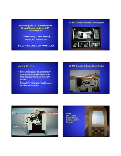
<strong>Surveying</strong> <strong>and</strong> <strong>QC</strong> <strong>of</strong> <strong>Stereotactic</strong><br />
<strong>Breast</strong> <strong>Biopsy</strong> <strong>Units</strong> <strong>for</strong> <strong>ACR</strong><br />
Accreditation<br />
AAPM Spring Clinical Meeting<br />
Phoenix, AZ March 17, 2013<br />
Melissa C. Martin, M.S., F<strong>ACR</strong>, FAAPM, FACMP<br />
Learning Objectives<br />
Become familiar with the recommendations <strong>and</strong><br />
requirements <strong>of</strong> the <strong>ACR</strong> <strong>Stereotactic</strong> <strong>Breast</strong><br />
<strong>Biopsy</strong> Accreditation Program (SBBAP) - 1999<br />
Quality Control Manual In<strong>for</strong>mation <strong>for</strong> image<br />
quality, patient dose, <strong>and</strong> needle placement<br />
accuracy<br />
Become familiar with the operation <strong>and</strong><br />
per<strong>for</strong>mance <strong>of</strong> SBB systems - both prone table<br />
<strong>and</strong> upright add-on systems<br />
LORAD Prone Table<br />
LORAD <strong>Stereotactic</strong> <strong>Breast</strong> <strong>Biopsy</strong> System<br />
LORAD <strong>Stereotactic</strong> <strong>Breast</strong> <strong>Biopsy</strong> System<br />
LORAD<br />
<strong>Stereotactic</strong><br />
<strong>Breast</strong> <strong>Biopsy</strong><br />
System Control<br />
Pendant
Siemens (Fischer)<br />
MammoVision <strong>Biopsy</strong> System<br />
Siemens (Fischer) MammoVision <strong>Biopsy</strong><br />
System<br />
Siemens Mammomat Inspiration SBB Add-On<br />
Affirm <strong>Biopsy</strong> Guidance System<br />
the next generation <strong>of</strong> breast biopsy<br />
State‐<strong>of</strong>‐the‐art solution <strong>for</strong> upright<br />
biopsy procedures<br />
Advanced ergonomic design<br />
Advanced, Ergonomic Design<br />
easy to use <strong>and</strong> install<br />
Novel features to minimize procedure<br />
steps <strong>and</strong> simplify workflow<br />
Versatile <strong>and</strong> flexible solution <strong>for</strong> any<br />
setting<br />
Plat<strong>for</strong>m <strong>for</strong> future advances in biopsy<br />
Lightweight, compact device<br />
– ~7 kg (~15lb)<br />
Balanced design<br />
Easy grasp h<strong>and</strong>les<br />
Installs easily, in just seconds
Enhanced Visualization<br />
intuitive targeting<br />
Uses a novel 10°angle to enter the breast<br />
Fully Integrated System<br />
increased efficiency<br />
• Add‐on to any Dimensions*<br />
• Utilizes existing detector <strong>and</strong> compression<br />
mechanism<br />
– Superb image quality<br />
– Large field <strong>of</strong> view simplifies positioning<br />
70 cm SID –longest in the industry!<br />
– Provides com<strong>for</strong>table working space <strong>and</strong><br />
better patient access<br />
– Allows easier, faster installation <strong>of</strong> biopsy<br />
devices<br />
• Allows you to biopsy under the same<br />
imaging modality<br />
*Affirm comes st<strong>and</strong>ard with a single gantry 2D biopsy license . Additional licenses are available <strong>for</strong> purchase.<br />
Simplified Workflow<br />
streamlined procedures<br />
– <strong>Biopsy</strong> device is removed from path <strong>of</strong> x‐ray <strong>for</strong><br />
unobstructed view <strong>of</strong> lesions<br />
– Uses intuitive, Cartesian targeting<br />
» Allows user to think in Cartesian space <strong>for</strong><br />
targeting<br />
» S<strong>of</strong>tware automatically factors in angle,<br />
making adjustments seamless to users<br />
Fully integrated user interface<br />
– All activities per<strong>for</strong>med on the easy‐to‐use<br />
Dimensions AWS<br />
Automated image acquisition<br />
– C‐arm moves automatically to the appropriate<br />
location <strong>for</strong> stereo views<br />
– Requires minimal steps<br />
– Shortens procedure time<br />
Accurate <strong>and</strong> efficient<br />
– Targeting s<strong>of</strong>tware removes guesswork<br />
– Provides visual feedback <strong>of</strong> needle placement<br />
Versatility <strong>and</strong> Flexibility<br />
wide range <strong>of</strong> use<br />
All Selenia Dimensions are biopsy‐ready <strong>and</strong> tomosynthesis capable<br />
– Dimensions with AWS 5000 may require display upgrade (minimum 2<br />
MP medical grade grayscale monitor)<br />
Affirm is compatible with wide array <strong>of</strong> biopsy devices<br />
– Pre‐programmed <strong>for</strong> Hologic’s ATEC <strong>and</strong> Eviva<br />
Selenia Dimensions with AWS 8000 Selenia Dimensions with AWS 5000<br />
<strong>ACR</strong> SBB Program Statistics<br />
As <strong>of</strong> January 1, 2012, <strong>ACR</strong> has accredited<br />
1052 units at 1020 facilities providing<br />
<strong>Stereotactic</strong> <strong>Breast</strong> <strong>Biopsy</strong> Procedures<br />
In 2011, the first attempt pass rate <strong>for</strong> new<br />
or renewing units was 82%. Almost all<br />
facilities pass on their second attempt at<br />
accreditation after taking appropriate<br />
corrective action to improve image quality<br />
<strong>ACR</strong> SBB Program Statistics<br />
The process typically takes 4 to 6 months.<br />
The review process takes approximately 90<br />
days after the <strong>ACR</strong> receives the submitted<br />
material.<br />
There are currently no MQSA requirements<br />
<strong>for</strong> personnel per<strong>for</strong>ming SBB procedures<br />
but there are training <strong>and</strong> experience<br />
requirements <strong>for</strong> accreditation by the <strong>ACR</strong>
<strong>ACR</strong> Accreditation <strong>of</strong> SBB <strong>Units</strong><br />
Currently, mammography units used<br />
exclusively <strong>for</strong> SBB procedures are not<br />
required to be certified under MQSA<br />
Facilities must have an accredited SBB<br />
program to be named as a Center <strong>of</strong><br />
Excellence <strong>for</strong> <strong>Breast</strong> Care by the <strong>ACR</strong><br />
Goals <strong>of</strong> <strong>QC</strong> <strong>for</strong> <strong>Stereotactic</strong> <strong>Breast</strong> <strong>Biopsy</strong><br />
To ensure that image quality in <strong>Stereotactic</strong><br />
<strong>Breast</strong> <strong>Biopsy</strong> equals or exceeds that <strong>of</strong><br />
screening <strong>and</strong> diagnostic mammography<br />
To ensure that equipment designed<br />
specifically <strong>for</strong> Stereo <strong>Breast</strong> <strong>Biopsy</strong><br />
per<strong>for</strong>ms properly<br />
To ensure that needle localizations are<br />
accurate<br />
General Requirements <strong>for</strong> SBBAP<br />
Qualified TEAM: Physicians, Technologist,<br />
<strong>and</strong> Medical Physicist<br />
Equipment: Table or “add-on”; film or<br />
digital<br />
QA Program, Manual, <strong>and</strong> Committee<br />
Technologist’s <strong>QC</strong> Testing - daily, weekly,<br />
monthly, semi-annual - 6 tests<br />
Medical Physicist’s <strong>QC</strong> Testing - acceptance<br />
<strong>and</strong> annual - 11 tests<br />
Quality Control:<br />
Medical Physicist’s Evaluation<br />
Acceptance Test Be<strong>for</strong>e Patient Use<br />
Report Required as Part <strong>of</strong> <strong>ACR</strong> Application<br />
Annually Thereafter<br />
The 1999 <strong>ACR</strong> SBB Quality Control Manual<br />
has a section <strong>for</strong> the Medical Physicist with<br />
suggested Test Procedures, Forms, <strong>and</strong><br />
Summary Report Format<br />
Detailed instructions on 11 Required<br />
Physicist’s tests<br />
<strong>ACR</strong> Quality Control Manual<br />
• Mammography <strong>QC</strong> Manual<br />
(1990, 1992, 1994, 1999)<br />
• <strong>Stereotactic</strong> <strong>Breast</strong> <strong>Biopsy</strong><br />
<strong>QC</strong> Manual (1999)<br />
• Sent free to all facilities<br />
in program<br />
• To purchase, call <strong>ACR</strong> Pubs: (800) 227-7762<br />
• <strong>QC</strong> <strong>for</strong>ms available to anyone on Web site
Rad Tech <strong>QC</strong> Tests<br />
Mammo <strong>QC</strong> Tests Also Apply if Screen-Film Used<br />
Localization Accuracy - daily<br />
Phantom Image - weekly<br />
Hardcopy Output Quality - monthly, if app<br />
Visual Checklist - monthly<br />
Compression Force - semi-annually<br />
Repeat Analysis - semi-annually<br />
Zero Alignment Test – be<strong>for</strong>e ea patient, if app<br />
Medical Physicist’s Quality Control Tests<br />
1. <strong>Stereotactic</strong> Unit Assembly Evaluation<br />
2. Collimation Assessment<br />
3. Focal Spot Per<strong>for</strong>mance & Digital System<br />
Limiting Resolution<br />
4. kVp Accuracy <strong>and</strong> Reproducibility<br />
5. Beam Quality Assessment (HVL)<br />
6. AEC or Manual Exposure Per<strong>for</strong>mance<br />
Medical Physicist’s Quality Control Tests<br />
7A. Uni<strong>for</strong>mity <strong>of</strong> Screen Speed<br />
7B. Digital Receptor Uni<strong>for</strong>mity<br />
8. <strong>Breast</strong> Entrance Exposure, Average<br />
Gl<strong>and</strong>ular Dose, <strong>and</strong> Exposure Reproducibility<br />
9. Image Quality Evaluation<br />
10. Artifact Evaluation<br />
11. Localization Accuracy Test<br />
<strong>Stereotactic</strong> Unit Assembly Evaluation<br />
Collimation Assessment<br />
LORAD Upright<br />
<strong>Biopsy</strong> Paddle
Measurement Tools Available on Screen<br />
Collimation Assessment<br />
Collimation Assessment SID = 84 cm <strong>for</strong><br />
LORAD SBB Tables<br />
Collimator Assessment <strong>for</strong> Add-On <strong>Units</strong><br />
Actual Opening in<br />
Metal<br />
Compression<br />
Plate is 5 x 5 cm<br />
area<br />
Digital Limiting Resolution/Focal Spot<br />
Digital Limiting Resolution/Focal Spot
Digital Limiting Resolution/Focal Spot<br />
Hologic requires at least 7 lp/mm resolution<br />
kVp Accuracy <strong>and</strong> Reproducibility<br />
kVp Accuracy <strong>and</strong> Reproducibility<br />
Beam Quality Assessment<br />
Beam Quality Specifications <strong>for</strong> SBB <strong>Units</strong><br />
The minimum acceptable Half-Value Layer measurement on a<br />
digital or film/screen SBB unit is<br />
Action<br />
Limit: If measured HVL < (kVp/100) (in mm Al)<br />
or<br />
if measured HVL > (kVp/100) + C (in mm Al)<br />
where C = 0.12 <strong>for</strong> Mo/Mo, C = 0.19 <strong>for</strong> Mo/Rh, <strong>and</strong> C = 0.22 <strong>for</strong> Rh/Rh,<br />
then seek<br />
service<br />
correction.<br />
Image Quality Evaluation (Phantom)<br />
Objective: Ensure Image Quality <strong>for</strong> SBB meets or<br />
exceeds that <strong>of</strong> mammography, <strong>and</strong> to detect<br />
temporal changes in image quality<br />
Procedure: Same as <strong>for</strong> Mammography, except<br />
<strong>ACR</strong> phantom must be imaged in 4 separate<br />
quadrants <strong>for</strong> digital because <strong>of</strong> small field <strong>of</strong> view
Two Types <strong>of</strong> Approved Phantoms<br />
“Mini” <strong>Stereotactic</strong> <strong>Breast</strong> <strong>Biopsy</strong><br />
Accreditation Phantom<br />
Nuclear Associates 18-250<br />
“Mini” <strong>Stereotactic</strong> <strong>Breast</strong><br />
<strong>Biopsy</strong> Accreditation Phantom<br />
Chest Wall Side<br />
SBBAP Testing Criteria<br />
Dose <strong>and</strong> Phantom<br />
Mammography Accreditation<br />
Phantom<br />
RMI 156<br />
Nuclear Associates 18-220<br />
• Dose<br />
- Must be less than 300 mrads (3 mGy)<br />
• Phantom image quality<br />
MAP Phantom Mini Phantom<br />
F/S Digital F/S Digital<br />
Fibers 4.0 5.0 2.0 3.0<br />
Speck<br />
Groups<br />
3.0 4.0 2.0 3.0<br />
Masses 3.0 3.5 2.0 2.5<br />
RMI 156<br />
Accreditation<br />
Phantom<br />
Image Quality <strong>for</strong> SBB <strong>Units</strong><br />
LORAD<br />
Upright<br />
Add-on<br />
with Mini-<br />
Phantom<br />
<strong>for</strong> Image<br />
Quality<br />
Test<br />
RMI 156 or NA 18-220<br />
-MAP Phantom
Nuclear Associates<br />
Digital Mini Phantom<br />
AEC or Manual Exposure Per<strong>for</strong>mance<br />
2 cm<br />
Thickness<br />
AEC or Manual Exposure Per<strong>for</strong>mance<br />
4 cm<br />
Thickness -<br />
Also used <strong>for</strong><br />
Uni<strong>for</strong>mity<br />
<strong>and</strong> Artifacts<br />
AEC or Manual Exposure Per<strong>for</strong>mance<br />
6 cm Thickness 8 cm Thickness<br />
Statistics Tools <strong>for</strong> AEC<br />
Per<strong>for</strong>mance <strong>and</strong><br />
Uni<strong>for</strong>mity Evaluation
AEC or Manual Exposure Per<strong>for</strong>mance<br />
AEC or Manual Exposure Control<br />
Per<strong>for</strong>mance Requirement<br />
Action Limit (Digital): If the signal range<br />
exceeds ±20% <strong>of</strong> signal <strong>for</strong> 4 cm phantom,<br />
revise technique chart.<br />
<br />
Action Limit (Screen-Film): If the density<br />
range exceeds ±0.15 <strong>of</strong> mean, revise<br />
technique chart.<br />
Digital Receptor Uni<strong>for</strong>mity Requirements<br />
For <strong>Units</strong> with ROI statistics measurement<br />
capability:<br />
Action Limit: If SNR(I) / SNR(Center) is ><br />
1.15 or < 0.85, seek service correction.<br />
Digital Receptor Uni<strong>for</strong>mity Requirements<br />
For <strong>Units</strong> without ROI statistics<br />
measurement capability:<br />
Action Limits: If geometric pincushioning > 1 cm<br />
from edge <strong>of</strong> image or<br />
If non-uni<strong>for</strong>m areas (w/o black dots) ><br />
10% <strong>of</strong> image or<br />
If line w/o black dots > 1/4 length <strong>of</strong> image,<br />
seek service correction<br />
Digital Receptor Uni<strong>for</strong>mity<br />
Digital Receptor Uni<strong>for</strong>mity - Image Statistics
<strong>Breast</strong> Entrance Exposure, Average Gl<strong>and</strong>ular<br />
Dose, <strong>and</strong> Exposure Reproducibility<br />
Same Procedure as <strong>for</strong> Mammography<br />
Recommended Signal Level <strong>for</strong> Digital<br />
Digital Matrix Sizes<br />
Per<strong>for</strong>mance Criteria:<br />
a) Coefficient <strong>of</strong> Variation < 0.05<br />
b) Av. Gl<strong>and</strong>ular Dose < 3 mGy <strong>for</strong><br />
Screen/Film <strong>and</strong> <strong>for</strong> Digital Image Receptors<br />
Entrance Exposure/Mid-Gl<strong>and</strong>ular Doses<br />
Entrance Exposure/Mid-Gl<strong>and</strong>ular Doses<br />
Entrance<br />
Exposure<br />
Measurements<br />
on LORAD Addon<br />
<strong>Units</strong><br />
Entrance Exposure/Mid-Gl<strong>and</strong>ular Doses<br />
Artifact Evaluation
11. Localization Accuracy:<br />
Gelatin Phantom<br />
Objective: To assure that the biopsy needle is<br />
accurately placed <strong>for</strong> sampling as directed from the<br />
stereotactic scout images<br />
Technologist to per<strong>for</strong>m test<br />
Physicist to observe <strong>and</strong> analyze results<br />
End-to-End test which supplements the daily in-air<br />
positioning accuracy test<br />
Localization Accuracy: Gelatin Phantom<br />
Method<br />
1. Position Needle:<br />
- Target Lesion Using Stereo Views<br />
- Position Core Needle to Proper X, Y, <strong>and</strong> Z<br />
Coordinates<br />
2. Verify Needle Position:<br />
- Acquire Stereo Pre-fire Images<br />
- Needle Tip should be within Lesion<br />
3. Fire Gun<br />
Localization Accuracy: Gelatin Phantom<br />
Method<br />
4. Verify Post-Fire Position<br />
- Acquire Post-Fire Stereo Images<br />
- Needle Tip should be beyond Center <strong>of</strong> Lesion<br />
5. Verify Sampling <strong>of</strong> Lesion<br />
- Examine Contents <strong>of</strong> Core Sample<br />
Gelatin Phantom <strong>Biopsy</strong><br />
Gelatin <strong>Biopsy</strong> Images<br />
Localization Accuracy
Clinical SBB Procedures - Upright System<br />
Images are acquired on the same Full Field Digital Detector<br />
with the same image processing as original<br />
screening/diagnostic images<br />
First step is to acquire a scout image to confirm lesion<br />
positioning<br />
Clinical SBB Procedures - Upright System<br />
<strong>Stereotactic</strong> image with needle<br />
in Pre-Fire Position<br />
<strong>Stereotactic</strong> image post clip<br />
placement<br />
Question # 1<br />
Questions<br />
The half-value layer acceptable <strong>for</strong> 28 kVp with<br />
a Mo/Mo target/filter on a <strong>Stereotactic</strong><br />
<strong>Breast</strong> <strong>Biopsy</strong> Unit is<br />
A: 0.25 mm Al<br />
B: 0.27 mm Al<br />
C: 0.35 mm Al<br />
D: 0.43 mm Al
Question # 1<br />
The half-value layer acceptable <strong>for</strong> 28 kVp with a<br />
Mo/Mo target/filter on a <strong>Stereotactic</strong> <strong>Breast</strong> <strong>Biopsy</strong><br />
Unit is<br />
C: 0.35 mm Al<br />
Ref: <strong>ACR</strong> <strong>QC</strong> Manual <strong>for</strong> <strong>Stereotactic</strong> <strong>Breast</strong> <strong>Biopsy</strong><br />
(1999), p. 68-69.<br />
Question # 2:<br />
The Mean Gl<strong>and</strong>ular Dose calculated <strong>for</strong> a Mo/Mo<br />
target/filter combination on a <strong>Stereotactic</strong> <strong>Breast</strong><br />
<strong>Biopsy</strong> Unit <strong>for</strong> a 512 x 512 matrix may not exceed<br />
A: 150 mrad<br />
B: 200 mrad<br />
C: 250 mrad<br />
D: 300 mrad<br />
Question # 2<br />
The Mean Gl<strong>and</strong>ular Dose calculated <strong>for</strong> a Mo/Mo<br />
target/filter combination on a <strong>Stereotactic</strong> <strong>Breast</strong><br />
<strong>Biopsy</strong> Unit <strong>for</strong> a 512 x 512 matrix may not exceed<br />
D: 300 mrad<br />
Ref: <strong>QC</strong> Manual <strong>for</strong> <strong>Stereotactic</strong> <strong>Breast</strong> <strong>Biopsy</strong> (1999),<br />
page 80.<br />
Question # 3<br />
Which <strong>of</strong> the following statements is correct<br />
regarding regulatory compliance requirements <strong>of</strong><br />
SBB units?<br />
A: All SBB units must meet MQSA requirements<br />
B: All SBB units must be Accredited by the <strong>ACR</strong><br />
C: All SBB units must meet State Regulatory<br />
Requirements<br />
D: All SBB units are exempt from all MQSA <strong>and</strong><br />
State Regulatory Requirements<br />
Question # 3<br />
Which <strong>of</strong> the following statements is correct<br />
regarding regulatory compliance requirements <strong>of</strong><br />
SBB units?<br />
C: All SBB units must meet State Regulatory<br />
Requirements<br />
Ref: <strong>ACR</strong> SBB Accreditation FAQ Website<br />
Question # 4<br />
When using the “Mini” phantom <strong>for</strong> SBB unit image<br />
quality evaluation, which <strong>of</strong> the following options is<br />
the minimum acceptable image quality scores when<br />
viewed on a digital imaging system?<br />
A: 3 fibers, 3 speck groups, 2.5 masses<br />
B: 3 fibers, 3 speck groups, 3 masses<br />
C: 3 fibers, 3 speck groups, 4 masses<br />
D: 3.5 fibers, 3 speck groups, 3.5 masses
Question # 4<br />
When using the “Mini” phantom <strong>for</strong> SBB unit image<br />
quality evaluation, which <strong>of</strong> the following options is<br />
the minimum acceptable image quality scores when<br />
viewed on a digital imaging system?<br />
A: 3 fibers, 3 speck groups, 2.5 masses<br />
Ref: <strong>ACR</strong> <strong>Stereotactic</strong> <strong>Breast</strong> <strong>Biopsy</strong> Quality Control<br />
Manual (1999), page 91.<br />
Question # 5<br />
When evaluating the AEC or Manual Exposure Control Per<strong>for</strong>mance<br />
with a uni<strong>for</strong>m density phantom varying from 2 to 8 cm in thickness,<br />
the mean signal value range should not exceed which <strong>of</strong> the<br />
following?<br />
A: For phantoms <strong>of</strong> 2 to 6 cm thickness, the mean signal should be<br />
within +/- 15% <strong>of</strong> the mean signal value <strong>for</strong> the 4 cm phantom.<br />
B: For phantoms <strong>of</strong> 2 to 6 cm thickness, the mean signal should be<br />
within +/- 20% <strong>of</strong> the mean signal value <strong>for</strong> the 4 cm phantom.<br />
C: For phantoms <strong>of</strong> 4 to 8 cm thickness, the mean signal should be<br />
within +/- 15% <strong>of</strong> the mean signal value <strong>for</strong> the 4 cm phantom.<br />
D. For phantoms <strong>of</strong> 4 to 8 cm thickness, the mean signal should be<br />
within +/- 20% <strong>of</strong> the mean signal value <strong>for</strong> the 4 cm phantom.<br />
Question # 5<br />
When evaluating the AEC or Manual Exposure Control Per<strong>for</strong>mance<br />
with a uni<strong>for</strong>m density phantom varying from 2 to 8 cm in thickness,<br />
the mean signal value range should not exceed which <strong>of</strong> the<br />
following? .<br />
B: For phantoms <strong>of</strong> 2 to 6 cm thickness, the mean signal should be<br />
within +/- 20% <strong>of</strong> the mean signal value <strong>for</strong> the 4 cm phantom.<br />
Ref: <strong>ACR</strong> <strong>Stereotactic</strong> <strong>Breast</strong> <strong>Biopsy</strong> Quality Control Manual (1999),<br />
page 71.<br />
Melissa C. Martin, M.S., F<strong>ACR</strong>, FAAPM, FACMP<br />
Therapy Physics, Inc.<br />
879 West 190th St., Ste 400, Gardena, CA 90248<br />
Office: 310-217-4114 Cell: 310-612-8127<br />
e-mail: Melissa@TherapyPhysics.com


![SBRT&StereoscopicIGRT_2012 [Compatibility Mode]](https://img.yumpu.com/16889220/1/184x260/sbrtstereoscopicigrt-2012-compatibility-mode.jpg?quality=85)