Evidence based green synthesis of nanoparticles - Advanced ...
Evidence based green synthesis of nanoparticles - Advanced ...
Evidence based green synthesis of nanoparticles - Advanced ...

Create successful ePaper yourself
Turn your PDF publications into a flip-book with our unique Google optimized e-Paper software.
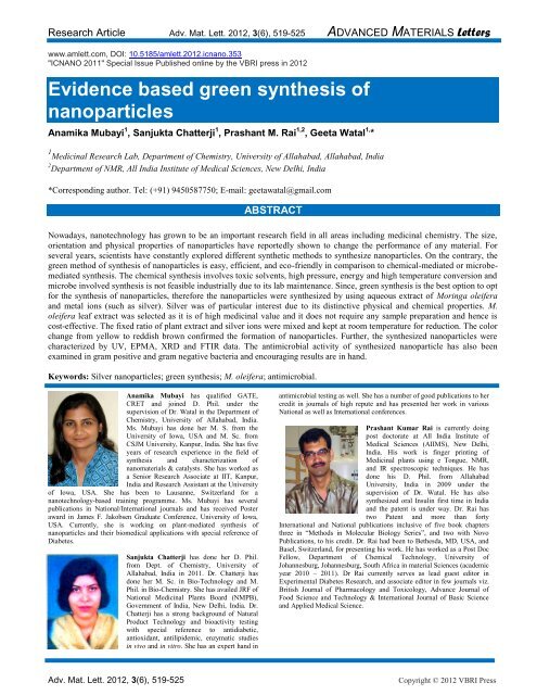
Research Article Adv. Mat. Lett. 2012, 3(6), 519-525 ADVANCED MATERIALS Letters<br />
www.amlett.com, DOI: 10.5185/amlett.2012.icnano.353<br />
"ICNANO 2011" Special Issue Published online by the VBRI press in 2012<br />
<strong>Evidence</strong> <strong>based</strong> <strong>green</strong> <strong>synthesis</strong> <strong>of</strong><br />
<strong>nanoparticles</strong><br />
Anamika Mubayi 1 , Sanjukta Chatterji 1 , Prashant M. Rai 1,2 , Geeta Watal 1, *<br />
1 Medicinal Research Lab, Department <strong>of</strong> Chemistry, University <strong>of</strong> Allahabad, Allahabad, India<br />
2 Department <strong>of</strong> NMR, All India Institute <strong>of</strong> Medical Sciences, New Delhi, India<br />
*Corresponding author. Tel: (+91) 9450587750; E-mail: geetawatal@gmail.com<br />
ABSTRACT<br />
Nowadays, nanotechnology has grown to be an important research field in all areas including medicinal chemistry. The size,<br />
orientation and physical properties <strong>of</strong> <strong>nanoparticles</strong> have reportedly shown to change the performance <strong>of</strong> any material. For<br />
several years, scientists have constantly explored different synthetic methods to synthesize <strong>nanoparticles</strong>. On the contrary, the<br />
<strong>green</strong> method <strong>of</strong> <strong>synthesis</strong> <strong>of</strong> <strong>nanoparticles</strong> is easy, efficient, and eco-friendly in comparison to chemical-mediated or microbemediated<br />
<strong>synthesis</strong>. The chemical <strong>synthesis</strong> involves toxic solvents, high pressure, energy and high temperature conversion and<br />
microbe involved <strong>synthesis</strong> is not feasible industrially due to its lab maintenance. Since, <strong>green</strong> <strong>synthesis</strong> is the best option to opt<br />
for the <strong>synthesis</strong> <strong>of</strong> <strong>nanoparticles</strong>, therefore the <strong>nanoparticles</strong> were synthesized by using aqueous extract <strong>of</strong> Moringa oleifera<br />
and metal ions (such as silver). Silver was <strong>of</strong> particular interest due to its distinctive physical and chemical properties. M.<br />
oleifera leaf extract was selected as it is <strong>of</strong> high medicinal value and it does not require any sample preparation and hence is<br />
cost-effective. The fixed ratio <strong>of</strong> plant extract and silver ions were mixed and kept at room temperature for reduction. The color<br />
change from yellow to reddish brown confirmed the formation <strong>of</strong> <strong>nanoparticles</strong>. Further, the synthesized <strong>nanoparticles</strong> were<br />
characterized by UV, EPMA, XRD and FTIR data. The antimicrobial activity <strong>of</strong> synthesized nanoparticle has also been<br />
examined in gram positive and gram negative bacteria and encouraging results are in hand.<br />
Keywords: Silver <strong>nanoparticles</strong>; <strong>green</strong> <strong>synthesis</strong>; M. oleifera; antimicrobial.<br />
Anamika Mubayi has qualified GATE,<br />
CRET and joined D. Phil. under the<br />
supervision <strong>of</strong> Dr. Watal in the Department <strong>of</strong><br />
Chemistry, University <strong>of</strong> Allahabad, India.<br />
Ms. Mubayi has done her M. S. from the<br />
University <strong>of</strong> Iowa, USA and M. Sc. from<br />
CSJM University, Kanpur, India. She has five<br />
years <strong>of</strong> research experience in the field <strong>of</strong><br />
<strong>synthesis</strong> and characterization <strong>of</strong><br />
nanomaterials & catalysts. She has worked as<br />
a Senior Research Associate at IIT, Kanpur,<br />
India and Research Assistant at the University<br />
<strong>of</strong> Iowa, USA. She has been to Lausanne, Switzerland for a<br />
nanotechnology-<strong>based</strong> training programme. Ms. Mubayi has several<br />
publications in National/International journals and has received Poster<br />
award in James F. Jakobsen Graduate Conference, University <strong>of</strong> Iowa,<br />
USA. Currently, she is working on plant-mediated <strong>synthesis</strong> <strong>of</strong><br />
<strong>nanoparticles</strong> and their biomedical applications with special reference <strong>of</strong><br />
Diabetes.<br />
Sanjukta Chatterji has done her D. Phil.<br />
from Dept. <strong>of</strong> Chemistry, University <strong>of</strong><br />
Allahabad, India in 2011. Dr. Chatterji has<br />
done her M. Sc. in Bio-Technology and M.<br />
Phil. in Bio-Chemistry. She has availed JRF <strong>of</strong><br />
National Medicinal Plants Board (NMPB),<br />
Government <strong>of</strong> India, New Delhi, India. Dr.<br />
Chatterji has a strong background <strong>of</strong> Natural<br />
Product Technology and bioactivity testing<br />
with special reference to antidiabetic,<br />
antioxidant, antilipidemic, enzymatic studies<br />
in vivo and in vitro. She has an expert hand in<br />
antimicrobial testing as well. She has a number <strong>of</strong> good publications to her<br />
credit in journals <strong>of</strong> high repute and has presented her work in various<br />
National as well as International conferences.<br />
Prashant Kumar Rai is currently doing<br />
post doctorate at All India Institute <strong>of</strong><br />
Medical Sciences (AIIMS), New Delhi,<br />
India. His work is finger printing <strong>of</strong><br />
Medicinal plants using e Tongue, NMR,<br />
and IR spectroscopic techniques. He has<br />
done his D. Phil. from Allahabad<br />
University, India in 2009 under the<br />
supervision <strong>of</strong> Dr. Watal. He has also<br />
synthesized oral Insulin first time in India<br />
and the patent is under way. Dr. Rai has<br />
two Patent and more than forty<br />
International and National publications inclusive <strong>of</strong> five book chapters<br />
three in “Methods in Molecular Biology Series”, and two with Novo<br />
Publications, to his credit. Dr. Rai had been to Bethesda, MD, USA, and<br />
Basel, Switzerland, for presenting his work. He has worked as a Post Doc<br />
Fellow, Department <strong>of</strong> Chemical Technology, University <strong>of</strong><br />
Johannesburg, Johannesburg, South Africa in material Sciences (academic<br />
year 2010 – 2011). Dr Rai currently serves as lead guest editor in<br />
Experimental Diabetes Research, and associate editor in few journals viz.<br />
British Journal <strong>of</strong> Pharmacology and Toxicology, Advance Journal <strong>of</strong><br />
Food Science and Technology & International Journal <strong>of</strong> Basic Science<br />
and Applied Medical Science.<br />
Adv. Mat. Lett. 2012, 3(6), 519-525 Copyright © 2012 VBRI Press
Geeta Watal, a graduate from BHU, did<br />
her post-graduation in Chemistry from the<br />
University <strong>of</strong> Allahabad in 1979. Dr. Watal<br />
is actively engaged in Medicinal Research<br />
specifically in treating Diabetes and its<br />
complications. Ten <strong>of</strong> her research students<br />
have been awarded D. Phil. and six are still<br />
working under her able guidance. She had<br />
been invited by the American Diabetes<br />
Technology Society to Philadelphia,<br />
Pennsylvania, USA in Oct.2004, where she<br />
presented her work. She had also been to<br />
Corvallis, Oregon, USA in July 2005 and to<br />
Arlington, Virginia, USA in Aug, 2006 for presenting her work in annual<br />
meets <strong>of</strong> American Society <strong>of</strong> Pharmacognosy. She has handled a number<br />
<strong>of</strong> projects funded by UGC, ICMR and NMPB. In connection with her<br />
collaborative research work, she has travelled to New Paltz campus, New<br />
York University, USA in Oct, 2007 and Harvard University, Washington,<br />
USA in Oct, 2008. Her National collaborators are from All India Institute<br />
<strong>of</strong> Medical Sciences, New Delhi and Delhi University. Dr. Watal has five<br />
Patents for her inventions and more than eighty publications in journals <strong>of</strong><br />
high repute along with three book chapters in “<strong>Advanced</strong> Protocols in<br />
Oxidative Stress; Methods in Molecular Biology Series” to her credit. Dr.<br />
Watal is currently involved in a screening program <strong>of</strong> herbal extracts from<br />
India, and other parts <strong>of</strong> the globe, for their bioactivities. She has invented<br />
a number <strong>of</strong> highly effective medications by unfolding the mystery<br />
involved in the role <strong>of</strong> phytoelements and plant mediated synthesized<br />
compounds at Nano level. Her motto is “Integration <strong>of</strong> natural medicine<br />
into conventional treatments”.<br />
Introduction<br />
The prospect <strong>of</strong> exploiting natural resources for metal<br />
nanoparticle <strong>synthesis</strong> has become to be a competent and<br />
environmentally benign approach [1]. Green <strong>synthesis</strong> <strong>of</strong><br />
<strong>nanoparticles</strong> is an eco-friendly approach which might pave<br />
the way for researchers across the globe to explore the<br />
potential <strong>of</strong> different herbs in order to synthesize<br />
<strong>nanoparticles</strong> [2]. Silver <strong>nanoparticles</strong> have been reported<br />
to be synthesized from various parts <strong>of</strong> herbal plants viz.<br />
bark <strong>of</strong> Cinnamom,[3] Neem leaves,[4-5] Tannic acid[6]<br />
and various plant leaves [7].<br />
Metal <strong>nanoparticles</strong> have received significant attention<br />
in recent years owing to their unique properties and<br />
practical applications [8, 9]. In recent times, several groups<br />
have been reported to achieve success in the <strong>synthesis</strong> <strong>of</strong><br />
Au, Ag and Pd <strong>nanoparticles</strong> obtained from extracts <strong>of</strong><br />
plant parts, e.g., leaves [10], lemongrass [11], neem leaves<br />
[12-13] and others [14]. These researchers have not only<br />
been able to synthesize <strong>nanoparticles</strong> but also obtained<br />
particles <strong>of</strong> exotic shapes and morphologies [12]. The<br />
impressive success in this field has opened up avenues to<br />
develop “<strong>green</strong>er” methods <strong>of</strong> synthesizing metal<br />
<strong>nanoparticles</strong> with perfect structural properties using mild<br />
starting materials. Traditionally, the chemical and physical<br />
methods used to synthesize silver <strong>nanoparticles</strong> are<br />
expensive and <strong>of</strong>ten raise questions <strong>of</strong> environmental risk<br />
because <strong>of</strong> involving the use <strong>of</strong> toxic, hazardous chemicals<br />
[15].<br />
Also, majority <strong>of</strong> the currently prevailing synthetic<br />
methods are usually dependent on the use <strong>of</strong> organic<br />
solvents because <strong>of</strong> hydrophobicity <strong>of</strong> the capping agents<br />
used [16]. Recently, the search for cleaner methods <strong>of</strong><br />
<strong>synthesis</strong> has ushered in developing bio-inspired<br />
approaches. Bio-inspired methods are advantageous<br />
compared to other synthetic methods as, they are<br />
economical and restrict the use <strong>of</strong> toxic chemicals as well<br />
Mubayi et al.<br />
as high pressure, energy and temperatures [17].<br />
Nanoparticles may be synthesized either intracellularly or<br />
extracellularly employing yeast, fungi bacteria or plant<br />
materials which have been found to have diverse<br />
applications.<br />
Silver <strong>nanoparticles</strong> (AgNPs) have been proven to<br />
possess immense importance and thus, have been<br />
extensively studied [18-20]. AgNPs find use in several<br />
applications such as electrical conducting, catalytic,<br />
sensing, optical and antimicrobial properties [21]. In the<br />
last some years, there has been an upsurge in studying<br />
AgNPs on account <strong>of</strong> their inherent antimicrobial efficacy<br />
[22]. They are also being seen as future generation<br />
therapeutic agents against several drug-resistant microbes<br />
[23]. Physicochemical methods for synthesizing AgNPs<br />
thus, pose problems due to use <strong>of</strong> toxic solvents, high<br />
energy consumption and generation <strong>of</strong> by-products.<br />
Accordingly, there is an urgent need to develop<br />
environment-friendly procedures for synthesizing AgNPs<br />
[24]. Plant extracts have shown large prospects in AgNP<br />
<strong>synthesis</strong> [20].<br />
Moringa oleifera (M. oleifera) (Family: Moringaceae,<br />
English name: drumstick tree) has been reported to be<br />
essentially used as an ingredient <strong>of</strong> the Indian diet since<br />
ages. It is cultivated almost all over India and its leaves and<br />
fruits are traditionally used as vegetables. Almost all parts<br />
<strong>of</strong> the plant have been utilized in the traditional system <strong>of</strong><br />
medicine. The plant leaves have also been reported for its<br />
antitumor, cardioprotective, hypotensive, wound and eye<br />
healing properties [25]. AgNPs synthesized from the<br />
aqueous extract <strong>of</strong> M. oleifera leaves in hot condition, have<br />
been reported in literature [26]. In the present study,<br />
<strong>synthesis</strong> <strong>of</strong> AgNPs in cold condition has been reported,<br />
reducing the silver ions present in the silver nitrate solution<br />
by the aqueous extract <strong>of</strong> M. oleifera leaves. Further, these<br />
biologically synthesized <strong>nanoparticles</strong> were found to be<br />
considerably sensitive to different pathogenic bacterial<br />
strains tested.<br />
Experimental<br />
Materials<br />
Chemicals used in the present study were <strong>of</strong> highest purity<br />
and purchased from Sigma-Aldrich (New Delhi, India);<br />
Merck and Himedia (Mumbai, India). M. oleifera leaves<br />
were collected locally from University <strong>of</strong> Allahabad,<br />
Allahabad, Uttar Pradesh, India.<br />
Preparation <strong>of</strong> plant extract<br />
Plant leaf extract <strong>of</strong> M. oleifera was prepared by taking 5 g<br />
<strong>of</strong> the leaves and properly washed in distilled water. They<br />
were then cut into fine pieces and taken in a 250 mL<br />
Erlenmeyer flask with 100 mL <strong>of</strong> sterile distilled water.<br />
The mixture was boiled for 5 min before finally filtering it.<br />
The extract thus obtained was stored at 4 °C and used<br />
within a week [7].<br />
Synthesis <strong>of</strong> silver <strong>nanoparticles</strong><br />
The aqueous solution <strong>of</strong> 1 mM silver nitrate (AgNO3) was<br />
prepared to synthesize AgNPs. 190 mL <strong>of</strong> aqueous solution<br />
Adv. Mat. Lett. 2012, 3(6), 519-525 Copyright © 2012 VBRI press 520
Research Article Adv. Mat. Lett. 2012, 3(6), 519-525 ADVANCED MATERIALS Letters<br />
<strong>of</strong> 1 mM AgNO3 was slowly added to10 mL <strong>of</strong> M. oleifera<br />
aqueous leaf extract while stirring, for reduction into Ag<br />
ions and kept at room temperature for 6 h [7, 27].<br />
UV-Vis spectra analysis<br />
UV-Vis spectrum <strong>of</strong> the reaction medium recorded the<br />
reduction <strong>of</strong> pure Ag + ions at 6 h after diluting the sample<br />
with distilled water. UV-Vis spectral analysis was<br />
performed by using UV-Vis double beam<br />
spectrophotometer [UV-1700 PharmaSpec UV-Vis<br />
Spectrophotometer (Shimadzu)].<br />
XRD (X-ray diffraction) measurement<br />
The AgNP solution was repeatedly centrifuged at 5000 rpm<br />
for 20 min, re-dispersed with distilled water and<br />
lyophilized to obtain pure AgNPs pellets. The dried<br />
mixture <strong>of</strong> AgNPs was collected to determine the formation<br />
<strong>of</strong> AgNPs by X’Pert Pro x-ray diffractometer (PANalytical<br />
BV, The Netherlands) operated at a voltage <strong>of</strong> 30 kV and a<br />
current <strong>of</strong> 30 mA with CuKα radiation in a θ- 2θ<br />
configuration.<br />
EPMA analysis<br />
EPMA-WDS analyses <strong>of</strong> carbon coated samples were<br />
performed using an Electron Probe Micro Analysis JEOL<br />
Superprobe (JEOL JXA-8100, Japan) with X-ray<br />
wavelength dispersive spectroscopy.<br />
FTIR (Fourier Transform Infrared) analysis<br />
The earlier centrifuged and redispersed AgNPs obtained<br />
have removed any free residual biomass. Subsequently, the<br />
dried powder was obtained by lyophilizing the purified<br />
suspension. The resulting lyophilized powder was<br />
examined by Infrared (IR) spectra, recorded on a Bruker<br />
Vector-22 Infrared spectrophotometer using KBr pellets.<br />
Bacterial strains <strong>of</strong> Klebsiella pneumoniae (Gramnegative),<br />
Staphylococcus aureus (Gram-positive),<br />
Pseudomonas aeruginosa (Gram-negative), Enterococcus<br />
faecalis (Gram-negative) and Escherichia coli (Gramnegative)<br />
were clinical isolates obtained from the<br />
Department <strong>of</strong> Biotechnology, All India Institute <strong>of</strong><br />
Medical Sciences (AIIMS), New Delhi, India and the<br />
microbiologist <strong>of</strong> the department confirmed the identity<br />
<strong>based</strong> on microscopic examination, Gram’s character, and<br />
biochemical test pr<strong>of</strong>ile. Bacterial stocks were maintained<br />
and stored as 1 ml aliquots at -80ºC in Luria Bertani (LB)<br />
broth for all the five bacterial strains. Bacterial stocks were<br />
revived from -80ºC and grown in Luria Bertani (LB) broth<br />
for Klebsiella pneumoniae, Staphylococcus aureus,<br />
Pseudomonas aeruginosa, Enterococcus faecalis and<br />
Escherichia coli. All cultures were grown overnight at<br />
37ºC ± 0.5°C, pH 7.4 in a shaker incubator (190-220 rpm).<br />
Their sensitivity to the reference drug, Streptomycin<br />
(Sigma-Aldrich, New Delhi, India) was also checked.<br />
Luria Bertani broth (Himedia), Luria Bertani agar<br />
(Himedia) standard antibiotic and Streptomycin (Himedia)<br />
were used in antimicrobial sensitivity testing. Briefly,<br />
Luria Bertani (LB) broth/agar medium was used to<br />
cultivate bacteria. Fresh overnight cultures <strong>of</strong> inoculum (50<br />
μl) <strong>of</strong> each culture were spread on to LB agar plates. Sterile<br />
paper discs <strong>of</strong> 5 mm diameter (containing 30 μl <strong>of</strong> AgNPs)<br />
along with the standard antibiotic, Streptomycin containing<br />
discs were placed in each plate. Antimicrobial activities <strong>of</strong><br />
the synthesized AgNPs were determined, using the agar<br />
disc diffusion assay method [27].<br />
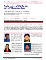
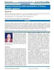
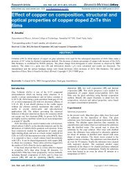
Fig. 1. UV-Vis absorption spectrum <strong>of</strong> AgNPs synthesized by treating<br />
1mM AgNO3 solution with M. oleifera leaf extract after 6 hrs.<br />
Results and discussion<br />
UV -Visible studies<br />
UV–Vis spectroscopy is an important technique to<br />
establish the formation and stability <strong>of</strong> metal <strong>nanoparticles</strong><br />
in aqueous solution [28].The relationship between UVvisible<br />
radiation absorbance characteristics and the<br />
absorbate’s size and shape is well-known. Consequently,<br />
shape and size <strong>of</strong> <strong>nanoparticles</strong> in aqueous suspension can<br />
be assessed by UV-visible absorbance studies.<br />
Fig. 1 depicts the absorbance spectra <strong>of</strong> reaction<br />
mixture containing aqueous silver nitrate solution (1 mM)<br />
and M. oleifera leaf broth (prepared from 5 g leaf material).<br />
The absorption spectra obtained reveal the production <strong>of</strong><br />
AgNPs within 6 h. On adding the afore-mentioned plant<br />
broth to AgNO3 solution, the solution changed from<br />
yellowish orange to brown. The final color turns deep and<br />
finally, brownish with passage <strong>of</strong> time. The intensity <strong>of</strong> the<br />
absorbance was found to increase as the reaction proceeded<br />
further.<br />
AgNPs displaying intense yellowish brown colour in<br />
water arises from the surface plasmons. This is due to the<br />
dipole oscillation arising when an electromagnetic field in<br />
the visible range is coupled to the collective oscillations <strong>of</strong><br />
conduction electrons. It is an established fact that metal<br />
<strong>nanoparticles</strong> ranging from 2 to 100 nm in size demonstrate<br />
strong and broad surface plasmon peak [29]. The optical<br />
absorption spectra <strong>of</strong> metal <strong>nanoparticles</strong> that are governed<br />
by surface plasmon resonances (SPR), move towards<br />
elongated wavelengths, with the increase in particle size.<br />
The absorption band position is also strongly dependent on<br />
dielectric constant <strong>of</strong> the medium and surface-adsorbed<br />
species [30].<br />
As postulated by Mie's theory, spherical <strong>nanoparticles</strong><br />
results in a single SPR band in the absorption spectra. On<br />
the other hand, anisotropic particles provide two or more<br />
Adv. Mat. Lett. 2012, 3(6), 519-525 Copyright © 2012 VBRI press
SPR bands depending on the particle shape [31]. In the<br />
present study, a reaction mixture confirms a single SPR<br />
band disclosing spherical shape <strong>of</strong> AgNPs. The reduction<br />
<strong>of</strong> Ag+ ions was validated by performing qualitative<br />
analysis for free Ag + ions presence with NaCl in the<br />
supernatant obtained after centrifugation <strong>of</strong> the reaction<br />
mixture. The AgNPs obtained from the reaction mixture<br />
consisting <strong>of</strong> 1mM AgNO3 and leaf extract, were purified<br />
and further examined. The AgNPs were found to be<br />
amazingly stable even after 6 months.<br />
Rapid <strong>synthesis</strong> <strong>of</strong> steady AgNPs using leaf broth (20<br />
g <strong>of</strong> leaf biomass) and 1 mM aqueous AgNO3 have been<br />
reported by Sastry et al. [10] Shivshankar et al. [11] have<br />
reported rapid <strong>synthesis</strong> <strong>of</strong> stable gold, silver and bimetallic<br />
Ag/Au core shell <strong>nanoparticles</strong> using 20 g <strong>of</strong> leaf<br />
biomass <strong>of</strong> 1mM aqueous AgNO3. Similarly, Pratap et al.<br />
[29] reported <strong>synthesis</strong> <strong>of</strong> gold and silver <strong>nanoparticles</strong><br />
with leaf extract using ammonia as a speed-up agent for<br />
<strong>synthesis</strong> <strong>of</strong> silver. All the above research papers used leaf<br />
broth made by boiling finely chopped fresh leaves. This<br />
procedure involves incessant agitation <strong>of</strong> the broth after<br />
addition <strong>of</strong> salt solution.<br />
The reduction <strong>of</strong> the silver ions is moderately rapid at<br />
ambient conditions. This is innovative and interesting to<br />
the field <strong>of</strong> material science as, the evaluated leaf biomass<br />
was found to have the capability to reduce metal ions at<br />
ambient circumstances. Furthermore, the biomass handling<br />
and processing is less rigid since, it does not involve<br />
boiling for long hours or successive treatment.<br />
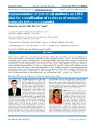
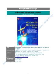
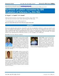
Fig. 2. XRD pattern <strong>of</strong> the <strong>green</strong> synthesized AgNPs.<br />
XRD studies<br />
Fig. 2 shows the XRD-spectrum <strong>of</strong> purified sample <strong>of</strong><br />
AgNPs. The peaks observed in the spectrum at 2θ values <strong>of</strong><br />
38.07°, 44.15° and 64.49° corresponds to 111, 200 and 220<br />
planes for silver, respectively [15].<br />
Some unidentified peaks were also observed near the<br />
characteristic peaks. A peak at 46º is possibly due to<br />
crystalline nature <strong>of</strong> the capping agent [32, 10]. This<br />
clearly shows that the AgNPs are crystalline in nature due<br />
to reduction <strong>of</strong> Ag + ions by M. oleifera leaf extract. The<br />
AgNPs were centrifuged and redispersed in distilled water<br />
several times before XRD and EPMA analysis, as<br />
mentioned earlier. This excludes the possibility <strong>of</strong> any free<br />
compound/protein present that might lead to independent<br />
crystallization and thus, resulting in Bragg’s reflections.<br />
Usually, the particle size is responsible for the broadening<br />
<strong>of</strong> peaks in the XRD pattern <strong>of</strong> solids [33]. The noise due<br />
Mubayi et al.<br />
to the protein shell surrounding the <strong>nanoparticles</strong> is visible<br />
from the spectrum [34].<br />
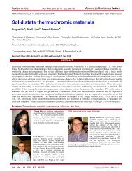
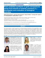
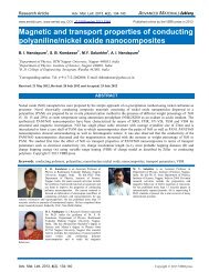
Fig. 3. EPMA micrograph <strong>of</strong> the AgNPs<br />
EPMA<br />
An EPMA was employed to examine the structure <strong>of</strong> the<br />
<strong>nanoparticles</strong> that were synthesized. Representative EPMA<br />
micrographs <strong>of</strong> the reaction mixtures comprising 10 ml <strong>of</strong><br />
M. oleifera leaf extract and 1 mM <strong>of</strong> silver nitrate<br />
magnified 30,000 times are shown in Fig. 3. From the<br />
figure, it is clear that the AgNPs adhered to nano-clusters.<br />
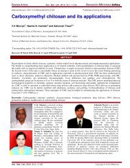
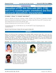
Fig. 4. FTIR spectra <strong>of</strong> dried powder <strong>of</strong> (a) M. oleifera leaf extract (b)<br />
<strong>nanoparticles</strong> synthesized by leaf extract solution.<br />
FTIR studies<br />
The exact procedures <strong>of</strong> bio-reduction is not fully<br />
comprehended and have reported that the reduction <strong>of</strong> Ag +<br />
to Ag <strong>nanoparticles</strong> takes place possibly in the presence <strong>of</strong><br />
the enzyme, NADPH-dependent dehydrogenase [32]. The<br />
precise direction in which the electrons are transported is a<br />
matter requiring investigation. Moreover, the information<br />
concerning environment being responsible for high stability<br />
<strong>of</strong> metal <strong>nanoparticles</strong>, is not widely available. FTIR<br />
investigation <strong>of</strong> isolated AgNPs free from proteins and<br />
water-soluble compounds was performed in this direction.<br />
The analysis <strong>of</strong> IR spectra throws light on biomolecules<br />
bearing different functionalities present in fundamental<br />
system. Representative spectra (Fig. 4b) clearly show the<br />
purified <strong>nanoparticles</strong> showed the presence <strong>of</strong> bands due to<br />
Adv. Mat. Lett. 2012, 3(6), 519-525 Copyright © 2012 VBRI press 522
Research Article Adv. Mat. Lett. 2012, 3(6), 519-525 ADVANCED MATERIALS Letters<br />
O–H stretching (~3315cm−1), aldehydic CH stretching<br />
(~2914 cm −1 ), C=C group (~1627 cm −1 ) and C–O stretch<br />
(~1043 cm −1 ).<br />
The FTIR spectra <strong>of</strong> biomass (Fig. 4a) show bands<br />
~3403, ~2919, ~2048, ~1604, ~1504, ~1402, ~1270 and<br />
~1045 cm -1 . The band ~1045 cm -1 can be allotted to the<br />
ether linkages or -C-O-,11,14 (Fig. 4a). The band at ~1045<br />
cm-1 largely might be due to the -C-O- groups <strong>of</strong> the<br />
polyols viz. flavones, terpenoids and the polysaccharides<br />
present in the biomass [14]. The absorbance band ~1604<br />
cm-1 (Fig. 4a) is associated with the stretching vibration <strong>of</strong><br />
-C=C- or aromatic groups [11, 14]. The band ~2048 may<br />
be due to C=O stretching vibrations <strong>of</strong> the carbonyl<br />
functional group in ketones, aldehydes, and carboxylic<br />
acids.10,29,14 Also, the spectrum (Fig. 4a) also reveals an<br />
intense band ~2919 cm -1 allocated to the asymmetric<br />
stretching vibration <strong>of</strong> sp 3 hybridized -CH2 groups [32].<br />
The band ~1627 cm -1 (Fig. 4b) is due to amide-I bond<br />
<strong>of</strong> proteins, indicating predominant surface capping species<br />
having -C=O functionality which are mainly responsible<br />
for stabilization. A broad intense band ~3400 cm −1 in both<br />
the spectra can be contributed to the N-H stretching<br />
frequency arising from the peptide linkages present in the<br />
proteins <strong>of</strong> the extract [32]. The shoulders around the band<br />
can be specified as the overtone <strong>of</strong> the amide-II band and<br />
the stretching frequency <strong>of</strong> the O-H band, possibly arising<br />
from the carbohydrates and/or proteins present in the<br />
sample. The flattening <strong>of</strong> the shoulders in Fig 1b indicates<br />
decrease in the concentration <strong>of</strong> the peptide linkages in the<br />
solution [32]. The spectra Fig. 4b also shows broad<br />
asymmetric band ~2100 cm −1 that can be assigned to the N-<br />
H stretching band in the free amino groups <strong>of</strong> AgNPs. The<br />
bands <strong>of</strong> functional groups such as -C-O-C- , -C-O- and -<br />
C=O are obtained from the heterocyclic water soluble<br />
compounds in the biomass, which as observed in the IR<br />
spectra <strong>of</strong> biomass is in good agreement with the value<br />
reported in the literature.<br />
Gold <strong>nanoparticles</strong> are thoroughly investigated in the<br />
polyol <strong>synthesis</strong> and both oxygen and nitrogen atoms <strong>of</strong><br />
pyrrolidone unit can assist the adsorption <strong>of</strong> PVP on to the<br />
surface <strong>of</strong> metal nanostructures to safeguard the<br />
synthesized <strong>nanoparticles</strong> [35]. Similarly, the oxygen atoms<br />
here might assist the adsorption <strong>of</strong> the heterocyclic<br />
components on to the particle surface in stabilizing the<br />
<strong>nanoparticles</strong>. It is also obvious from the two spectra <strong>of</strong><br />
biomass and AgNPs that the flavones lead to the bioreduction.<br />
Flavones could be adhered to the surface <strong>of</strong> the<br />
metal <strong>nanoparticles</strong>, probably by interaction through πelectrons<br />
<strong>of</strong> carbonyl groups in the absence <strong>of</strong> other strong<br />
ligating agents in adequate concentrations [11].<br />
Antibacterial studies<br />
Antibacterial activity <strong>of</strong> biogenic AgNPs was evaluated by<br />
using standard Zone <strong>of</strong> Inhibition (ZOI) microbiology<br />
assay. The <strong>nanoparticles</strong> showed inhibition zone against<br />
almost all the studied bacteria (Table 1, Fig 5).<br />
Maximum ZOI was found to be 12 mm for<br />
Escherichia coli (Clinical isolate) and no ZOI for<br />
Pseudomonas aeruginosa. Whereas, the other three<br />
bacterial strains <strong>of</strong> Staphylococcus aureus, Klebsiella<br />
pneumoniae and Enterococcus faecalis showed ZOI <strong>of</strong> 7, 9<br />
and 6 mm whereas, ZOI <strong>of</strong> the standard antibiotic,<br />
streptomycin obtained against Escherichia coli was found<br />
to be 11 mm (Table 1).<br />
Fig. 5. Antimicrobial activity <strong>of</strong> silver <strong>nanoparticles</strong> synthesized from M.<br />
oleifera leaf extract against microorganism (bacteria).<br />
Table 1. Zone <strong>of</strong> Inhibition <strong>of</strong> AgNPs <strong>synthesis</strong>ed from M. oleifera leaf<br />
extract.<br />
Bacterial Strains Zone <strong>of</strong> Inhibition (mm)<br />
AgNPs Reference drug*<br />
Staphylococcus aureus 7 16<br />
Escherichia coli 12 11<br />
Klebsiella pneumoniae 9 14<br />
Enterococcus faecalis 6 17<br />
Pseudomonas aeruginosa - 18<br />
AgNO3 which is readily soluble in water has been<br />
exploited as an antiseptic agent for many decades [36].<br />
Dilute solution <strong>of</strong> silver nitrate has been used since the 19th<br />
century to treat infections and burns [37]. The exact<br />
mechanism <strong>of</strong> the antibacterial effect <strong>of</strong> silver ions is<br />
partially understood. Literature survey reveals that the<br />
positive charge on the Ag ion is crucial for its antimicrobial<br />
activity. The antibacterial activity is probably derived,<br />
through the electrostatic attraction between negativelycharged<br />
cell membrane <strong>of</strong> microorganism and positivelycharged<br />
<strong>nanoparticles</strong> [38-40]. However, Sondi and<br />
Salopek-Sondi [41] reported that the antimicrobial activity<br />
<strong>of</strong> AgNPs on Gram-negative bacteria was dependent on the<br />
concentration <strong>of</strong> AgNPs and was closely associated with<br />
the formation <strong>of</strong> pits in the cell wall <strong>of</strong> bacteria.<br />
Accumulation <strong>of</strong> the AgNPs in the pits results in the<br />
permeability <strong>of</strong> the cell membrane, causing cell death.<br />
Similarly, Amro et al. [42] suggested that depletion <strong>of</strong> the<br />
silver metal from the outer membrane may cause<br />
progressive release <strong>of</strong> lipopolysaccharide molecules and<br />
membrane proteins. This results in the formation <strong>of</strong><br />
irregularly-shaped pits and hence increases the membrane<br />
permeability. Similar mechanism has been reported to be<br />
operative by Sondi and Salopek-Sondi [41] in the<br />
membrane structure <strong>of</strong> during treatment with Ag<br />
Adv. Mat. Lett. 2012, 3(6), 519-525 Copyright © 2012 VBRI press
<strong>nanoparticles</strong>. Recently, Kim and co-workers [43] have<br />
reported that the silver <strong>nanoparticles</strong> generate free radicals<br />
that are responsible for damaging the membrane. They also<br />
speculated that the free radicals are developed from the<br />
surface <strong>of</strong> the AgNPs. Lee et al. [44] investigated the<br />
antibacterial effect <strong>of</strong> nanosized silver colloidal solution<br />
against padding the solution on textile fabrics. Shrivastava<br />
et al. [45] studied antibacterial activity against E. coli<br />
(ampicillin resistant), and S. aureus (multi-drug resistant).<br />
They reported that the effect was dose-dependent and was<br />
more pronounced against gram-negative organisms than<br />
gram-positive ones. They found that the major mechanism<br />
through which AgNPs manifest antibacterial properties was<br />
either by anchoring or penetrating the bacterial cell wall,<br />
and modulating cellular signaling by dephosphorylating<br />
putative key peptide substrates on tyrosine residues [45].<br />
Similarly, Chun-Nam and coworkers [46] reported that the<br />
AgNPs target the bacterial membrane, leading to a<br />
dissipation <strong>of</strong> the proton motive force resulting in the<br />
collapse <strong>of</strong> the membrane potential. They also proposed<br />
that the AgNPs mediated antibacterial effects in a much<br />
more efficient physiochemical manner than Ag+ ions. The<br />
antibacterial efficacy <strong>of</strong> the biogenic AgNPs reported in the<br />
present study may be ascribed to the mechanism described<br />
above but, it still remains to clarify the exact effect <strong>of</strong> the<br />
<strong>nanoparticles</strong> on important cellular metabolism like DNA,<br />
RNA and protein <strong>synthesis</strong>.<br />
Conclusion<br />
The present study represents a clean, non-toxic as well as<br />
eco-friendly procedure for synthesizing AgNPs. The<br />
capping around each particle provides regular chemical<br />
environment formed by the bio-organic compound present<br />
in the M. oleifera leaf broth, which may be chiefly<br />
responsible for the particles to become stabilized. This<br />
technique gives us a simple and efficient way for the<br />
<strong>synthesis</strong> <strong>of</strong> <strong>nanoparticles</strong> with tunable optical properties<br />
governed by particle size. From the <strong>of</strong> nanotechnology<br />
point <strong>of</strong> view, this is a noteworthy development for<br />
synthesizing AgNPs economically. In conclusion, this<br />
<strong>green</strong> chemistry approach toward the <strong>synthesis</strong> <strong>of</strong> AgNPs<br />
possesses several advantages viz, easy process by which<br />
this may be scaled up, economic viability, etc. Applications<br />
<strong>of</strong> such eco-friendly <strong>nanoparticles</strong> in bactericidal, wound<br />
healing and other medical and electronic applications,<br />
makes this method potentially stimulating for the largescale<br />
<strong>synthesis</strong> <strong>of</strong> other inorganic materials, like<br />
nanomaterials. Toxicity studies <strong>of</strong> M. oleifera-mediated<br />
synthesized AgNPs are also underway.<br />
Acknowledgement<br />
The first author (A. M.) acknowledges UGC (University Grants<br />
Commission), Government <strong>of</strong> India, New Delhi, India for<br />
providing financial assistance in the form <strong>of</strong> fellowship. We<br />
sincerely thank National Centre <strong>of</strong> Experimental Mineralogy and<br />
Petrology, University <strong>of</strong> Allahabad, Allahabad, UP, India for<br />
EPMA and XRD analysis and Indian Institute <strong>of</strong> Technology,<br />
Kanpur for FTIR.<br />
Reference<br />
Mubayi et al.<br />
1. Bhattacharya, D.; Gupta, R. K.; Crit. Rev. Biotechnol. 2005, 25,<br />
199.<br />
DOI: 10.1080/07388550500361994.<br />
2. Savithramma, N.; Rao, M. L.; Devi, P. S. J. Biol. Sci. 2011, 11, 39.<br />
DOI: 10.3923/jbs.2011.39.45<br />
3. Sathishkumar, M.; Sneha, K.; Won, S. W.; Cho, C. W.; Kim, S.;<br />
Yun Y. S. Colloids Surf. B 2009, 73, 332.<br />
DOI: 10.1016/j.colsurfb.2009.06.005<br />
4. Tripathi, A.; Chandrasekaran, N.; Raichur, A. M.; Mukherjee, A. J.<br />
Biomed. Nanotechnol. 2009, 5, 93.<br />
DOI: 10.1166/jbn.2009.038<br />
5. Prathna, T. C.; Chandrasekaran, N.; Raichur A. M.; Mukherjee, A.<br />
Colloids Surf. B 2011, 82, 152.<br />
DOI:10.1016/j.colsurfb.2010.08.036<br />
6. 6. Sivaraman, S. K.; Elango, I.; Kumar, S.; Santhanam, V. Curr. Sci.<br />
2009, 97, 1055.<br />
7. Song, J. Y.; Kim B. S. Bioproc. Biosyst. Eng. 2009, 32, 79.<br />
DOI: 10.1007/s00449-008-0224-6.<br />
8. Ahmad, A.; Mukherjee, P.; Senapati, S.; Mandal, D.; Khan, M. I.;<br />
Kumar, R.; Sastry, M. Colloids Surf. B 2003, 28, 313.<br />
DOI: 10.1016/S0927-7765(02)00174-1<br />
9. Shahverdi, A.R.; Minaeian, S.; Shahverdi, H.R.; Jamalifar, H.; Nohi,<br />
A.A. Proc. Biochem. 2007, 2, 919.<br />
DOI: 10.1016/j.procbio.2007.02.005<br />
10. Shankar, S. S.; Ahmad, A.; Sastry, M. Biotechnol. Prog. 2003, 19,<br />
1627.<br />
DOI: 10.1021/bp034070w.<br />
11. Shankar, S. S.; Rai, A.; Ahmad, A.; Sastry, M. J. Colloid Interf. Sci.<br />
2004, 75, 496.<br />
DOI: 10.1016/j.jcis.2004.03.003<br />
12. Shankar, S. S.; Rai, A.; Ahmad, A.; Sastry, M. Chem. Mater. 2005,<br />
17, 566.<br />
DOI: 10.1021/cm048292g<br />
13. Chandran, S.P.; Chaudhary, M.; Pasricha, R.; Ahmad, A.; Sastry, M.<br />
Biotechnol. Prog. 2006, 22, 577.<br />
DOI: 10.1021/bp0501423<br />
14. Huang, J.; Li, Q.; Sun, D.; Lu, Y.; Su, Y.; Yang, X.; Wang, H.;<br />
Wang, Y.; Shao, W.; He, N.; Hong, J.; Chen, C. Nanotechnology<br />
2007, 18, 105104.<br />
DOI:10.1088/0957-4484/18/10/105104<br />
15. Tripathy, A.; Raichur, A. M.; Chandrasekaran, N.; Prathna, T.C.;<br />
Mukherjee, A.; J. Nanopart. Res. 2010, 12, 237.<br />
DOI: 10.1007/s11051-009-9602-5<br />
16. Raveendran, P.; Fu, J.; Wallen, S.L. J. Am. Chem. Soc. 2003, 125,<br />
13940.<br />
DOI: 10.1021/ja029267j<br />
17. Parashar, U. K.; Saxena, P. S.; Srivastava, A. Dig. J. Nanomater.<br />
Bios. 2009, 4, 159.<br />
18. Nair, L. S.; Laurencin, C. T. J. Biomed. Nanotechnol. 2007, 3, 301.<br />
DOI: 10.1166/jbn.2007.041<br />
19. Zhang, Y. W.; Peng, H. S.; Huang, W.; Zhou, Y. F.; Yan, D. Y. J.<br />
Colloid Interf. Sci. 2008, 325, 371.<br />
DOI: 10.1016/j.jcis.2008.05.063<br />
20. Sharma, V.K.; Yngard, R.A.; Lin, Y. Adv. Colloid Interf. Sci. 2009,<br />
145, 83.<br />
DOI: 10.1016/j.cis.2008.09.002<br />
21. Abou El-Nour MM, Eftaiha A, Al-Warthan A, Ammar RAA. Arab<br />
J. Chem. 2010, 3: 182, 135.<br />
DOI: 10.1016/j.arabjc.2010.04.008<br />
22. Choi, O.; Deng, K.K.; Kim, N.J.; Ross Jr., L.; Surampalli, R.Y.; Hu,<br />
Z. Water Res. 2008, 42, 3066.<br />
DOI: 10.1016/j.watres.2008.02.021<br />
23. Rai, M.; Yadav, A.; Gade, A. Biotechnol. Adv. 2009, 27, 76.<br />
DOI: 10.1016/j.biotechadv.2008.09.002<br />
24. Thakkar, K. N.; Mhatre, S. S.; Parikh, R.Y. Nanomedicine NBM<br />
2010, 6, 257.<br />
DOI:10.1016/j.nano.2009.07.002<br />
25. Rathi, B.; Bodhankar, S.; Deheti, A.M. Indian J. Exp. Biol. 2006, 44,<br />
898.<br />
26. Prasad, TNVKV; Elumalai, E.K. Asian Pac. J. Trop. Biomed. 2011,<br />
439.<br />
DOI: 10.1016/S2221-1691(11)60096-8<br />
27. Jain, D.; Daima, H. K.; Kachhwaha, S.; Kothari S. L. Dig. J.<br />
Nanomater. Bios. 2009, 4, 557.<br />
28. Philip, D.; Unni, C.; Aromal, S. A.; Vidhu V. K. Spectrochim. Acta<br />
Adv. Mat. Lett. 2012, 3(6), 519-525 Copyright © 2012 VBRI press 524
Research Article Adv. Mat. Lett. 2012, 3(6), 519-525 ADVANCED MATERIALS Letters<br />
A 2011, 78, 899.<br />
DOI: 10.1016/j.saa.2010.12.060<br />
29. Prathap, S. C.; Chaudhary, M.; Pasricha, R.; Ahmad A.; Sastry, M.<br />
Biotechnol. Prog. 2006, 22, 577.<br />
DOI: 10.1021/ bp0501423.<br />
30. Xia, Y.; Halas, N. J. Mrs. Bull. 2005, 30, 338.<br />
DOI: 10.1557/mrs2005.96<br />
31. Mie, G.; Ann. D. Physik. 1908, 25, 377.<br />
DOI: 10.1002/andp. 19083300302.<br />
32. Mukherjee, P.; Roy, M.; Mandal, B. P.; Dey, G. K.; Mukherjee, P.<br />
K.; Ghatak, J.; Tyagi, A. K.; Kale, S. P. Nanotechnology 2008, 19,<br />
075103.<br />
DOI: 10.1088/0957-4484/19/7/075103<br />
33. Jenkins, R.; Snyder, R. L. Introduction to X-ray powder<br />
diffractometry; Winefordner, J. D. (Ed.); Wiley: USA, 1996, pp.<br />
403+xxiii.<br />
DOI: 10.1002/(SICI)1097-4539(199707)26:43.0.CO;2-4<br />
34. Vigneshwaran, N.; Ashtaputre, N. M.; Varadarajan, P. V.; Nachane,<br />
R. P.; Paralikar, K. M.; Balasubramanya, R. H. Mater. Lett. 2007,<br />
61, 1413.<br />
DOI: 10.1016/j.matlet. 2006.07.042.<br />
35. Xiong, Y.; Washio, I.; Chen, J.; Cai, H.; Li, Z. Y.; Xia, Y. Langmuir<br />
2006, 22, 8563.<br />
DOI: 10.1021/la061323x.<br />
36. Lansdown, A. B. J. Wound Care 2002, 11, 125.<br />
37. Fox Jr. C. L., Arch. Surg. 1968, 96, 184.<br />
38. Hamouda, T.; Myc, A.; Donovan, B.; Shih, A.; Reuter, J. D.; Baker<br />
Jr, J. R. Microbiol. Res. 2000, 156, 1.<br />
DOI: 10.1078/0944-5013-00069.<br />
39. Dibrov, P.; Dzioba, J.; Gosink, K. K.; Hase, C. C. Antimicrob.<br />
Agents Chemother. 2002, 46, 2668.<br />
DOI: 10.1128/AAC.46.8.2668-2670.2002.<br />
40. Dragieva, I.; Stoeva, S.; Stoimenov, P.; Pavlikianov E.; Klabunde,<br />
K. Nanostruct. Mater. 1999, 12, 267.<br />
DOI: 10.1016/S0965-9773(99)00114-2.<br />
41. Sondi, I.; Salopek-Sondi, B. J. Colloid Interf. Sci. 2004, 275, 177.<br />
DOI: 10.1016/j.jcis.2004.02.012.<br />
42. Amro, N. A.; Kotra, L. P.; Wadu-Mesthrige, K.; Bulychev, A.;<br />
Mobashery S.; Liu, G. Langmuir 2000, 16, 2789.<br />
DOI: 10.1021/la991013x.<br />
43. Kim, J. S.; Kuk, E.; Yu, K. N.; Kim, J. H.; Park, S. J.; Lee, H. J.;<br />
Kim, S. H.; Park, Y. K.; Park, Y. H.; Hwang, C. Y.; Kim, Y. K.;<br />
Lee, Y. S.; Jeong D. H.; Cho, M. H. Nanomed.: Nanotechnol. Biol.<br />
Med. 2007, 3, 95.<br />
DOI: 10.1016/j.nano.2006.12.001<br />
44. Lee, H. J.; S. Y. Yeo and S. H. Jeong, J. Mater. Sci. 2003, 38, 2199.<br />
DOI: 10.1023/A:1023736416361.<br />
45. Shrivastava, S.; Bera, T.; Roy, A.; Singh, G.; Ramachandrarao, P.;<br />
Dash, D. Nanotechnology 2007, 18, 225103.<br />
DOI: 10.1088/0957-4484/18/22/225103.<br />
46. Chun-Nam, L.; Chi-Ming, H.; Rong, C.; He. Qing-Yu, Wing-Yiu,<br />
Y.; S. Hongzhe, Paul Kwong-Hang, T.; Jen-Fu C.; Chi-Ming, C. J.<br />
Proteome Res. 2006, 5, 916.<br />
DOI: 10.1021/pr0504079.<br />
Adv. Mat. Lett. 2012, 3(6), 519-525 Copyright © 2012 VBRI press